Chapter 1: Sexual Reproduction in Flowering Plants
In the late 1890s, a Russian botanist named Sergei Nawaschin sat at his microscope, patiently watching what happened inside the ovule of a lily flower after pollination. Botanists already knew that a male gamete fused with the egg to start a new plant. But Nawaschin noticed something no one had reported before. The pollen tube did not deliver just one male gamete that mattered — it delivered two, and both of them fused with something inside the embryo sac. One joined the egg, as expected. The second fused with two other nuclei sitting in the centre of the embryo sac.
This was strange. Fertilisation was supposed to be a single fusion event. Why would a flower need two? Nawaschin had stumbled upon a process found nowhere else in the living world — a double fusion that flowering plants, and only flowering plants, perform every time they make a seed. That second fusion, it turned out, was the flower’s way of preparing a packed lunch of stored food for the baby plant before the baby had even begun to grow.
That single observation, made through a simple lens, opened up the whole story of how the flowers around us turn into fruits and seeds. In this chapter, we follow that story from the flower bud to the ripe seed.
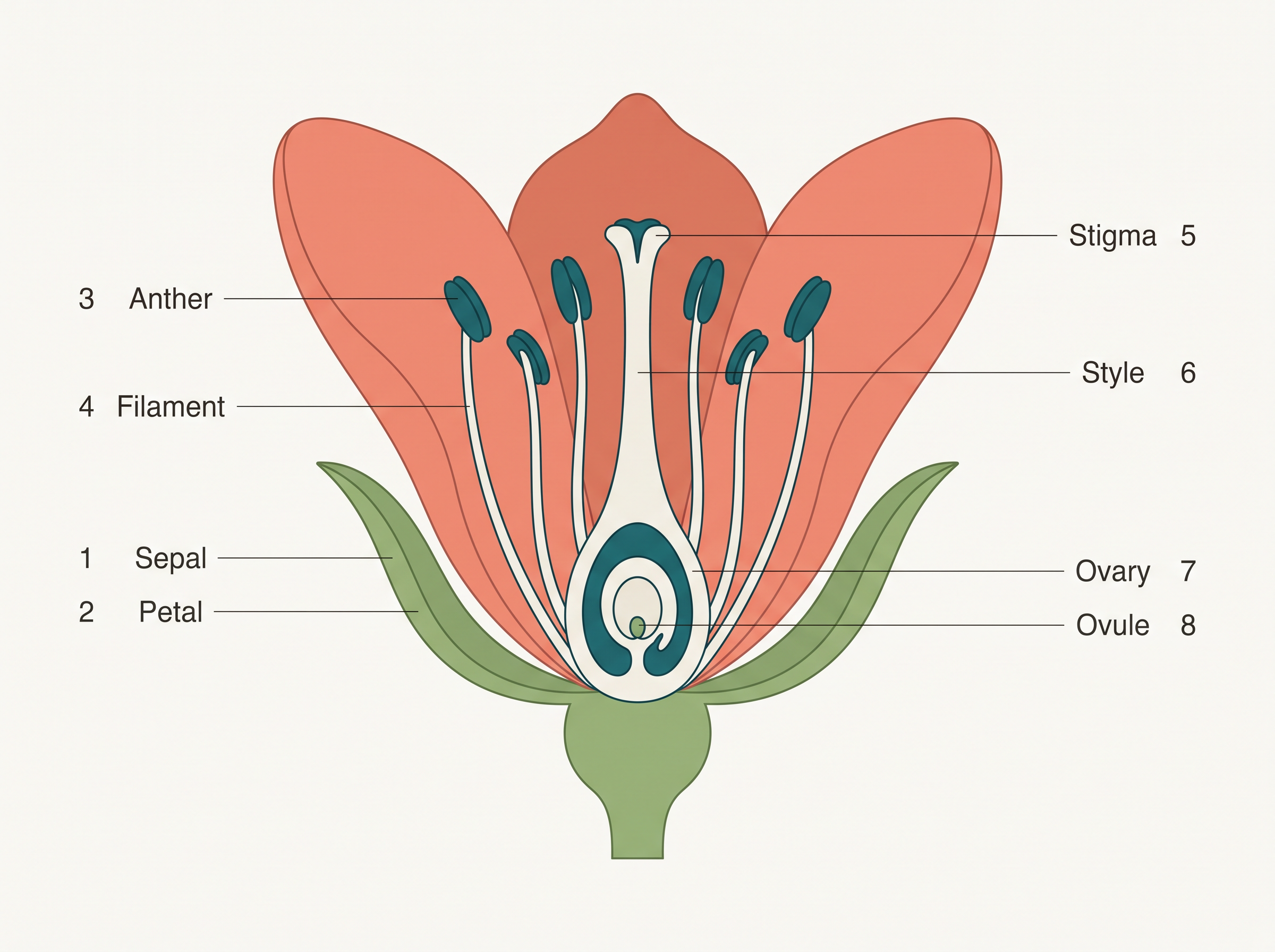
Fig. 1.0 – L.S. of a typical flower.
By the end of this chapter, you will be able to answer each of these questions yourself.
1.1 Flower – A Fascinating Organ of Angiosperms
Human beings have shared an intimate bond with flowers for as long as we can remember. We value them for their beauty, use them to decorate our homes, and give them meaning in social, religious and cultural life. Over the ages, flowers have become symbols through which we express our deepest feelings — love, affection, happiness, and even grief and mourning.
This human fascination is understandable, but it tells only half the story. Angiosperms, the flowering plants, are plants whose seeds develop enclosed within fruits, and in every one of them the flower is far more than a decoration. To a biologist, a flower is a morphological and embryological marvel — the very site where sexual reproduction takes place.
The reproductive purpose of a flower
In earlier classes you studied the parts of a typical flower — the protective and attractive outer whorls, and the two inner whorls that carry out reproduction. Figure 1.0 will help you recall these parts. Notice how the showy outer whorls surround the smaller but far more important reproductive whorls at the centre.
Everything that follows in this chapter — pollination, fertilisation, the making of fruits and seeds — begins in these central whorls. So before we trace those events, it is worth pausing on where exactly the male and female reproductive units arise.
1.2 Pre-fertilisation: Structures and Events
Long before a flower is ever visible on a plant, the plant has already “decided” to flower. This decision is set in motion by several hormonal and structural changes inside the plant. These changes cause a small mound of dividing cells, the floral primordium, to differentiate and develop. From these primordia arise inflorescences — the arrangements of flowers on a branch — which bear first the floral buds and then the open flowers.
Within each flower, two vital reproductive whorls differentiate and develop: the androecium and the gynoecium. The androecium is a whorl of stamens and represents the male reproductive organ of the flower. The gynoecium represents the female reproductive organ. Everything that leads up to fertilisation — the making of male and female gametes, and their meeting through pollination — happens in and around these two whorls. That is why they are called the pre-fertilisation structures, and the processes within them the pre-fertilisation events.
1.2.1 Stamen, Microsporangium and Pollen Grain
A typical stamen has two parts, as shown in Figure 1.1a. There is a long, slender stalk called the filament, and a terminal, usually two-lobed structure called the anther. The lower end of the filament is attached either to the thalamus (the tip of the flower stalk that bears the whorls) or to a petal.
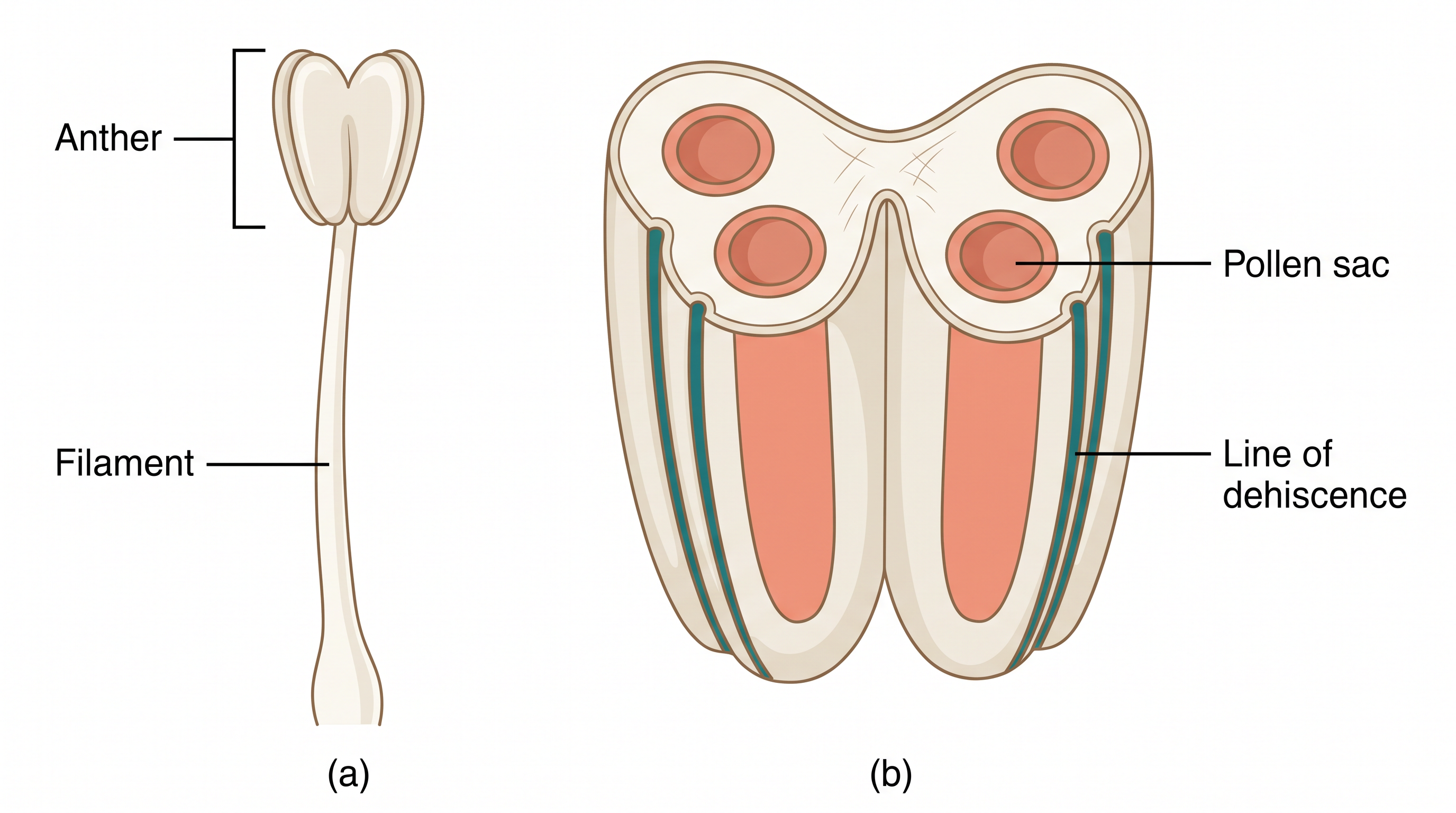
Fig. 1.1 – (a) A typical stamen and (b) 3D section of an anther.
The number and length of stamens vary a great deal from one species to another. If you collected one stamen each from ten different flowers and arranged them on a slide, the range of sizes and shapes would be striking. Examining them under a dissecting microscope reveals just how varied anthers can be in shape and in the way they are attached.
Inside the anther
A typical angiosperm anther is bilobed, and each lobe contains two theca — so the anther is described as dithecous (Figure 1.1b). A lengthwise groove usually runs down the anther, separating the two theca.
To understand the tissues inside, we look at a transverse section (a cut across the anther), shown in Figure 1.2a. Here the bilobed nature is very clear. The anther is a four-sided (tetragonal) structure with four microsporangia — the pollen-forming chambers — located at its four corners, two in each lobe.
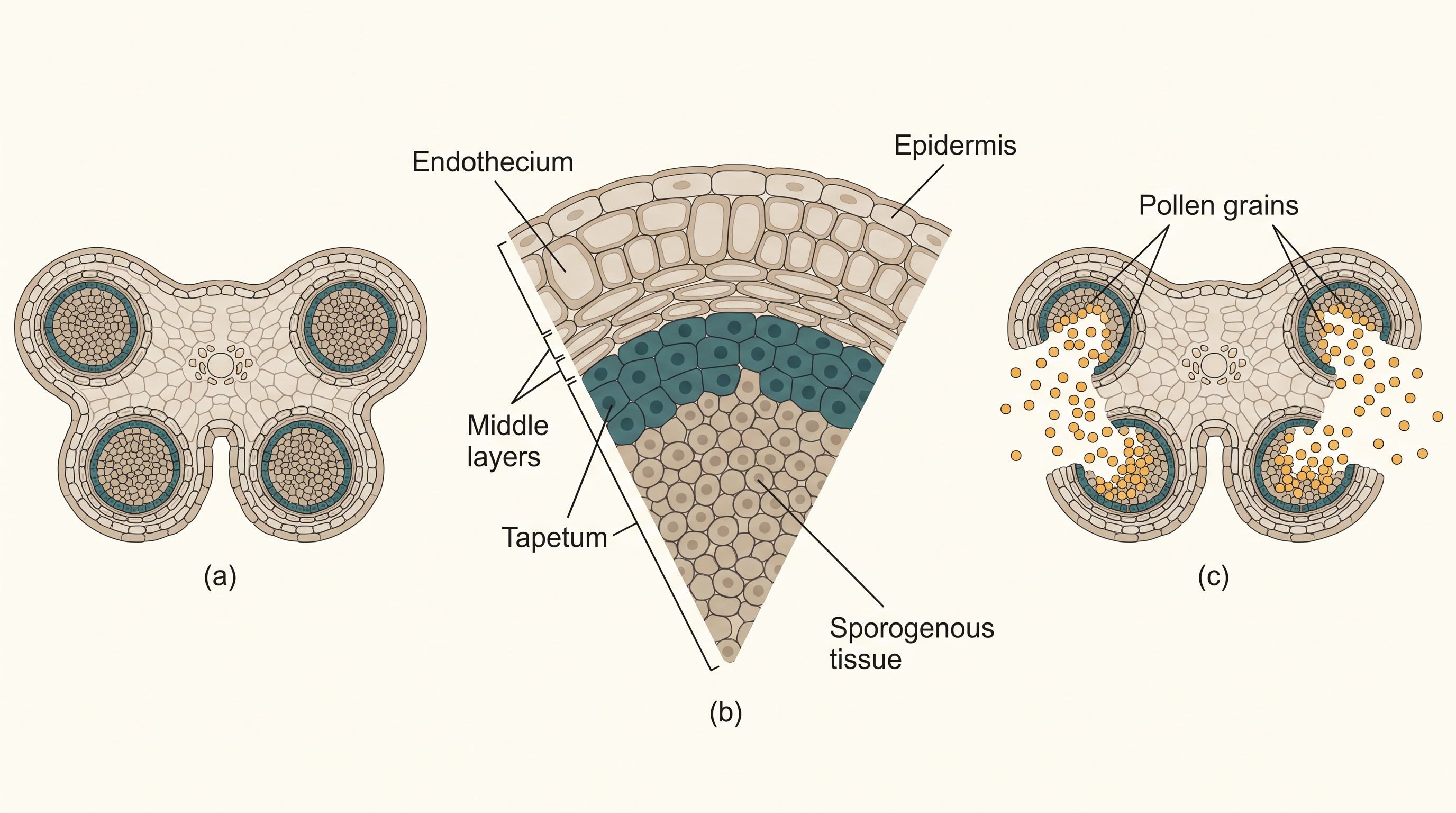
Fig. 1.2 – T.S. of an anther and its wall layers.
As the anther matures, the microsporangia develop into pollen sacs. These extend along the whole length of the anther and become packed with pollen grains.
Structure of a microsporangium
In transverse section, a typical microsporangium looks almost circular. It is usually surrounded by four wall layers (Figure 1.2b): the epidermis on the outside, then the endothecium, the middle layers, and finally the innermost layer, the tapetum.
The outer three layers — epidermis, endothecium and middle layers — carry out protection. They also help the anther to split open (dehisce) so that the pollen can be released.
The innermost layer, the tapetum, has a nourishing role. It provides food to the developing pollen grains. Tapetal cells are easy to recognise: they have dense cytoplasm and usually contain more than one nucleus. This multinucleate condition arises because the nucleus divides without the cell dividing afterwards.
Microsporogenesis
When the anther is still young, the centre of each microsporangium is filled with a group of tightly packed, similar-looking cells called the sporogenous tissue.
As the anther develops, the cells of this sporogenous tissue divide by meiosis. Meiosis is a special “reduction division” that halves the chromosome number, so that a diploid (\(2n\)) cell gives rise to haploid (\(n\)) products. Each meiotic division here produces a group of four cells called a microspore tetrad.
Because every cell of the sporogenous tissue is capable of forming a microspore tetrad, each such cell is a potential pollen mother cell (also called a microspore mother cell, PMC). The whole process of forming microspores from a pollen mother cell through meiosis is called microsporogenesis. This sequence is summarised in FC 1.1.
Flowchart
FC 1.1 – Microsporogenesis: sporogenous tissue → pollen mother cell / PMC (\(2n\)) → meiosis → microspore tetrad (four cells, \(n\)) → microspores separate → mature pollen grains (male gametophyte, \(n\))
As the anther matures and dries, the four microspores of each tetrad separate from one another and develop into pollen grains. Several thousand microspores, and hence pollen grains, form inside a single microsporangium. They are all released when the anther dehisces (Figure 1.2c).
The pollen grain
Pollen grains represent the male gametophyte — the tiny, gamete-producing generation of the plant’s life cycle. If you touch the opened anthers of Hibiscus or almost any flower, a yellowish powder of pollen grains sticks to your fingers. Sprinkle some on a drop of water on a slide and look under a microscope: the variety of sizes, shapes, colours and surface designs across different species is remarkable (Figure 1.3).
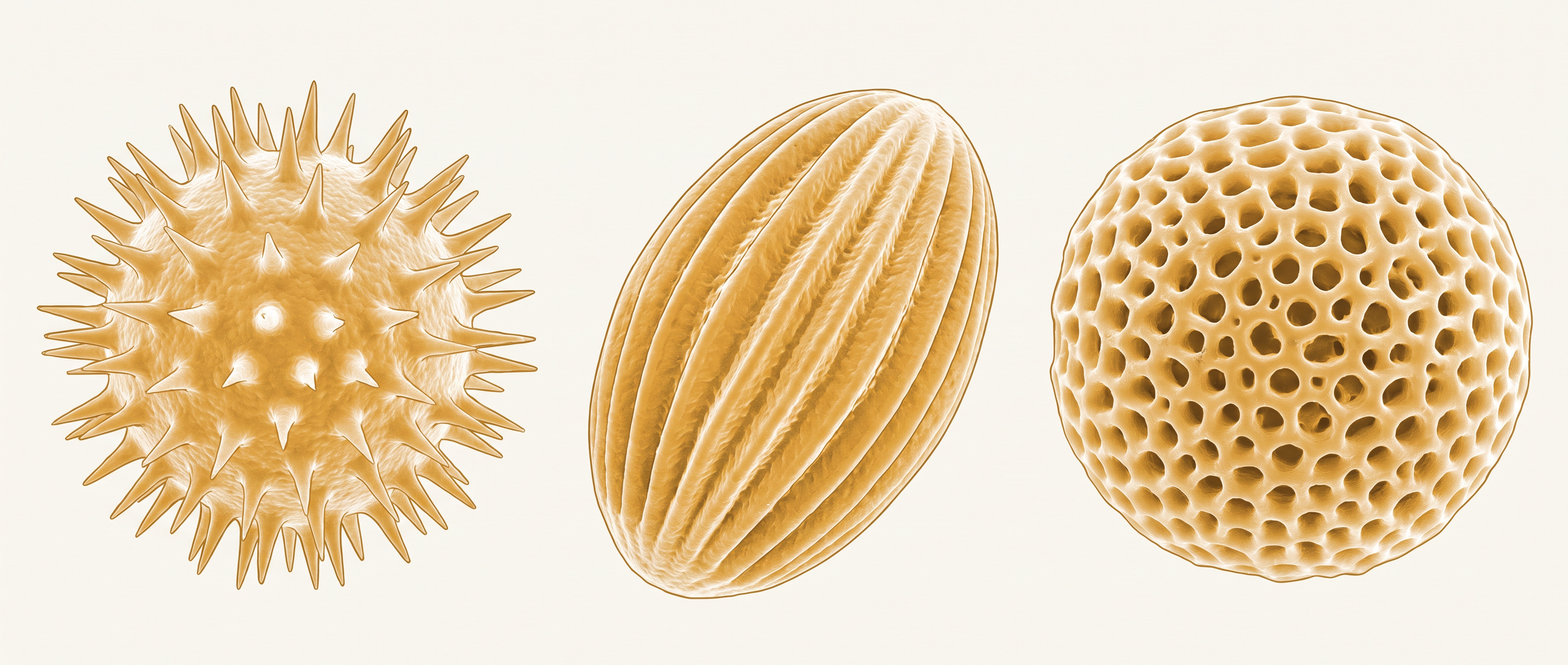
Fig. 1.3 – Diversity of pollen grain surfaces.
Pollen grains are generally spherical and measure about 25–50 micrometres (µm) across. Each has a prominent two-layered wall. The tough outer layer is the exine, made of sporopollenin — one of the most resistant organic materials known. Sporopollenin can withstand high temperatures and strong acids and alkalis, and no enzyme is yet known that can break it down.
Because sporopollenin is so durable, pollen grains are extremely well preserved as fossils. The exine is not uniform, though: it carries distinct openings called germ pores where sporopollenin is absent. It is through these thin spots that the pollen tube will later emerge.
Beneath the exine lies the inner wall, the intine. This is a thin, continuous layer made of cellulose and pectin. Inside the intine, the cytoplasm of the pollen grain is bounded by a plasma membrane.
When a pollen grain is mature, it usually contains two cells — a large vegetative cell and a small generative cell (Figure 1.4b). The vegetative cell is bigger, holds abundant stored food, and has a large, irregularly shaped nucleus. The generative cell is small, spindle-shaped, has dense cytoplasm, and floats within the cytoplasm of the vegetative cell.
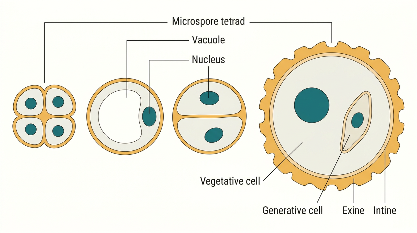
Fig. 1.4 – Maturation of a pollen grain.
In more than 60 per cent of angiosperms, pollen grains are shed at this two-celled stage. In the remaining species, the generative cell divides by mitosis to form two male gametes even before the pollen is shed — so these grains are released at a three-celled stage.
Pollen and human life
Pollen grains of many species trigger severe allergies and bronchial problems in some people, sometimes leading to long-term respiratory disorders such as asthma and bronchitis. A well-known offender is Parthenium, or carrot grass, which entered India as a contaminant along with imported wheat and has since spread almost everywhere, causing widespread pollen allergy.
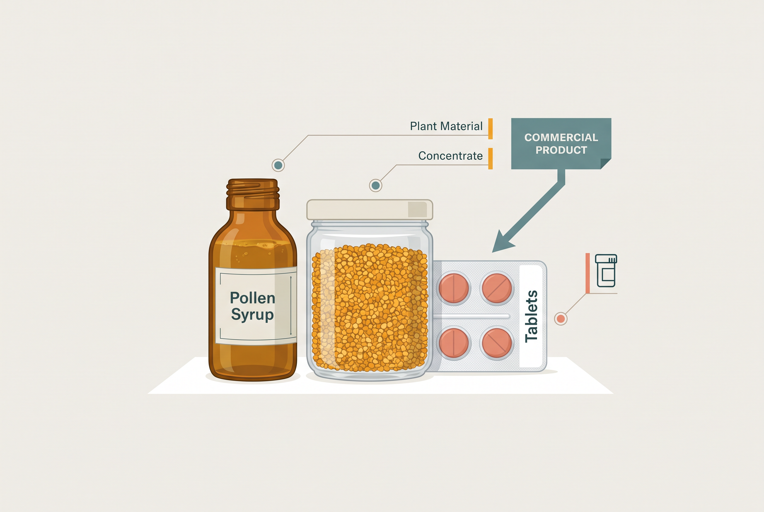
Fig. 1.5 – Commercial pollen supplements.
Once shed, pollen grains must land on a stigma before they lose their viability if they are to bring about fertilisation. How long a pollen grain stays viable varies widely and depends partly on temperature and humidity. In cereals such as rice and wheat, pollen loses viability within about 30 minutes of release. In some members of the families Rosaceae, Leguminoseae and Solanaceae, pollen stays viable for months.
1.2.2 The Pistil, Megasporangium (ovule) and Embryo sac
The gynoecium is the female reproductive part of the flower. It may consist of a single pistil (monocarpellary) or of more than one pistil (multicarpellary). When there is more than one pistil, they may be fused together — a condition called syncarpous (Figure 1.6b) — or they may remain free, which is called apocarpous (Figure 1.6c).
Each pistil has three parts (Figure 1.6a): the stigma, the style and the ovary. The stigma sits at the top and serves as the landing platform for pollen grains. The style is the elongated, slender part just below the stigma. The swollen basal part is is the ovary.
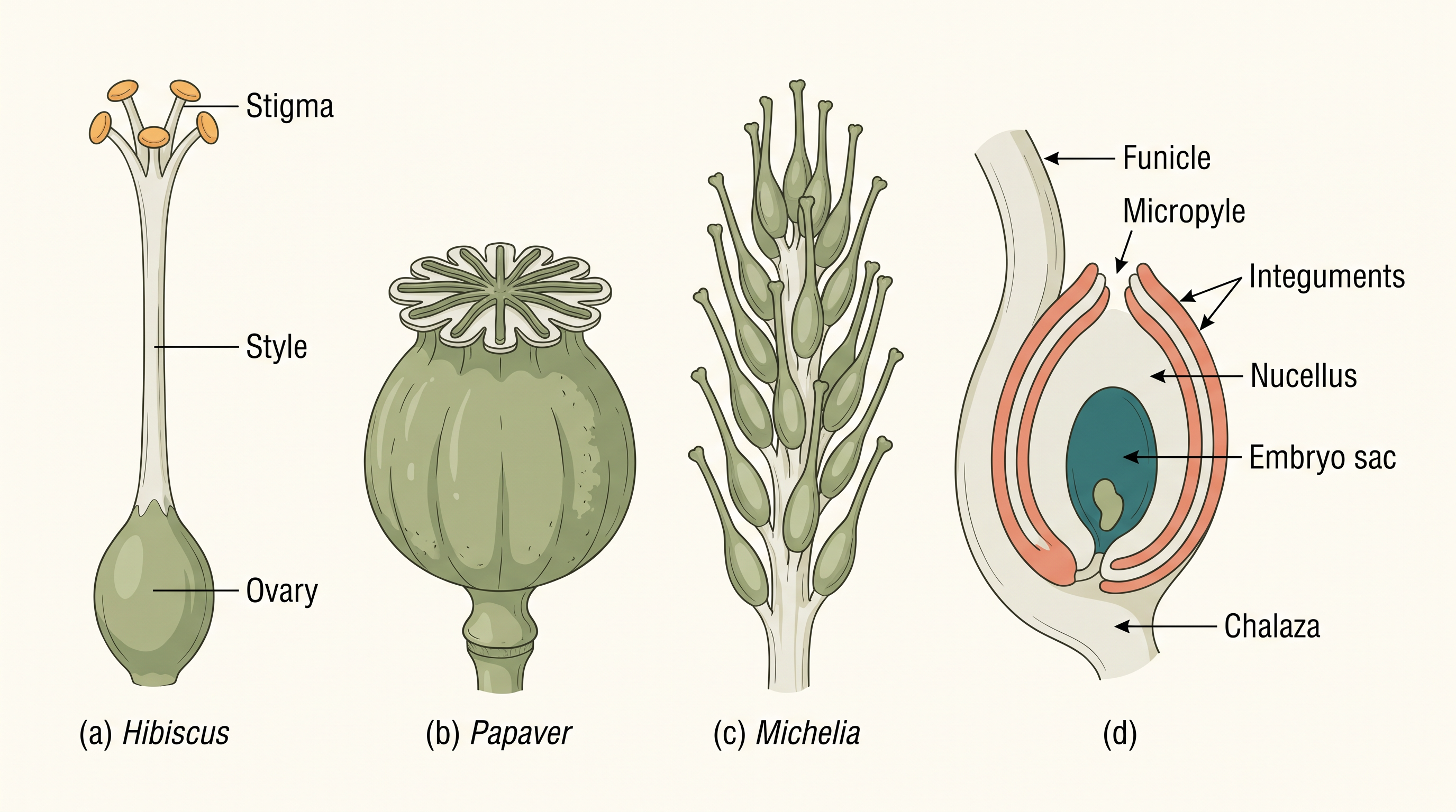
Fig. 1.6 – Diversity of pistils and an anatropous ovule.
Inside the ovary is a cavity called the ovarian cavity or locule. On its inner wall is the placenta, the tissue to which ovules are attached. (You may recall the definition and the types of placentation from Class XI.) Arising from the placenta are the megasporangia, commonly called ovules. The number of ovules in an ovary ranges from just one (as in wheat, paddy and mango) to many (as in papaya, water melon and orchids).
The megasporangium (ovule)
Let us look closely at a typical angiosperm ovule (Figure 1.6d). The ovule is a small structure attached to the placenta by a stalk called the funicle. The body of the ovule joins the funicle at a region called the hilum, so the hilum is really the junction between the ovule and its stalk.
Each ovule is wrapped in one or two protective coverings called integuments. The integuments enclose the ovule almost completely, except at the tip, where they leave a small opening called the micropyle. Directly opposite the micropylar end is the chalaza, which represents the base of the ovule.
Enclosed within the integuments is a mass of cells called the nucellus. Nucellar cells are packed with reserve food. Embedded in the nucellus is the embryo sac, or female gametophyte. An ovule generally has just a single embryo sac, and this develops from one megaspore.
Megasporogenesis
The formation of megaspores from a megaspore mother cell is called megasporogenesis. In the micropylar region of the nucellus, an ovule usually sets aside a single megaspore mother cell (MMC). This is a large cell with dense cytoplasm and a prominent nucleus.
The MMC then undergoes meiosis. Because meiosis is a reduction division, it produces four haploid megaspores from the one diploid MMC (Figure 1.7a).
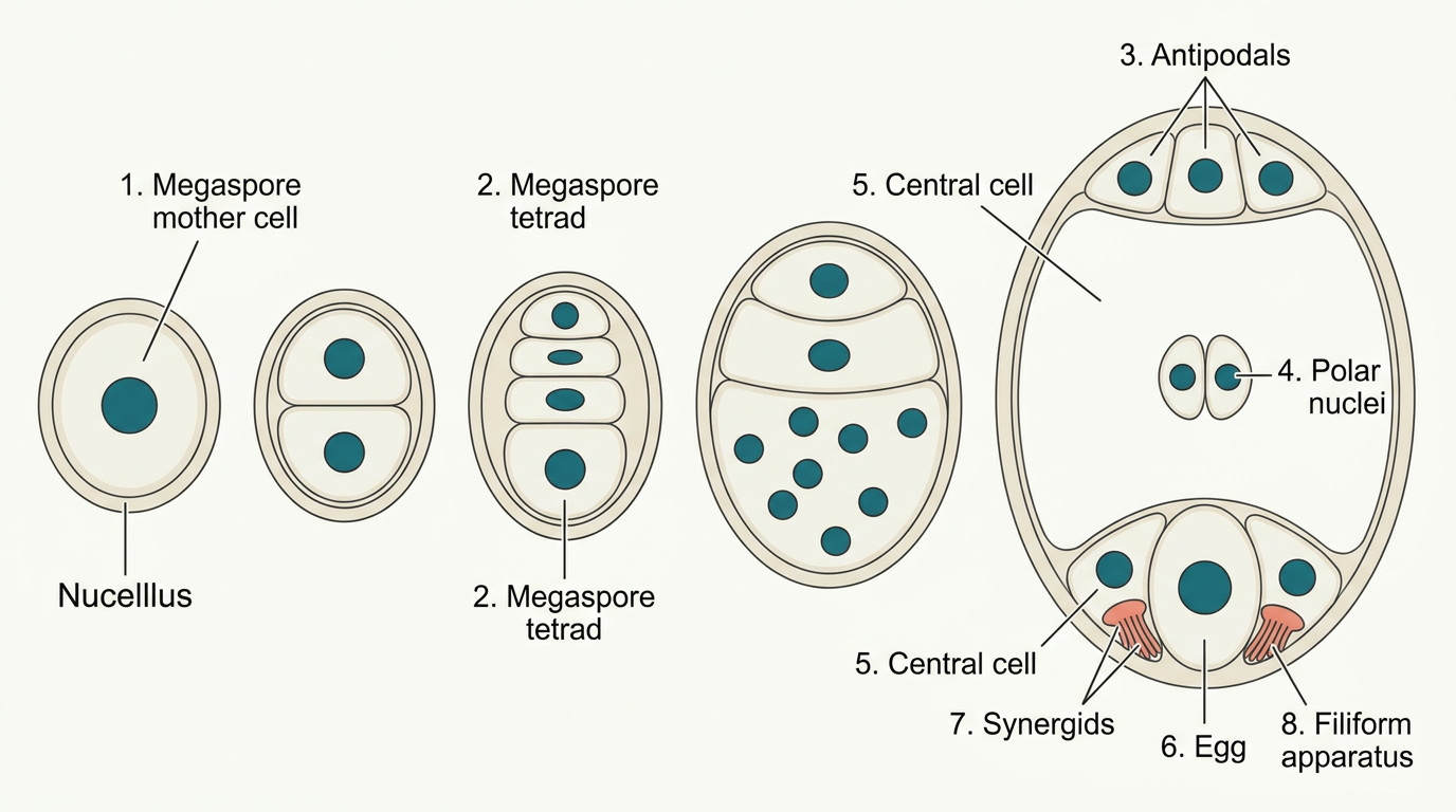
Fig. 1.7 – Development of the female gametophyte.
The female gametophyte
In most flowering plants, only one of the four megaspores stays functional; the other three degenerate. This single functional megaspore develops into the female gametophyte, or embryo sac. Because the embryo sac forms from just one megaspore, this pattern is called monosporic development.
Now let us follow how the embryo sac itself is built (Figure 1.7b). The nucleus of the functional megaspore divides by mitosis to give two nuclei, which move to opposite poles — this is the 2-nucleate embryo sac. Two more rounds of mitotic nuclear division follow, producing first the 4-nucleate and then the 8-nucleate stage. This whole sequence is shown in FC 1.2.
Flowchart
FC 1.2 – Megasporogenesis and female gametophyte: megaspore mother cell / MMC (\(2n\)) → meiosis → four megaspores (\(n\)) → three degenerate, one functional megaspore → mitosis → 2-nucleate → mitosis → 4-nucleate → mitosis → 8-nucleate → cell walls laid down → mature embryo sac (7-celled, 8-nucleate)
An important detail is that these mitotic divisions are strictly free-nuclear: each nuclear division is not immediately followed by the laying down of a cell wall. Only after the 8-nucleate stage are cell walls formed, organising the structure into the typical female gametophyte.
Look carefully at how the cells are arranged inside the mature embryo sac (Figure 1.7b, c). Six of the eight nuclei become surrounded by cell walls and organised into six cells. The remaining two nuclei, called the polar nuclei, stay free (without their own cell walls) in the large central cell, sitting just below the egg apparatus.
There is a fixed pattern to this arrangement. At the micropylar end, three cells group together to form the egg apparatus. The egg apparatus consists of two synergids and one egg cell. Each synergid carries special thickenings at its micropylar tip called the filiform apparatus, which later help guide the pollen tube into the synergid.
At the opposite, chalazal end lie three cells called the antipodals. In the middle is the large central cell with its two polar nuclei. So, although a typical angiosperm embryo sac at maturity has eight nuclei, it has only seven cells.
1.2.3 Pollination
In the previous sections you saw that the male gametes are produced in the pollen grain and the female gamete (the egg) in the embryo sac. Both kinds of gametes are non-motile — they cannot swim or move on their own. So how are they brought together for fertilisation?
The answer is pollination. The transfer of pollen grains, shed from the anther, to the stigma of a pistil is called pollination. Flowering plants have evolved an astonishing variety of adaptations to achieve it, very often using external agents to carry their pollen.
Kinds of pollination
Depending on the source of the pollen, pollination is divided into three types, summarised in FC 1.3.
Flowchart
FC 1.3 – Kinds of pollination (by source of pollen): Pollination → Autogamy (pollen and stigma of the same flower) / Geitonogamy (another flower of the same plant) / Xenogamy (a flower of a different plant)
Autogamy is pollination within the same flower — pollen is transferred from the anther to the stigma of that very flower (Figure 1.8a). In an ordinary flower that opens and exposes its anthers and stigma, complete autogamy is rather rare. It needs two conditions to be met: the release of pollen and the receptivity of the stigma must be synchronised in time, and the anthers and stigma must lie close enough together for self-pollen to reach the stigma.
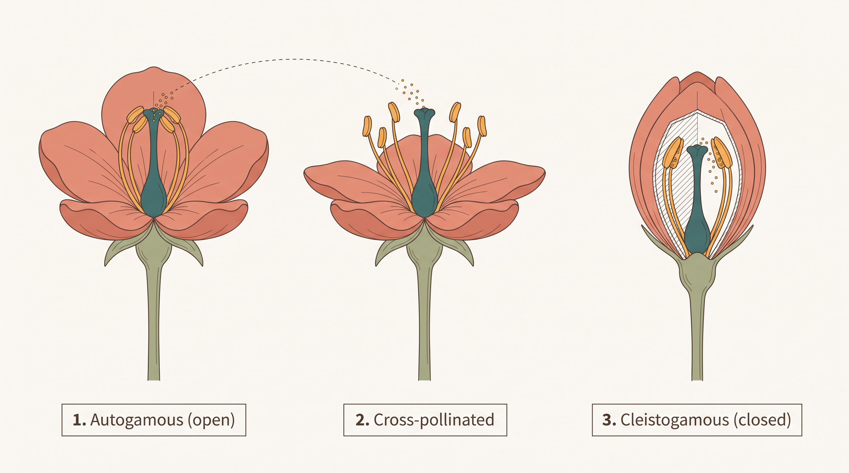
Fig. 1.8 – Flowers showing different types of pollination.
Some plants, such as Viola (the common pansy), Oxalis and Commelina, produce two kinds of flowers. Chasmogamous flowers are like ordinary flowers, with exposed anthers and stigma. Cleistogamous flowers never open at all (Figure 1.8c); their anthers and stigma stay close together, and when the anthers split open inside the closed bud, pollen falls directly onto the stigma.
Because a cleistogamous flower never opens, no foreign pollen can reach its stigma, so it is always self-pollinated (autogamous). Such flowers give assured seed-set even when no pollinators are present.
Geitonogamy is the transfer of pollen from the anther of one flower to the stigma of another flower on the same plant. In terms of mechanics it is a form of cross-pollination, because it needs a pollinating agent, but genetically it behaves just like autogamy, since the pollen still comes from the same plant.
Xenogamy is the transfer of pollen from the anther of one plant to the stigma of a flower on a genetically different plant (Figure 1.8b). This is the only type of pollination that brings genetically different pollen to the stigma, and therefore the only one that gives true genetic mixing.
Agents of pollination
Plants use two abiotic agents (wind and water) and one biotic agent (animals) to move pollen. Most plants rely on biotic agents; only a small proportion depend on the abiotic ones.
In both wind and water pollination, a pollen grain reaching the stigma is largely a matter of chance. To make up for this uncertainty and the heavy loss of pollen involved, such flowers produce enormous quantities of pollen compared with the small number of ovules available.
Pollination by wind is the more common of the two abiotic types. It requires pollen grains that are light and non-sticky, so that air currents can carry them. Wind-pollinated flowers usually have well-exposed stamens, so pollen is easily swept into the air (Figure 1.9), and large, often feathery stigmas that can trap floating pollen. They frequently have a single ovule per ovary and many flowers packed into an inflorescence. A familiar example is the corn cob: the “silky” ears you see are simply the stigmas and styles waving in the wind to catch pollen. Wind pollination is very common in grasses.
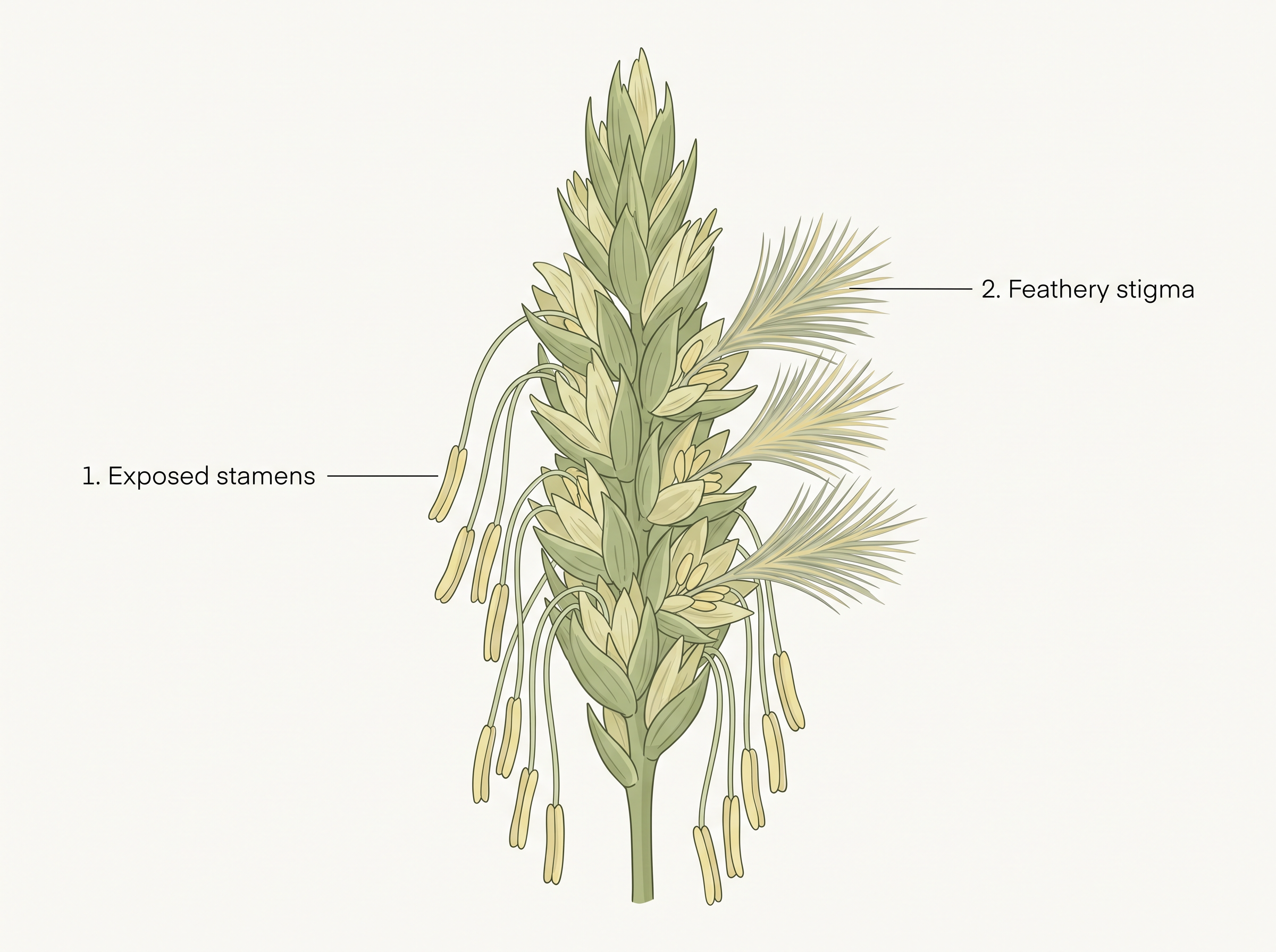
Fig. 1.9 – A wind-pollinated plant.
Pollination by water is quite rare in flowering plants — limited to about 30 genera, mostly monocotyledons. By contrast, water is the regular means of transporting male gametes in lower plant groups such as algae, bryophytes and pteridophytes. In fact, the need for water to move their male gametes is thought to be one reason the distribution of some bryophytes and pteridophytes is restricted.
Examples of water-pollinated plants include Vallisneria and Hydrilla in fresh water, and marine sea-grasses such as Zostera. Not every aquatic plant uses water for pollination, though. In many, such as water hyacinth and water lily, the flowers rise above the water surface and are pollinated by insects or wind, just like land plants.
In Vallisneria, the female flower reaches the water surface on a long stalk, while the male flowers or pollen grains are released onto the surface. Carried passively by water currents (Figure 1.10a), some pollen eventually reaches the female flowers and their stigmas. In sea-grasses, the female flowers stay submerged and the pollen is released within the water. Here the pollen grains are often long and ribbon-like, drifting passively until some reach a stigma. In most water-pollinated species, the pollen is shielded from wetting by a mucilaginous covering.
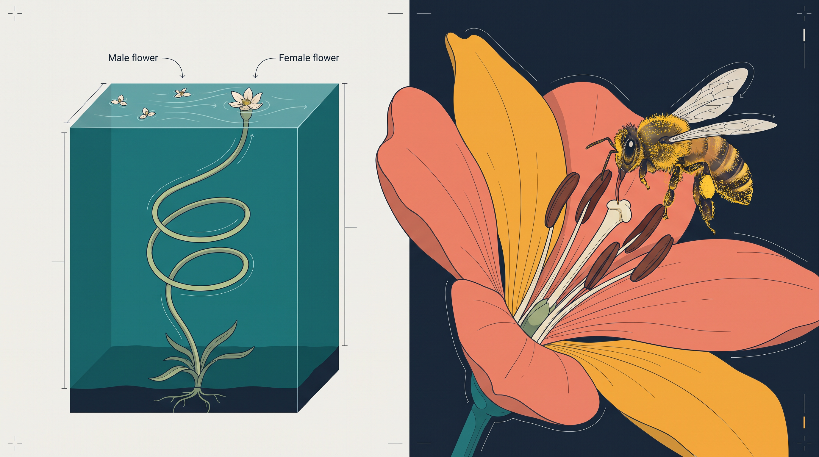
Fig. 1.10 – Pollination by (a) water in Vallisneria and (b) insects.
Both wind- and water-pollinated flowers tend not to be colourful and do not produce nectar. Since they do not rely on animals, there is no need to invest in advertisements or rewards.
Pollination by animals is used by the majority of flowering plants. Common animal pollinators include bees, butterflies, flies, beetles, wasps, ants, moths, birds (such as sunbirds and hummingbirds) and bats (Figure 1.10b). Among all of these, insects — and especially bees — are the dominant pollinators. Even larger animals have been recorded as pollinators in some species, including certain primates (lemurs), tree-dwelling (arboreal) rodents, and reptiles such as gecko lizards and garden lizards.
Flowers pollinated by animals are often specially adapted to one particular kind of animal. Most insect-pollinated flowers are large, colourful, fragrant and rich in nectar. When individual flowers are small, many of them cluster into an inflorescence to become more conspicuous together.
Animals are drawn to flowers by colour and/or fragrance. Flowers pollinated by flies and beetles may even give off foul odours to attract them. To keep the animals coming, flowers must offer rewards, and the usual rewards are nectar and pollen grains. While collecting these rewards, the visiting animal brushes against the anthers and stigma. Its body picks up a coating of pollen — which is generally sticky in animal-pollinated flowers — and when the animal later touches the stigma of another flower, pollination is achieved.
In some species the reward is not food but a safe place to lay eggs. An example is Amorphophallus, whose flower is about 6 feet tall — among the tallest flowers in the world.
Outbreeding devices
Most flowering plants bear hermaphrodite (bisexual) flowers, in which pollen can easily fall on the stigma of the same flower. Continued self-pollination leads to inbreeding depression — a loss of vigour in the offspring. To avoid this, flowering plants have evolved several devices that discourage self-pollination and encourage cross-pollination.
In some species, pollen release and stigma receptivity are not synchronised: either the pollen is released before the stigma is receptive, or the stigma becomes receptive well before the pollen is released. In others, the anther and stigma are placed at different positions, so pollen cannot easily reach the stigma of the same flower. Both of these devices prevent autogamy.
A third device is self-incompatibility. This is a genetic mechanism that prevents self-pollen — from the same flower or from other flowers of the same plant — from fertilising the ovules, by blocking pollen germination or pollen tube growth in the pistil.
A fourth device is the production of unisexual flowers. If both male and female flowers are borne on the same plant (a condition called monoecious, as in castor and maize), autogamy is prevented, but geitonogamy is not. In species such as papaya, the male and female flowers are on different plants, so each plant is either male or female — a condition called dioecious. This prevents both autogamy and geitonogamy.
Pollen–pistil interaction
Pollination does not guarantee that the “right” pollen has arrived. Often, pollen of the wrong type — from another species, or self-pollen in a self-incompatible plant — also lands on the stigma. The pistil, however, has the ability to recognise whether the pollen is compatible (right type) or incompatible (wrong type).
If the pollen is compatible, the pistil accepts it and promotes the post-pollination events that lead to fertilisation. If it is incompatible, the pistil rejects it by preventing the pollen from germinating on the stigma or by stopping the pollen tube from growing in the style. This acceptance-or-rejection is the outcome of a continuous chemical “dialogue” between the pollen grain and the pistil. Only in recent years have botanists begun to identify the pollen and pistil chemicals responsible for this recognition.
Following a compatible pollination, the pollen grain germinates on the stigma and puts out a pollen tube through one of its germ pores (Figure 1.11a). The contents of the pollen grain move into this tube, which then grows through the tissues of the stigma and style and reaches the ovary (Figure 1.11b, c).
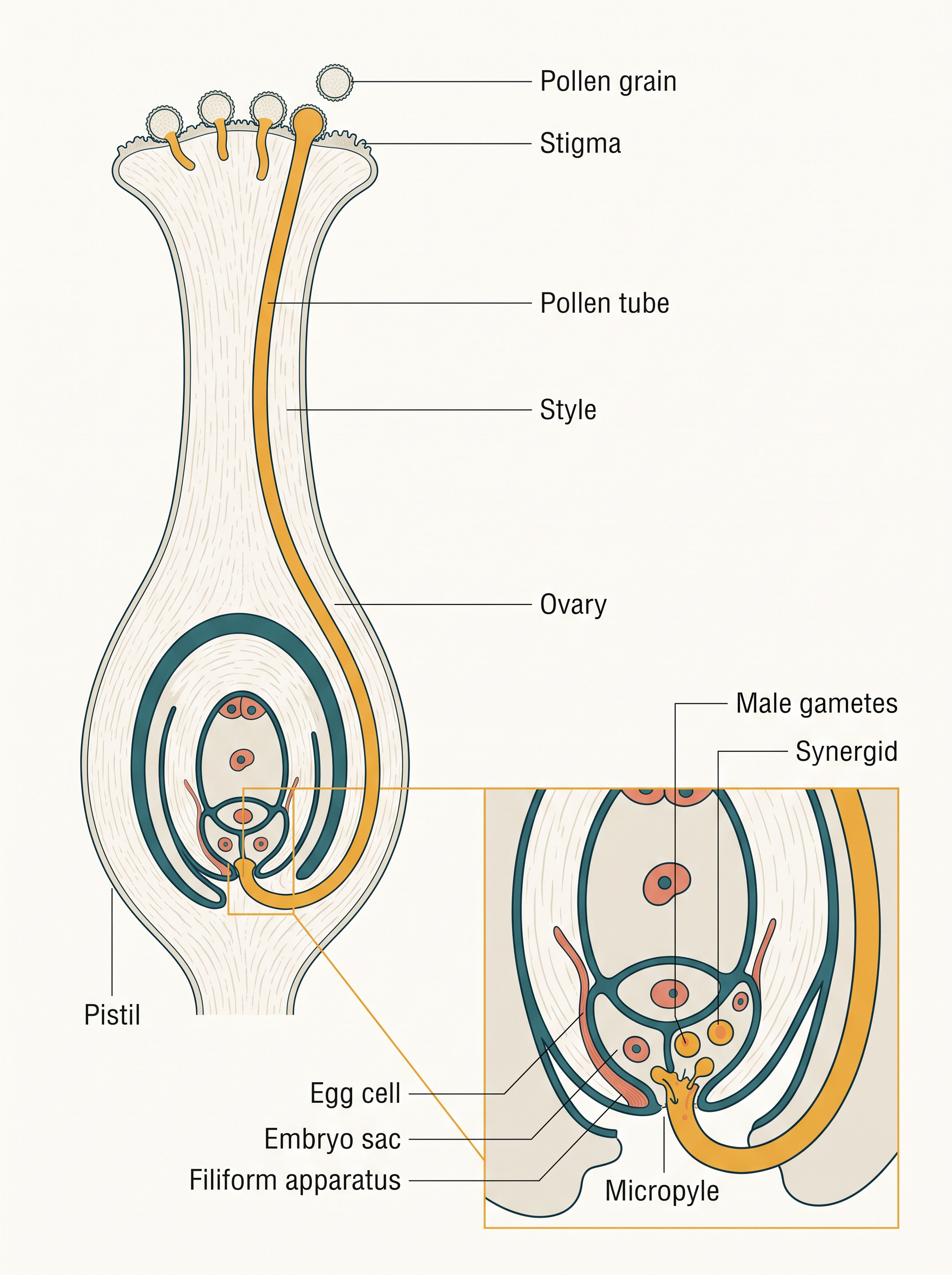
Fig. 1.11 – Pollen–pistil interaction and pollen tube entry.
Recall the two shedding conditions of pollen. In plants that shed pollen at the two-celled stage, the generative cell divides to form the two male gametes while the pollen tube is still growing through the stigma. In plants that shed pollen at the three-celled stage, the two male gametes are already present and are carried along from the very beginning.
After reaching the ovary, the pollen tube enters the ovule through the micropyle and then pushes into one of the synergids through the filiform apparatus (Figure 1.11d, e). Studies show that the filiform apparatus at the micropylar end of the synergid guides the entry of the pollen tube. All these events — from the landing of pollen on the stigma until the pollen tube enters the ovule — are together called pollen–pistil interaction. This whole journey is traced in FC 1.4.
Flowchart
FC 1.4 – Pollen–pistil interaction (compatible pollen): pollen lands on stigma → recognised as compatible → pollen germinates → pollen tube emerges through a germ pore → grows through stigma and style → reaches ovary → enters ovule through the micropyle → enters a synergid via the filiform apparatus → releases two male gametes
Understanding pollen–pistil interaction is useful in practice, because a plant breeder who knows how to manipulate it can sometimes push through even incompatible pollinations to obtain desired hybrids.
Artificial hybridisation
A plant breeder often wants to cross different species, or even genera, to combine desirable characters into a commercially “superior” variety. Such artificial hybridisation is one of the major approaches to crop improvement. In these crossing experiments, it is essential that only the chosen pollen is used, and that the stigma is protected from unwanted pollen. This is achieved by two techniques: emasculation and bagging.
If the female parent bears bisexual flowers, the anthers must be removed from the flower bud before they dehisce, using a pair of forceps. This removal of anthers is called emasculation. The emasculated flower is then covered with a bag of suitable size, usually made of butter paper, to keep unwanted pollen off its stigma. This covering is called bagging. When the stigma of the bagged flower becomes receptive, mature pollen collected from the desired male parent is dusted onto it, the flower is rebagged, and the fruit is allowed to develop.
If the female parent bears unisexual flowers, emasculation is not needed, because there are no anthers to remove. Here the female flower buds are simply bagged before they open. When the stigma becomes receptive, the desired pollen is applied and the flower is rebagged.
1.3 Double Fertilisation
Once the pollen tube has entered one of the synergids, it releases its two male gametes into the cytoplasm of that synergid. This delivery sets up two separate fusion events, one after the other, deep inside the embryo sac.
One of the male gametes moves towards the egg cell and fuses with its nucleus. This fusion of a male gamete with the egg is called syngamy, and it produces a diploid (\(2n\)) cell — the zygote. Here one haploid male gamete (\(n\)) unites with the haploid egg (\(n\)), restoring the diploid number.
The second male gamete travels instead towards the two polar nuclei sitting in the central cell, and fuses with both of them (Figure 1.12a). Because this fuses three haploid nuclei at once — one male gamete (\(n\)) and two polar nuclei (\(n + n\)) — it is called triple fusion, and it produces a triploid (\(3n\)) primary endosperm nucleus (PEN).
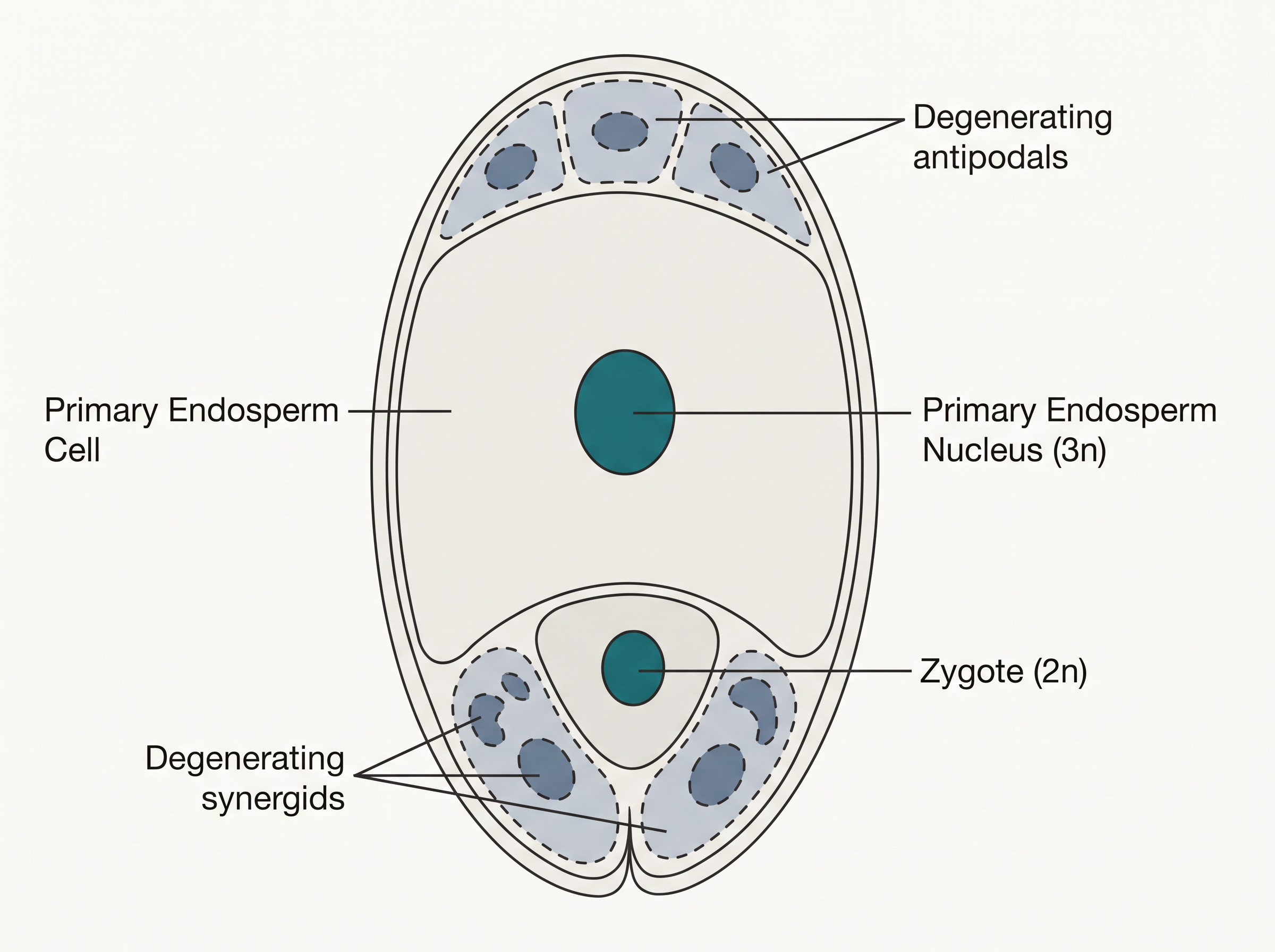
Fig. 1.12 – (a) The fertilised embryo sac.
Since two distinct fusions — syngamy and triple fusion — take place within a single embryo sac, the whole phenomenon is called double fertilisation. This double event is unique to flowering plants and occurs nowhere else in the living world. The sequence is summarised in FC 1.5.
Flowchart
FC 1.5 – Double fertilisation: pollen tube discharges two male gametes into a synergid → gamete 1 fuses with egg (syngamy) → diploid zygote (\(2n\)); gamete 2 fuses with the two polar nuclei (triple fusion) → triploid primary endosperm nucleus, PEN (\(3n\))
After triple fusion, the central cell becomes the primary endosperm cell (PEC), which goes on to develop into the endosperm. The zygote, meanwhile, develops into the embryo. So the two fusion products have two different destinies: one builds the future plant, and the other builds its food store.
1.4 Post-fertilisation: Structures and Events
Double fertilisation is not the end of the story — it is the trigger for a whole series of developments. Once the zygote and the primary endosperm nucleus have formed, the flower begins converting its parts into a seed and a fruit. The events that follow — the development of the endosperm and the embryo, the maturation of the ovule(s) into seed(s), and of the ovary into a fruit — are together called the post-fertilisation events.
1.4.1 Endosperm
An interesting rule of timing applies here: endosperm development begins before embryo development. This ordering is not accidental. By building the food-store first, the plant ensures that the embryo has assured nutrition ready and waiting as soon as it starts to grow.
The primary endosperm cell divides repeatedly to form a triploid endosperm tissue. The cells of this tissue are packed with reserve food materials, and this stored food is used to nourish the developing embryo.
How the endosperm forms
In the most common pattern, the primary endosperm nucleus (PEN) undergoes several successive nuclear divisions without cell walls forming in between, producing many free nuclei. This stage is called the free-nuclear endosperm. Later, cell walls are laid down and the endosperm becomes cellular. The number of free nuclei formed before this cellularisation varies greatly from species to species.
The fate of the endosperm differs between species. In some plants it is completely consumed by the developing embryo before the seed matures, as in pea, groundnut and beans. In others it persists in the mature seed and is used up only during germination, as in castor and coconut.
1.4.2 Embryo
The embryo develops at the micropylar end of the embryo sac, where the zygote lies. Most zygotes begin dividing only after a certain amount of endosperm has already formed — again, an adaptation that guarantees the embryo a food supply.
Although mature seeds differ enormously, the early stages of embryo development — a process called embryogeny — are similar in both monocotyledons and dicotyledons. Figure 1.13b shows these stages in a dicot. The zygote first forms a proembryo, and then passes through the globular and heart-shaped stages before becoming the mature embryo.
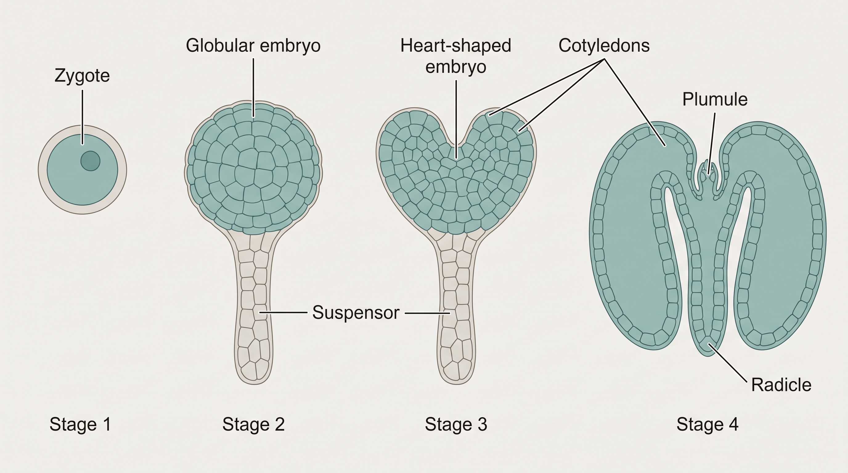
Fig. 1.13 – (b) Stages in dicot embryo development.
Flowchart
FC 1.6 – Embryogeny in a dicot: zygote → proembryo → globular embryo → heart-shaped embryo → mature embryo
The dicot embryo
A typical dicotyledonous embryo (Figure 1.14a) consists of an embryonal axis and two cotyledons. The part of the embryonal axis lying above the level of the cotyledons is the epicotyl, which ends in the plumule or stem tip. The cylindrical part below the level of the cotyledons is the hypocotyl, which ends at its lower end in the radicle or root tip. The root tip itself is covered by a protective root cap.
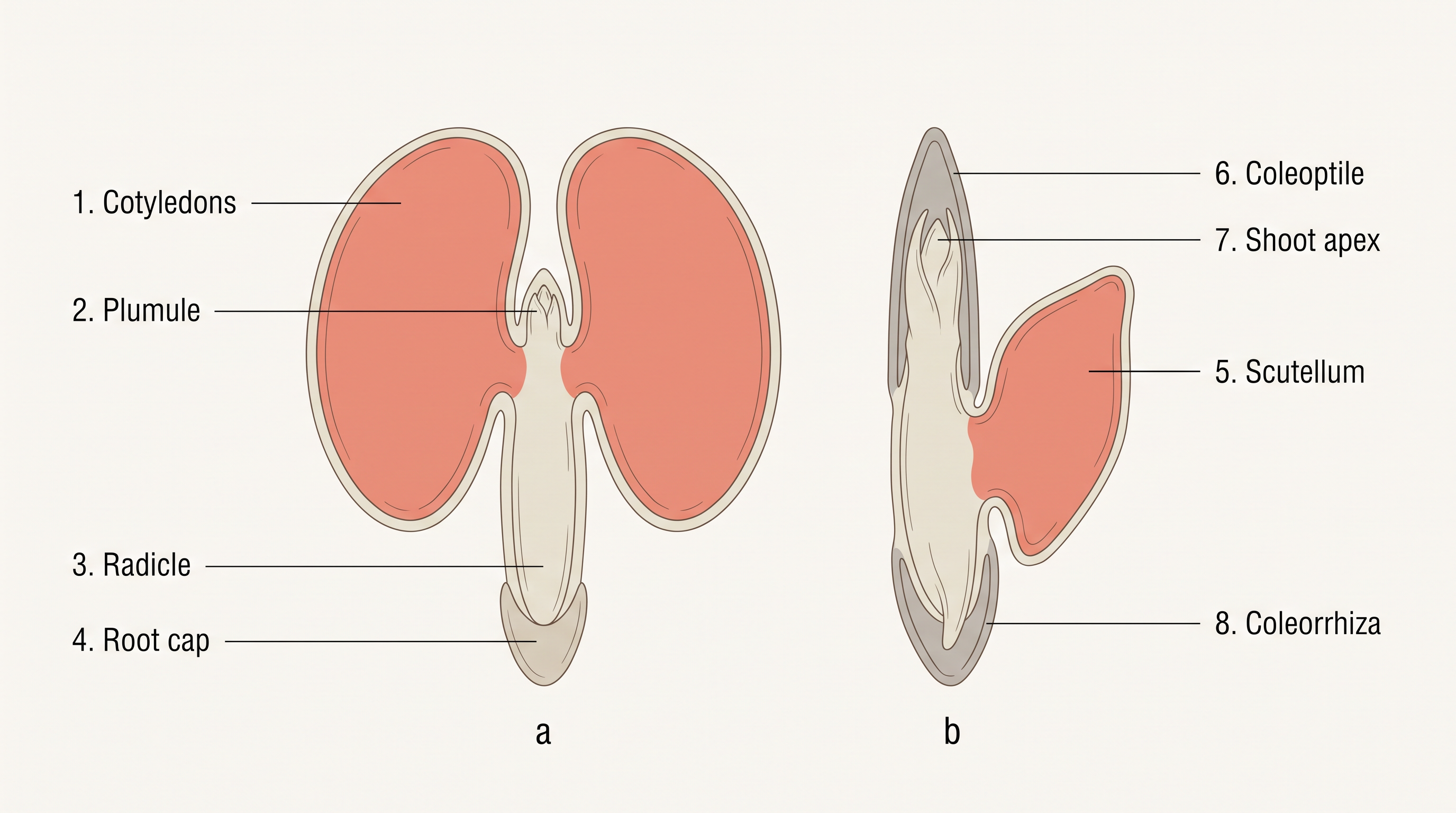
Fig. 1.14 – Mature (a) dicot and (b) monocot embryos.
The monocot embryo
Embryos of monocotyledons (Figure 1.14b) have only one cotyledon. In the grass family, this single cotyledon is called the scutellum and is placed to one side (lateral) of the embryonal axis.
At its lower end, the embryonal axis carries the radicle and root cap, enclosed in an undifferentiated sheath called the coleorrhiza. Above the point where the scutellum is attached lies the epicotyl, which bears a shoot apex and a few leaf primordia enclosed within a hollow, protective structure called the coleoptile.
1.4.3 Seed
In angiosperms, the seed is the final product of sexual reproduction, and is often described simply as a fertilised ovule. Seeds are formed inside fruits. A seed typically consists of a seed coat (or coats), one or more cotyledons, and an embryo axis.
The cotyledons (Figure 1.15a) are usually simple, thick and swollen structures, made bulky by the food reserves stored in them — as in legumes.
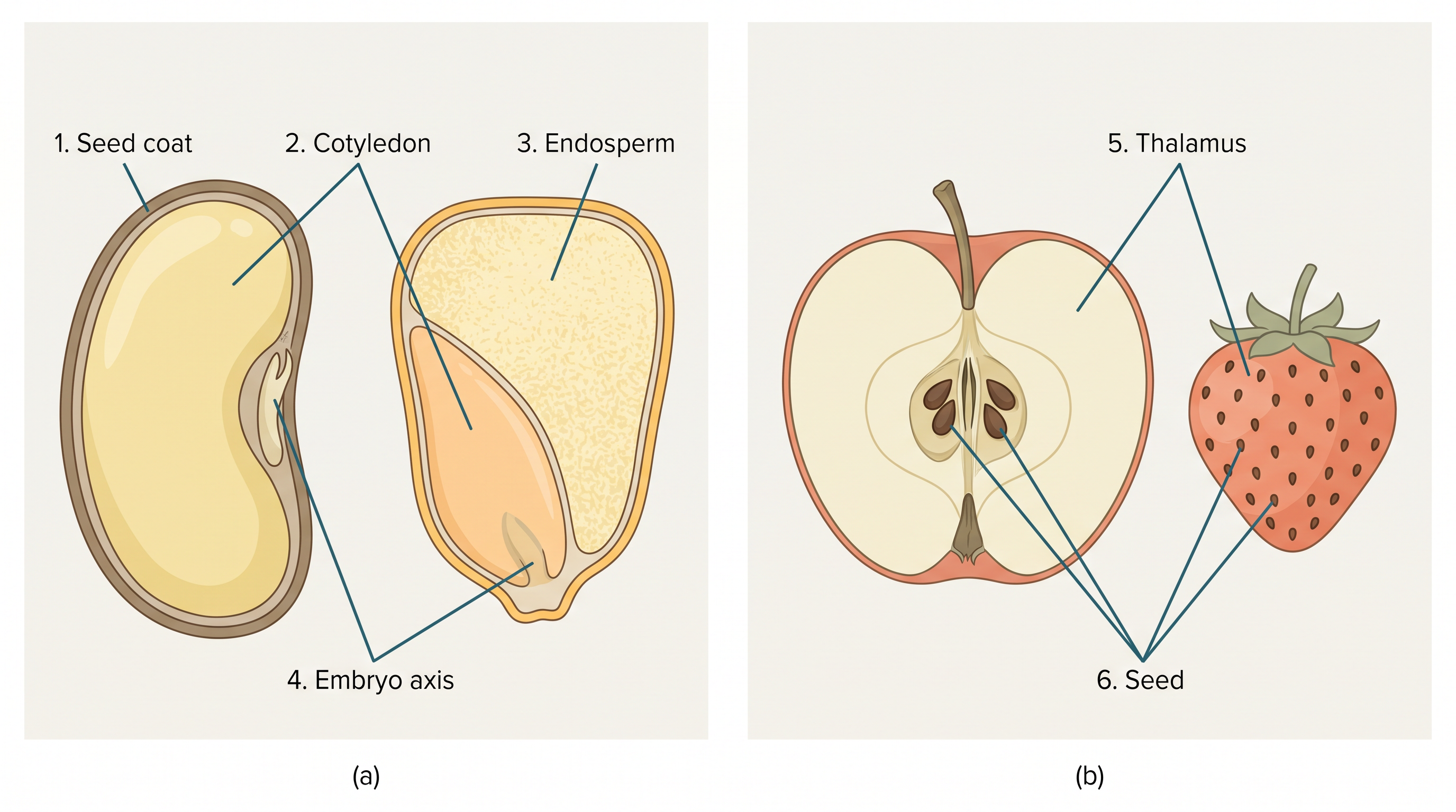
Fig. 1.15 – (a) Seed structures and (b) false fruits.
Types of seed
Depending on whether endosperm remains at maturity, mature seeds are of two kinds. Non-albuminous (ex-albuminous) seeds have no residual endosperm, because it is completely consumed during embryo development — as in pea and groundnut. Albuminous seeds retain part of the endosperm, because it is not fully used up during embryo development — as in wheat, maize, barley and castor.
Occasionally, remnants of the nucellus also persist in the seed, as in black pepper and beet. This leftover, persistent nucellus is called the perisperm.
From ovule to seed
As the seed forms, the integuments of the ovule harden into tough, protective seed coats (Figure 1.15a). The micropyle survives as a small pore in the seed coat, and this pore later allows oxygen and water to enter the seed during germination.
As the seed ripens, its water content falls and it becomes relatively dry (about 10–15 per cent moisture by mass). The general metabolic activity of the embryo slows right down. The embryo may then enter a resting state called dormancy, or — if conditions are favourable, with enough moisture, oxygen and a suitable temperature — it may germinate straight away.
From ovary to fruit
As ovules mature into seeds, the ovary develops into a fruit; the two transformations proceed side by side. The wall of the ovary becomes the wall of the fruit, called the pericarp. Fruits may be fleshy, as in guava, orange and mango, or dry, as in groundnut and mustard. Many fruits have evolved special mechanisms to disperse their seeds. This whole set of post-fertilisation changes is summarised in FC 1.7.
Flowchart
FC 1.7 – Post-fertilisation fates: zygote → embryo; primary endosperm cell → endosperm; ovule → seed; integuments → seed coat; ovary → fruit; ovary wall → pericarp
True fruits, false fruits and parthenocarpy
In most plants, by the time the fruit develops, the other floral parts wither and fall off. But in a few species, such as apple, strawberry and cashew, the thalamus also contributes to the fruit. Such fruits are called false fruits (Figure 1.15b). Fruits that develop only from the ovary are called true fruits.
Although fruits usually result from fertilisation, a few species form fruits without it. Such fruits are called parthenocarpic fruits, and banana is a well-known example. Parthenocarpy can also be induced by applying growth hormones, and such fruits are seedless.
Why seeds matter
Seeds bring several advantages to angiosperms. Because pollination and fertilisation do not depend on water, seed formation is more dependable. Seeds also carry better strategies for dispersal, helping the species reach and colonise new habitats.
With their sufficient food reserves, seeds nourish the young seedling until it can photosynthesise on its own, and the hard seed coat protects the young embryo. Being products of sexual reproduction, seeds also carry new genetic combinations, which is the source of variation.
Seeds are, in fact, the very basis of our agriculture. The drying-out and dormancy of mature seeds are what make it possible to store them — so that they can be used as food throughout the year and sown to raise the next season’s crop. Imagine agriculture with seeds that germinated immediately after forming and could not be stored at all.
The remarkable longevity of seeds
How long a seed stays alive after dispersal varies enormously. Some species lose viability within a few months; many stay viable for several years; and a few can survive for hundreds of years.
Finally, it is worth pausing on the sheer reproductive capacity of flowering plants. Ask yourself: How many eggs are in one embryo sac? How many embryo sacs in one ovule? How many ovules in one ovary? How many ovaries in one flower? How many flowers on a whole tree — and so on. The numbers multiply astonishingly.
Some fruits carry vast numbers of seeds. Orchid fruits contain thousands of tiny seeds each, as do the fruits of parasitic species such as Orobanche and Striga. And think of the tiny seed of Ficus: from that minute structure grows an enormous tree that itself produces billions of seeds — a remarkable example of a tiny structure generating a huge biomass over the years.
1.5 Apomixis and Polyembryony
Everything we have followed so far — pollination, double fertilisation, seed formation — assumes that seeds are products of fertilisation. Yet a few flowering plants break this rule. Some species of the family Asteraceae and some grasses have evolved a special mechanism to produce seeds without fertilisation. This is called apomixis.
You have already seen that fruit production without fertilisation is called parthenocarpy (as in banana). Apomixis is its counterpart for seeds: it is a form of asexual reproduction that closely mimics sexual reproduction, because it still produces seeds — just without the fusion of gametes.
How apomictic seeds develop
Apomictic seeds can develop in more than one way. In some species, the egg cell is formed as a diploid cell without reduction division (that is, without meiosis) and then develops directly into the embryo, without any fertilisation.
More often — as in many Citrus and mango varieties — the extra embryos come from a different source. Some of the nucellar cells surrounding the embryo sac begin to divide, push their way into the embryo sac, and develop into embryos. In such species, a single ovule ends up containing many embryos.
The occurrence of more than one embryo in a single seed is called polyembryony.
Why apomixis matters for agriculture
Hybrid varieties of many of our food and vegetable crops are grown on a large scale because they have hugely increased productivity. But hybrids come with a problem: hybrid seeds must be produced fresh every year. If seeds collected from a hybrid crop are sown, the offspring segregate — they no longer keep the hybrid’s desirable characters. Producing hybrid seed each year is expensive, which makes the seed costly for farmers.
1.6 Summary
- Flowers are the seat of sexual reproduction in angiosperms. Within a flower, the androecium (made of stamens) is the male reproductive part, and the gynoecium (made of pistils) is the female reproductive part.
- A typical anther is bilobed, dithecous (two theca per lobe) and tetrasporangiate (four microsporangia). Pollen grains develop inside the microsporangia. Each microsporangium is surrounded by four wall layers — the epidermis, endothecium, middle layers and the innermost tapetum, which nourishes the developing pollen.
- The sporogenous tissue at the centre of the microsporangium undergoes meiosis (microsporogenesis) to form tetrads of microspores. Each microspore then matures into a pollen grain.
- A pollen grain represents the male gametophytic generation. It has a two-layered wall: the tough outer exine, made of sporopollenin and bearing germ pores, and the inner intine of cellulose and pectin. At shedding, a pollen grain may have two cells (a vegetative cell and a generative cell) or three cells (a vegetative cell and two male gametes).
- The pistil has three parts — the stigma, the style and the ovary. Ovules are borne inside the ovary. An ovule has a stalk (funicle), one or two protective integuments, and an opening called the micropyle. Its central tissue is the nucellus, in which the archesporium differentiates.
- A cell of the archesporium — the megaspore mother cell — divides by meiosis (megasporogenesis), and one of the resulting megaspores develops into the embryo sac (the female gametophyte). The mature embryo sac is 7-celled and 8-nucleate: at the micropylar end lies the egg apparatus (two synergids and one egg cell), at the chalazal end are three antipodals, and at the centre is a large central cell with two polar nuclei.
- Pollination is the transfer of pollen grains from the anther to the stigma. Pollinating agents are either abiotic (wind and water) or biotic (animals).
- Pollen–pistil interaction covers all events from the landing of pollen on the stigma until the pollen tube enters the embryo sac (if the pollen is compatible), or the inhibition of the pollen (if it is incompatible). After a compatible pollination, the pollen grain germinates, the pollen tube grows through the style, enters the ovule, and discharges two male gametes into one of the synergids.
- Angiosperms show double fertilisation, because two fusions occur in each embryo sac: syngamy (a male gamete + egg → diploid zygote) and triple fusion (a male gamete + two polar nuclei → triploid primary endosperm nucleus). The zygote develops into the embryo and the primary endosperm cell forms the endosperm. Endosperm formation always precedes the development of the embryo. This overall sequence is summarised in FC 1.8.
Flowchart
FC 1.8 – Overall sequence of sexual reproduction in a flowering plant: microsporogenesis (pollen grains) and megasporogenesis (embryo sac) → pollination → pollen–pistil interaction (pollen tube reaches embryo sac) → double fertilisation (syngamy + triple fusion) → zygote → embryo, and primary endosperm cell → endosperm → ovule → seed, ovary → fruit
- The developing embryo passes through the proembryo, globular and heart-shaped stages before maturing. A mature dicotyledonous embryo has two cotyledons and an embryonal axis with an epicotyl and a hypocotyl. Embryos of monocotyledons have a single cotyledon. After fertilisation, the ovary develops into the fruit and the ovules develop into seeds.
- Apomixis, found in some angiosperms and particularly in grasses, is the formation of seeds without fertilisation. Apomicts offer several advantages in horticulture and agriculture.
- Polyembryony is the occurrence of more than one embryo in a single seed.
1.7 Points to Ponder
- Wind- and water-pollinated flowers are usually small, dull-coloured and produce no nectar, while animal-pollinated flowers are often large, colourful, fragrant and rich in nectar. Why should the pollinating agent make such a difference to what a flower “invests” in?
- The plant forms its food-store (the endosperm) only after fertilisation begins, and the zygote usually waits for some endosperm to form before it starts dividing. Why might it be advantageous for a plant to build the food supply only after a viable fertilisation, rather than in advance?
- Cleistogamous flowers guarantee seed-set even without any pollinator, but they can never be cross-pollinated. Under what circumstances might this assured self-pollination help a species, and when might the lack of cross-pollination become a disadvantage for its long-term survival?
- Apomictic embryos are genetically identical to the parent plant. If apomixis could be built into hybrid crop varieties, what advantages might farmers gain — and what might be lost in terms of the genetic variation that sexual reproduction normally provides?
- In algae, bryophytes and pteridophytes, water is needed to carry the male gametes, but flowering plants have made reproduction independent of water. How might this freedom from water have helped angiosperms spread across so many land habitats?
- A single tiny seed of Ficus can grow into an enormous tree that itself produces billions of seeds. What does this extraordinary reproductive capacity suggest about the chances that any one seed has of actually growing into a mature plant in nature?
- Self-incompatibility lets a pistil reject its own pollen even though that pollen is perfectly “healthy.” Why might a plant evolve a mechanism to refuse its own genetically compatible pollen?
1.8 NCERT Questions
- Name the parts of an angiosperm flower in which development of male and female gametophyte take place.
- Differentiate between microsporogenesis and megasporogenesis. Which type of cell division occurs during these events? Name the structures formed at the end of these two events.
- Arrange the following terms in the correct developmental sequence: Pollen grain, sporogenous tissue, microspore tetrad, pollen mother cell, male gametes.
- With a neat, labelled diagram, describe the parts of a typical angiosperm ovule.
- What is meant by monosporic development of female gametophyte?
- With a neat diagram explain the 7-celled, 8-nucleate nature of the female gametophyte.
- What are chasmogamous flowers? Can cross-pollination occur in cleistogamous flowers? Give reasons for your answer.
- Mention two strategies evolved to prevent self-pollination in flowers.
- What is self-incompatibility? Why does self-pollination not lead to seed formation in self-incompatible species?
- What is bagging technique? How is it useful in a plant breeding programme?
- What is triple fusion? Where and how does it take place? Name the nuclei involved in triple fusion.
- Why do you think the zygote is dormant for sometime in a fertilised ovule?
- Differentiate between:
- hypocotyl and epicotyl;
- coleoptile and coleorrhiza;
- integument and testa;
- perisperm and pericarp.
- Why is apple called a false fruit? Which part(s) of the flower forms the fruit?
- What is meant by emasculation? When and why does a plant breeder employ this technique?
- If one can induce parthenocarpy through the application of growth substances, which fruits would you select to induce parthenocarpy and why?
- Explain the role of tapetum in the formation of pollen-grain wall.
- What is apomixis and what is its importance?
1.9 Check Your Concepts
- Assertion–Reason. Assertion: Pollen grains can be recovered as fossils millions of years old. Reason: The exine of the pollen grain is made of sporopollenin, which resists high temperature, strong acids and alkalis, and is not degraded by any known enzyme. State whether the assertion and reason are true, and whether the reason correctly explains the assertion.
- Why is the tapetum considered essential for the formation of healthy pollen grains, even though it is only a wall layer and not part of the sporogenous tissue itself?
- A pollen grain is shed at the two-celled stage in one plant and at the three-celled stage in another. In which of the two will the two male gametes be formed inside the pollen tube, and why?
- Sequence-ordering. Arrange the following in the correct order of a pollen tube’s journey after a compatible pollination: enters a synergid through the filiform apparatus, pollen germinates on the stigma, pollen tube reaches the ovary, pollen tube enters the ovule through the micropyle, pollen tube grows through the style.
- Match the following:
| Column A | Column B |
|---|---|
| a. Synergids | i. three, at the chalazal end |
| b. Antipodals | ii. two, in the central cell |
| c. Polar nuclei | iii. two, at the micropylar end with filiform apparatus |
| d. Egg cell | iv. single female gamete |
- The mature embryo sac has eight nuclei but only seven cells. Explain how this is possible.
- State the ploidy (haploid or diploid) of each of the following, giving a reason: the nucellus, the megaspore mother cell, the functional megaspore, and the female gametophyte.
- Both geitonogamy and xenogamy require a pollinating agent, yet only one of them gives genetically different offspring. Which one, and why does the other not, even though the pollen is carried between flowers?
- Wind-pollinated flowers and animal-pollinated flowers differ strikingly in colour, scent and nectar. Explain, in terms of function, why each set of features suits its pollinating agent.
- What happens if the anthers of a bisexual flower are not removed before performing an artificial cross? Name the technique used to prevent this problem and describe how it is carried out.
- Distinguish between a monoecious and a dioecious plant, and state which of the two can prevent both autogamy and geitonogamy.
- Assertion–Reason. Assertion: Cleistogamous flowers always produce seeds even in the complete absence of pollinators. Reason: Cleistogamous flowers never open, so only self-pollen can reach their stigma. State whether both are true and whether the reason explains the assertion.
- Double fertilisation is described as an event “unique to flowering plants.” Identify the two fusion events involved, name their products with ploidy, and explain what each product develops into.
- Why is it biologically advantageous that endosperm development begins before embryo development in most angiosperms?
- The coconut is often used to illustrate the two forms of endosperm. Identify which part of a tender coconut is free-nuclear endosperm and which is cellular endosperm.
- Differentiate between albuminous and non-albuminous seeds, giving one example of each, and state what determines which type a seed becomes.
- Diagram-labelling. Draw a labelled diagram of a typical dicotyledonous embryo and mark the following: cotyledons, epicotyl, plumule, hypocotyl and radicle.
- A gardener finds several embryos inside a single orange seed. Name this phenomenon, explain how such extra embryos arise in many Citrus and mango varieties, and state whether these embryos are genetically identical to the parent.
- “What is good for the seed industry is not always good for genetic variation.” In the context of turning hybrid crops into apomicts, explain the benefit to farmers and the possible loss in terms of variation.
- What happens if the pollen landing on a stigma is incompatible? Describe the two ways in which the pistil can reject such pollen.