Chapter 5: Molecular Basis of Inheritance
In the winter of 1869, a young Swiss physician named Friedrich Miescher was working in a laboratory in Tübingen, Germany, trying to understand what cells are actually made of. He needed a steady supply of cells, and he found one in an unlikely place — the pus-soaked bandages discarded from a nearby surgical clinic. Pus is full of white blood cells, and white blood cells have large nuclei.
Miescher carefully separated the nuclei and treated them with different chemicals. To his surprise, he isolated a substance that behaved unlike any protein he knew. It was mildly acidic and unusually rich in phosphorus. Because it came from the nucleus, he called it “nuclein” — the substance we now call DNA.
Miescher had no idea that he was holding the master molecule of life. He did not know that this faintly acidic material carried the instructions to build an entire organism, or that it could copy itself with astonishing precision. It would take nearly eighty more years, and the work of many scientists, before the world understood what nuclein truly was: the molecular basis of inheritance. This chapter follows that long detective story — and the molecule at the centre of it.
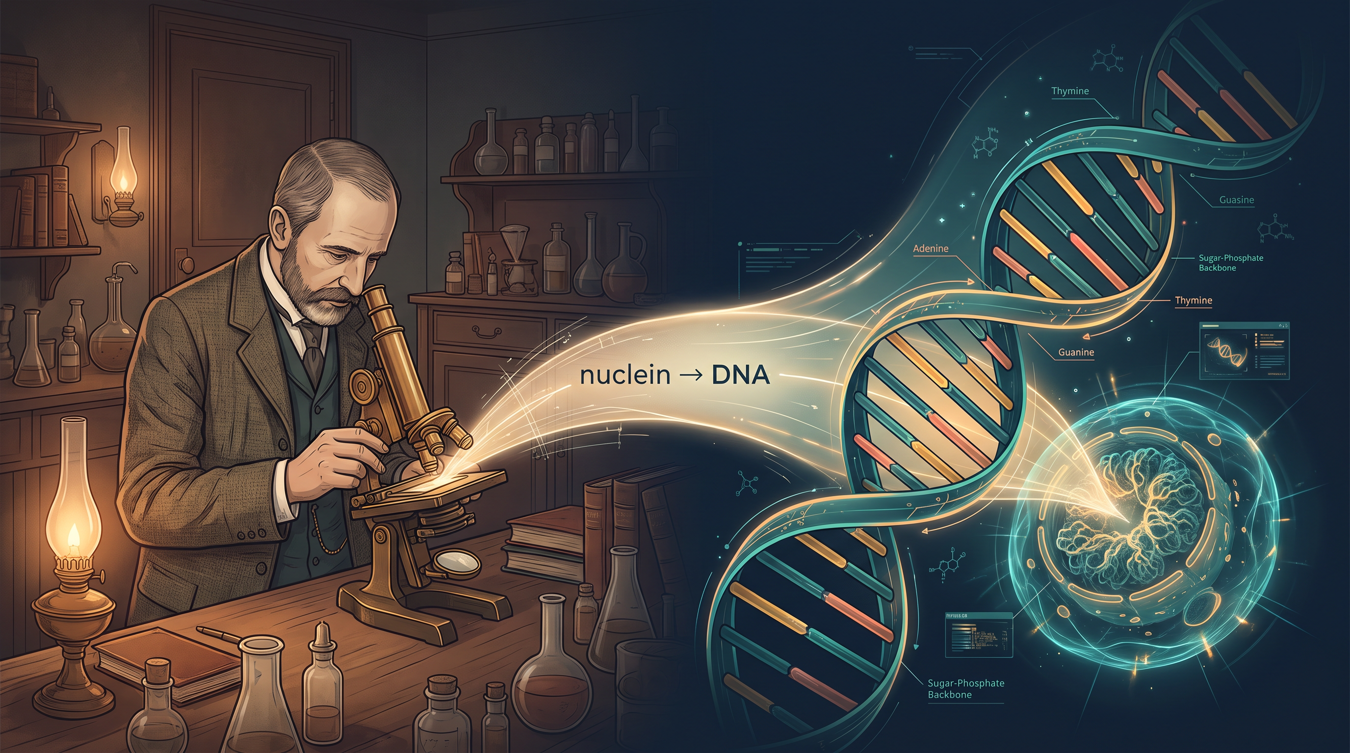
Fig. 5.0 – From nuclein to DNA.
By the end of this chapter, you will be able to answer each of these questions yourself.
5.1 The DNA
DNA, or deoxyribonucleic acid, is a very long polymer made of repeating units called deoxyribonucleotides. A polymer simply means a large molecule built by joining many small identical units in a chain, the way a long train is built from many coaches. The small units here are nucleotides, and when the sugar in them is deoxyribose, we call them deoxyribonucleotides.
The length of a DNA molecule is usually described by the number of nucleotides it contains, or by the number of base pairs (bp) — a base pair being one pair of nucleotides, one from each of the two strands. This length is a fixed characteristic of each organism, almost like a signature.
A few real examples make this clear. A tiny virus called bacteriophage φX174 has just \(5386\) nucleotides. Bacteriophage lambda has \(48502\) base pairs. The bacterium Escherichia coli has about \(4.6 \times 10^6\) base pairs. The haploid content of human DNA — that is, the DNA in a single set of chromosomes — is about \(3.3 \times 10^9\) base pairs. From a small virus to a human being, the amount of DNA rises enormously. Let us now look closely at how such a long polymer is actually built.
5.1.1 Structure of Polynucleotide Chain
Building Blocks: The Nucleotide
Let us recall the chemical structure of a polynucleotide chain, which can be either DNA or RNA. Every nucleotide is made of three parts joined together: a nitrogenous base, a pentose sugar (a five-carbon sugar — ribose in RNA and deoxyribose in DNA), and a phosphate group.
The nitrogenous bases are of two kinds. Purines are the larger, double-ring bases — Adenine (A) and Guanine (G). Pyrimidines are the smaller, single-ring bases — Cytosine (C), Uracil (U) and Thymine (T). Cytosine is found in both DNA and RNA. Thymine is present only in DNA, while Uracil takes its place in RNA.
From Nucleoside to Polynucleotide Chain
When a nitrogenous base attaches to the –OH group at the 1′C (first carbon) of the pentose sugar through an N-glycosidic linkage, the unit formed is called a nucleoside. Examples are adenosine or deoxyadenosine, guanosine or deoxyguanosine, cytidine or deoxycytidine, and uridine or deoxythymidine. (The “deoxy-” versions carry deoxyribose and belong to DNA.)
When a phosphate group then links to the –OH at the 5′C (fifth carbon) of a nucleoside through a phosphoester linkage, a nucleotide (or deoxynucleotide, depending on the sugar) is formed.
Two nucleotides join together through a 3′–5′ phosphodiester linkage to form a dinucleotide. In this bond, the phosphate bridges the 3′ carbon of one sugar and the 5′ carbon of the next. When many more nucleotides are joined in this way, a long polynucleotide chain is produced, as shown in Fig. 5.1.
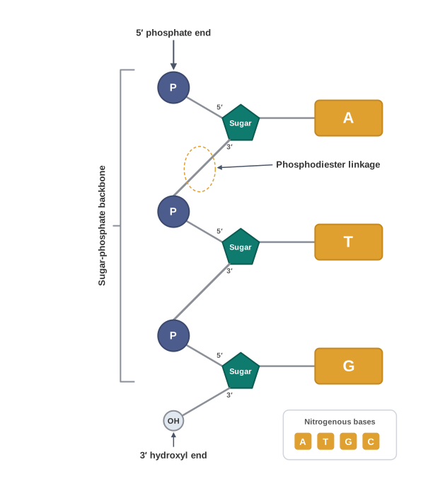
Fig. 5.1 – A single polynucleotide chain.
Such a chain has a definite direction, and the two ends are not the same. At one end there is a free phosphate group on the 5′ carbon of the sugar — this is called the 5′-end. At the other end the sugar has a free –OH on its 3′ carbon — this is the 3′-end. The alternating sugar and phosphate units form a strong backbone, and the nitrogenous bases stick out sideways from this backbone.
How RNA Differs
RNA is built the same way but differs in two small yet important ways. First, every nucleotide in RNA has an extra –OH group at the 2′ position of the ribose sugar. Second, RNA uses uracil in place of thymine (thymine is chemically 5-methyl uracil, so the two are close cousins).
The First Glimpse — and the Double Helix
DNA was first noticed long before its structure was understood. An acidic substance obtained from the nucleus was identified by Friedrich Meischer in 1869 and named “Nuclein.” Because such a long, delicate polymer was extremely hard to isolate in one piece, working out its exact three-dimensional structure remained a puzzle for a very long time.
The breakthrough came in 1953, when James Watson and Francis Crick proposed their famous Double Helix model of DNA. They built it using the X-ray diffraction data produced by Maurice Wilkins and Rosalind Franklin. Their model rested on the idea of base pairing between the two strands. It also fitted an earlier observation by Erwin Chargaff, who had found that in double-stranded DNA the amount of Adenine equals that of Thymine, and the amount of Guanine equals that of Cytosine.
This base pairing gives the two strands a special property: they are complementary to each other. If you know the sequence of bases on one strand, you can immediately predict the sequence on the other. This also means that each strand of a parental DNA can act as a template — a pattern to copy from — for building a new strand. The two daughter DNA molecules made this way would be exact copies of the parent. In one stroke, this explained how genetic information could be faithfully passed on.
Salient Features of the Double Helix
The Watson–Crick double-helix structure can be summarised in five key features:
It is made of two polynucleotide chains. The backbone is formed by sugar and phosphate, and the bases point inward.
The two chains have anti-parallel polarity. This means that if one chain runs in the \(5′ \rightarrow 3′\) direction, the other runs \(3′ \rightarrow 5′\).
The bases of the two strands pair through hydrogen bonds (H-bonds), forming base pairs. Adenine forms two hydrogen bonds with Thymine, and vice-versa; Guanine bonds with Cytosine through three hydrogen bonds. As a result, a purine always lies opposite a pyrimidine. This keeps the distance between the two strands almost uniform along the whole helix, as shown in Fig. 5.2.
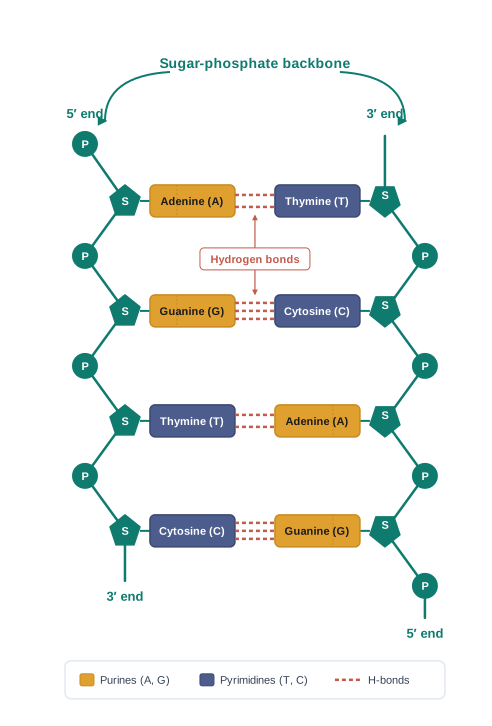
Fig. 5.2 – A double-stranded polynucleotide chain.
The two chains are coiled together in a right-handed fashion. The pitch of the helix (the length of one full turn) is \(3.4\) nm, where a nanometre is one-billionth of a metre (\(10^{-9}\) m). There are roughly \(10\) base pairs in each turn, so the distance between two adjacent base pairs is about \(0.34\) nm.
The plane of each base pair stacks over the plane of the next one in the helix. This base stacking, along with the hydrogen bonds, gives the helical structure its stability (Fig. 5.3).
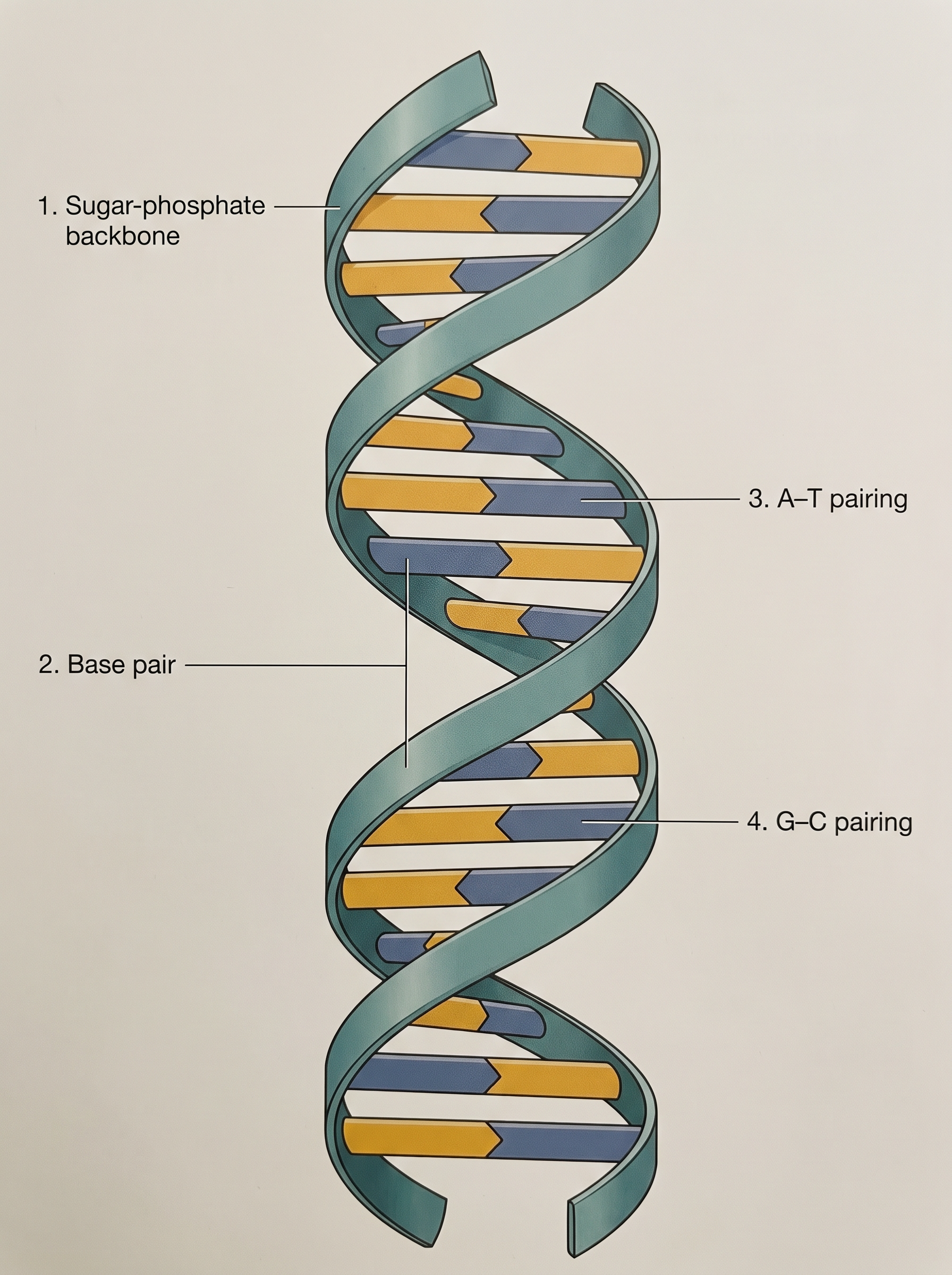
Fig. 5.3 – The DNA double helix.
The Central Dogma
The simplicity of the double-helix model, and how neatly it explained heredity, made it revolutionary. Very soon after, Francis Crick proposed the Central dogma of molecular biology. It states the normal direction in which genetic information flows in a cell: \(DNA \rightarrow RNA \rightarrow Protein\). DNA is copied into RNA (transcription), and RNA directs the making of protein (translation). This flow is summarised in FC 5.1.
Flowchart
FC 5.1 – Central dogma: DNA →(replication, DNA copies itself) DNA; DNA →(transcription) RNA →(translation) Protein; special branch in some viruses: RNA →(reverse transcription) DNA.
In some viruses, the flow can run in the reverse direction, that is, from RNA back to DNA.
5.1.2 Packaging of DNA Helix
Why Packaging Is a Problem
We just saw that adjacent base pairs are \(0.34\) nm apart. If we multiply this distance by the total number of base pairs in a typical human cell, we can estimate the length of its DNA:
\[6.6 \times 10^9 \text{ bp} \times 0.34 \times 10^{-9} \text{ m/bp} \approx 2.2 \text{ m}\]
So the DNA in a single human cell, stretched out, would be about \(2.2\) metres long. Yet it must fit inside a nucleus that is only about \(10^{-6}\) m across. Packing more than two metres of thread into such a tiny space is like folding a very long rope into a matchbox. How does the cell manage it?
Packaging in Prokaryotes
In prokaryotes such as E. coli, there is no true, membrane-bound nucleus. Even so, the DNA is not left scattered loosely through the cell. DNA carries a negative charge, and it is held together by certain positively charged proteins in a region called the nucleoid. Within the nucleoid, the DNA is arranged in large loops that are held in place by these proteins.
Packaging in Eukaryotes
In eukaryotes, the organisation is far more elaborate. Here the cell uses a set of positively charged, basic proteins called histones.
A protein gains its charge from the amino acids in it that have charged side chains. Histones are especially rich in two basic amino acids, lysine and arginine, both of which carry positive charges on their side chains. This is why histones are positively charged overall.
Eight histone molecules group together to form a unit called a histone octamer. The negatively charged DNA then wraps around this positively charged histone octamer — opposite charges attract — to form a structure called a nucleosome, shown in Fig. 5.4a. A typical nucleosome holds about \(200\) base pairs of DNA.
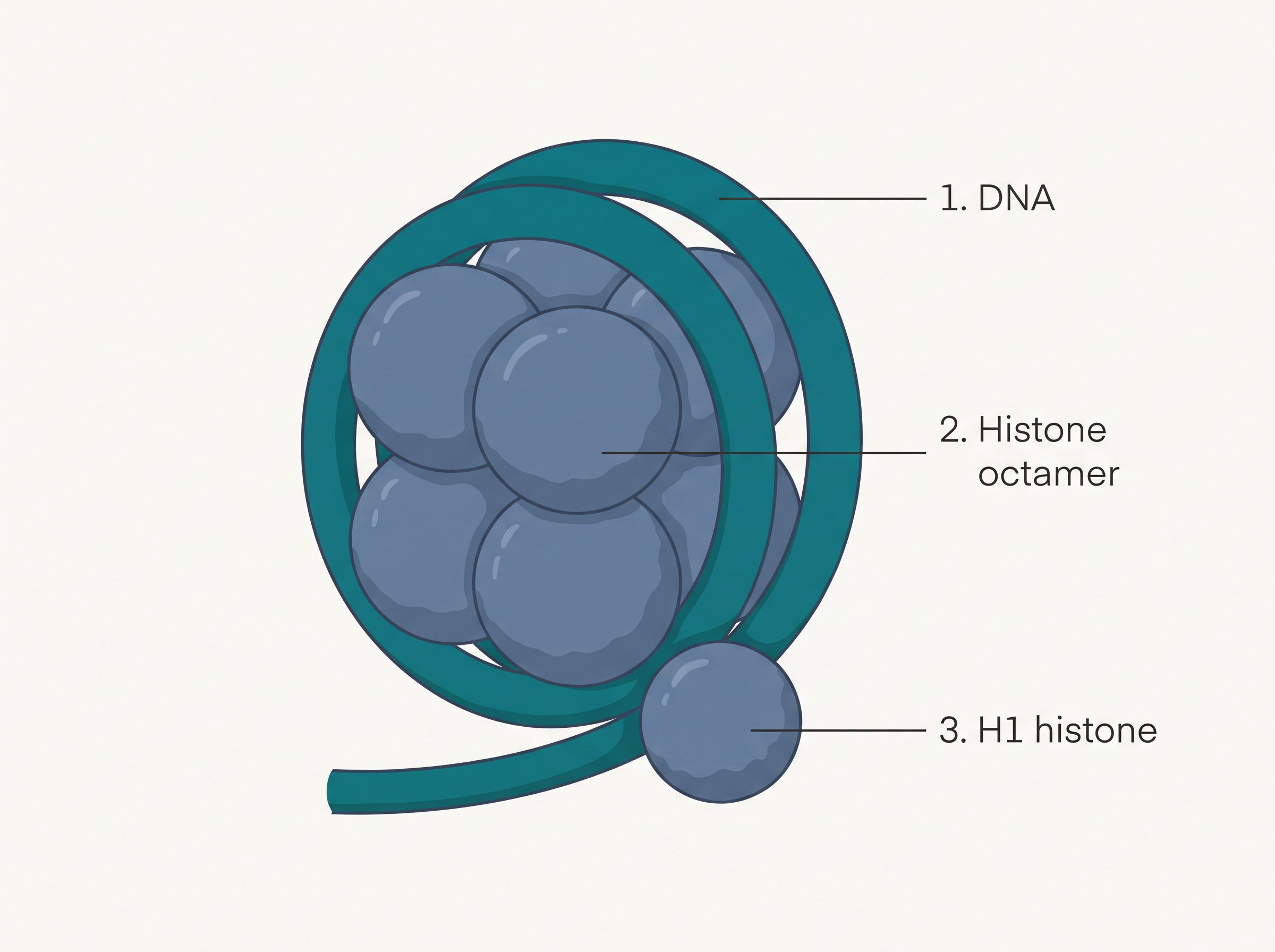
Fig. 5.4a – Structure of a nucleosome.
Nucleosomes are the repeating units of a thread-like, stainable material in the nucleus called chromatin. Under an electron microscope (EM), the nucleosomes along the chromatin look like beads threaded on a string, as seen in Fig. 5.4b.
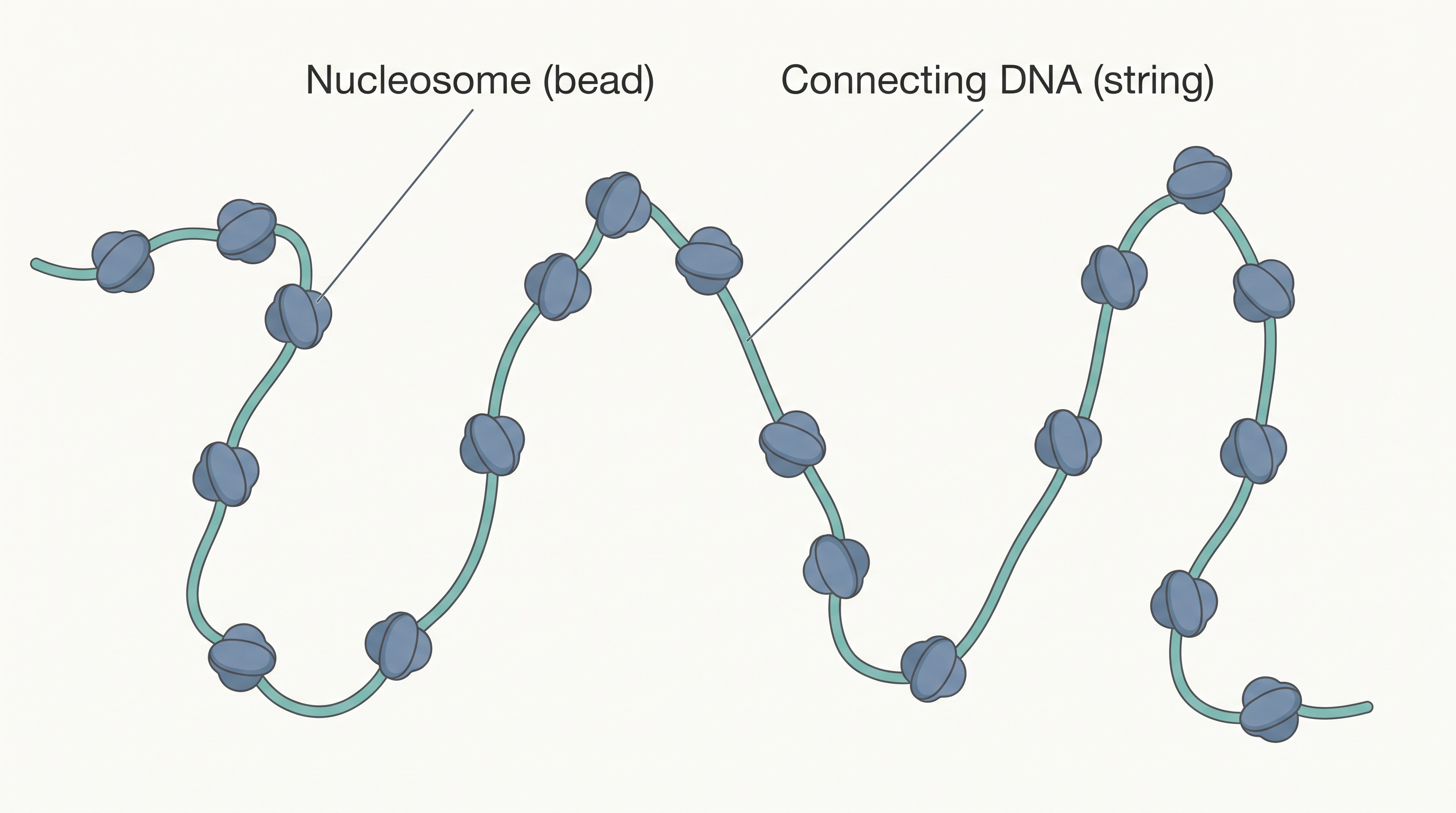
Fig. 5.4b – Beads-on-string appearance of chromatin.
From Chromatin to Chromosome
The beads-on-string chromatin is coiled and folded still further to form chromatin fibres. During the metaphase stage of cell division, these fibres become tightly coiled and condensed to form chromosomes. This step-by-step compaction is summarised in FC 5.2.
Flowchart
FC 5.2 – DNA packaging hierarchy: DNA double helix → wraps around histone octamer to form nucleosome → nucleosomes form ‘beads-on-string’ chromatin → chromatin coils into chromatin fibre → condenses at metaphase into chromosome.
Packing chromatin at these higher levels needs an extra set of proteins, referred to collectively as Non-histone Chromosomal (NHC) proteins.
Within a typical nucleus, chromatin is not packed evenly everywhere. Some regions are loosely packed and stain lightly; these are called euchromatin. Other regions are more densely packed and stain darkly; these are called heterochromatin. Importantly, euchromatin is transcriptionally active (its genes can be read to make RNA), whereas heterochromatin is inactive.
5.2 The Search for Genetic Material
By the late nineteenth century, two great ideas had appeared at almost the same time. Miescher had found nuclein in the nucleus, and Gregor Mendel had worked out the principles of inheritance you studied in the previous chapter. Yet the two ideas were not yet connected. Nobody had shown that nuclein — that is, DNA — was actually the material that carries inheritance. That proof took a surprisingly long time to arrive.
By around 1926, the search for the mechanism of inheritance had reached the level of molecules. The work of Gregor Mendel, Walter Sutton, Thomas Hunt Morgan and many others had already narrowed the hunt down to the chromosomes inside the nucleus of most cells. But one crucial question remained unanswered: which molecule on the chromosome was actually the genetic material — was it protein, or was it DNA?
Transforming Principle
In 1928, Frederick Griffith carried out a series of experiments with the bacterium Streptococcus pneumoniae, which causes pneumonia. During these experiments he witnessed something remarkable — a living bacterium seemed to change its very form, a change we now call transformation.
When Streptococcus pneumoniae is grown on a culture plate, some bacteria form smooth, shiny colonies (the S strain) while others form rough colonies (the R strain). The difference is a coat: S-strain bacteria have a mucous coat made of polysaccharide (a sugar polymer), while R-strain bacteria do not. This coat matters, because the S strain is virulent — able to cause disease. Mice injected with the S strain die of pneumonia, but mice injected with the R strain survive.
Griffith could kill the bacteria by heating them. He found that when heat-killed S-strain bacteria were injected into mice, the mice did not die. But when he injected a mixture of heat-killed S bacteria and living R bacteria, the mice died — and, astonishingly, he recovered living S bacteria from the dead mice. The whole set of experiments is summarised in FC 5.3.
Flowchart
FC 5.3 – Griffith’s transformation experiment: (1) live S strain → inject mice → mice die; (2) live R strain → inject mice → mice live; (3) heat-killed S strain → inject mice → mice live; (4) heat-killed S strain + live R strain → inject mice → mice die, and living S bacteria recovered.
Griffith concluded that the R-strain bacteria had somehow been transformed by the heat-killed S strain. Some “transforming principle,” passed from the dead S bacteria, had enabled the living R strain to build a smooth polysaccharide coat and turn virulent. This could only mean that genetic material had been transferred from the dead cells to the living ones. However, his experiments did not reveal the chemical nature of that transforming principle.
Biochemical Characterisation of Transforming Principle
Before the work of Oswald Avery, Colin MacLeod and Maclyn McCarty (carried out between 1933 and 1944), most scientists believed the genetic material was a protein. These three researchers set out to find the actual chemical identity of Griffith’s “transforming principle.”
Their approach was direct. They purified different biochemicals — proteins, DNA, RNA and others — from heat-killed S cells, and tested which of them could transform living R cells into S cells. They discovered that DNA alone, taken from the S bacteria, could cause the R bacteria to transform.
They then used enzymes as precise tools. Protein-digesting enzymes (proteases) and RNA-digesting enzymes (RNases) did not stop transformation — so the transforming substance was neither a protein nor RNA. But digestion with DNase (a DNA-digesting enzyme) did stop transformation. This pointed clearly to DNA as the cause of transformation. They concluded that DNA is the hereditary material — though, at the time, not all biologists were convinced.
5.2.1 The Genetic Material is DNA
The unquestionable proof that DNA is the genetic material came from the experiments of Alfred Hershey and Martha Chase in 1952. They worked with bacteriophages — viruses that infect bacteria (the word means “bacteria-eaters”).
When a bacteriophage attaches to a bacterium, its genetic material enters the bacterial cell. The bacterial cell then treats this incoming viral genetic material as if it were its own, and starts manufacturing many new virus particles. Hershey and Chase asked a sharp question: when the phage infects the bacterium, is it the protein or the DNA of the virus that actually enters the cell?
To answer this, they used radioactive labels. A radioactive isotope is a form of an element that gives off radiation and can therefore be tracked. They grew one batch of viruses on a medium containing radioactive phosphorus, and another batch on a medium containing radioactive sulfur. This choice was clever and rested on one key chemical fact: DNA contains phosphorus but no sulfur, while protein contains sulfur but no phosphorus. So viruses grown with radioactive phosphorus had radioactive DNA (but not radioactive protein), and viruses grown with radioactive sulfur had radioactive protein (but not radioactive DNA).
The radioactive phages were allowed to attach to E. coli bacteria. As infection proceeded, the empty viral coats were shaken off the bacteria by whirling them in a blender, and the virus particles were then separated from the bacteria by spinning in a centrifuge. The full sequence of steps is shown in FC 5.4.
Flowchart
FC 5.4 – Hershey–Chase procedure: grow phages on radioactive ³²P (labels DNA) or radioactive ³⁵S (labels protein) → allow phages to infect E. coli → agitate in a blender to detach viral coats → centrifuge to separate bacteria from viral coats → measure radioactivity in bacteria vs supernatant.
The results were decisive. Bacteria infected with viruses that had radioactive DNA became radioactive, showing that the DNA had passed from the virus into the bacteria. But bacteria infected with viruses that had radioactive protein were not radioactive, showing that the viral protein had stayed outside and never entered. DNA, therefore, is the genetic material that is passed from the virus to the bacterium, as illustrated in Fig. 5.5.
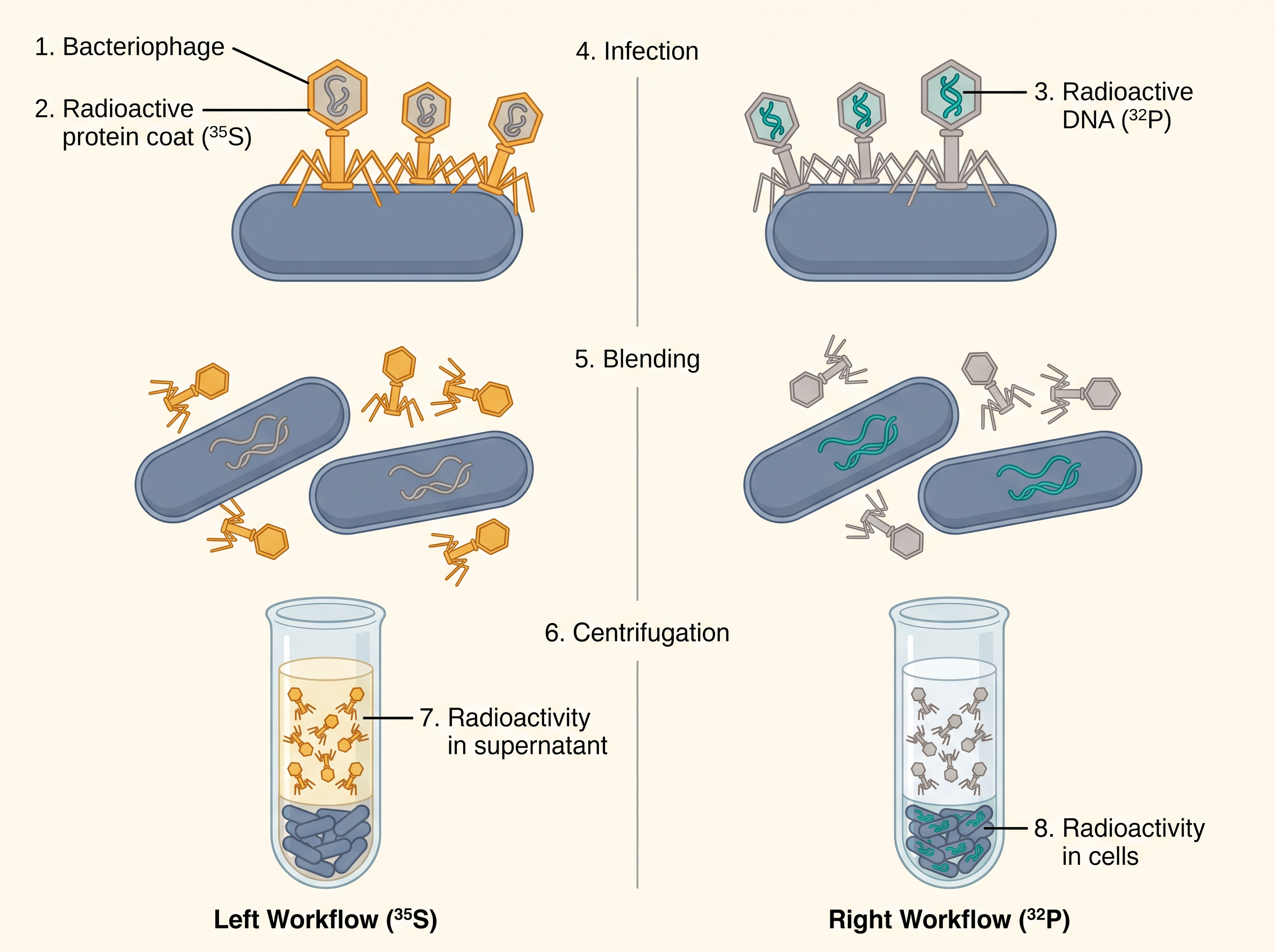
Fig. 5.5 – The Hershey–Chase experiment.
5.2.2 Properties of Genetic Material (DNA versus RNA)
From the discussion above, it is clear that the long debate — protein versus DNA — was firmly settled by the Hershey–Chase experiment. It became an established fact that DNA acts as the genetic material.
However, it later became clear that in some viruses RNA, not DNA, is the genetic material. Examples include the Tobacco Mosaic virus and the QB bacteriophage. This raises a deeper question: why is DNA the predominant genetic material, while RNA more often performs the dynamic jobs of messenger and adapter? The answer lies in the chemical differences between the two nucleic acids, which you met in Section 5.1.
A molecule that is to serve as genetic material must satisfy four conditions:
It should be able to make a copy of itself (replication).
It should be chemically and structurally stable.
It should allow slow changes (mutations) that are needed for evolution.
It should be able to express itself in the form of “Mendelian characters” — that is, it must control observable traits.
Let us examine each condition in turn.
Can It Replicate?
Because of the rule of base pairing and complementarity, both DNA and RNA can direct their own duplication — each strand can guide the making of a matching new strand. Other molecules in the cell, such as proteins, fail at this very first condition, since they cannot copy themselves.
Is It Stable?
The genetic material must be stable enough not to change with the different stages of an organism’s life cycle, with age, or with changes in its physiology. Griffith’s transforming principle already hinted at this stability: heat killed the bacteria, yet it did not destroy the genetic properties they carried.
This is now easily explained by the structure of DNA. When the two complementary strands are separated by heat, they can come back together and pair up again once suitable conditions are provided. Furthermore, the –OH group at the 2′ position of every nucleotide in RNA is a reactive group. It makes RNA labile — easily broken down. RNA is also known to be catalytic, and therefore reactive. DNA, by contrast, is chemically less reactive and structurally more stable. Among the two nucleic acids, DNA is the better genetic material.
The presence of thymine in DNA (in place of RNA’s uracil) also adds extra stability to DNA. The detailed reason involves the process of DNA repair, which you will study in higher classes.
Can It Mutate?
Both DNA and RNA can undergo mutation. However, because RNA is unstable, it mutates at a faster rate. As a result, viruses that carry an RNA genome and have a short life span tend to mutate and evolve more quickly than others.
Can It Express Itself?
RNA can directly serve as a template for making proteins, so it can express characters easily. DNA, on the other hand, depends on RNA to make proteins — the entire protein-synthesising machinery of the cell has evolved around RNA.
Putting all of this together: both RNA and DNA can function as genetic material, but because DNA is more stable, it is preferred for the long-term storage of genetic information. RNA, being more suited to quick action, is better for the transmission and expression of that information.
5.3 RNA World
Once we accept that DNA is the genetic material and that RNA came first in evolution, an obvious question follows: which of the two was the very first genetic material of life? This question is taken up in detail in the chapter on chemical evolution, so here we shall only highlight a few key facts and ideas.
The answer, based on the evidence we have today, is that RNA was the first genetic material. There is now enough evidence to suggest that essential life processes — such as metabolism, translation and splicing — all evolved around RNA. (Metabolism is the sum of the chemical reactions that keep a cell alive; translation is the making of protein; splicing is a way of processing RNA that you will meet later in this chapter.)
In those early times, RNA played a double role. It acted as the genetic material, and it also acted as a catalyst — a molecule that speeds up a chemical reaction. In fact, there are still some important biochemical reactions in living systems that are carried out by RNA catalysts rather than by protein enzymes.
Why DNA Replaced RNA
Being a catalyst made RNA reactive, and being reactive made it unstable. For long-term storage of genetic information, this was a serious weakness. Over evolutionary time, therefore, DNA evolved from RNA through chemical modifications that made it more stable.
DNA also gained a further advantage. Because it is double-stranded, each of its two strands is a complementary copy of the other. This allowed cells to evolve a process of repair: if one strand is damaged, the intact complementary strand can be used as a template to correct it.
Flowchart
FC 5.5 – The RNA-to-DNA evolutionary story: RNA (acts as genetic material + catalyst) → catalytic activity makes RNA reactive and unstable → DNA evolves from RNA with stabilising chemical changes → DNA becomes double-stranded and gains repair → stable long-term storage of information.
5.4 Replication
While proposing the double-helical structure of DNA, Watson and Crick did not stop at structure alone — they immediately suggested how DNA might copy itself. In their original 1953 paper, they famously remarked that the specific base pairing they had proposed at once hinted at a possible mechanism for copying the genetic material. In other words, the very shape of DNA suggested how it could be duplicated.
Their scheme was simple and elegant. The two strands of the double helix would separate, and each old strand would act as a template for building a new complementary strand. After copying is complete, every DNA molecule would carry one parental (old) strand and one newly synthesised strand. Because each daughter molecule keeps (“conserves”) one of the two original strands, this scheme was named semiconservative DNA replication, shown in Fig. 5.6.
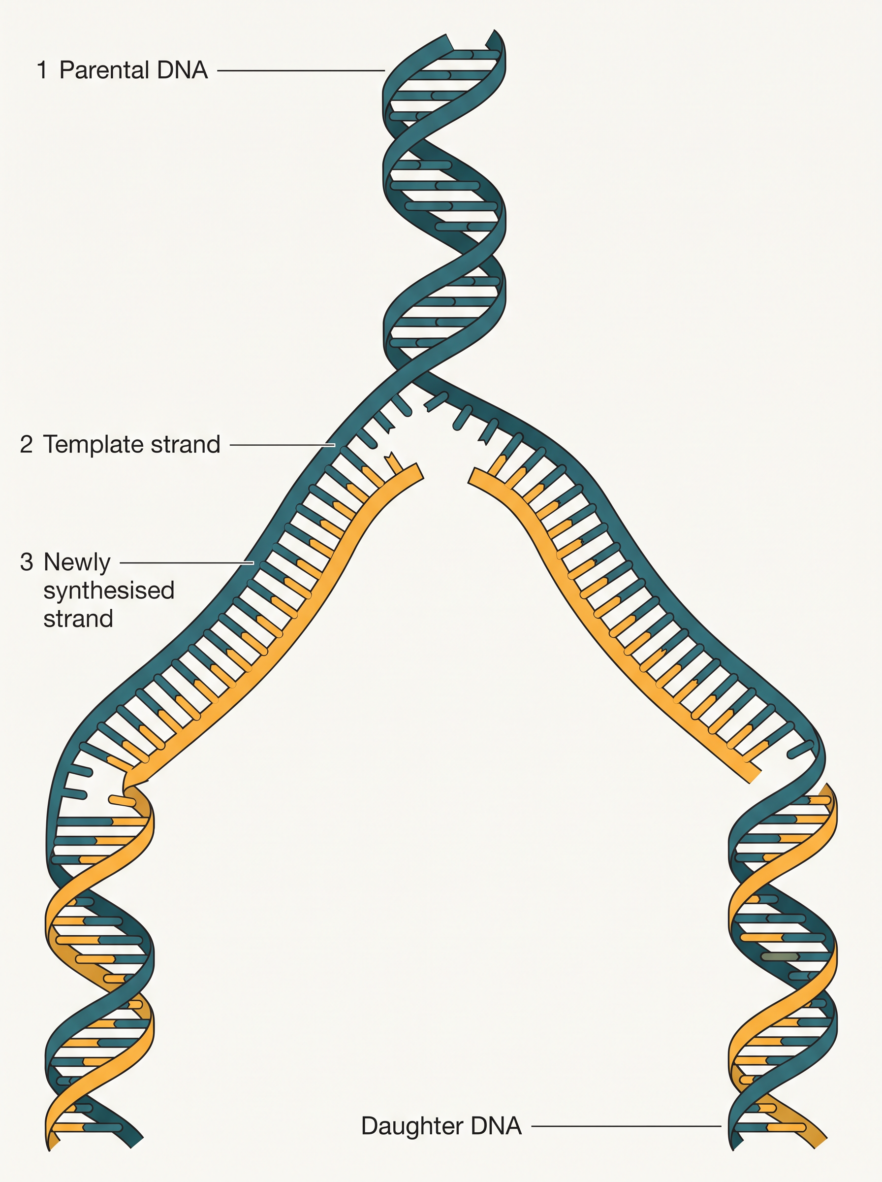
Fig. 5.6 – Semiconservative DNA replication.
5.4.1 The Experimental Proof
It is now firmly proven that DNA replicates semiconservatively. This was shown first in Escherichia coli, and afterwards in higher organisms such as plants and human cells.
The Meselson–Stahl Experiment
Matthew Meselson and Franklin Stahl performed a landmark experiment in 1958 to test whether replication is really semiconservative.
First, they grew E. coli for many generations in a medium containing \(^{15}NH_4Cl\), in which \(^{15}N\) (a heavy isotope of nitrogen) was the only nitrogen source. Over these generations, the \(^{15}N\) became built into the newly made DNA, making the DNA “heavy.” This heavy DNA could be told apart from ordinary DNA by spinning it in a cesium chloride (CsCl) density gradient, where molecules settle according to their density. (Note that \(^{15}N\) is not radioactive; it is separated from ordinary \(^{14}N\) only on the basis of density.)
Next, they shifted the cells into a medium containing normal \(^{14}NH_4Cl\). At definite time intervals, as the cells kept dividing, they took samples and extracted the DNA that remained as double-stranded helices. Each sample was run on a CsCl gradient to measure its density, as shown in Fig. 5.7.
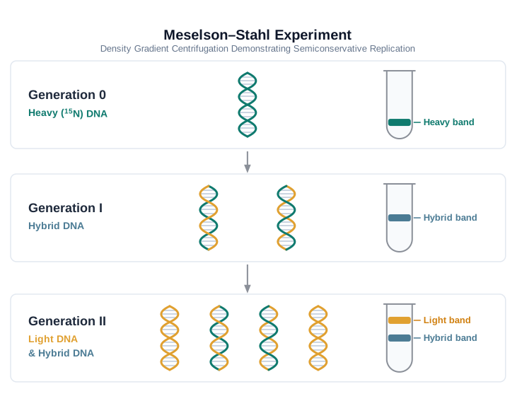
Fig. 5.7 – The Meselson–Stahl experiment.
The results were exactly what semiconservative replication predicts. DNA extracted one generation after the shift from \(^{15}N\) to \(^{14}N\) medium — that is, after 20 minutes, since E. coli divides every 20 minutes — had a hybrid (intermediate) density. DNA extracted after one more generation — after 40 minutes, the second generation — was made of equal amounts of this hybrid DNA and of light DNA.
Confirmation in Plants
Very similar experiments, this time using radioactive thymidine to track newly made DNA in the chromosomes, were carried out on Vicia faba (faba beans) by Taylor and colleagues, also in 1958. These experiments proved that the DNA in chromosomes, too, replicates semiconservatively.
5.4.2 The Machinery and the Enzymes
The Main Enzyme: DNA Polymerase
In living cells such as E. coli, replication needs a set of catalysts (enzymes). The main enzyme is called DNA-dependent DNA polymerase, because it uses a DNA template to catalyse the polymerisation (joining together) of deoxynucleotides into a new strand.
These enzymes are remarkably efficient, because they must join a huge number of nucleotides in a very short time. Consider that E. coli has about \(4.6 \times 10^6\) bp (compare this with a human, whose diploid content is about \(6.6 \times 10^9\) bp), yet it completes replication within 18 minutes. That works out to an average rate of roughly \(2000\) bp per second.
Speed alone is not enough — the enzyme must also work with very high accuracy. Any mistake made during replication would result in a mutation. In addition, replication is energetically very expensive. The deoxyribonucleoside triphosphates (dNTPs) used in the process serve a dual purpose: they act as the raw materials (substrates) for the new strand, and they also supply the energy for the polymerisation reaction. This is because the two terminal phosphates in a deoxyribonucleoside triphosphate are high-energy phosphates, just as they are in ATP.
The Replication Fork
Besides DNA polymerase, many additional enzymes are needed to finish replication accurately. For a long DNA molecule, the two strands cannot be pulled apart along their entire length at once, because that would need enormous energy. Instead, replication takes place within a small opening in the helix called the replication fork, which moves along as copying proceeds.
Now a complication arises. DNA-dependent DNA polymerase can add nucleotides in only one direction, that is \(5′ \rightarrow 3′\). Because the two template strands run in opposite directions, this one-way rule affects the two strands differently, as illustrated in Fig. 5.8.
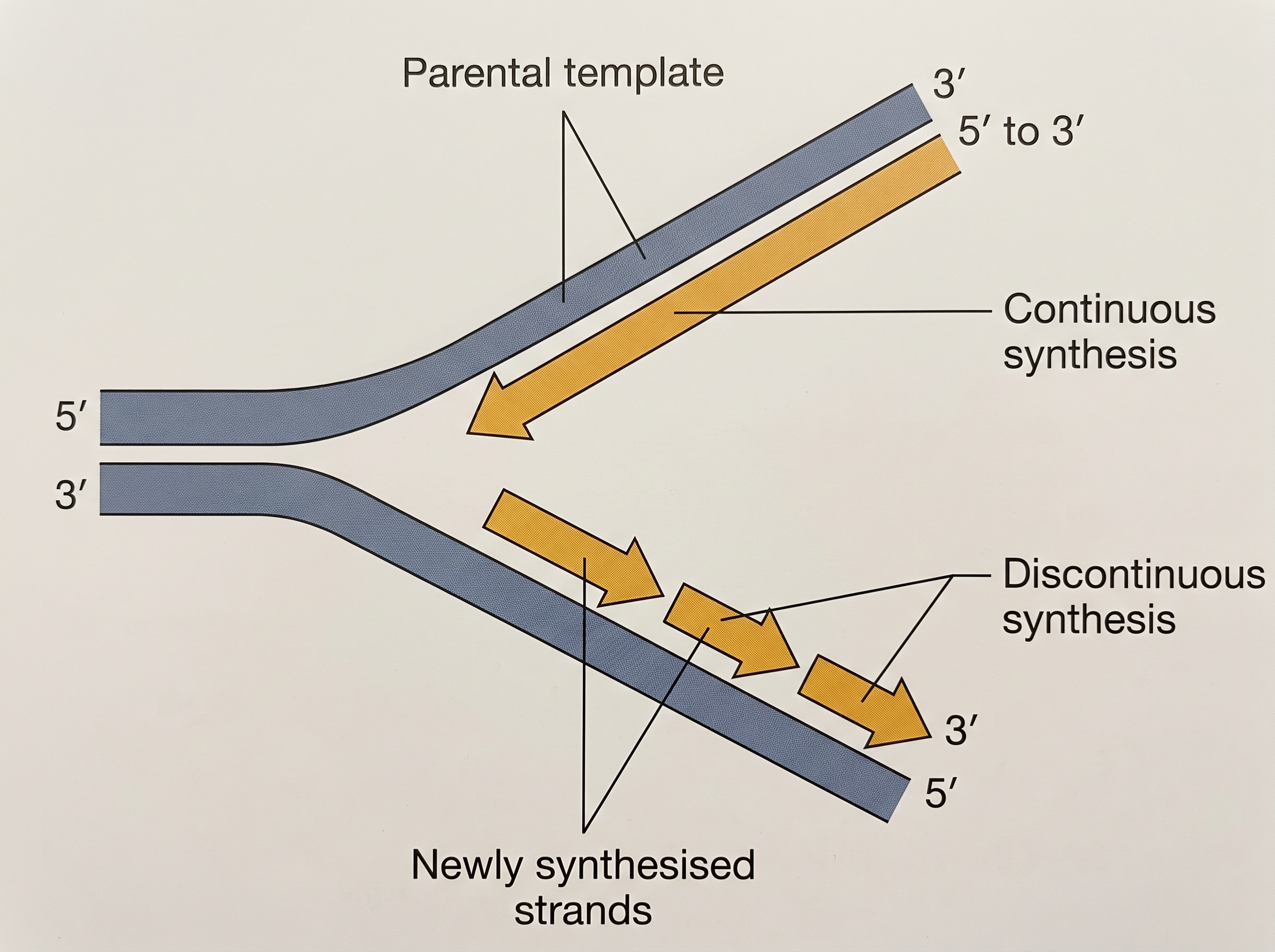
Fig. 5.8 – A DNA replication fork.
On the template strand that has the polarity \(3′ \rightarrow 5′\), the new strand can be made smoothly and without a break — this is continuous replication. On the other template, which has the polarity \(5′ \rightarrow 3′\), the new strand has to be made in short pieces — this is discontinuous replication. The short, discontinuously made fragments are later joined together by the enzyme DNA ligase.
Where Replication Begins
DNA polymerases cannot start replication on their own, and replication does not begin at random points along the DNA. There is a definite region in the E. coli DNA where replication starts, called the origin of replication.
This origin has a very practical importance in the laboratory. In recombinant DNA work, if a piece of DNA is to be multiplied inside a cell, it needs a carrier called a vector — and it is the vector that provides the origin of replication.
Replication in Eukaryotes
Not every detail of replication is understood well. In eukaryotes, the replication of DNA takes place during the S-phase of the cell cycle. The copying of DNA and the cell-division cycle must be tightly coordinated with each other. If a cell fails to divide after its DNA has been replicated, the result is polyploidy — a chromosomal anomaly in which the cell ends up with extra sets of chromosomes. You will learn the detailed nature of the origin and the events happening there in higher classes.
5.5 Transcription
Transcription is the process of copying genetic information from one strand of DNA into RNA. Here too, the principle of complementarity governs the process — with one small change. Now adenine (adenosine) pairs with uracil instead of thymine, because RNA uses uracil in place of thymine.
There is an important difference from replication. In replication, once the process begins, the entire DNA of the organism gets duplicated. In transcription, only a segment of the DNA — and only one of its two strands — is copied into RNA. Because of this selectivity, the cell must clearly mark the boundaries: which region of DNA, and which strand, is to be transcribed.
Why Only One Strand Is Copied
Why are both strands not copied during transcription? There are two clear reasons.
First, if both strands acted as templates, they would produce two RNA molecules with different sequences (remember, complementary does not mean identical). If these then coded for proteins, the amino acid sequences would differ. One segment of DNA would end up coding for two different proteins, which would badly complicate the machinery that transfers genetic information.
Second, if two RNA molecules were produced at the same time from the two strands, they would be complementary to each other. They would pair up to form a double-stranded RNA. This would stop the RNA from being translated into protein, and the whole exercise of transcription would be wasted.
5.5.1 Transcription Unit
A transcription unit in DNA is defined mainly by three regions:
a Promoter,
the Structural gene, and
a Terminator.
Template and Coding Strands
There is a fixed convention for naming the two DNA strands within the structural gene of a transcription unit. Since the two strands have opposite polarity, and DNA-dependent RNA polymerase (the enzyme that makes RNA) can add nucleotides only in the \(5′ \rightarrow 3′\) direction, only one strand can serve as the template.
The strand with polarity \(3′ \rightarrow 5′\) acts as the template, and is therefore called the template strand. The other strand, with polarity \(5′ \rightarrow 3′\), has the same sequence as the RNA (except that it carries thymine where RNA has uracil). This strand is displaced during transcription and is not copied — yet, oddly, it is called the coding strand. By convention, all reference points of a transcription unit are described using the coding strand.
To make this concrete, here is a hypothetical stretch of a transcription unit:
\[3′\text{-ATGCATGCATGCATGCATGCATGC-}5′ \quad \text{Template Strand}\] \[5′\text{-TACGTACGTACGTACGTACGTACG-}3′ \quad \text{Coding Strand}\]
Promoter and Terminator
The promoter and the terminator flank the structural gene in a transcription unit, as shown in Fig. 5.9.
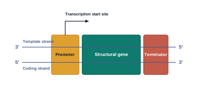
Fig. 5.9 – Schematic of a transcription unit.
The promoter lies towards the 5′-end (upstream) of the structural gene (the reference being the polarity of the coding strand). It is a DNA sequence that provides the binding site for RNA polymerase. Importantly, the very presence of a promoter is what defines which strand is the template and which is the coding strand. If the promoter and terminator were to swap positions, the definitions of coding and template strands would be reversed.
The terminator lies towards the 3′-end (downstream) of the coding strand and usually marks the end of transcription. There are also additional regulatory sequences that may lie further upstream or downstream of the promoter; some of their properties are discussed under the regulation of gene expression.
5.5.2 Transcription Unit and the Gene
A gene is defined as the functional unit of inheritance. Although there is no doubt that genes are located on the DNA, it is surprisingly difficult to define a gene strictly in terms of a DNA sequence. For instance, a DNA sequence that codes for a tRNA or an rRNA molecule also counts as a gene, even though it does not code for a protein.
Cistron: Monocistronic and Polycistronic
If we define a cistron as a segment of DNA coding for one polypeptide, then the structural gene in a transcription unit can be described as monocistronic (mostly in eukaryotes) or polycistronic (mostly in bacteria or prokaryotes).
Split Genes: Exons and Introns
In eukaryotes, the monocistronic structural genes have interrupted coding sequences — that is, the genes are split. The coding or expressed sequences are called exons; these are the parts that appear in the mature (processed) RNA. The exons are interrupted by introns (intervening sequences), which do not appear in the mature RNA. This split-gene arrangement makes it even harder to define a gene neatly in terms of a DNA segment.
Regulatory Sequences
The inheritance of a character is also influenced by the promoter and regulatory sequences of a structural gene. For this reason, these regulatory sequences are sometimes loosely called regulatory genes, even though they do not themselves code for any RNA or protein.
5.5.3 Types of RNA and the process of Transcription
The Three Types of RNA
In bacteria, there are three major types of RNA: mRNA (messenger RNA), tRNA (transfer RNA) and rRNA (ribosomal RNA). All three are needed to make a protein in the cell. The mRNA provides the template (the message to be read); the tRNA brings amino acids and reads the genetic code; and the rRNAs play a structural and catalytic role during translation.
The Three Steps of Transcription
In bacteria, a single DNA-dependent RNA polymerase carries out the transcription of all types of RNA. The process happens in three steps, summarised in FC 5.6.
Flowchart
FC 5.6 – Transcription in bacteria: Initiation (RNA polymerase + sigma factor bind the promoter) → Elongation (polymerase opens the helix and adds ribonucleotides 5′→3′ following complementarity) → Termination (polymerase reaches the terminator; with the rho factor, the newly made RNA and the polymerase fall off).
RNA polymerase first binds to the promoter and starts transcription — this is Initiation. It uses nucleoside triphosphates as its substrate and builds RNA in a template-dependent manner, following the base-pairing rules. It also helps open up the DNA helix and continues to add nucleotides — this is Elongation. Only a short stretch of the newly made RNA stays attached to the enzyme at any time. When the polymerase reaches the terminator region, the newly formed (nascent) RNA falls off, and so does the RNA polymerase. This is Termination.
An interesting question is how a single RNA polymerase manages all three steps — initiation, elongation and termination. The answer is that the RNA polymerase, on its own, is really only good at elongation. To start and stop, it borrows help. It associates briefly with an initiation-factor (σ, the sigma factor) to initiate, and with a termination-factor (ρ, the rho factor) to terminate. Associating with these factors changes the specificity of the polymerase so that it either initiates or terminates, as shown in Fig. 5.10.
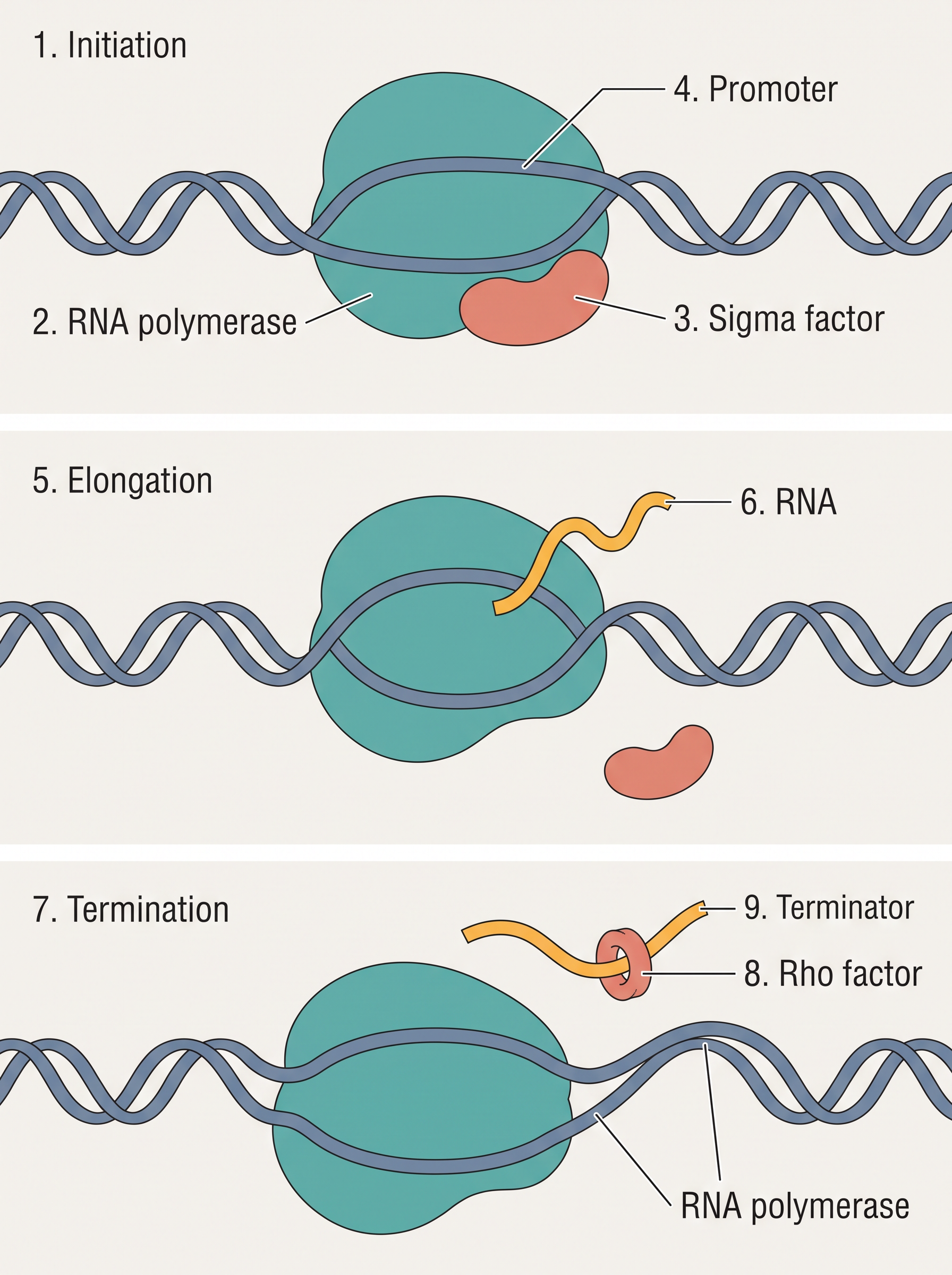
Fig. 5.10 – Process of transcription in bacteria.
Coupled Transcription and Translation in Bacteria
In bacteria, the mRNA does not need any processing to become active. Also, because there is no separation of cytosol and nucleus in bacteria, transcription and translation take place in the same compartment. As a result, translation can often begin even before the mRNA is fully transcribed. In this way, transcription and translation can be coupled in bacteria.
Transcription in Eukaryotes
In eukaryotes, there are two additional complexities.
The first is that there are at least three RNA polymerases in the nucleus (in addition to the RNA polymerase found in organelles), and there is a clear division of labour among them. RNA polymerase I transcribes the rRNAs (28S, 18S and 5.8S). RNA polymerase III is responsible for tRNA, 5srRNA, and snRNAs (small nuclear RNAs). RNA polymerase II transcribes the precursor of mRNA, the heterogeneous nuclear RNA (hnRNA).
Splicing, Capping and Tailing
The second complexity is that the primary transcripts in eukaryotes contain both the exons and the introns and are non-functional. So they must be processed. In splicing, the introns are removed and the exons are joined together in a defined order.
The hnRNA also undergoes two more changes, called capping and tailing. In capping, an unusual nucleotide (methyl guanosine triphosphate) is added to the 5′-end of the hnRNA. In tailing, a string of adenylate residues (200–300 of them) is added at the 3′-end in a template-independent manner. It is this fully processed hnRNA, now called mRNA, that is transported out of the nucleus for translation, as shown in Fig. 5.11. The full sequence of eukaryotic processing is summarised in FC 5.7.
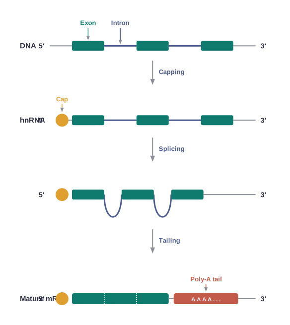
Fig. 5.11 – Transcription and processing in eukaryotes.
Flowchart
FC 5.7 – Eukaryotic mRNA processing: primary transcript / hnRNA (contains exons + introns) → splicing (introns removed, exons joined) → capping (methyl guanosine triphosphate added at 5′-end) → tailing (200–300 adenylate residues added at 3′-end) → mature mRNA → transported out of nucleus for translation.
The significance of these complexities is only now being understood. The split-gene arrangement is probably an ancient feature of the genome. The presence of introns is a reminder of very early life, and the process of splicing reflects the past dominance of the RNA-world. In recent times, the study of RNA and RNA-dependent processes has gained ever greater importance.
5.6 Genetic Code
During replication and transcription, one nucleic acid was copied to make another nucleic acid. Both processes are easy to picture, because they rest on the simple idea of complementarity — base pairing with base.
Translation is different, and harder. Translation requires transferring information from a polymer of nucleotides (the mRNA) into a polymer of amino acids (the protein). But here there is no complementarity between nucleotides and amino acids, and none could even be imagined in theory — a base cannot “pair” with an amino acid. Even so, there was plenty of evidence that changes in the nucleic acid (the genetic material) led to changes in the amino acids of proteins. This led scientists to propose a genetic code — a set of rules by which the nucleotide sequence directs the amino acid sequence during protein synthesis.
Cracking the Code
If working out the biochemical nature of the genetic material and the structure of DNA was exciting, then proposing and deciphering the genetic code was the most challenging task of all. It genuinely needed scientists from many fields — physicists, organic chemists, biochemists and geneticists.
It was George Gamow, a physicist, who reasoned that since there are only 4 bases and they must code for 20 amino acids, the code must use a combination of bases. He suggested that the code should be made of three nucleotides. This was a bold idea, because a code of three bases gives \(4^3 = 4 \times 4 \times 4 = 64\) possible codons — far more than the 20 needed.
Proving that the codon is actually a triplet was even more daunting. The chemical method developed by Har Gobind Khorana was instrumental in making RNA molecules with defined base combinations (homopolymers and copolymers). Marshall Nirenberg’s cell-free system for protein synthesis finally allowed the code to be deciphered. The enzyme discovered by Severo Ochoa (polynucleotide phosphorylase) was also helpful, as it could join ribonucleotides into RNA of defined sequence in a template-independent manner. Piecing it all together, a “checkerboard” of the genetic code was drawn up, given in Table 5.1.
The Codon Table
Table 5.1 lists all 64 codons and the amino acids they specify.
Table 5.1: The Codons for the Various Amino Acids
| 1st base | 2nd = U | 2nd = C | 2nd = A | 2nd = G | 3rd base |
|---|---|---|---|---|---|
| U | UUU Phe | UCU Ser | UAU Tyr | UGU Cys | U |
| U | UUC Phe | UCC Ser | UAC Tyr | UGC Cys | C |
| U | UUA Leu | UCA Ser | UAA Stop | UGA Stop | A |
| U | UUG Leu | UCG Ser | UAG Stop | UGG Trp | G |
| C | CUU Leu | CCU Pro | CAU His | CGU Arg | U |
| C | CUC Leu | CCC Pro | CAC His | CGC Arg | C |
| C | CUA Leu | CCA Pro | CAA Gln | CGA Arg | A |
| C | CUG Leu | CCG Pro | CAG Gln | CGG Arg | G |
| A | AUU Ile | ACU Thr | AAU Asn | AGU Ser | U |
| A | AUC Ile | ACC Thr | AAC Asn | AGC Ser | C |
| A | AUA Ile | ACA Thr | AAA Lys | AGA Arg | A |
| A | AUG Met | ACG Thr | AAG Lys | AGG Arg | G |
| G | GUU Val | GCU Ala | GAU Asp | GGU Gly | U |
| G | GUC Val | GCC Ala | GAC Asp | GGC Gly | C |
| G | GUA Val | GCA Ala | GAA Glu | GGA Gly | A |
| G | GUG Val | GCG Ala | GAG Glu | GGG Gly | G |
Features of the Genetic Code
The genetic code has these salient features:
The codon is a triplet. Of the 64 codons, 61 code for amino acids, and 3 do not code for any amino acid — these function as stop codons.
Some amino acids are coded by more than one codon; because of this, the code is said to be degenerate.
The codon is read on the mRNA in a continuous, contiguous fashion. There are no punctuations (no gaps or markers between codons).
The code is nearly universal. For example, from bacteria to humans, UUU codes for phenylalanine (Phe). Some exceptions to this rule are found in mitochondrial codons and in some protozoans.
AUG has dual functions. It codes for methionine (Met), and it also acts as the initiator codon (the “start” signal).
UAA, UAG and UGA are the stop (terminator) codons.
5.6.1 Mutations and Genetic Code
The relationship between genes and DNA is best understood through mutation studies. (You studied mutation and its effects in Chapter 4.) The effects of large deletions and rearrangements in a segment of DNA are easy to grasp — they may cause the loss or gain of a gene, and therefore of a function. Here, we focus on point mutations.
Point Mutations and Sickle Cell Anaemia
A classical example of a point mutation is a change of a single base pair in the gene for the beta-globin chain, which changes one amino acid residue from glutamate to valine. This single change results in the disease known as sickle cell anaemia.
Frameshift Mutations
The effect of point mutations that insert or delete a base can be understood using a simple sentence made of three-letter words, just like the triplet code:
RAM HAS RED CAP
If we insert a letter B between HAS and RED and re-group into three-letter words, it reads:
RAM HAS BRE DCA P
If we insert two letters at the same place, say BI, it now reads:
RAM HAS BIR EDC AP
But if we insert three letters together, say BIG, the sentence reads:
RAM HAS BIG RED CAP
The same can be shown by deleting letters. Deleting R, E and D one by one and re-grouping into three-letter words gives:
RAM HAS EDC AP
RAM HAS DCA P
RAM HAS CAP
The conclusion is clear. Inserting or deleting one or two bases shifts the reading frame from the point of insertion or deletion onwards — such mutations are called frameshift insertion or deletion mutations. In contrast, when three bases (or a multiple of three) are inserted or deleted, this adds or removes one or more whole codons — hence one or more whole amino acids — and the reading frame stays unchanged from that point onwards.
5.6.2 tRNA – the Adapter Molecule
From the very moment the code was proposed, Francis Crick realised there had to be some mechanism to read the code and, at the same time, link it to the correct amino acid. This was necessary because amino acids themselves have no structural features that would let them read the code directly. He therefore postulated an adapter molecule — one that could read the code on one side and bind to a specific amino acid on the other.
That adapter turned out to be the tRNA, which was already known (then called sRNA, or soluble RNA) even before the genetic code was worked out; its role as an adapter was recognised only later.
The tRNA has an anticodon loop, which carries bases complementary to the codon on the mRNA. It also has an amino acid acceptor end, to which the amino acid binds. Each amino acid has its own specific tRNA, as shown in Fig. 5.12. For starting protein synthesis, there is a special initiator tRNA. Notably, there are no tRNAs for the stop codons — which is why the stop codons end translation.
Structure of tRNA
In Fig. 5.12, the secondary structure of tRNA is drawn to look like a clover-leaf. In its actual three-dimensional form, however, tRNA is a compact molecule shaped like an inverted L.
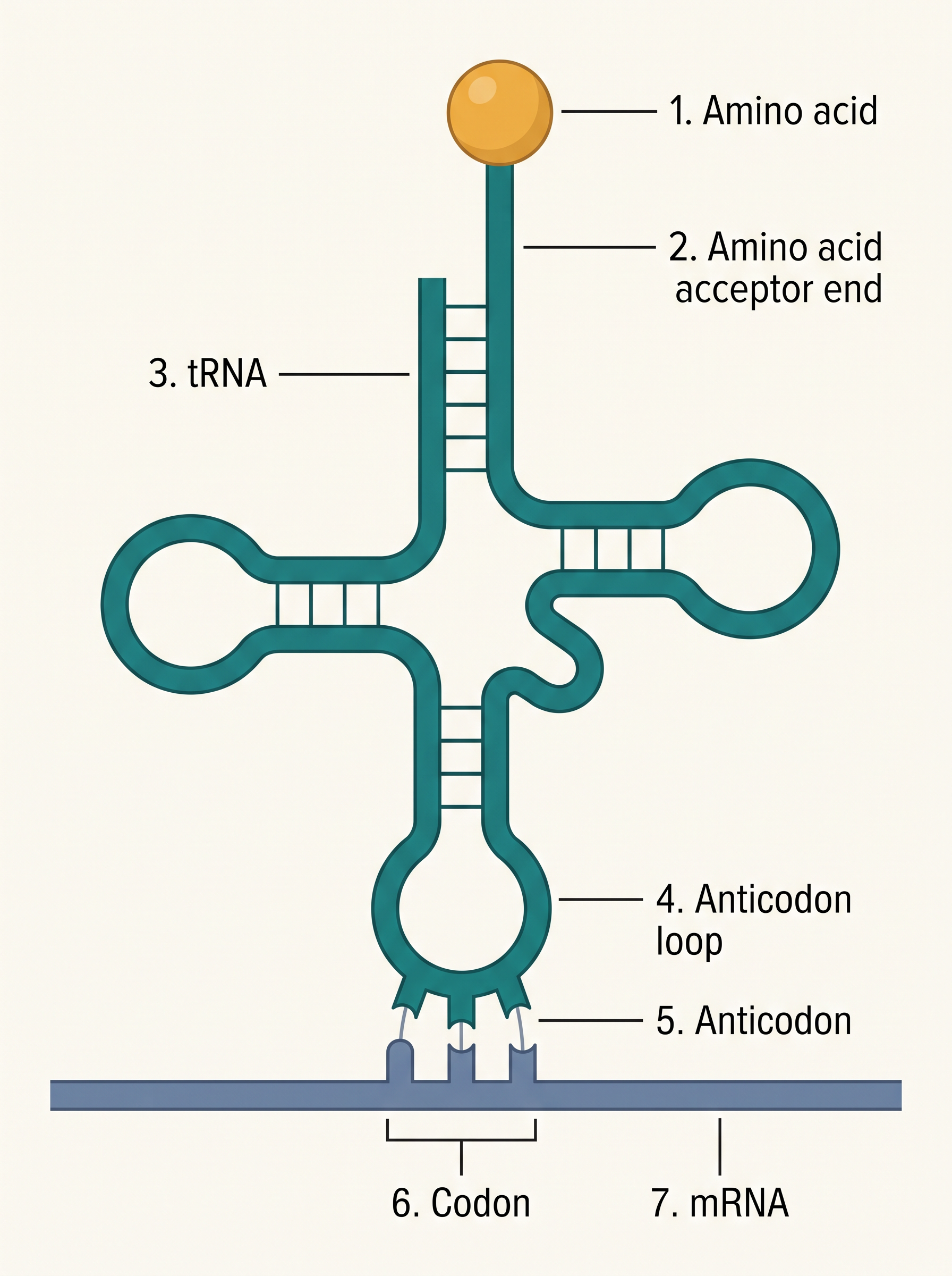
Fig. 5.12 – Structure of the tRNA adapter molecule.
5.7 Translation
Translation is the process of joining amino acids together, one by one, to form a polypeptide, as shown in Fig. 5.13. The order and sequence of these amino acids is decided entirely by the sequence of bases in the mRNA. In other words, the coded message in the mRNA is finally “read out” as a chain of amino acids.
The amino acids in a polypeptide are joined by a bond called the peptide bond (the same bond you met in the chemistry of proteins in Class XI). Forming a peptide bond needs energy. So, in the very first phase, amino acids are activated using ATP and are attached to their matching (cognate) tRNA.
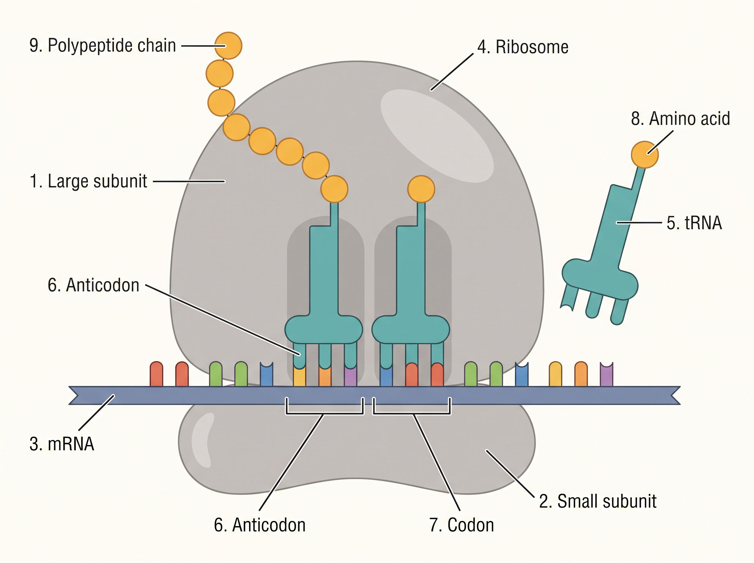
Fig. 5.13 – Translation of mRNA on a ribosome.
Charging of tRNA
This step of activating an amino acid and linking it to its correct tRNA is called charging of tRNA, or more precisely, aminoacylation of tRNA.
The value of charging becomes clear at the next stage. If two such charged tRNAs are brought close enough together, the formation of a peptide bond between their amino acids is favoured energetically. The presence of a catalyst then speeds up this peptide-bond formation.
The Ribosome: the Protein Factory
The cellular factory responsible for making proteins is the ribosome. A ribosome is built from structural RNAs (rRNAs) and about 80 different proteins. In its resting (inactive) state, it exists as two separate subunits — a large subunit and a small subunit.
When the small subunit meets an mRNA, the process of translating that mRNA into protein begins. The large subunit has two sites where incoming amino acids (carried by tRNAs) can bind, so that two amino acids are held close enough to form a peptide bond between them.
Remarkably, the ribosome itself acts as the catalyst for peptide-bond formation. In bacteria, it is the 23S rRNA that does this catalysis — making it an enzyme made of RNA, that is, a ribozyme.
The Steps of Translation
Before translation can start, we must know where to start and stop. A translational unit in the mRNA is the stretch of RNA flanked by the start codon (AUG) and a stop codon, which codes for a polypeptide. Besides this, the mRNA carries extra sequences that are not translated, called untranslated regions (UTRs). UTRs are present at both the 5′-end (before the start codon) and the 3′-end (after the stop codon), and they are needed for efficient translation.
The whole process runs through three stages, summarised in FC 5.8.
Flowchart
FC 5.8 – Translation: charging of tRNA (amino acid + ATP + tRNA) → Initiation (ribosome binds mRNA at start codon AUG, recognised only by initiator tRNA) → Elongation (charged tRNAs pair by anticodon–codon base pairing; ribosome moves codon by codon; amino acids added one by one to the growing polypeptide) → Termination (release factor binds the stop codon; the complete polypeptide is released from the ribosome).
For initiation, the ribosome binds to the mRNA at the start codon (AUG), which is recognised only by the initiator tRNA. The ribosome then moves into the elongation phase of protein synthesis. During elongation, complexes made of an amino acid linked to its tRNA bind, one after another, to the appropriate codon on the mRNA, by forming complementary base pairs between the codon and the tRNA anticodon. The ribosome moves from codon to codon along the mRNA, and amino acids are added one by one, building a polypeptide whose sequence is dictated by the DNA and represented by the mRNA.
Finally, in termination, a release factor binds to the stop codon. This ends translation and releases the completed polypeptide from the ribosome.
5.8 Regulation of Gene Expression
Regulation of gene expression is a broad term. It refers to the many ways in which a cell controls how much, and when, a gene is expressed. Since the expression of a gene finally results in a polypeptide (protein), this control can be applied at several different steps.
Levels of Regulation in Eukaryotes
In eukaryotes, regulation can be exerted at four levels:
at the transcriptional level (controlling the formation of the primary transcript),
at the processing level (controlling splicing),
at the transport of mRNA from the nucleus to the cytoplasm, and
at the translational level.
Why regulate at all? Because genes are expressed only to carry out a particular function. For example, if the enzyme beta-galactosidase is made by E. coli, it is used to break down (hydrolyse) the disaccharide lactose into galactose and glucose, which the bacterium then uses for energy. So if there is no lactose around, the bacterium has no reason to keep making beta-galactosidase. In short, it is the metabolic, physiological or environmental conditions that decide whether a gene is switched on. The very development of an embryo into an adult, too, is the result of switching different sets of genes on and off in a coordinated way.
Regulation in Prokaryotes: Operators and Repressors
In prokaryotes, the main point of control is the rate of transcriptional initiation — that is, how readily transcription is started.
Within a transcription unit, the activity of RNA polymerase at a promoter is itself controlled by accessory proteins that affect whether the polymerase can recognise the start site. These regulatory proteins can act in two ways: positively, as activators (helping transcription), or negatively, as repressors (blocking it).
In many cases, whether the promoter is accessible is decided by proteins binding to a DNA sequence called the operator. The operator usually lies right next to the promoter, and in most cases the operator sequence binds a repressor protein. Each operon has its own specific operator and its own specific repressor. For instance, the lac operator is present only in the lac operon, and it interacts only with the lac repressor.
5.8.1 The Lac operon
The working of the lac operon was worked out through the close teamwork of a geneticist, Francois Jacob, and a biochemist, Jacque Monod. They were the first to explain a transcriptionally regulated system.
In the lac operon (where lac stands for lactose), a polycistronic structural gene is controlled by a common promoter and regulatory genes. This kind of arrangement — several genes under one control unit — is called an operon. A few examples are the lac operon, trp operon, ara operon, his operon and val operon.
Genes of the Lac Operon
The lac operon has one regulatory gene, the i gene (here i stands for inhibitor, not inducer), and three structural genes: z, y and a.
The i gene codes for the repressor of the lac operon. The z gene codes for beta-galactosidase (β-gal), which breaks lactose into its two units, galactose and glucose. The y gene codes for permease, which increases the cell’s permeability to β-galactosides (so lactose can get in). The a gene codes for a transacetylase. All three gene products are needed for the metabolism of lactose. In most operons, similarly, the genes work together in the same or a related metabolic pathway, as shown in Fig. 5.14.
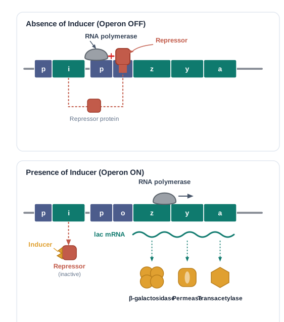
Fig. 5.14 – Regulation of the lac operon.
Lactose as the Inducer
Lactose is the substrate for the enzyme beta-galactosidase, and it controls the switching on and off of the operon. Because of this, lactose is called the inducer.
When there is no preferred carbon source such as glucose, and lactose is provided in the growth medium, the lactose is carried into the cells by permease. (A very low level of lac operon expression must always be present in the cell — otherwise no permease would exist and lactose could never enter in the first place.) Once inside, lactose induces the operon.
How the Operon Is Switched On and Off
The repressor of the operon is made all the time (constitutively) from the i gene. This repressor protein binds to the operator region and prevents RNA polymerase from transcribing the operon — so the operon stays off.
When an inducer such as lactose (or allolactose) is present, it interacts with the repressor and inactivates it. The inactivated repressor can no longer sit on the operator. RNA polymerase now gains access to the promoter, and transcription proceeds — the operon is switched on. Essentially, the lac operon can be seen as the regulation of enzyme synthesis by its own substrate. This on/off logic is summarised in FC 5.9.
Flowchart
FC 5.9 – Control of the lac operon: if lactose absent → repressor (from i gene) binds operator → RNA polymerase blocked → operon OFF (no β-galactosidase). If lactose present → lactose/allolactose binds and inactivates repressor → operator free → RNA polymerase transcribes z, y, a → β-galactosidase, permease, transacetylase made → lactose is metabolised.
Negative Regulation
Because the operon is controlled by a repressor that blocks transcription, this kind of control is called negative regulation.
The lac operon is also under positive regulation, but that is beyond the scope of discussion at this level.
5.9 Human Genome Project
In the previous sections you learnt that it is the sequence of bases in DNA that decides the genetic information of an organism. In other words, the genetic make-up of an individual lies in its DNA sequence. It follows that if two individuals differ, their DNA sequences must also differ, at least in some places.
These ideas led scientists to a grand ambition: to find out the complete DNA sequence of the entire human genome. This became possible only after genetic engineering techniques allowed any piece of DNA to be isolated and cloned, and after simple, fast methods for determining DNA sequences became available. With these tools in hand, a very ambitious project of sequencing the human genome was launched in 1990.
The Human Genome Project (HGP) was rightly called a mega project. Its sheer scale becomes clear from a few numbers. The human genome has about \(3 \times 10^9\) bp. If sequencing cost about US $3 per bp (the estimated cost at the start), the total cost would be roughly 9 billion US dollars. And if the sequence were printed in books — 1000 letters per page and 1000 pages per book — it would take about 3300 books just to store the DNA sequence from a single human cell. Handling such an enormous amount of data needed high-speed computers for storage, retrieval and analysis. For this reason, HGP was closely tied to the rapid growth of a new field called Bioinformatics.
Goals of HGP
Some of the important goals of the HGP were:
to identify all the approximately 20,000–25,000 genes in human DNA;
to determine the sequences of the 3 billion chemical base pairs that make up human DNA;
to store this information in databases;
to improve tools for data analysis;
to transfer related technologies to other sectors, such as industries; and
to address the ethical, legal and social issues (ELSI) that might arise from the project.
The Human Genome Project was a 13-year project, coordinated by the U.S. Department of Energy and the National Institute of Health. During its early years, the Wellcome Trust (U.K.) became a major partner, and further contributions came from Japan, France, Germany, China and others. The project was completed in 2003.
Knowledge about how DNA variations affect us can lead to revolutionary new ways to diagnose, treat and one day prevent the many disorders that affect humans. Besides revealing human biology, studying the DNA of other organisms tells us about their natural abilities, which can help in health care, agriculture, energy and environmental clean-up. Many non-human model organisms have also been sequenced — including bacteria, yeast, Caenorhabditis elegans (a free-living, non-pathogenic nematode), Drosophila (the fruit fly), and plants such as rice and Arabidopsis.
How the Genome Was Sequenced
The methods used two major approaches. One approach focused on identifying only those genes that are expressed as RNA; these are known as Expressed Sequence Tags (ESTs). The other took the “blind” approach of sequencing the whole genome — every coding and non-coding stretch — and only later assigning functions to different regions, a step called Sequence Annotation.
For sequencing, the total DNA from a cell was isolated and cut into many random fragments of relatively small size (recall that DNA is a very long polymer, and there are technical limits to sequencing very long pieces). These fragments were cloned in a suitable host using specialised vectors. Cloning amplified (made many copies of) each fragment so that it could then be sequenced easily. The commonly used hosts were bacteria and yeast, and the vectors used were BAC (bacterial artificial chromosomes) and YAC (yeast artificial chromosomes).
The fragments were then sequenced using automated DNA sequencers based on a method developed by Frederick Sanger. (Remember that Sanger is also credited with developing a method to determine the amino acid sequences of proteins.) These sequences were arranged using overlapping regions between them, which is why overlapping fragments had to be generated in the first place. Aligning all these pieces was far beyond human ability, so specialised computer programs were developed, as suggested in Fig. 5.15. The sequences were then annotated and assigned to each chromosome. This whole workflow is summarised in FC 5.10.
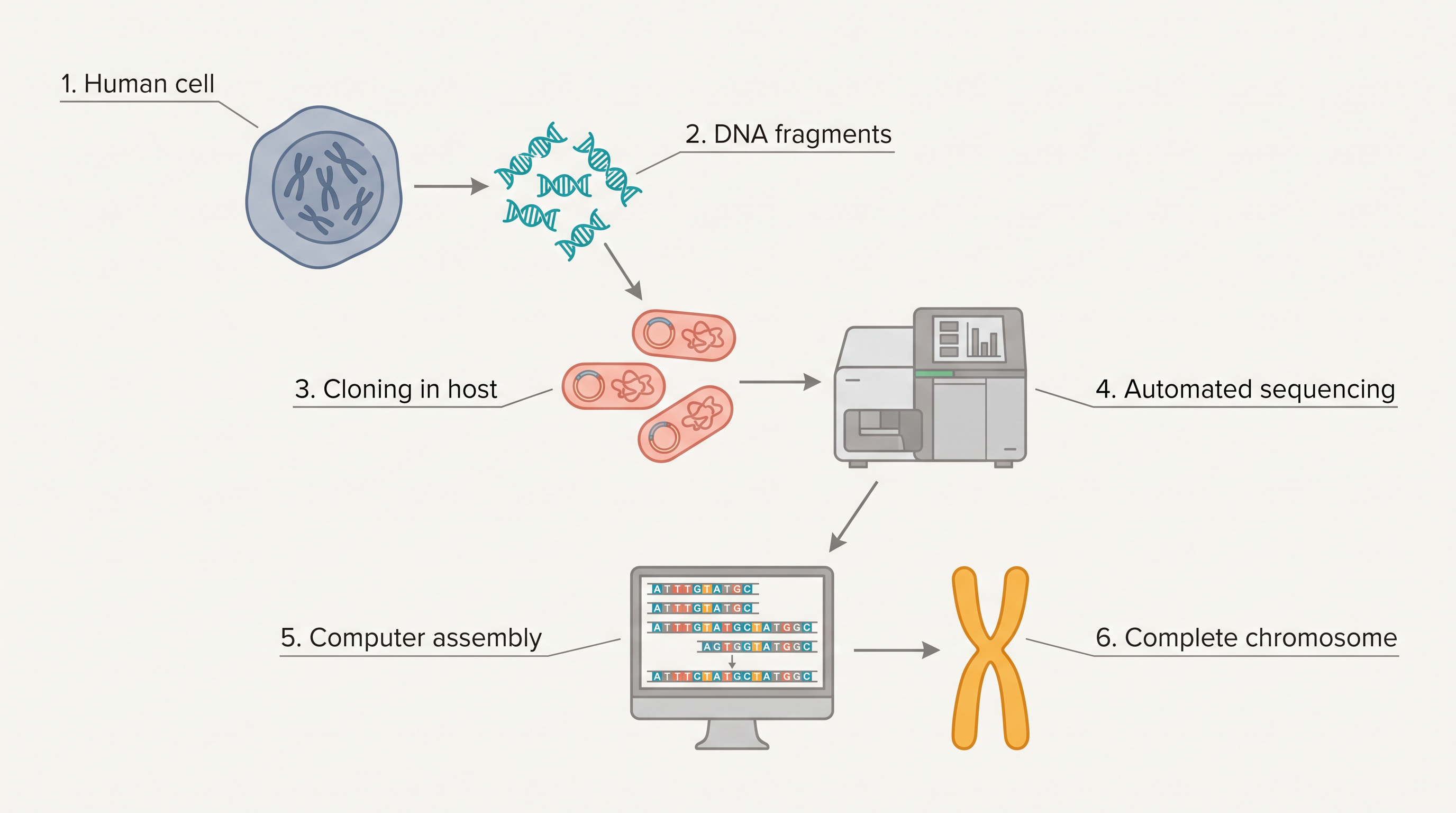
Fig. 5.15 – Sequencing workflow in the Human Genome Project.
Flowchart
FC 5.10 – Genome sequencing workflow: isolate total DNA → cut into random small fragments → clone in host (bacteria/yeast) using BAC/YAC vectors → amplify each fragment → sequence using automated (Sanger-based) sequencers → align overlapping fragments using computer programs → annotate and assign sequences to chromosomes.
The sequence of chromosome 1 was completed only in May 2006 — the last of the 24 human chromosomes (22 autosomes plus X and Y) to be sequenced. Another challenging task was assigning genetic and physical maps to the genome. This was done using information on the polymorphism of restriction endonuclease recognition sites, and on certain repetitive DNA sequences known as microsatellites. (One use of polymorphism in repetitive DNA — DNA fingerprinting — is explained in the next section.)
5.9.1 Salient Features of Human Genome
Some of the key observations from the Human Genome Project are:
The human genome contains 3164.7 million bp.
The average gene consists of about 3000 bases, but gene sizes vary greatly — the largest known human gene, dystrophin, has 2.4 million bases.
The total number of genes is estimated at about 30,000 — much lower than earlier estimates of 80,000 to 1,40,000 genes. Almost all (99.9 per cent) of the nucleotide bases are exactly the same in all humans.
The functions are unknown for over 50 per cent of the discovered genes.
Less than 2 per cent of the genome codes for proteins.
Repeated sequences make up a very large portion of the human genome.
Repetitive sequences are stretches of DNA repeated many times — sometimes hundreds to thousands of times. They are thought to have no direct coding function, but they shed light on chromosome structure, dynamics and evolution.
Chromosome 1 has the most genes (2968), and the Y chromosome has the fewest (231).
Scientists have identified about 1.4 million locations where single-base DNA differences occur in humans. These are called SNPs — single nucleotide polymorphisms, pronounced “snips.” This information promises to transform how we find chromosomal locations of disease-linked sequences and how we trace human history.
5.9.2 Applications and Future Challenges
Drawing real meaning from these DNA sequences will shape biological research for decades to come, deepening our understanding of living systems. This is an enormous task that will need the skill and creativity of tens of thousands of scientists worldwide, in both the public and private sectors.
Perhaps the greatest impact of having the human genome sequence is that it allows a completely new way of doing biology. In the past, researchers could study only one or a few genes at a time. Now, with whole-genome sequences and new high-throughput technologies, they can approach questions systematically and on a much broader scale. They can study all the genes in a genome at once — for example, all the transcripts in a particular tissue, organ or tumour — or explore how tens of thousands of genes and proteins work together in interconnected networks to run the chemistry of life.
5.10 DNA Fingerprinting
As we saw in the previous section, 99.9 per cent of the base sequence is the same among all humans. It is the remaining tiny fraction — the differences in DNA sequence — that make every individual unique in their appearance.
If one wants to find the genetic differences between two individuals, or among individuals in a population, sequencing the whole DNA every time would be a slow and expensive task. Imagine trying to compare two sets of \(3 \times 10^6\) base pairs. DNA fingerprinting offers a much quicker way to compare the DNA of any two individuals.
DNA fingerprinting works by looking at differences in certain specific regions of DNA called repetitive DNA. In these regions, a small stretch of DNA is repeated many times over.
When genomic DNA is spun in a density gradient centrifuge, these repetitive sequences separate out as small, distinct peaks, while the bulk of the DNA forms one major peak. Because these small peaks appear as separate (“satellite”) peaks alongside the main peak, they are called satellite DNA. Depending on their base composition (A:T rich or G:C rich), the length of the segment, and the number of repeated units, satellite DNA is grouped into categories such as micro-satellites and mini-satellites.
These satellite sequences normally do not code for any protein, yet they form a large part of the human genome. Importantly, they show a high degree of polymorphism, and this is what makes them the basis of DNA fingerprinting. Since DNA taken from any tissue of a person — blood, hair-follicle, skin, bone, saliva, sperm and so on — shows the same degree of polymorphism, it becomes a very useful tool for identification in forensic (crime-investigation) work. Moreover, because these polymorphisms are inherited from parents to children, DNA fingerprinting also forms the basis of paternity testing in cases of dispute.
Understanding DNA Polymorphism
Because polymorphism in DNA is the foundation of both genetic mapping and DNA fingerprinting, it is important to understand what it means.
Polymorphism arises due to mutations. (Recall the different kinds of mutations and their effects from Chapter 4 and from earlier sections of this chapter.) New mutations may appear in an individual either in somatic cells (body cells) or in germ cells (the cells that produce gametes in sexually reproducing organisms). If a germ-cell mutation does not seriously reduce the individual’s ability to have offspring who can pass on that mutation, it can spread to other members of the population through sexual reproduction.
Traditionally, an allelic sequence variation is called a DNA polymorphism if more than one variant (allele) at a locus (a fixed position on a chromosome) occurs in the human population with a frequency greater than 0.01. In simple terms, an inheritable mutation seen in a population at high frequency is a DNA polymorphism. Such variation is more likely to be found in non-coding DNA, because mutations there usually have no immediate effect on an individual’s ability to reproduce. These mutations keep accumulating generation after generation, forming one basis of variability.
Polymorphisms range from a single nucleotide change all the way to very large-scale changes. Such polymorphisms play a very important role in evolution and speciation, which you will study in higher classes.
The Technique of DNA Fingerprinting
The technique of DNA fingerprinting was initially developed by Alec Jeffreys. He used a satellite DNA as a probe that showed a very high degree of polymorphism. This was called a Variable Number of Tandem Repeats (VNTR).
The technique, as used in its early form, was based on Southern blot hybridisation using a radiolabelled VNTR as a probe (a probe is a labelled DNA piece that binds to its complementary sequence). It involved these steps:
isolation of DNA;
digestion of DNA by restriction endonucleases (enzymes that cut DNA at specific sites);
separation of the DNA fragments by electrophoresis (which sorts fragments by size);
transferring (blotting) of the separated fragments onto synthetic membranes, such as nitrocellulose or nylon;
hybridisation using a labelled VNTR probe; and
detection of the hybridised fragments by autoradiography (exposing the labelled bands on a film).
These steps are summarised in FC 5.11, and the overall scheme is shown in Fig. 5.16.
Flowchart
FC 5.11 – DNA fingerprinting: isolate DNA → digest with restriction endonucleases → separate fragments by electrophoresis → blot fragments onto a nitrocellulose/nylon membrane → hybridise with a labelled VNTR probe → detect bands by autoradiography → read the banding pattern.

Fig. 5.16 – Schematic of DNA fingerprinting.
VNTR and the Banding Pattern
The VNTR belongs to a class of satellite DNA called the mini-satellite. In it, a small DNA sequence is arranged one after another (tandemly) in many copies. The number of these copies varies from chromosome to chromosome within an individual, and the number of repeats shows a very high degree of polymorphism. As a result, the size of a VNTR can range from 0.1 to 20 kb (kilobases).
Because of this variation, after hybridisation with the VNTR probe, the autoradiogram shows many bands of differing sizes. Together, these bands form a characteristic pattern for each individual’s DNA, as in Fig. 5.16. This pattern differs from one individual to another in a population — the only exception being monozygotic (identical) twins, who share the same DNA.
The sensitivity of the technique has been increased by using the polymerase chain reaction (PCR), which you will study in Chapter 9. As a result, DNA from even a single cell is now enough to carry out a DNA fingerprinting analysis.
Applications
Besides its use in forensic science, DNA fingerprinting has much wider applications, such as in determining population and genetic diversities. Today, many different probes are used to generate DNA fingerprints.
5.11 Summary
Nucleic acids are long polymers of nucleotides. DNA stores genetic information, while RNA mostly helps to transfer and express that information.
Both DNA and RNA can act as genetic material, but DNA — being chemically and structurally more stable — is the better one. In evolution, however, RNA came first, and DNA was later derived from RNA.
The hallmark of the double-stranded helical structure of DNA is the hydrogen bonding between bases on opposite strands. Adenine pairs with Thymine through two H-bonds, and Guanine pairs with Cytosine through three H-bonds. This makes one strand complementary to the other.
DNA replicates semiconservatively — each daughter molecule keeps one old strand and gains one new one — and this process is guided by complementary hydrogen bonding.
A segment of DNA that codes for RNA can, in simple terms, be called a gene.
During transcription, one strand of DNA acts as a template to direct the synthesis of a complementary RNA.
In bacteria, the transcribed mRNA is functional and can be translated directly.
In eukaryotes, the gene is split: coding sequences (exons) are interrupted by non-coding sequences (introns). The introns are removed and the exons are joined by splicing to make a functional RNA.
The mRNA carries base sequences that are read three at a time (the triplet genetic code) to specify each amino acid.
The genetic code is read, again by complementarity, by tRNA — the adapter molecule. There are specific tRNAs for every amino acid. Each tRNA carries a specific amino acid at one end and pairs, through its anticodon, with the codon on the mRNA.
The site of translation (protein synthesis) is the ribosome, which binds the mRNA and provides a platform for joining amino acids. One of the rRNAs catalyses peptide-bond formation — an example of an RNA enzyme, or ribozyme.
Because translation is built around RNA, it suggests that life itself began around RNA.
Transcription and translation are energetically very expensive, so they must be tightly regulated. Regulation of transcription is the primary step in controlling gene expression.
In bacteria, several genes are arranged and regulated together in units called operons. The lac operon is the model operon; it carries the genes for lactose metabolism and is regulated by the amount of lactose present — so it can be seen as the regulation of enzyme synthesis by its substrate.
The Human Genome Project was a mega project that aimed to sequence every base in the human genome. It yielded a great deal of new information and opened up many new fields, such as bioinformatics.
DNA fingerprinting is a technique to find variations among individuals of a population at the DNA level. It is based on the polymorphism of DNA sequences and has immense applications in forensic science, genetic biodiversity and evolutionary biology.
Flowchart
FC 5.12 – The chapter’s central flow: DNA →(replication) DNA (semiconservative) ; DNA →(transcription, one strand as template) RNA →(processing in eukaryotes: splicing, capping, tailing) mRNA →(translation on ribosome, tRNA reads codons) polypeptide/protein ; with regulation acting mainly at the transcription step.
5.12 Points to Ponder
DNA is more stable than RNA, yet evolution is thought to have begun with RNA. Why might life have started with the less stable of the two molecules, and what advantage did switching to DNA later provide?
The genetic code is a triplet. If a fourth base had somehow existed in nature, would the code still need to be a triplet? Think about how the number of bases and the number of amino acids together decide the size of a codon.
The code is described as “degenerate” (many codons for one amino acid) and “nearly universal” (almost the same in all organisms). How might each of these properties protect an organism against the harmful effects of mutations?
Eukaryotic genes are interrupted by introns that are later spliced out. Making and then removing these sequences seems wasteful. What benefits might a split-gene arrangement offer that could outweigh this apparent waste?
In transcription, only one strand of DNA is copied, and only a segment at a time — unlike replication, where the whole molecule is duplicated. Why does the cell handle these two “copying” processes so differently?
The lac operon makes the enzymes for lactose only when lactose is present. Consider how such “make it only when needed” control helps a bacterium compete for survival in a changing environment.
The Human Genome Project set aside effort and funds to study the ethical, legal and social issues (ELSI) of the work. What kinds of concerns do you think such a project should anticipate, and why decide on them before, rather than after, the science is done?
DNA fingerprinting relies on non-coding, highly variable regions rather than genes. Reflect on why the “junk”-looking parts of the genome turn out to be so powerful for telling individuals apart.
Almost all (99.9 per cent) of human DNA is identical between people, and less than 2 per cent of it codes for proteins. What does this suggest about where human individuality and complexity actually come from?
5.13 NCERT Questions
Group the following as nitrogenous bases and nucleosides: Adenine, Cytidine, Thymine, Guanosine, Uracil and Cytosine.
If a double-stranded DNA has 20 per cent of cytosine, calculate the per cent of adenine in the DNA.
If the sequence of one strand of DNA is written as follows: 5′-ATGCATGCATGCATGCATGCATGCATGC-3′. Write down the sequence of the complementary strand in 5′→3′ direction.
If the sequence of the coding strand in a transcription unit is written as follows: 5′-ATGCATGCATGCATGCATGCATGCATGC-3′. Write down the sequence of the mRNA.
Which property of the DNA double helix led Watson and Crick to hypothesise the semi-conservative mode of DNA replication? Explain.
Depending upon the chemical nature of the template (DNA or RNA) and the nature of the nucleic acid synthesised from it (DNA or RNA), list the types of nucleic acid polymerases.
How did Hershey and Chase differentiate between DNA and protein in their experiment while proving that DNA is the genetic material?
Differentiate between the following:
- Repetitive DNA and Satellite DNA
- mRNA and tRNA
- Template strand and Coding strand
List two essential roles of the ribosome during translation.
In the medium where E. coli was growing, lactose was added, which induced the lac operon. Then, why does the lac operon shut down some time after the addition of lactose in the medium?
Explain (in one or two lines) the function of the following:
- Promoter
- tRNA
- Exons
Why is the Human Genome Project called a mega project?
What is DNA fingerprinting? Mention its application.
Briefly describe the following:
- Transcription
- Polymorphism
- Translation
- Bioinformatics
5.14 Check Your Concepts
Assertion (A): In DNA, a purine always pairs with a pyrimidine. Reason (R): This keeps the distance between the two strands of the helix almost uniform. State whether A and R are true, and whether R correctly explains A.
A region of DNA rich in Guanine–Cytosine pairs is more stable than a region rich in Adenine–Thymine pairs. Give the reason based on hydrogen bonding.
The DNA in a human cell is about 2.2 m long, yet it fits inside a nucleus only about 10⁻⁶ m across. Arrange the levels of DNA packaging in the correct order that makes this possible, starting from the DNA double helix and ending at the chromosome.
What would happen to DNA packaging in a eukaryotic nucleus if histone proteins lost their positive charge? Explain your reasoning.
Match each scientist (or team) with their contribution:
| Column I | Column II |
|---|---|
| a. Griffith | i. proved semiconservative replication in E. coli |
| b. Avery, MacLeod and McCarty | ii. demonstrated bacterial transformation |
| c. Hershey and Chase | iii. showed the transforming principle is DNA |
| d. Meselson and Stahl | iv. proved DNA is the genetic material using radioactively labelled phages |
In the Hershey–Chase experiment, why were radioactive phosphorus and radioactive sulfur chosen as the two labels? What property of DNA and protein made this choice work?
Assertion (A): RNA mutates at a faster rate than DNA. Reason (R): The 2′-OH group present at every nucleotide in RNA makes it more reactive and less stable. Evaluate A and R.
Give reasons why DNA, rather than RNA, is preferred as the genetic material for the long-term storage of information.
During transcription, only one strand of a DNA segment is copied, whereas during replication the entire DNA is duplicated. Explain why the cell treats these two “copying” processes differently.
Distinguish between the template strand and coding strand of a transcription unit. Which of the two has the same sequence as the mRNA (except for the base change)?
Arrange the following events of eukaryotic mRNA formation in the correct sequence: capping, splicing, transcription of hnRNA, tailing, transport of mature mRNA to the cytoplasm.
What would happen to the final protein if an intron were not removed during the splicing of a eukaryotic primary transcript?
The genetic code is said to be degenerate and nearly universal. Explain what each term means, giving one example for each.
A single base-pair substitution changes glutamate to valine in the beta-globin chain. Name the resulting disorder, and explain how a change in just one amino acid can lead to disease.
Distinguish between a frameshift mutation and a substitution (point) mutation, based on their effect on the reading frame.
Why is the 23S rRNA of the bacterial ribosome described as a ribozyme? Which reaction of translation does it catalyse?
Assertion (A): A very low level of lac operon expression must always be present in the cell. Reason (R): Permease is needed for lactose to enter the cell and induce the operon. Evaluate A and R.
Explain why the control of the lac operon by its repressor is called negative regulation.
In a schematic diagram of a transcription unit, label the following: promoter, structural gene, terminator, template strand and coding strand. Also indicate the direction of transcription.
Why does DNA fingerprinting rely on highly polymorphic, non-coding repetitive DNA rather than on protein-coding genes?
Match each eukaryotic RNA polymerase with the RNA it transcribes:
| Column I | Column II |
|---|---|
| a. RNA polymerase I | i. precursor of mRNA (hnRNA) |
| b. RNA polymerase II | ii. rRNAs (28S, 18S and 5.8S) |
| c. RNA polymerase III | iii. tRNA, 5S rRNA and snRNAs |
- Why can transcription and translation be coupled in bacteria, but not in eukaryotes?
5.15 Practice With Numericals
A double-stranded DNA molecule contains 22 per cent adenine. Calculate the percentages of thymine, guanine and cytosine in this DNA.
A DNA double helix has \(1 \times 10^4\) base pairs. Given that the distance between two adjacent base pairs is 0.34 nm, calculate the length of this DNA molecule in nanometres and in micrometres.
E. coli completes the replication of its \(4.6 \times 10^6\) bp genome in about 18 minutes. Calculate the approximate average rate of polymerisation in base pairs per second.
E. coli grown for many generations in \(^{15}N\) medium is transferred to \(^{14}N\) medium. After three rounds of replication, what fraction of the DNA molecules would be fully “light” and what fraction would be “hybrid”?
The human genome contains about \(3 \times 10^9\) base pairs. If less than 2 per cent of the genome codes for protein, estimate the maximum number of base pairs that are protein-coding.
If the cost of sequencing were US $3 per base pair, estimate the total cost of sequencing a genome of \(3 \times 10^9\) base pairs.
A codon is a group of three bases chosen from 4 possible bases. Calculate how many different codons are possible. Then calculate how many would be possible if the code were a doublet (two bases), and state whether that would be enough to code for 20 amino acids.
The coding region of an mRNA (excluding the stop codon) contains 900 nucleotides. How many amino acids will the resulting polypeptide contain?