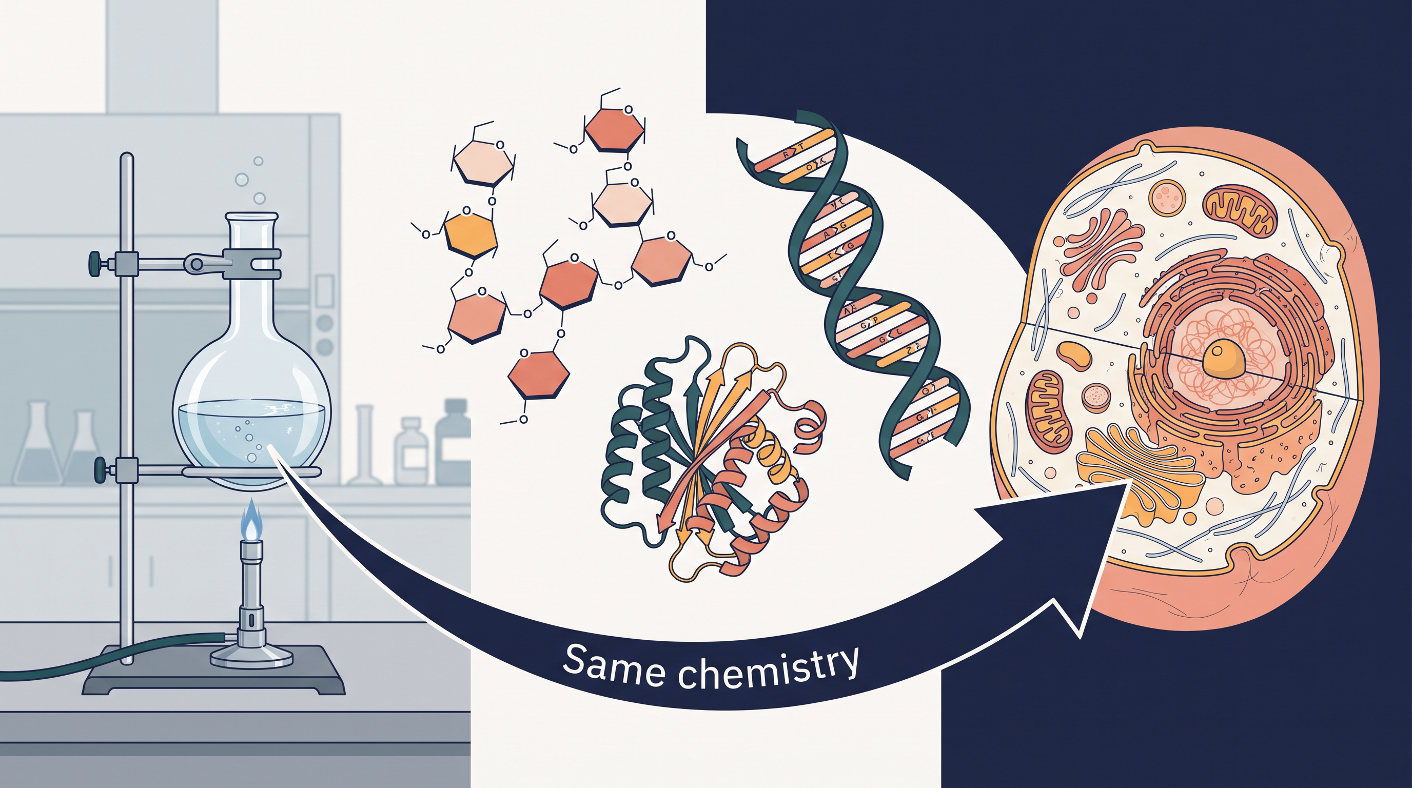
In the 1820s, chemists were sure of one thing: the compounds found inside living things could never be made in a laboratory. Sugars, urea, fats and proteins were believed to carry a mysterious “vital force” that only a living body could supply. A young German chemist, Friedrich Wöhler, was not trying to overturn that belief. He was simply heating ammonium cyanate, an ordinary inorganic salt, in his laboratory. What he obtained instead was urea — the very compound excreted by animals, including humans.
That single experiment quietly changed chemistry. If a substance made by a kidney could also be made from a bottled salt on a laboratory bench, then the molecules of life obeyed the same rules as every other molecule. There was no separate chemistry for living matter.
Nearly two centuries later, we take this idea further. A living cell grows, repairs itself and reproduces — yet it is built entirely from non-living atoms and molecules. Carbohydrates, proteins, nucleic acids, vitamins and hormones are ordinary organic compounds with ordinary functional groups. What makes them extraordinary is how their structures fit their jobs. Study of these molecules and their reactions inside the body is called biochemistry, and this chapter is your first look at it.
Q1. Glucose clearly contains an aldehyde group — yet it fails Schiff’s test, a standard test for aldehydes. How can a molecule hide its own functional group? Q2. Starch and cellulose are both long chains of glucose. Why can we digest a bowl of rice but not a sheet of paper? Q3. When an egg is boiled, the clear egg white turns into a firm white solid — and cooling it never brings the liquid back. What exactly has changed in the protein, and why is the change one-way? Q4. Vitamin C must be eaten almost every day, while vitamin A can be stored for months. What difference in their molecules decides this? Q5. The two strands of DNA have different base sequences, yet they are called complementary. How can two unlike strands each carry the same message?
Keep these questions in mind. As you work through this chapter, you will be able to reason out every one of them for yourself.
10.1 Carbohydrates
Carbohydrates are made mainly by plants, and they form one of the largest families of naturally occurring organic compounds. Cane sugar, glucose and starch are everyday examples — the sugar in your tea, the energy in your bloodstream, and the bulk of the rice on your plate all belong to this single family.
Most carbohydrates have a general formula \(\\mathrm{C\_x(H\_2O)\_y}\). Written this way, they look like carbon atoms attached to water molecules, which is exactly why early chemists called them hydrates of carbon — and the name “carbohydrate” stuck. Glucose, for instance, has the molecular formula \(\\mathrm{C\_6H\_{12}O\_6}\), which can be rewritten as \(\\mathrm{C\_6(H\_2O)\_6}\) and fits the pattern neatly.
Why the old definition failed
The trouble is that this formula-based definition is neither reliable nor complete. Acetic acid, \(\\mathrm{CH\_3COOH}\), can be written as \(\\mathrm{C\_2(H\_2O)\_2}\) and fits the general formula perfectly — yet it is a carboxylic acid, not a carbohydrate at all. Working the other way round, rhamnose, \(\\mathrm{C\_6H\_{12}O\_5}\), is a genuine carbohydrate but simply does not fit the \(\\mathrm{C\_x(H\_2O)\_y}\) pattern.
So chemists stopped defining carbohydrates by their formula and started defining them by their chemistry. A very large number of reactions showed that these compounds always carry specific functional groups: several hydroxyl (–OH) groups together with either an aldehyde (–CHO) group or a ketone (\(>\\mathrm{C=O}\)) group. This is a far more dependable basis for a definition.
Carbohydrates are optically active polyhydroxy aldehydes or ketones, or compounds that give such units on hydrolysis.
Read that definition slowly, because every word in it is doing work. “Polyhydroxy” means many –OH groups. “Aldehyde or ketone” is the second functional group that must be present. “Optically active” means the molecule can rotate the plane of plane-polarised light, which happens because these molecules contain asymmetric (chiral) carbon atoms. And the final phrase — “or compounds that give such units on hydrolysis” — is what allows big molecules like starch and cellulose, which have no free –CHO group of their own, to still be called carbohydrates.
Some carbohydrates taste sweet, and these are also called sugars. The most common sugar used in our homes is sucrose, while the sugar present in milk is lactose. Carbohydrates as a whole are also called saccharides.
The word saccharide comes from the Greek sakcharon, meaning sugar. Several individual sugars are named just as directly: lactose from the Latin lac (milk), because it is found in milk; maltose from malt, from which it was first obtained; and dextrose, an old name for glucose, simply because glucose rotates plane-polarised light to the right (dextro).
10.1.1 Classification of Carbohydrates
Since hydrolysis is so central to what a carbohydrate is, it is also the most sensible basis for classifying carbohydrates. The question we ask is simple: when this compound is broken apart by water (with the help of acid or an enzyme), how many simple sugar units does it give? On this basis, carbohydrates are broadly divided into three groups, as summarised in FC 10.1.
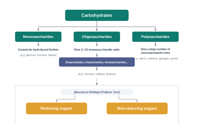
(i) Monosaccharides. A carbohydrate that cannot be hydrolysed further into a simpler polyhydroxy aldehyde or ketone unit is called a monosaccharide. These are the building blocks of the whole family. About 20 monosaccharides are known to occur in nature; glucose, fructose and ribose are common examples.
A monosaccharide is a carbohydrate that cannot be hydrolysed further to give a simpler unit of polyhydroxy aldehyde or ketone.
(ii) Oligosaccharides. Carbohydrates that yield two to ten monosaccharide units on hydrolysis are called oligosaccharides. They are further classified as disaccharides, trisaccharides, tetrasaccharides and so on, depending on exactly how many monosaccharide units they release.
Among these, disaccharides are by far the most common. The two monosaccharide units obtained from a disaccharide may be the same or different. One molecule of sucrose, on hydrolysis, gives one molecule of glucose and one molecule of fructose — two different units. Maltose, on the other hand, gives two molecules of glucose only.
An oligosaccharide is a carbohydrate that yields two to ten monosaccharide units on hydrolysis.
(iii) Polysaccharides. Carbohydrates that yield a large number of monosaccharide units on hydrolysis are called polysaccharides. Starch, cellulose, glycogen and gums are common examples. Polysaccharides are not sweet in taste, and for this reason they are also called non-sugars.
A polysaccharide is a carbohydrate that yields a large number of monosaccharide units on hydrolysis.
Reducing and non-reducing sugars
There is a second, completely different way of sorting carbohydrates, and it depends on how they behave with two mild oxidising reagents you have already met in the chemistry of aldehydes and ketones — Fehling’s solution and Tollens’ reagent. Both reagents are reduced by an aldehyde group (and, in sugars, by a keto group that can rearrange into one).
All those carbohydrates which reduce Fehling’s solution and Tollens’ reagent are called reducing sugars. Those that do not are called non-reducing sugars. All monosaccharides — whether they are aldoses or ketoses — are reducing sugars.
Carbohydrates that reduce Fehling’s solution and Tollens’ reagent are called reducing sugars; those that do not are called non-reducing sugars.
Fehling’s solution and Tollens’ reagent (Both are mild oxidising agents used as tests, not as preparative reagents → Fehling’s solution, on warming with a reducing sugar, gives a reddish-brown precipitate of copper(I) oxide; Tollens’ reagent gives a silver mirror on the inner wall of the test tube → they are the standard way to decide whether a given sugar is reducing or non-reducing, and therefore whether its aldehyde or keto group is free or locked up in a linkage.)
This reducing-sugar test moved out of the chemistry laboratory and into the clinic. For decades, the standard bedside check for glucose in urine — an indicator of uncontrolled diabetes — was a Fehling’s-type (Benedict’s) test: a few drops of urine warmed with the blue reagent, and a colour change towards brick-red meant sugar was present. Modern hospitals use enzyme-based strips and blood-glucose meters instead, but the underlying chemistry being exploited is the same one you are studying here: glucose has a free aldehyde group, so it can be oxidised, so it acts as a reducing agent.
(question only — no solution) Sucrose does not reduce Fehling’s solution, but its hydrolysis products do. Explain what must have changed at the molecular level, and name the two products formed.
10.1.2 Monosaccharides
Monosaccharides are classified further on two counts: the number of carbon atoms they contain, and the functional group they carry.
If a monosaccharide contains an aldehyde group, it is known as an aldose; if it contains a keto group, it is known as a ketose. The number of carbon atoms is also folded into the name — a three-carbon sugar is a triose, a six-carbon sugar is a hexose, and so on. Put the two pieces of information together and the name tells you almost everything: an aldohexose is a six-carbon sugar with an aldehyde group, while a ketohexose is a six-carbon sugar with a keto group. The full pattern is set out in Table 10.1.
Table 10.1: Different Types of Monosaccharides
| 3 |
Triose |
Aldotriose |
Ketotriose |
| 4 |
Tetrose |
Aldotetrose |
Ketotetrose |
| 5 |
Pentose |
Aldopentose |
Ketopentose |
| 6 |
Hexose |
Aldohexose |
Ketohexose |
| 7 |
Heptose |
Aldoheptose |
Ketoheptose |
10.1.2.1 Glucose
Glucose occurs freely in nature as well as in the combined form. It is present in sweet fruits and in honey, and ripe grapes contain it in large amounts. In the laboratory and in industry it is prepared as follows.
Preparation of Glucose
1. From sucrose (cane sugar). If sucrose is boiled with dilute \(\\mathrm{HCl}\) or \(\\mathrm{H\_2SO\_4}\) in alcoholic solution, glucose and fructose are obtained in equal amounts.
\[\\mathrm{C\_{12}H\_{22}O\_{11}} + \\mathrm{H\_2O} \\xrightarrow{\\ \\mathrm{H^+}\\ } \\mathrm{C\_6H\_{12}O\_6} + \\mathrm{C\_6H\_{12}O\_6}\]
Here \(\\mathrm{C\_{12}H\_{22}O\_{11}}\) is sucrose, and the two products are glucose and fructose respectively. The \(\\mathrm{H^+}\) above the arrow is the acid acting as a catalyst: it speeds up the breaking of the linkage between the two sugar units without being consumed.
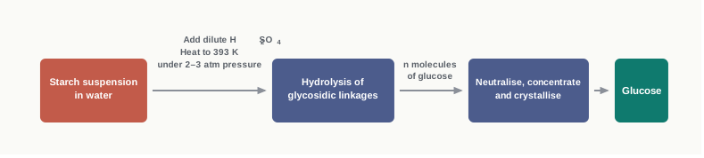
2. From starch. Commercially, glucose is obtained by hydrolysis of starch, boiling it with dilute \(\\mathrm{H\_2SO\_4}\) at 393 K under pressure.
\[(\\mathrm{C\_6H\_{10}O\_5})\_n + n,\\mathrm{H\_2O} \\xrightarrow\[\\ \\text{393 K};\\ 2\\text{--}3\\ \\text{atm}\\ ]{\\ \\mathrm{H^+}\\ } n,\\mathrm{C\_6H\_{12}O\_6}\]
In this equation \((\\mathrm{C\_6H\_{10}O\_5})\_n\) is starch or cellulose and the product is glucose. Note that a pressure of 2–3 atm is used along with the raised temperature: the reaction mixture must be heated well above the normal boiling point of water, and pressure keeps the water in the liquid state so that hydrolysis can continue. The industrial route is summarised in FC 10.2.
34.2 g of sucrose is completely hydrolysed with dilute acid. Calculate the mass of glucose obtained. (Molar masses: sucrose \(= 342\\ \\text{g mol}^{-1}\), glucose \(= 180\\ \\text{g mol}^{-1}\))
Begin with the balanced equation, because the mole ratio comes from it:
\[\\mathrm{C\_{12}H\_{22}O\_{11}} + \\mathrm{H\_2O} \\xrightarrow{\\ \\mathrm{H^+}\\ } \\mathrm{C\_6H\_{12}O\_6} + \\mathrm{C\_6H\_{12}O\_6}\]
One mole of sucrose gives one mole of glucose (and one mole of fructose).
Moles of sucrose taken:
\[n = \\frac{34.2\\ \\text{g}}{342\\ \\text{g mol}^{-1}} = 0.100\\ \\text{mol}\]
From the 1 : 1 mole ratio, moles of glucose formed \(= 0.100\\ \\text{mol}\).
Mass of glucose:
\[m = 0.100\\ \\text{mol} \\times 180\\ \\text{g mol}^{-1} = 18.0\\ \\text{g}\]
Answer: 18.0 g of glucose (and, alongside it, 18.0 g of fructose).
Notice that 34.2 g of sucrose gives 18.0 g + 18.0 g = 36.0 g of products, which is more than the mass of sucrose taken. There is no error here: the extra mass comes from the water molecule added during hydrolysis.
Structure of Glucose
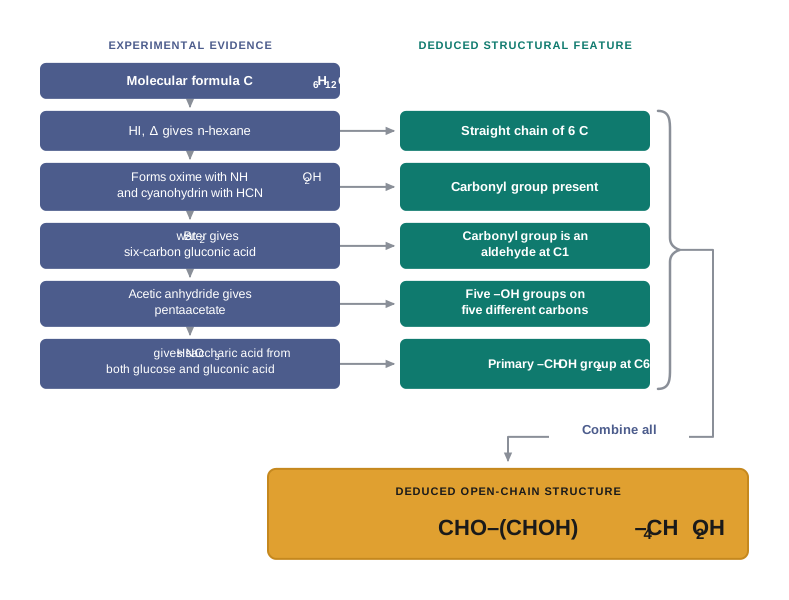
Glucose is an aldohexose and is also known as dextrose. It is the monomer of many of the larger carbohydrates, namely starch and cellulose, and it is probably the most abundant organic compound on earth. Its structure was not guessed — it was assigned on the basis of the six lines of experimental evidence given below. The open-chain structure of glucose is shown in Fig. 10.3.
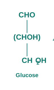
1. Molecular formula. Its molecular formula was found to be \(\\mathrm{C\_6H\_{12}O\_6}\).
2. Straight chain of six carbons. On prolonged heating with \(\\mathrm{HI}\), glucose forms n-hexane. This tells us that all six carbon atoms are linked in a straight chain, because a branched arrangement would have given a branched alkane instead.
\[\\mathrm{HOCH\_2(CHOH)\_4CHO} \\xrightarrow{\\ \\mathrm{HI},\\ \\Delta\\ } \\mathrm{CH\_3!-!CH\_2!-!CH\_2!-!CH\_2!-!CH\_2!-!CH\_3}\]
The product is n-hexane. Hot hydroiodic acid is a powerful reducing agent here: it strips away every oxygen-containing group and leaves only the carbon skeleton behind — which is exactly why the experiment reveals the shape of that skeleton.
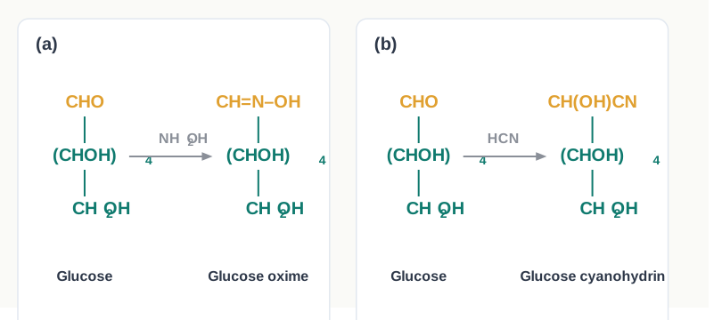
3. Presence of a carbonyl group. Glucose reacts with hydroxylamine to form an oxime, and it adds a molecule of hydrogen cyanide to give a cyanohydrin. Both of these are classic reactions of a carbonyl group, and together they confirm the presence of \(>\\mathrm{C=O}\) in glucose. The two reactions are drawn in Fig. 10.4.
\[\\mathrm{HOCH\_2(CHOH)\_4CHO} \\xrightarrow{\\ \\mathrm{NH\_2OH}\\ } \\mathrm{HOCH\_2(CHOH)\_4CH{=}N!-!OH}\]
\[\\mathrm{HOCH\_2(CHOH)\_4CHO} \\xrightarrow{\\ \\mathrm{HCN}\\ } \\mathrm{HOCH\_2(CHOH)\_4CH(OH)CN}\]
4. The carbonyl group is an aldehyde group. Glucose gets oxidised to a six-carbon carboxylic acid, gluconic acid, on reaction with a mild oxidising agent like bromine water. This indicates that the carbonyl group is present as an aldehydic group — a ketone would not be oxidised under such mild conditions, and if the chain had been cut, the acid would have had fewer than six carbon atoms. The oxidation is shown in Fig. 10.6.
\[\\mathrm{HOCH\_2(CHOH)\_4CHO} \\xrightarrow{\\ \\mathrm{Br\_2\\ water}\\ } \\mathrm{HOCH\_2(CHOH)\_4COOH}\]
The product is gluconic acid.
bromine water (A mild oxidising agent → used in aqueous solution at room temperature → it oxidises only the aldehyde group of a sugar to a carboxylic acid group, leaving the primary alcoholic –CH₂OH group untouched. This selectivity is precisely why it is the reagent of choice for proving that the carbonyl group in glucose is an aldehyde and for preparing gluconic acid.)
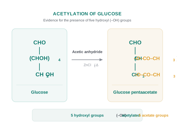
5. Five –OH groups, on five different carbons. Acetylation of glucose with acetic anhydride gives glucose pentaacetate. Since five acetyl groups are taken up, glucose must contain five –OH groups. Further, since the pentaacetate exists as a stable compound, these five –OH groups should be attached to different carbon atoms — two –OH groups on the same carbon atom would make the molecule unstable and it would simply lose water. The reaction is drawn in Fig. 10.5.
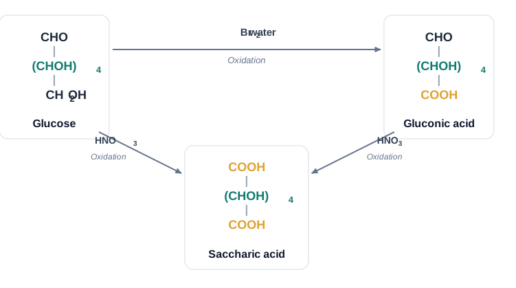
6. Presence of a primary alcoholic group. On oxidation with nitric acid, glucose as well as gluconic acid both yield the same dicarboxylic acid, saccharic acid. Think about what that means. Gluconic acid already has a –COOH group at one end; to end up with a dicarboxylic acid, the other end must also have been oxidised to –COOH. Only a primary alcoholic (–CH₂OH) group can be oxidised to –COOH in this way, so glucose must contain a primary alcoholic group at the far end of the chain. This double oxidation is drawn in Fig. 10.6.
\[\\mathrm{HOCH\_2(CHOH)\_4CHO} \\xrightarrow{\\ \\mathrm{HNO\_3}\\ } \\mathrm{HOOC(CHOH)\_4COOH}\]
\[\\mathrm{HOCH\_2(CHOH)\_4COOH} \\xrightarrow{\\ \\mathrm{HNO\_3}\\ } \\mathrm{HOOC(CHOH)\_4COOH}\]
The product in both cases is saccharic acid.
dilute nitric acid (A stronger oxidising agent than bromine water → warm dilute HNO₃ attacks both ends of the sugar chain → it oxidises the –CHO group and the primary –CH₂OH group to –COOH, giving a dicarboxylic acid. Remember the pair as a contrast: Br₂ water = one end only (gluconic acid); HNO₃ = both ends (saccharic acid). Questions in board and entrance papers very often test exactly this difference.)
A six-carbon monosaccharide X** gives n-hexane with hot \(\\mathrm{HI}\), forms an oxime with hydroxylamine, but on oxidation with bromine water it does not give a carboxylic acid. What can you conclude about the functional group in X?**
Take the evidence one clue at a time.
n-Hexane with hot \(\\mathrm{HI}\) shows that the six carbon atoms are in a straight, unbranched chain.
Oxime formation with hydroxylamine shows that a carbonyl group (\(>\\mathrm{C=O}\)) is present.
Failure to be oxidised by bromine water is the deciding clue. Bromine water is a mild oxidising agent and oxidises an aldehyde group to –COOH. If no acid is formed, the carbonyl group cannot be an aldehyde group.
Conclusion: X contains a keto group, not an aldehyde group. It is therefore a ketohexose (the same reasoning that identifies fructose), and not an aldohexose like glucose.
Configuration of glucose: the Fischer picture
Knowing which groups are present is only half the answer. Glucose contains four asymmetric carbon atoms, and the –OH groups on them can be arranged in space in several ways. The exact spatial arrangement of the different –OH groups was worked out by Fischer after studying many other properties of the molecule.
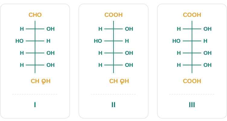
The correct configuration of glucose is represented as structure I in Fig. 10.8. Once glucose is fixed as I, gluconic acid follows as II (the top –CHO replaced by –COOH) and saccharic acid as III (both ends now –COOH).
Emil Fischer, working in Germany in the closing years of the nineteenth century, took on what looked like an impossible problem: sugars are colourless, sticky, chemically similar to one another, and there were no spectrometers to help. Working only with careful reactions and optical rotations, he deduced how the –OH groups of glucose and its relatives are arranged in space, and he invented the flat “cross” drawing — the Fischer projection — that let chemists put a three-dimensional molecule on paper without ambiguity. He received the Nobel Prize in Chemistry in 1902 for his work on sugars and purines. The projections you are learning to read in this section are his invention, still doing their job more than a century later.
The D– and L– notation
Glucose is correctly named as D(+)-glucose. The two labels in that name mean two entirely different things, and confusing them is one of the commonest mistakes students make.
The letter ‘D’ before the name represents the configuration — the spatial arrangement of groups. The sign ‘(+)’ represents the dextrorotatory nature of the molecule — the direction in which it actually rotates plane-polarised light, as measured in a polarimeter. It should be remembered that ‘D’ and ‘L’ have no relation with the optical activity of the compound. They are also not related to the small letters ‘d’ and ‘l’ (see Chapter 6). So a D-compound may be dextrorotatory or laevorotatory; you must measure to find out.
The letters ‘D’ or ‘L’ before the name of any compound indicate the relative configuration of a particular stereoisomer with respect to the configuration of some other compound whose configuration is already known. In the case of carbohydrates, that reference compound is a particular isomer of glyceraldehyde.
Glyceraldehyde contains one asymmetric carbon atom and therefore exists in two enantiomeric forms — (+)-glyceraldehyde and (–)-glyceraldehyde — which are mirror images of each other, as shown in Fig. 10.9.
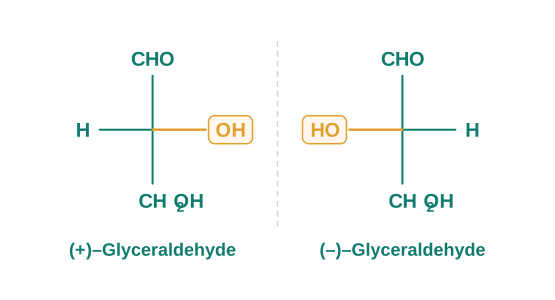
The (+) isomer of glyceraldehyde has the ‘D’ configuration. This means that when its structural formula is written on paper following specific conventions (which you will study in higher classes), the –OH group lies on the right-hand side in the structure. All those compounds which can be chemically correlated to the D(+) isomer of glyceraldehyde are said to have D-configuration, whereas those which can be correlated to the L(–) isomer of glyceraldehyde are said to have L-configuration. In the L(–) isomer, the –OH group is on the left-hand side, as you can see in the structure.
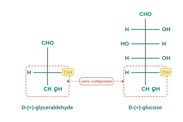
The prefix D or L before the name of a carbohydrate indicates its relative configuration with reference to glyceraldehyde — D if the –OH on the reference carbon lies on the right, L if it lies on the left. These letters describe configuration only, and say nothing about the direction of optical rotation, which is shown separately by (+) or (–).
Now, glucose has four asymmetric carbon atoms, not one — so which of them do we compare with glyceraldehyde? The rule is a definite one. For assigning the configuration of monosaccharides, it is the lowest asymmetric carbon atom which is compared. In (+)-glucose, the –OH on the lowest asymmetric carbon is on the right side, which is comparable to (+)-glyceraldehyde, so (+)-glucose is assigned the D-configuration. The other asymmetric carbon atoms of glucose are not considered for this comparison. Also, the structure of glucose and glyceraldehyde is written in a way that the most oxidised carbon (in this case –CHO) is at the top. Fig. 10.10 shows the two structures side by side with the compared carbon boxed.
(question only — no solution) D-(–)-fructose is laevorotatory, yet it carries the prefix D. Explain, in one or two sentences, why there is no contradiction here.
Cyclic Structure of Glucose
The open-chain structure I of glucose explained most of its properties — but not all of them. Three stubborn observations refused to fit.
1. Despite having the aldehyde group, glucose does not give Schiff’s test, and it does not form the hydrogensulphite addition product with \(\\mathrm{NaHSO\_3}\). Both of these are reactions that any ordinary aldehyde would give readily.
2. The pentaacetate of glucose does not react with hydroxylamine, indicating the absence of a free –CHO group. This is a striking result: acetylation should only block the –OH groups, so an aldehyde group, if it were truly free, should have survived and still formed an oxime.
3. Glucose is found to exist in two different crystalline forms, which are named as \(\\alpha\) and \(\\beta\). The \(\\alpha\)-form of glucose (m.p. 419 K) is obtained by crystallisation from a concentrated solution of glucose at 303 K, while the \(\\beta\)-form (m.p. 423 K) is obtained by crystallisation from a hot and saturated aqueous solution at 371 K. An open-chain structure with a fixed arrangement offers no way for one compound to have two distinct melting points.
This behaviour could not be explained by the open-chain structure I for glucose. It was therefore proposed that one of the –OH groups of the molecule may add to the –CHO group of the same molecule, forming a cyclic hemiacetal structure. This is simply the familiar addition of an alcohol to an aldehyde, except that here the alcohol and the aldehyde are parts of the same molecule, so the chain closes on itself into a ring.
It was found that glucose forms a six-membered ring in which the –OH at C-5 is involved in ring formation. This one proposal explains both problems at once: it accounts for the absence of a free –CHO group, and it accounts for the existence of glucose in two forms, shown in Fig. 10.11. These two cyclic forms exist in equilibrium with the open-chain structure.
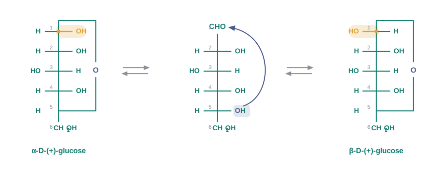
The two cyclic hemiacetal forms of glucose differ only in the configuration of the hydroxyl group at C1, which is called the anomeric carbon (the aldehyde carbon before cyclisation). Such isomers — the \(\\alpha\)-form and the \(\\beta\)-form — are called anomers.
Anomers are the two cyclic forms of a sugar that differ only in the configuration of the –OH group at the anomeric carbon (C1 in glucose, the carbon that was the carbonyl carbon before ring closure).
The six-membered cyclic structure of glucose is called the pyranose structure (\(\\alpha\)– or \(\\beta\)–), in analogy with pyran. Pyran is a cyclic organic compound with one oxygen atom and five carbon atoms in the ring. The cyclic structure of glucose is more correctly represented by the Haworth structure, given in Fig. 10.12, in which the ring is drawn as a flat hexagon seen edge-on, so that each –OH group can be shown clearly as pointing above or below the ring.
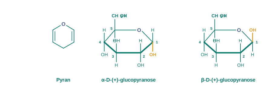
Q1. Glucose clearly contains an aldehyde group — yet it fails Schiff’s test, a standard test for aldehydes. How can a molecule hide its own functional group?
A. Because in solution glucose is almost entirely in its cyclic hemiacetal form. The –OH group at C-5 has added across the C1 aldehyde group and closed the molecule into a six-membered pyranose ring, so at any instant there is virtually no free –CHO group available. Schiff’s reagent and \(\\mathrm{NaHSO\_3}\) need a genuine free aldehyde group to react with, and they do not find one. The same explanation accounts for the pentaacetate: once the C1 –OH of the ring is acetylated, the ring can no longer open to release an aldehyde group at all, which is why the pentaacetate does not react with hydroxylamine.
(question only — no solution) The \(\\alpha\)- and \(\\beta\)-forms of D-glucose have different melting points (419 K and 423 K) yet the same molecular formula. State the relationship between them and identify the exact carbon atom at which they differ.
10.1.2.2 Fructose
Fructose is an important ketohexose. It is obtained along with glucose by the hydrolysis of the disaccharide sucrose. It is a natural monosaccharide found in fruits, honey and vegetables, and in its pure form it is used as a sweetener.
Structure of Fructose
Fructose also has the molecular formula \(\\mathrm{C\_6H\_{12}O\_6}\) — exactly the same as glucose. On the basis of its reactions it was found to contain a ketonic functional group at carbon number 2, and six carbons in a straight chain, as in the case of glucose. It belongs to the D-series and is a laevorotatory compound. It is therefore appropriately written as D-(–)-fructose. Its open-chain structure is shown in Fig. 10.13.
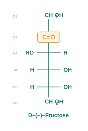
Here, then, is a clean example of structural isomerism among sugars: glucose and fructose have the same molecular formula but different functional groups, one an aldehyde at C1 and the other a ketone at C2.
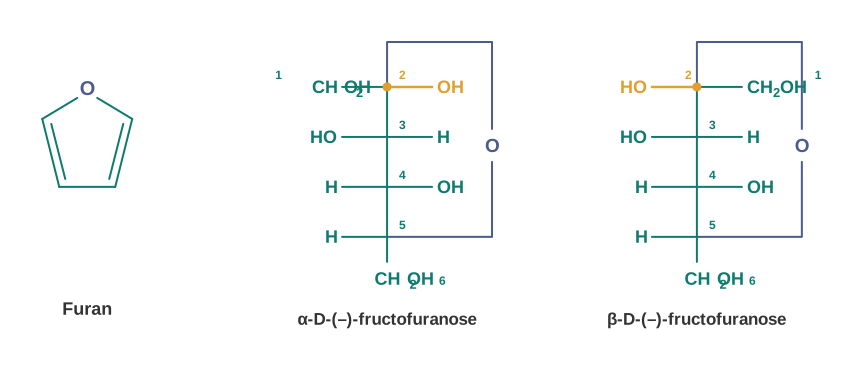
Fructose also exists in two cyclic forms, which are obtained by the addition of the –OH at C5 to the \(>\\mathrm{C=O}\) group. Count the atoms that end up in the ring and you will see that the ring thus formed is a five-membered ring, not a six-membered one as in glucose. It is named as the furanose structure, in analogy with the compound furan. Furan is a five-membered cyclic compound with one oxygen and four carbon atoms. The two cyclic forms are shown in Fig. 10.14.
The cyclic structures of the two anomers of fructose are more correctly represented by Haworth structures, shown in Fig. 10.15. Notice that fructose, like glucose, has an anomeric carbon — but in fructose it is C2, the carbon that carried the keto group before ring closure.
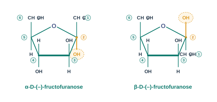
Glucose vs Fructose
| Molecular formula |
\(\\mathrm{C\_6H\_{12}O\_6}\) |
\(\\mathrm{C\_6H\_{12}O\_6}\) |
| Functional group |
Aldehyde (–CHO) at C1 |
Keto (\(>\\mathrm{C=O}\)) at C2 |
| Type of monosaccharide |
Aldohexose |
Ketohexose |
| Optical rotation |
Dextrorotatory, D-(+)-glucose |
Laevorotatory, D-(–)-fructose |
| Ring size on cyclisation |
Six-membered (pyranose), –OH at C5 adds to C1 |
Five-membered (furanose), –OH at C5 adds to C2 |
| Anomeric carbon |
C1 |
C2 |
| Reducing sugar? |
Yes |
Yes (all monosaccharides are reducing sugars) |
10.1.3 Disaccharides
You have already read that disaccharides, on hydrolysis with dilute acids or enzymes, yield two molecules of either the same or different monosaccharides. The two monosaccharide units are joined together by an oxide linkage, formed by the loss of a water molecule. Such a linkage between two monosaccharide units through an oxygen atom is called a glycosidic linkage.
A glycosidic linkage is the oxide (C–O–C) linkage formed between two monosaccharide units by the loss of a water molecule.
Now comes the key idea of this subsection — and it explains why some disaccharides reduce Fehling’s solution and others do not. In disaccharides, if the reducing groups of the monosaccharides, i.e. the aldehydic or ketonic groups, are themselves bonded into the glycosidic linkage, these are non-reducing sugars; sucrose is the standard example. On the other hand, sugars in which these functional groups are free are called reducing sugars — for example, maltose and lactose.
(i) Sucrose
One of the common disaccharides is sucrose, which on hydrolysis gives an equimolar mixture of D-(+)-glucose and D-(–)-fructose.
\[\\mathrm{C\_{12}H\_{22}O\_{11}} + \\mathrm{H\_2O} \\rightarrow \\mathrm{C\_6H\_{12}O\_6} + \\mathrm{C\_6H\_{12}O\_6}\]
Here sucrose gives D-(+)-glucose and D-(–)-fructose in equal molar amounts.
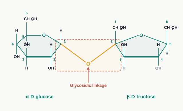
These two monosaccharides are held together by a glycosidic linkage between C1 of \(\\alpha\)-D-glucose and C2 of \(\\beta\)-D-fructose, as shown in Fig. 10.16. Look carefully at those two numbers: C1 is the anomeric carbon of glucose and C2 is the anomeric carbon of fructose. Since the reducing groups of glucose and fructose are both involved in glycosidic bond formation, sucrose is a non-reducing sugar.
Invert sugar
Sucrose is dextrorotatory, but after hydrolysis it gives dextrorotatory glucose and laevorotatory fructose. Now compare the magnitudes: the laevorotation of fructose (\(-92.4^\\circ\)) is more than the dextrorotation of glucose (\(+52.5^\\circ\)). Since the two are formed in equal amounts, the larger negative rotation wins and the resulting mixture is laevorotatory overall.
Thus, hydrolysis of sucrose brings about a change in the sign of rotation, from dextro (+) to laevo (–), and for this reason the product is named as invert sugar.
Invert sugar is the equimolar mixture of D-(+)-glucose and D-(–)-fructose obtained on hydrolysis of sucrose; it is named “invert” because the sign of optical rotation inverts from (+) to (–) during the hydrolysis.
Invert sugar is not a laboratory curiosity — it is an industrial workhorse. Honey is naturally rich in it, because bees carry an enzyme that hydrolyses the sucrose in nectar into glucose and fructose. Confectioners and bakers deliberately make or buy invert sugar for two practical reasons: fructose is sweeter than sucrose, so less sugar gives the same sweetness; and a mixture of two different sugars crystallises far less readily than a single pure sugar, which is why invert sugar keeps soft-centred sweets, jams and syrups smooth instead of grainy.
An aqueous solution of sucrose is dextrorotatory. It is boiled with a little dilute \(\\mathrm{HCl}\) and cooled, and the rotation is measured again. It is now found to be laevorotatory. Explain the observation and name the product.
Boiling with dilute acid hydrolyses the glycosidic linkage of sucrose:
\[\\mathrm{C\_{12}H\_{22}O\_{11}} + \\mathrm{H\_2O} \\xrightarrow{\\ \\mathrm{H^+}\\ } \\mathrm{C\_6H\_{12}O\_6} + \\mathrm{C\_6H\_{12}O\_6}\]
The products are D-(+)-glucose and D-(–)-fructose, formed in equal molar amounts.
Now compare their specific rotations. Glucose rotates the plane of polarised light to the right (\(+52.5^\\circ\)), while fructose rotates it to the left (\(-92.4^\\circ\)). Since equal amounts of the two are present, the net rotation of the mixture is decided by which contribution is larger in magnitude. The laevorotation of fructose is greater than the dextrorotation of glucose, so the mixture as a whole is laevorotatory.
Answer: The sign of rotation inverts from (+) to (–) because sucrose has been hydrolysed to an equimolar mixture of glucose and fructose in which fructose dominates the rotation. The product mixture is called invert sugar.
(ii) Maltose
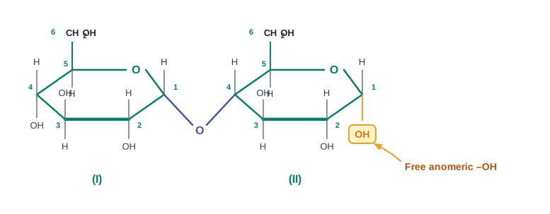
Another disaccharide, maltose, is composed of two \(\\alpha\)-D-glucose units, in which C1 of one glucose unit (I) is linked to C4 of another glucose unit (II), as drawn in Fig. 10.17.
Notice what has been left untouched. The C1 of the second glucose unit is not used in the linkage. A free aldehyde group can therefore be produced at C1 of the second glucose in solution — the ring can open — and maltose shows reducing properties. So maltose is a reducing sugar.
(iii) Lactose
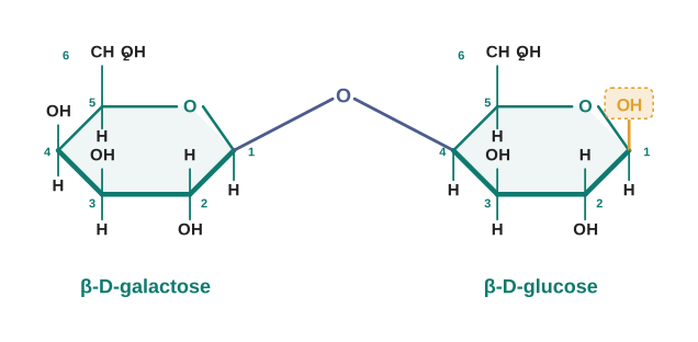
Lactose is more commonly known as milk sugar, since this disaccharide is found in milk. It is composed of \(\\beta\)-D-galactose and \(\\beta\)-D-glucose. The linkage is between C1 of galactose and C4 of glucose, as shown in Fig. 10.18.
Once again, the anomeric carbon (C-1) of the glucose unit is left free, so a free aldehyde group may be produced at C-1 of the glucose unit. Hence lactose is also a reducing sugar.
Sucrose vs Maltose vs Lactose
| Monosaccharide units |
\(\\alpha\)-D-glucose + \(\\beta\)-D-fructose |
Two \(\\alpha\)-D-glucose units |
\(\\beta\)-D-galactose + \(\\beta\)-D-glucose |
| Glycosidic linkage |
C1 of glucose to C2 of fructose |
C1 of one glucose to C4 of the other |
C1 of galactose to C4 of glucose |
| Anomeric carbons used up |
Both |
Only one |
Only one |
| Reducing or non-reducing |
Non-reducing |
Reducing |
Reducing |
| Common name / source |
Cane sugar, table sugar |
Obtained from malt |
Milk sugar, found in milk |
(question only — no solution) A disaccharide P** gives, on hydrolysis, one molecule of \(\\beta\)-D-galactose and one molecule of \(\\beta\)-D-glucose. P also gives a positive Tollens’ test. Identify P, state the carbon atoms involved in its glycosidic linkage, and explain why it is reducing while sucrose is not.**
10.1.4 Polysaccharides
Polysaccharides contain a large number of monosaccharide units joined together by glycosidic linkages. These are the most commonly encountered carbohydrates in nature. They mainly act as the food storage or structural materials of living organisms — which is a useful way to organise the three examples below: starch and glycogen store energy, while cellulose builds structure.
(i) Starch
Starch is the main storage polysaccharide of plants. It is the most important dietary source of energy for human beings. A high content of starch is found in cereals, roots, tubers and some vegetables.
Starch is a polymer of \(\\alpha\)-glucose and consists of two components — amylose and amylopectin.
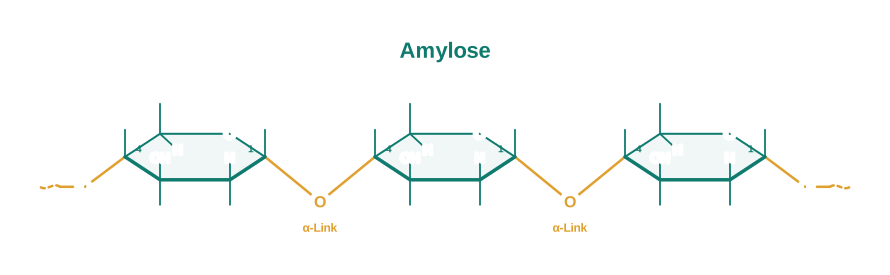
Amylose is the water-soluble component, which constitutes about 15–20% of starch. Chemically, amylose is a long unbranched chain with 200–1000 \(\\alpha\)-D-(+)-glucose units held together by C1–C4 glycosidic linkage, as drawn in Fig. 10.19.
Amylopectin is insoluble in water and constitutes about 80–85% of starch. It is a branched-chain polymer of \(\\alpha\)-D-glucose units in which the chain is formed by C1–C4 glycosidic linkage, whereas branching occurs by C1–C6 glycosidic linkage. That single difference — an occasional linkage through C6 instead of C4 — is what turns a straight chain into a tree-like, branched molecule, as shown in Fig. 10.20.
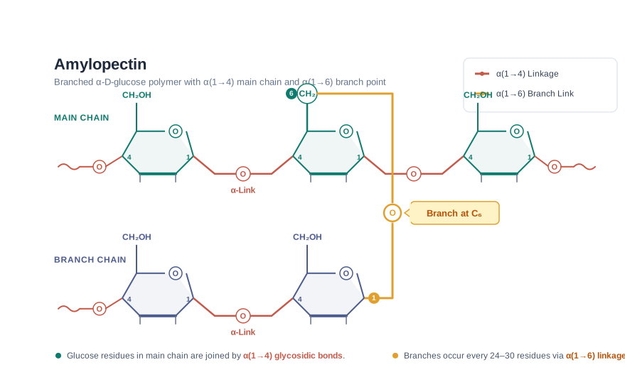
A sample of amylose has a molar mass of approximately \(1.62 \\times 10^5\\ \\text{g mol}^{-1}\). Estimate the number of glucose units in one such amylose chain, and check whether your answer is consistent with the range quoted for amylose.
In a polysaccharide chain, each glucose unit is joined to the next by loss of a water molecule. So the repeating unit of the chain is not \(\\mathrm{C\_6H\_{12}O\_6}\) (\(180\\ \\text{g mol}^{-1}\)) but \(\\mathrm{C\_6H\_{10}O\_5}\) — glucose minus one water molecule:
\[M(\\mathrm{C\_6H\_{10}O\_5}) = (6 \\times 12) + (10 \\times 1) + (5 \\times 16) = 72 + 10 + 80 = 162\\ \\text{g mol}^{-1}\]
Number of glucose units:
\[n = \\frac{1.62 \\times 10^5\\ \\text{g mol}^{-1}}{162\\ \\text{g mol}^{-1}} \\approx 1000\]
Answer: About 1000 glucose units. This sits at the upper end of the 200–1000 \(\\alpha\)-D-(+)-glucose units quoted for an amylose chain, so the result is consistent.
Notice the small but important step of using 162, not 180. Forgetting the water lost at each linkage is one of the commonest slips in polymer calculations.
(ii) Cellulose
Cellulose occurs exclusively in plants and it is the most abundant organic substance in the plant kingdom. It is a predominant constituent of the cell wall of plant cells.
Cellulose is a straight-chain polysaccharide composed only of \(\\beta\)-D-glucose units, which are joined by glycosidic linkage between C1 of one glucose unit and C4 of the next glucose unit, as shown in Fig. 10.21.
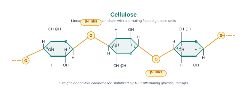
Now compare cellulose with amylose. Both are unbranched chains of glucose joined C1 to C4. The only structural difference is that amylose is built from \(\\alpha\)-D-glucose and cellulose from \(\\beta\)-D-glucose — a difference in the configuration at a single carbon atom. Yet one of them is our staple food and the other is wood and cotton. Few examples in chemistry show the structure–property link as sharply as this one.
Q2. Starch and cellulose are both long chains of glucose. Why can we digest a bowl of rice but not a sheet of paper?
A. Because of the configuration at the anomeric carbon. Starch is built from \(\\alpha\)-D-glucose units and cellulose from \(\\beta\)-D-glucose units, and this makes the two chains different in shape: the \(\\alpha\)-linked chain coils, while the \(\\beta\)-linked chain lies out straight and packs into stiff fibres. The enzymes in our digestive system are highly specific — they can hydrolyse \(\\alpha\)-glycosidic linkages but not \(\\beta\)-glycosidic linkages. So the starch in rice is broken down to glucose and absorbed, while the cellulose in paper (and in vegetable fibre) passes through us unchanged as roughage. Grazing animals manage it only because micro-organisms in their gut supply the enzyme that we lack.
(iii) Glycogen
The carbohydrates are stored in the animal body as glycogen. It is also known as animal starch, because its structure is similar to amylopectin and is rather more highly branched.
Glycogen is present in the liver, muscles and brain. When the body needs glucose, enzymes break the glycogen down to glucose. Glycogen is also found in yeast and fungi.
Starch vs Cellulose vs Glycogen
| Monomer unit |
\(\\alpha\)-D-glucose |
\(\\beta\)-D-glucose |
\(\\alpha\)-D-glucose |
| Chain structure |
Amylose is unbranched (C1–C4); amylopectin is branched (C1–C4 chain, C1–C6 branches) |
Straight, unbranched chain (C1–C4) |
Branched — similar to amylopectin but even more highly branched |
| Occurrence |
Plants: cereals, roots, tubers, vegetables |
Plants: cell wall (most abundant organic substance in the plant kingdom) |
Animals: liver, muscles, brain; also yeast and fungi |
| Biological role |
Food storage in plants; main dietary source of energy for humans |
Structural material |
Food storage in animals (“animal starch”) |
| Water solubility |
Amylose is water soluble; amylopectin is insoluble |
Insoluble |
— |
(question only — no solution) Amylopectin and cellulose are both polymers of glucose, yet only one of them is branched. State which one, name the linkage responsible for the branching, and identify the monomer unit of each.
10.1.5 Importance of Carbohydrates
Carbohydrates are essential for life in both plants and animals. They form a major portion of our food. Honey has been used for a long time as an instant source of energy by Vaids in the ayurvedic system of medicine.
Carbohydrates are used as storage molecules — as starch in plants and as glycogen in animals. The cell wall of bacteria and of plants is made up of cellulose. We build furniture and other articles from cellulose in the form of wood, and we clothe ourselves with cellulose in the form of cotton fibre. Carbohydrates also provide raw materials for many important industries like textiles, paper, lacquers and breweries.
There is one more role, and it is arguably the most far-reaching of all. Two aldopentoses — D-ribose and 2-deoxy-D-ribose (Section 10.5.1) — are present in nucleic acids. In other words, the very molecules that carry heredity are built around a five-carbon sugar. Finally, carbohydrates are found in the biosystem in combination with many proteins and lipids, forming molecules that do jobs neither could do alone.
10.1 Glucose and sucrose are soluble in water, but cyclohexane and benzene (simple six-membered ring compounds) are insoluble in water. Explain why.
10.2 What are the expected products of hydrolysis of lactose?
10.3 How do you explain the absence of an aldehyde group in the pentaacetate of D-glucose?
10.2 Proteins
Proteins are the most abundant biomolecules of the living system. Chief sources of proteins are milk, cheese, pulses, peanuts, fish and meat. They occur in every part of the body and form the fundamental basis of the structure and functions of life. They are also required for the growth and maintenance of the body.
The word protein is derived from the Greek word proteios, which means “primary” or “of prime importance” — a name that turned out to be a fair description of what these molecules actually do. All proteins are polymers of \(\\alpha\)-amino acids.
10.2.1 Amino Acids
Amino acids contain two functional groups in the same molecule: an amino group (–\(\\mathrm{NH\_2}\)) and a carboxyl group (–\(\\mathrm{COOH}\)). Everything characteristic about amino acids follows from this simple fact — one group is basic, the other acidic, and they are stuck together in the same molecule.
Depending upon the relative position of the amino group with respect to the carboxyl group, amino acids can be classified as \(\\alpha\), \(\\beta\), \(\\gamma\), \(\\delta\) and so on. In an \(\\alpha\)-amino acid, the –\(\\mathrm{NH\_2}\) group sits on the carbon atom immediately next to the –COOH group; in a \(\\beta\)-amino acid it sits one carbon further away, and so on. Only \(\\alpha\)-amino acids are obtained on hydrolysis of proteins. Amino acids may contain other functional groups also, carried on the side chain. The general structure of an \(\\alpha\)-amino acid is shown in Fig. 10.22.
\[\\mathrm{R!-!CH(NH\_2)!-!COOH} \\qquad (\\mathrm{R} = \\text{side chain})\]
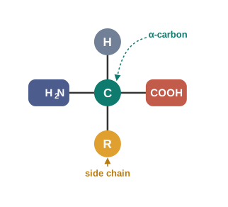
An \(\\alpha\)-amino acid is a compound in which the amino (–NH₂) group is attached to the carbon atom adjacent to the carboxyl (–COOH) group. Only \(\\alpha\)-amino acids are obtained on the hydrolysis of proteins.
How amino acids got their names
All \(\\alpha\)-amino acids have trivial names, which usually reflect a property of the compound or its source. Glycine is so named since it has a sweet taste (in Greek, glykos means sweet), and tyrosine was first obtained from cheese (in Greek, tyros means cheese).
Amino acids are generally represented by a three-letter symbol, and sometimes a one-letter symbol is also used — a convenience that becomes essential once you try to write out the sequence of a protein containing hundreds of residues. The structures of some commonly occurring amino acids, along with their 3-letter and 1-letter symbols, are given in Table 10.2.
Table 10.2: Natural Amino Acids (general structure: \(\\mathrm{H\_2N!-!CH(R)!-!COOH}\))
| 1. Glycine |
H |
Gly |
G |
| 2. Alanine |
–\(\\mathrm{CH\_3}\) |
Ala |
A |
| 3. Valine* |
\(\\mathrm{(H\_3C)\_2CH!-}\) |
Val |
V |
| 4. Leucine* |
\(\\mathrm{(H\_3C)\_2CH!-!CH\_2!-}\) |
Leu |
L |
| 5. Isoleucine* |
\(\\mathrm{H\_3C!-!CH\_2!-!CH(CH\_3)!-}\) |
Ile |
I |
| 6. Arginine* |
\(\\mathrm{HN{=}C(NH\_2)!-!NH!-!(CH\_2)\_3!-}\) |
Arg |
R |
| 7. Lysine* |
\(\\mathrm{H\_2N!-!(CH\_2)\_4!-}\) |
Lys |
K |
| 8. Glutamic acid |
\(\\mathrm{HOOC!-!CH\_2!-!CH\_2!-}\) |
Glu |
E |
| 9. Aspartic acid |
\(\\mathrm{HOOC!-!CH\_2!-}\) |
Asp |
D |
| 10. Glutamine |
\(\\mathrm{H\_2N!-!CO!-!CH\_2!-!CH\_2!-}\) |
Gln |
Q |
| 11. Asparagine |
\(\\mathrm{H\_2N!-!CO!-!CH\_2!-}\) |
Asn |
N |
| 12. Threonine* |
\(\\mathrm{H\_3C!-!CHOH!-}\) |
Thr |
T |
| 13. Serine |
\(\\mathrm{HO!-!CH\_2!-}\) |
Ser |
S |
| 14. Cysteine |
\(\\mathrm{HS!-!CH\_2!-}\) |
Cys |
C |
| 15. Methionine* |
\(\\mathrm{H\_3C!-!S!-!CH\_2!-!CH\_2!-}\) |
Met |
M |
| 16. Phenylalanine* |
\(\\mathrm{C\_6H\_5!-!CH\_2!-}\) |
Phe |
F |
| 17. Tyrosine |
\(\\mathrm{(\\textit{p})HO!-!C\_6H\_4!-!CH\_2!-}\) |
Tyr |
Y |
| 18. Tryptophan* |
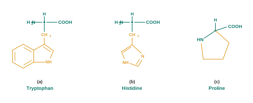
\(\\mathrm{-CH\_2!-}\) attached to an indole ring (see Fig. 10.23) |
Trp |
W |
| 19. Histidine* |
\(\\mathrm{-CH\_2!-}\) attached to an imidazole ring (see Fig. 10.23) |
His |
H |
| 20. Proline |
entire structure is cyclic — a ring joining the side chain back to the nitrogen (see Fig. 10.23) |
Pro |
P |
* essential amino acid
10.2.2 Classification of Amino Acids
Amino acids are classified as acidic, basic or neutral, depending upon the relative number of amino and carboxyl groups in their molecule. The logic is a simple count:
- an equal number of amino and carboxyl groups makes the amino acid neutral;
- more amino groups than carboxyl groups makes it basic;
- more carboxyl groups than amino groups makes it acidic.
Look back at Table 10.2 with this rule in mind and you can classify every entry yourself. Glutamic acid and aspartic acid carry an extra –COOH group in the side chain, so they are acidic; lysine and arginine carry extra nitrogen-containing basic groups, so they are basic; glycine, alanine and valine carry side chains with neither, so they are neutral.
Essential and non-essential amino acids
There is a second, entirely different way of classifying amino acids, and this one is about nutrition rather than about acidity.
The amino acids which can be synthesised in the body are known as non-essential amino acids. On the other hand, those which cannot be synthesised in the body and must be obtained through diet are known as essential amino acids (marked with an asterisk in Table 10.2).
Essential amino acids are those which cannot be synthesised in the body and must be supplied through the diet; non-essential amino acids are those which the body can synthesise for itself.
This is the chemistry behind a very old piece of Indian kitchen wisdom. Cereals such as rice and wheat are low in the essential amino acid lysine, while pulses such as dal are low in methionine but rich in lysine. Eaten separately, neither supplies the full set of essential amino acids well; eaten together, as dal with rice or roti, the shortfall in one is covered by the other. The same principle explains why a diet built on a single cereal alone can leave a person protein-deficient even when they are eating enough food by weight — quantity of protein is not the same thing as quality of protein.
Classify the following amino acids as acidic, basic or neutral, and give the reason in each case: (i) glutamic acid, (ii) lysine, (iii) alanine.
Count the acidic (–COOH) and basic (–\(\\mathrm{NH\_2}\) or other nitrogenous basic) groups in each molecule, including those in the side chain.
Glutamic acid, side chain \(\\mathrm{HOOC!-!CH\_2!-!CH\_2!-}\). Besides the –COOH and –\(\\mathrm{NH\_2}\) of the \(\\alpha\)-carbon, the side chain carries one more –COOH. Carboxyl groups (2) outnumber amino groups (1) → acidic.
Lysine, side chain \(\\mathrm{H\_2N!-!(CH\_2)\_4!-}\). Besides the \(\\alpha\)-amino and \(\\alpha\)-carboxyl groups, the side chain carries one more –\(\\mathrm{NH\_2}\). Amino groups (2) outnumber carboxyl groups (1) → basic.
Alanine, side chain –\(\\mathrm{CH\_3}\). The side chain carries no acidic or basic group, so there is one –COOH and one –\(\\mathrm{NH\_2}\) → equal numbers → neutral.
Answer: glutamic acid — acidic; lysine — basic; alanine — neutral.
Physical properties and the zwitter ion
Amino acids are usually colourless, crystalline solids. They are water-soluble, high-melting solids and behave like salts rather than like simple amines or carboxylic acids. That last observation is the clue to their structure, and it deserves a moment’s thought: an ordinary amine is a low-boiling, often smelly liquid, and an ordinary carboxylic acid is not a high-melting ionic-looking solid. Something ionic must be going on.
This behaviour is due to the presence of both an acidic (carboxyl group) and a basic (amino group) group in the same molecule. In aqueous solution, the carboxyl group can lose a proton and the amino group can accept a proton — and the simplest thing for the molecule to do is to hand the proton from one end to the other, within itself. This gives rise to a dipolar ion known as a zwitter ion, shown in Fig. 10.24. The zwitter ion is neutral overall, but contains both a positive and a negative charge.
\[\\mathrm{R!-!CH(NH\_2)!-!COOH} \\rightleftharpoons \\mathrm{R!-!CH(NH\_3^{+})!-!COO^{-}}\]
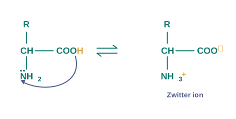
A zwitter ion is the dipolar form of an amino acid, carrying a positive charge on the protonated amino group (\(-\\mathrm{NH\_3^{+}}\)) and a negative charge on the deprotonated carboxyl group (\(-\\mathrm{COO^{-}}\)), while remaining electrically neutral as a whole.
In the zwitter ionic form, amino acids show amphoteric behaviour, as they react both with acids and with bases. Add acid, and the \(-\\mathrm{COO^{-}}\) end accepts a proton; add base, and the \(-\\mathrm{NH\_3^{+}}\) end gives one up. The same molecule can therefore act as an acid or as a base, depending on what it meets.
Optical activity of amino acids
Except glycine, all other naturally occurring \(\\alpha\)-amino acids are optically active, since the \(\\alpha\)-carbon atom is asymmetric — it carries four different groups: –COOH, –\(\\mathrm{NH\_2}\), –H and –R. Glycine is the single exception, because its side chain R is simply –H, which makes two of the four groups on the \(\\alpha\)-carbon identical, and the carbon is no longer asymmetric.
These optically active amino acids exist both in ‘D’ and ‘L’ forms. Most naturally occurring amino acids have L-configuration. L-amino acids are represented by writing the –\(\\mathrm{NH\_2}\) group on the left-hand side.
(question only — no solution) Out of the twenty amino acids in Table 10.2, only one is optically inactive. Identify it and explain, in terms of the groups attached to the \(\\alpha\)-carbon, why it is the exception.
10.2.3 Structure of Proteins
You have already read that proteins are the polymers of \(\\alpha\)-amino acids, and that they are connected to each other by a peptide bond or peptide linkage.
Chemically, a peptide linkage is an amide formed between a –COOH group and an –\(\\mathrm{NH\_2}\) group. The reaction between two molecules of similar or different amino acids proceeds through the combination of the amino group of one molecule with the carboxyl group of the other. This results in the elimination of a water molecule and the formation of a peptide bond, –CO–NH–.
A peptide linkage is the –CO–NH– (amide) bond formed between the carboxyl group of one amino acid and the amino group of another, with the elimination of a molecule of water.
The product of the reaction is called a dipeptide, because it is made up of two amino acids. For example, when the carboxyl group of glycine combines with the amino group of alanine, we get a dipeptide, glycylalanine (Gly-Ala), as shown in Fig. 10.25.
\[\\mathrm{H\_2N!-!CH\_2!-!COOH} + \\mathrm{H\_2N!-!CH(CH\_3)!-!COOH} \\xrightarrow{\\ -\\mathrm{H\_2O}\\ } \\mathrm{H\_2N!-!CH\_2!-!CO!-!NH!-!CH(CH\_3)!-!COOH}\]

From dipeptide to protein
If a third amino acid combines with a dipeptide, the product is called a tripeptide. A tripeptide contains three amino acids linked by two peptide linkages. Similarly, when four, five or six amino acids are linked, the respective products are known as tetrapeptide, pentapeptide or hexapeptide.
When the number of such amino acids is more than ten, then the products are called polypeptides. A polypeptide with more than a hundred amino acid residues, having a molecular mass higher than 10,000 u, is called a protein.
However, the distinction between a polypeptide and a protein is not very sharp. Polypeptides with fewer amino acids are likely to be called proteins if they ordinarily have a well-defined conformation of a protein — insulin, which contains 51 amino acids, is called a protein for exactly this reason.
A polypeptide is made of a single chain of 51 amino acid residues. (i) How many peptide linkages does it contain? (ii) How many molecules of water are eliminated when it is formed from its free amino acids?
Think of the amino acid residues as beads on a string. Each peptide linkage joins two neighbouring residues, and every linkage is formed by the loss of one water molecule.
- For a single unbranched chain, the number of linkages is always one less than the number of residues:
\[\\text{peptide linkages} = 51 - 1 = 50\]
- One water molecule is eliminated per peptide linkage formed, so:
\[\\text{water molecules eliminated} = 50\]
Answer: 50 peptide linkages and 50 molecules of water. (This is the arithmetic behind insulin, which contains 51 amino acids.)
(question only — no solution) Write the structures of the two different dipeptides that can be formed from one molecule of glycine and one molecule of alanine, name each of them, and mark the peptide linkage in each.
Classification by molecular shape
Proteins can be classified into two types on the basis of their molecular shape.
(a) Fibrous proteins. When the polypeptide chains run parallel and are held together by hydrogen and disulphide bonds, then a fibre-like structure is formed. Such proteins are generally insoluble in water. Some common examples are keratin (present in hair, wool and silk) and myosin (present in muscles).
(b) Globular proteins. This structure results when the chains of polypeptides coil around to give a spherical shape. These are usually soluble in water. Insulin and albumins are the common examples of globular proteins.
Fibrous proteins vs Globular proteins
| Arrangement of chains |
Polypeptide chains run parallel to one another |
Polypeptide chains coil around into a spherical shape |
| Held together by |
Hydrogen bonds and disulphide bonds |
Chains coil upon themselves |
| Overall shape |
Thread-like, fibre-like |
Spherical, ball-like |
| Solubility in water |
Generally insoluble |
Usually soluble |
| Examples |
Keratin (hair, wool, silk), myosin (muscles) |
Insulin, albumins |
The four levels of protein structure
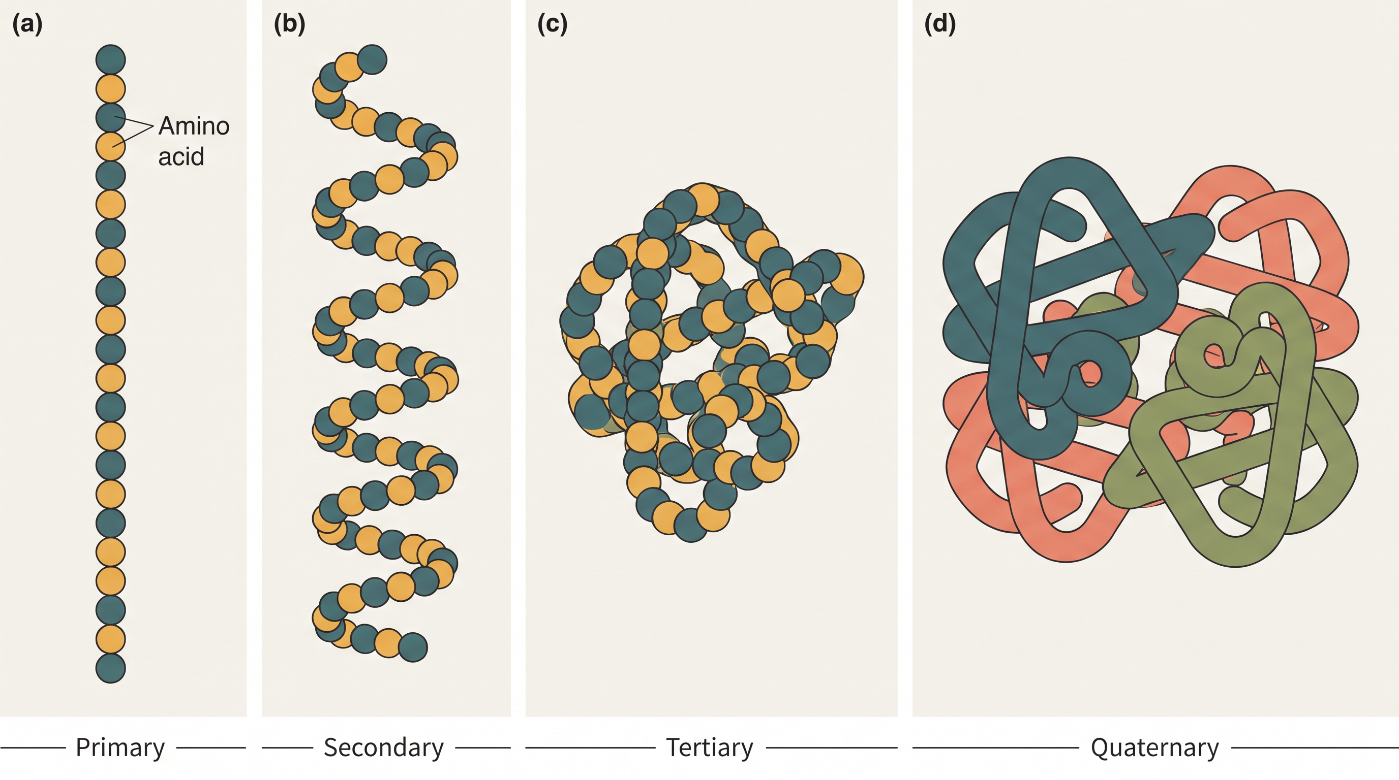
The structure and shape of proteins can be studied at four different levels — primary, secondary, tertiary and quaternary — each level being more complex than the previous one. It helps to think of them as answering four successive questions: which amino acids, in what order? how does the chain fold locally? how does the whole chain fold overall? and how do several chains fit together? The relationship is set out in FC 10.26.
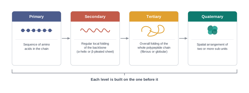
(i) Primary structure of proteins. Proteins may have one or more polypeptide chains. Each polypeptide in a protein has amino acids linked with each other in a specific sequence, and it is this sequence of amino acids that is said to be the primary structure of that protein. Any change in this primary structure, i.e. in the sequence of amino acids, creates a different protein.
For a long time it was not even clear that a protein had a single definite sequence — many chemists suspected proteins were ill-defined mixtures. Frederick Sanger, working in Cambridge in the years after the Second World War, chose insulin and set out to determine the exact order of its amino acids. He broke the protein into small fragments, identified the amino acid at the end of each fragment, and pieced the overlapping fragments back together like a jigsaw. It took him about a decade. The result — the complete sequence of insulin — proved that every molecule of a given protein has one exact, reproducible sequence, and it won him the Nobel Prize in Chemistry in 1958. This is the primary structure you are reading about, established for the first time.
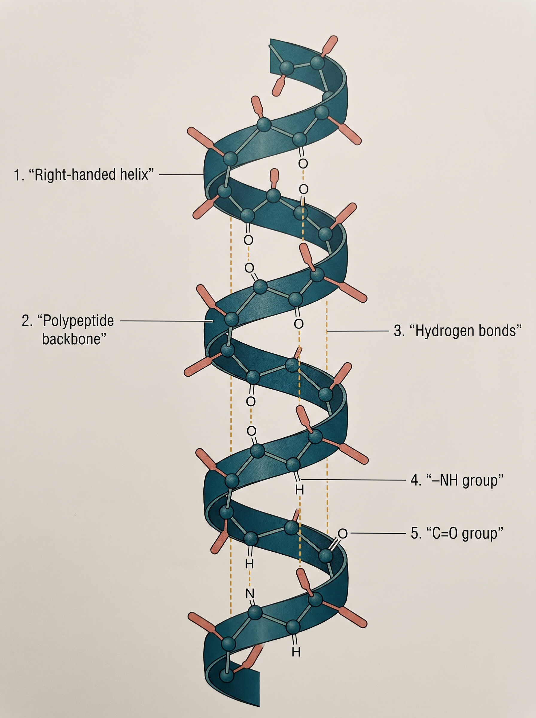
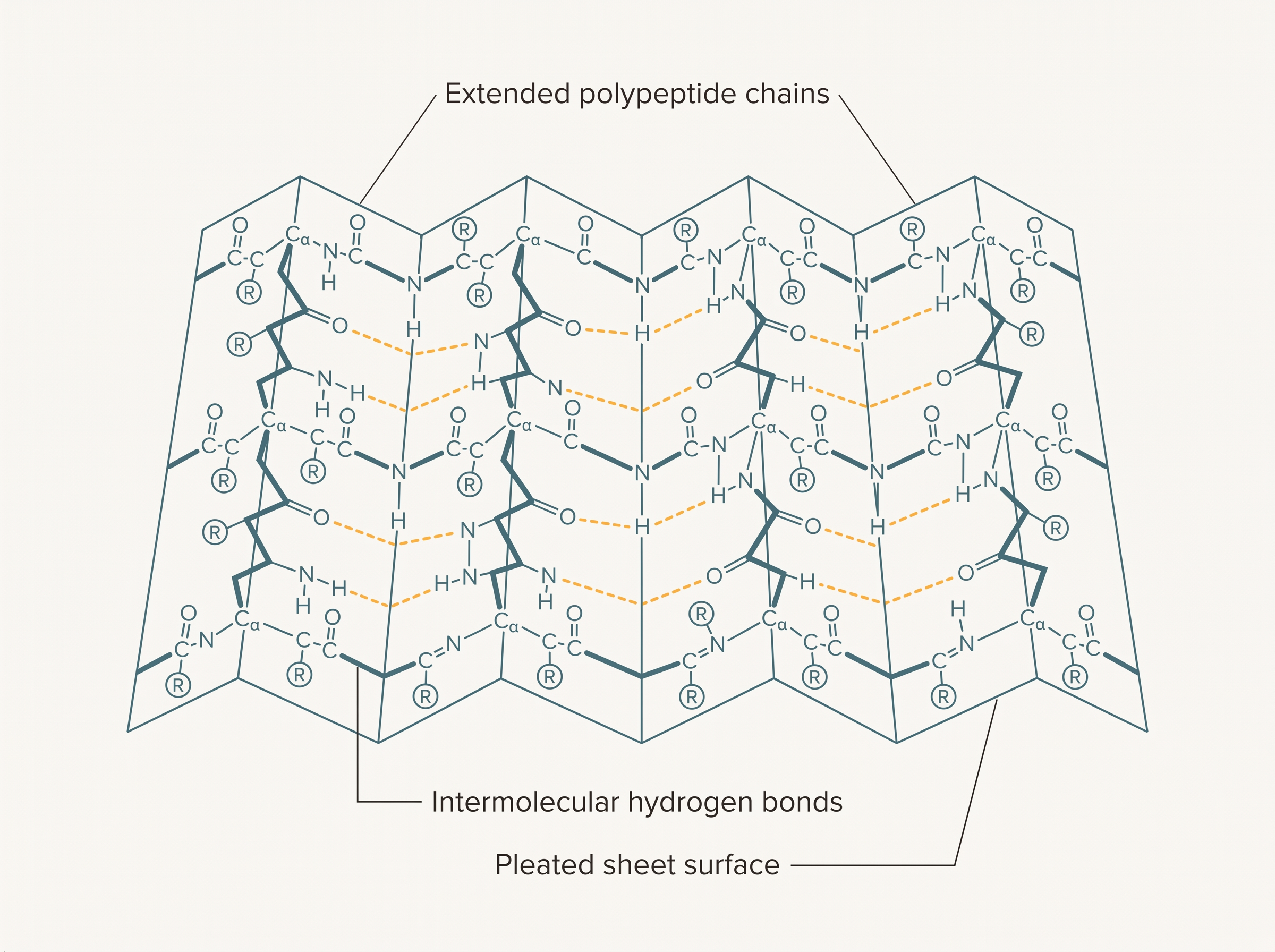
(ii) Secondary structure of proteins. The secondary structure of a protein refers to the shape in which a long polypeptide chain can exist. They are found to exist in two different types of structures, viz. \(\\alpha\)-helix and \(\\beta\)-pleated sheet structure. These structures arise due to the regular folding of the backbone of the polypeptide chain, due to hydrogen bonding between the \(>\\mathrm{C=O}\) and –NH– groups of the peptide bond.
The \(\\alpha\)-helix is one of the most common ways in which a polypeptide chain forms all possible hydrogen bonds, by twisting into a right-handed screw (helix) with the –NH group of each amino acid residue hydrogen bonded to the \(>\\mathrm{C=O}\) of an adjacent turn of the helix, as shown in Fig. 10.27.
In the \(\\beta\)-pleated sheet structure, all peptide chains are stretched out to nearly maximum extension and then laid side by side, where they are held together by intermolecular hydrogen bonds. The structure resembles the pleated folds of drapery, and is therefore known as a \(\\beta\)-pleated sheet. This is shown in Fig. 10.28.
α-Helix vs β-Pleated sheet
| Shape of the chain |
Coiled into a right-handed screw (helix) |
Stretched out to nearly maximum extension |
| Hydrogen bonding |
Within the same chain — –NH of one residue to \(>\\mathrm{C=O}\) of an adjacent turn |
Between neighbouring chains (intermolecular) |
| Arrangement |
One chain twisted on itself |
Chains laid side by side |
| Appearance |
Spring-like coil |
Pleated folds, like drapery |
The \(\\alpha\)-helix was not discovered by looking at a protein — it was worked out on paper. Linus Pauling, in the early 1950s, reasoned that the peptide unit is flat and that every –NH group in the backbone should be allowed to make a hydrogen bond if the structure is to be stable. Folding a paper model of the chain until those two conditions were satisfied gave a right-handed coil with a non-integral number of residues per turn — a result so unusual that it was at first met with scepticism, and so correct that it is now in every chemistry textbook, including this one.
(iii) Tertiary structure of proteins. The tertiary structure of proteins represents the overall folding of the polypeptide chains, i.e. the further folding of the secondary structure. It gives rise to the two major molecular shapes you have already met — fibrous and globular.
The main forces which stabilise the 2° and 3° structures of proteins are hydrogen bonds, disulphide linkages, van der Waals forces and electrostatic forces of attraction. Notice that none of these is a strong covalent bond except the disulphide linkage — a point worth remembering, because it explains why a protein’s shape can be destroyed so easily.
(iv) Quaternary structure of proteins. Some of the proteins are composed of two or more polypeptide chains, referred to as sub-units. The spatial arrangement of these sub-units with respect to each other is known as the quaternary structure.
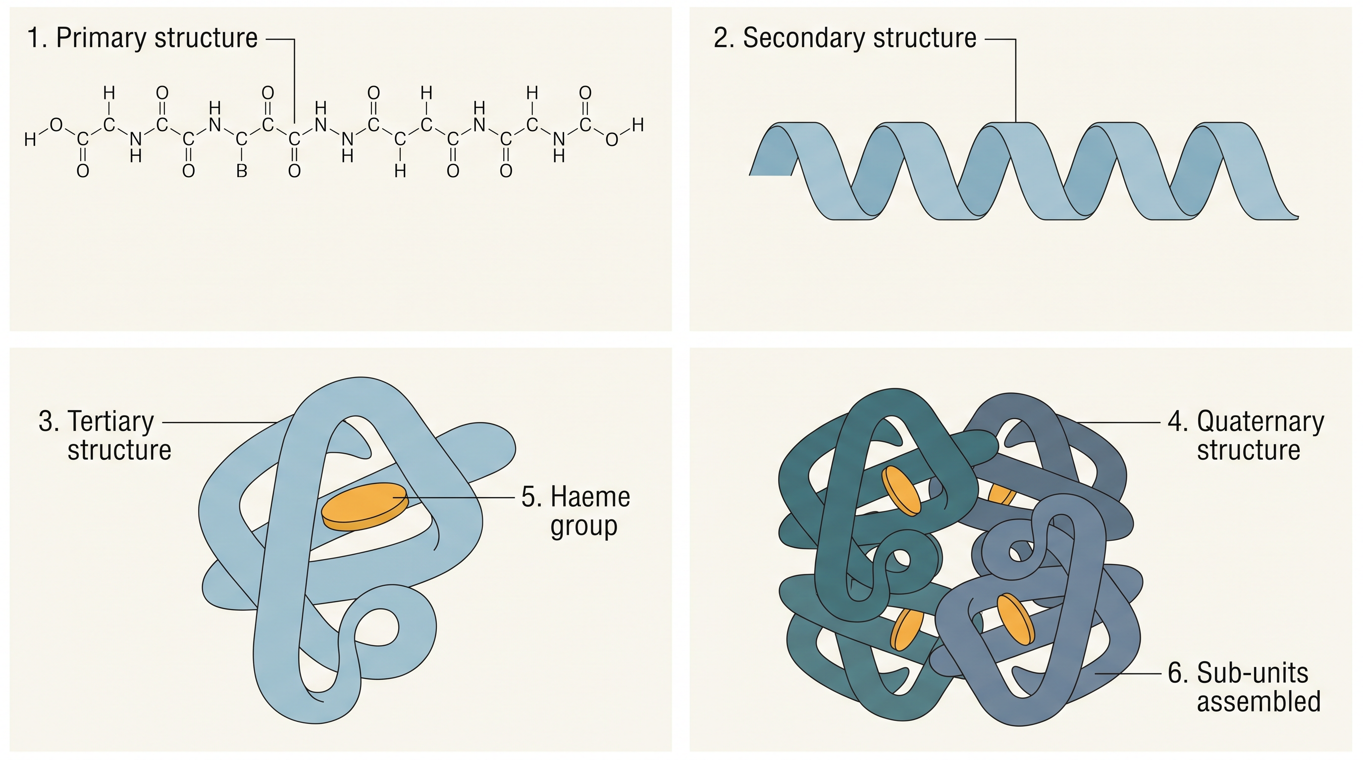
A diagrammatic representation of all these four structures is given in Fig. 10.29, where each coloured ball represents an amino acid. Haemoglobin, the oxygen-carrying protein of blood, is a real example in which all four levels can be seen at once, and its primary, secondary, tertiary and quaternary structures are shown in Fig. 10.30.
10.2.4 Denaturation of Proteins
A protein found in a biological system with a unique three-dimensional structure and biological activity is called a native protein.
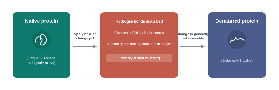
When a protein in its native form is subjected to a physical change like a change in temperature, or a chemical change like a change in pH, the hydrogen bonds are disturbed. Due to this, the globules unfold and the helix gets uncoiled, and the protein loses its biological activity. This is called denaturation of protein.
Denaturation is the loss of the three-dimensional structure (and hence the biological activity) of a native protein, brought about by a physical change such as heating or a chemical change such as a change in pH. During denaturation the secondary and tertiary structures are destroyed, but the primary structure remains intact.
Read the last clause of that definition again, because it is the whole point. The sequence of amino acids — the covalently bonded backbone — is untouched. What is lost is the shape, and with the shape goes the function. A protein that cannot hold its fold cannot do its job.
The coagulation of egg white on boiling is a common example of denaturation. Another example is the curdling of milk, which is caused by the formation of lactic acid by the bacteria present in milk — here it is a fall in pH, rather than heat, that does the damage.
Q3. When an egg is boiled, the clear egg white turns into a firm white solid — and cooling it never brings the liquid back. What exactly has changed in the protein, and why is the change one-way?
A. Egg white contains globular proteins dissolved in water. On boiling, the heat disturbs the hydrogen bonds that were holding the polypeptide chains in their neatly coiled, folded shape. The globules unfold and the helices uncoil — the secondary and tertiary structures are destroyed — and the unfolded chains tangle with one another and coagulate into a solid mass. This is denaturation. The primary structure, the sequence of amino acids joined by strong covalent peptide bonds, is unchanged; only the shape is gone. Cooling cannot restore it, because the countless weak interactions that once held the exact native fold cannot spontaneously re-form in the right pattern once the chains have tangled together — so the egg stays cooked.
Denaturation is used deliberately every day, and usually to destroy something. Rubbing a swab of alcohol on the skin before an injection works because ethanol denatures the proteins of the micro-organisms present, so they can no longer function. Sterilising instruments by boiling, and pasteurising milk by heating it, work the same way. The flip side is a safety warning worth taking seriously in a biology or biochemistry laboratory: an enzyme preparation, a blood sample or a vaccine that has been left in the sun, warmed carelessly, or exposed to strong acid or alkali may look perfectly unchanged and still be completely dead, because denaturation destroys function without necessarily destroying appearance.
(question only — no solution) Curd is made by adding a small amount of curd to warm milk and leaving it to set. Identify the chemical change responsible for the milk setting, name the substance produced by the bacteria, and state which levels of protein structure are affected.
10.4 The melting points and the solubility in water of amino acids are generally higher than those of the corresponding halo acids. Explain.
10.5 Where does the water present in the egg go after boiling the egg?
10.3 Enzymes
Life is possible due to the coordination of various chemical reactions in living organisms. An example is the digestion of food, the absorption of appropriate molecules, and ultimately the production of energy. This process involves a whole sequence of reactions, and all of them occur in the body under very mild conditions.
That phrase deserves a second look. In the laboratory, the hydrolysis of a disaccharide needs boiling with a mineral acid; in your body, the same bond is broken at about 310 K, at nearly neutral pH, in a matter of seconds. Something must be making these reactions possible without heat, pressure or strong acid. That something is a class of biological catalysts called enzymes.
Enzymes are biocatalysts — substances produced by living cells that speed up biochemical reactions in the body under mild conditions of temperature and pH, without themselves being consumed.
Almost all the enzymes are globular proteins — which is why this section follows the section on proteins, and why everything you learned about protein shape and denaturation applies directly here.
Specificity and naming
Enzymes are very specific for a particular reaction and for a particular substrate. A chemical catalyst like finely divided platinum will hydrogenate almost any double bond you offer it; an enzyme is not like that. It usually accepts one substrate, or one closely related family of substrates, and catalyses one reaction on it. (The substrate is simply the substance on which the enzyme acts.)
Because of this specificity, enzymes are generally named after the compound, or the class of compounds, upon which they work. For example, the enzyme that catalyses the hydrolysis of maltose into glucose is named maltase.
\[\\mathrm{C\_{12}H\_{22}O\_{11}} \\xrightarrow{\\ \\text{Maltase}\\ } 2,\\mathrm{C\_6H\_{12}O\_6}\]
Here maltose gives two molecules of glucose — the same reaction you met in Section 10.1, now carried out in the body at ordinary body temperature instead of by boiling with dilute acid.
Sometimes enzymes are also named after the reaction where they are used. For example, the enzymes which catalyse the oxidation of one substrate with the simultaneous reduction of another substrate are named as oxidoreductase enzymes. Whichever route is used, one rule holds: the ending of the name of an enzyme is -ase.
The word enzyme comes from the Greek for “in yeast” — a reminder of where the first hints of enzyme action were found. For most of the nineteenth century, chemists argued about whether fermentation required a living cell or merely a chemical agent. The question was settled in the closing years of that century by Eduard Buchner, who ground up yeast cells, filtered out every trace of living matter, and found that the cell-free juice still fermented sugar perfectly well. The catalytic power belonged to a substance, not to life itself — and he received the Nobel Prize in Chemistry in 1907 for this work. Biochemistry as a subject really begins here: whatever a cell can do, it does with molecules.
(question only — no solution) An enzyme catalyses the hydrolysis of a disaccharide and is found to have absolutely no effect on a closely similar disaccharide. Name the property of enzymes being illustrated, and state one structural feature of a disaccharide (from Section 10.1) that could make the two substrates different enough to matter.
10.3.1 Mechanism of Enzyme Action
Enzymes are needed only in small quantities for the progress of a reaction. This follows directly from the fact that they are catalysts: an enzyme is not used up in the reaction it speeds up. Having done its work on one substrate molecule, it is released unchanged and is free to act on the next. One molecule of enzyme can therefore process an enormous number of substrate molecules, one after another.
Similar to the action of chemical catalysts, enzymes are said to reduce the magnitude of the activation energy. Recall what activation energy means: it is the minimum extra energy that colliding reactant molecules must possess before they can be converted into products — the height of the energy barrier that stands between reactants and products. Lower the barrier, and a far greater fraction of molecules can cross it at the same temperature, so the reaction goes much faster.
For example, the activation energy for the acid hydrolysis of sucrose is \(6.22\\ \\text{kJ mol}^{-1}\), while the activation energy is only \(2.15\\ \\text{kJ mol}^{-1}\) when it is hydrolysed by the enzyme sucrase. The enzyme has not changed the reactants or the products in any way — it has simply provided an easier path between them, as you can see in the energy profile of Fig. 10.32.
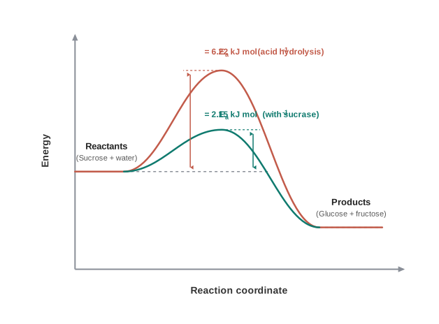
the two energy profiles for the hydrolysis of sucrose in Fig. 10.32 (Study the figure and answer: (a) Which curve corresponds to the enzyme-catalysed path, and how did you decide? (b) The reactants and the products sit at the same energy levels on both curves — what does this tell you about whether the enzyme changes the energy released by the reaction? (c) The enzyme lowers the barrier by about \(4\\ \\text{kJ mol}^{-1}\). Using what you know of the Arrhenius relation, would you expect the rate to increase by roughly this same factor, or by a much larger one? Give your reasoning.)
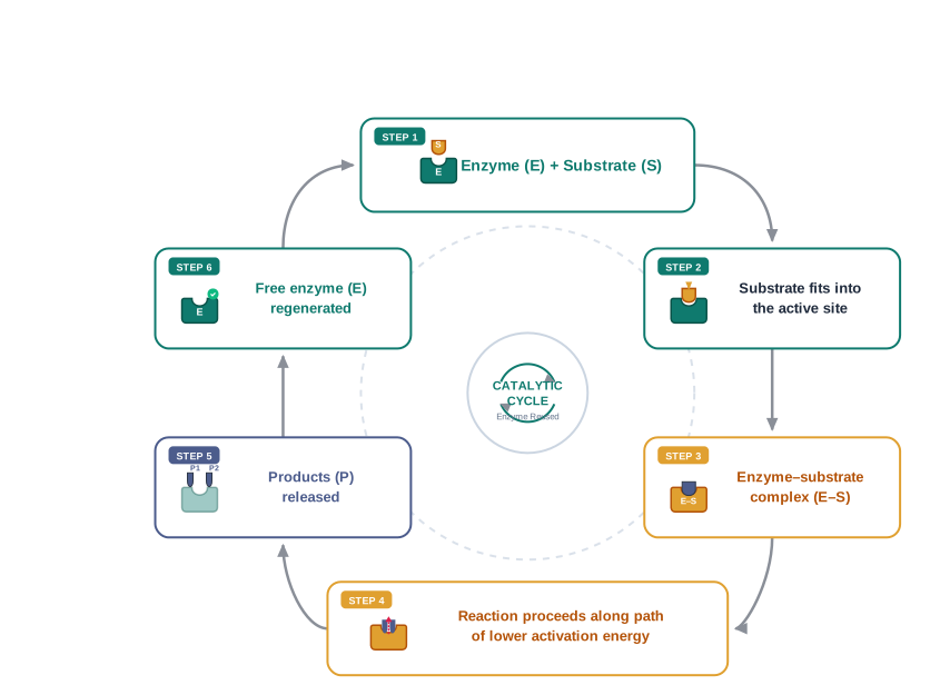
Mechanism for the enzyme action has been discussed earlier, in the chemistry of catalysis. The essential picture is worth recalling here, because it explains the specificity you have just read about. An enzyme molecule carries a small region of a particular shape, called the active site, into which only a substrate of the matching shape can fit. The substrate binds there, forming an enzyme–substrate complex; the reaction takes place on the bound substrate along the low-barrier path; the products then leave, and the free enzyme is ready for the next substrate molecule. The full cycle is set out in FC 10.33.
The activation energy for the acid hydrolysis of sucrose is \(6.22\\ \\text{kJ mol}^{-1}\), while for hydrolysis by sucrase it is \(2.15\\ \\text{kJ mol}^{-1}\). Assuming the pre-exponential factor \(A\) is the same in both cases, calculate how many times faster the enzyme-catalysed hydrolysis is at 298 K.
(\(R = 8.314\\ \\text{J K}^{-1}\\text{mol}^{-1}\))
Use the Arrhenius equation, which links the rate constant to the activation energy:
\[k = A,e^{-E\_a/RT}\]
Write it once for each path and take the ratio. Since \(A\) is the same for both, it cancels:
\[\\frac{k\_{\\text{enzyme}}}{k\_{\\text{acid}}} = \\frac{A,e^{-E\_{a(\\text{enzyme})}/RT}}{A,e^{-E\_{a(\\text{acid})}/RT}} = e^{\\left(E\_{a(\\text{acid})} - E\_{a(\\text{enzyme})}\\right)/RT}\]
Now put in the numbers. First find the difference in activation energies, converting kJ to J because \(R\) is in joules:
\[E\_{a(\\text{acid})} - E\_{a(\\text{enzyme})} = 6.22 - 2.15 = 4.07\\ \\text{kJ mol}^{-1} = 4070\\ \\text{J mol}^{-1}\]
\[RT = 8.314\\ \\text{J K}^{-1}\\text{mol}^{-1} \\times 298\\ \\text{K} = 2477.6\\ \\text{J mol}^{-1}\]
\[\\frac{k\_{\\text{enzyme}}}{k\_{\\text{acid}}} = e^{4070/2477.6} = e^{1.643} \\approx 5.2\]
Answer: The enzyme-catalysed hydrolysis is about 5.2 times faster than the acid-catalysed hydrolysis at 298 K, for the same value of \(A\).
Two things are worth noticing. First, the ratio depends on the difference in activation energies, not on their individual values. Second, because \(E\_a\) sits in an exponent, even a modest lowering of the barrier multiplies the rate — and the enzyme achieves this at body temperature, with no acid at all.
Enzymes vs ordinary chemical catalysts
| Chemical nature |
Almost all are globular proteins |
Usually metals, metal oxides or acids/bases |
| Specificity |
Highly specific — one enzyme, usually one substrate and one reaction |
Generally not specific; will catalyse a whole class of reactions |
| Conditions needed |
Mild — body temperature, nearly neutral pH |
Often high temperature and pressure, or strong acid |
| Effect on activation energy |
Lowers it (e.g. from \(6.22\) to \(2.15\\ \\text{kJ mol}^{-1}\) for sucrose hydrolysis) |
Lowers it |
| Quantity required |
Very small |
Small, but usually larger |
| Effect of heating strongly |
Denatured — loses activity permanently |
Usually unaffected; many need heat to work |
Look at the back of a packet of “biological” washing powder and you will find enzymes listed: a protease to break down protein stains such as blood, egg and gravy; an amylase for starch stains; and a lipase for grease. Each is doing in the washing machine exactly what it does in the body — hydrolysing a specific type of biomolecule — and it does so in warm water rather than hot. That last point is a genuine green-chemistry gain: a wash at 30 °C instead of 60 °C uses substantially less electricity for every load, in every machine, every day. The same logic drives the wider use of enzymes in industry, where an enzyme-catalysed step can replace one that would otherwise need a strong acid, a high temperature, and the waste treatment that goes with them.
(question only — no solution) Sucrose is hydrolysed in the laboratory by boiling with dilute \(\\mathrm{HCl}\), and in the body by the enzyme sucrase, giving the same two products. Name the products, state the type of linkage broken, and explain why the enzyme-catalysed reaction can be carried out at body temperature while the laboratory reaction cannot.
10.4 Vitamins
It has been observed that certain organic compounds are required in small amounts in our diet, but that their deficiency causes specific diseases. These compounds are called vitamins.
The word “small” is important here. A grown person eats hundreds of grams of carbohydrate, fat and protein every day, but needs only milligrams — sometimes micrograms — of a vitamin. Vitamins are not fuel and they are not building material; they are required for particular jobs, and when the tiny quantity needed is missing, a specific disease follows.
Most of the vitamins cannot be synthesised in our body, but plants can synthesise almost all of them, so they are considered as essential food factors. However, the bacteria of the gut can produce some of the vitamins required by us. All the vitamins are generally available in our diet.
Different vitamins belong to various chemical classes, and it is difficult to define them on the basis of structure. There is no single functional group, no single skeleton, that all vitamins share — they are grouped together by what they do, not by what they look like. They are generally regarded as organic compounds required in the diet in small amounts to perform specific biological functions for normal maintenance of optimum growth and health of the organism.
Vitamins are organic compounds required in the diet in small amounts to perform specific biological functions, necessary for the normal maintenance of optimum growth and health of the organism.
How vitamins are named
Vitamins are designated by alphabets — A, B, C, D and so on. Some of them are further named as sub-groups, e.g. \(\\mathrm{B\_1}\), \(\\mathrm{B\_2}\), \(\\mathrm{B\_6}\), \(\\mathrm{B\_{12}}\), because what was first thought to be a single vitamin B later turned out to be several different compounds occurring together.
An excess of vitamins is also harmful, and vitamin pills should not be taken without the advice of a doctor.
The term “Vitamine” was coined from the words vital + amine, since the earlier identified compounds had amino groups. Later work showed that most of them did not contain amino groups, so the letter ‘e’ was dropped, and the term vitamin is used these days.
The naming story has an author: the term vitamine was coined in the early twentieth century by the biochemist Casimir Funk, who was working on the substance in rice husk that cured beri-beri. He was right that these factors were vital, and wrong that they were all amines — chemistry has kept the name and quietly dropped the ‘e’.
Long before anyone knew what a vitamin was, a Scottish naval surgeon named James Lind ran what is often called the first controlled clinical trial. Scurvy was killing more sailors on long voyages than battles were. In the mid-eighteenth century, aboard ship, Lind divided sailors suffering from scurvy into small groups and gave each group a different supposed remedy — cider, vinegar, seawater, oranges and lemons among them. Only the men given the citrus fruit recovered. He had no idea that a molecule, ascorbic acid, was responsible; it would be nearly two centuries before vitamin C was isolated. But British ships eventually began carrying citrus, and one of the great killers of the age of sail simply disappeared. Every time you read “Scurvy (bleeding gums)” in the table below, this is the history behind that one line.
10.4.1 Classification of Vitamins
Vitamins are classified into two groups depending upon their solubility in water or fat. This looks like a small, almost trivial physical property to build a classification on — but it turns out to decide how the body handles the vitamin, how long it is kept, and how often you must eat it. The classification is set out in FC 10.34.
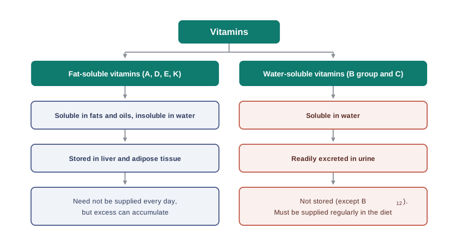
(i) Fat-soluble vitamins. Vitamins which are soluble in fats and oils but insoluble in water are kept in this group. These are vitamins A, D, E and K. They are stored in the liver and in adipose (fat-storing) tissues.
(ii) Water-soluble vitamins. The B group vitamins and vitamin C are soluble in water, so they are grouped together. Water-soluble vitamins must be supplied regularly in the diet, because they are readily excreted in the urine and cannot be stored (except vitamin \(\\mathrm{B\_{12}}\)) in our body.
Follow the cause and effect through, because this is the heart of the section. A fat-soluble vitamin dissolves in the fatty tissue it meets, and simply stays there — the body has a store to draw on. A water-soluble vitamin dissolves in the watery fluids of the body, is carried to the kidneys, and leaves in the urine — so there is no store, and today’s supply is gone by tomorrow. This is drawn in Fig. 10.35.
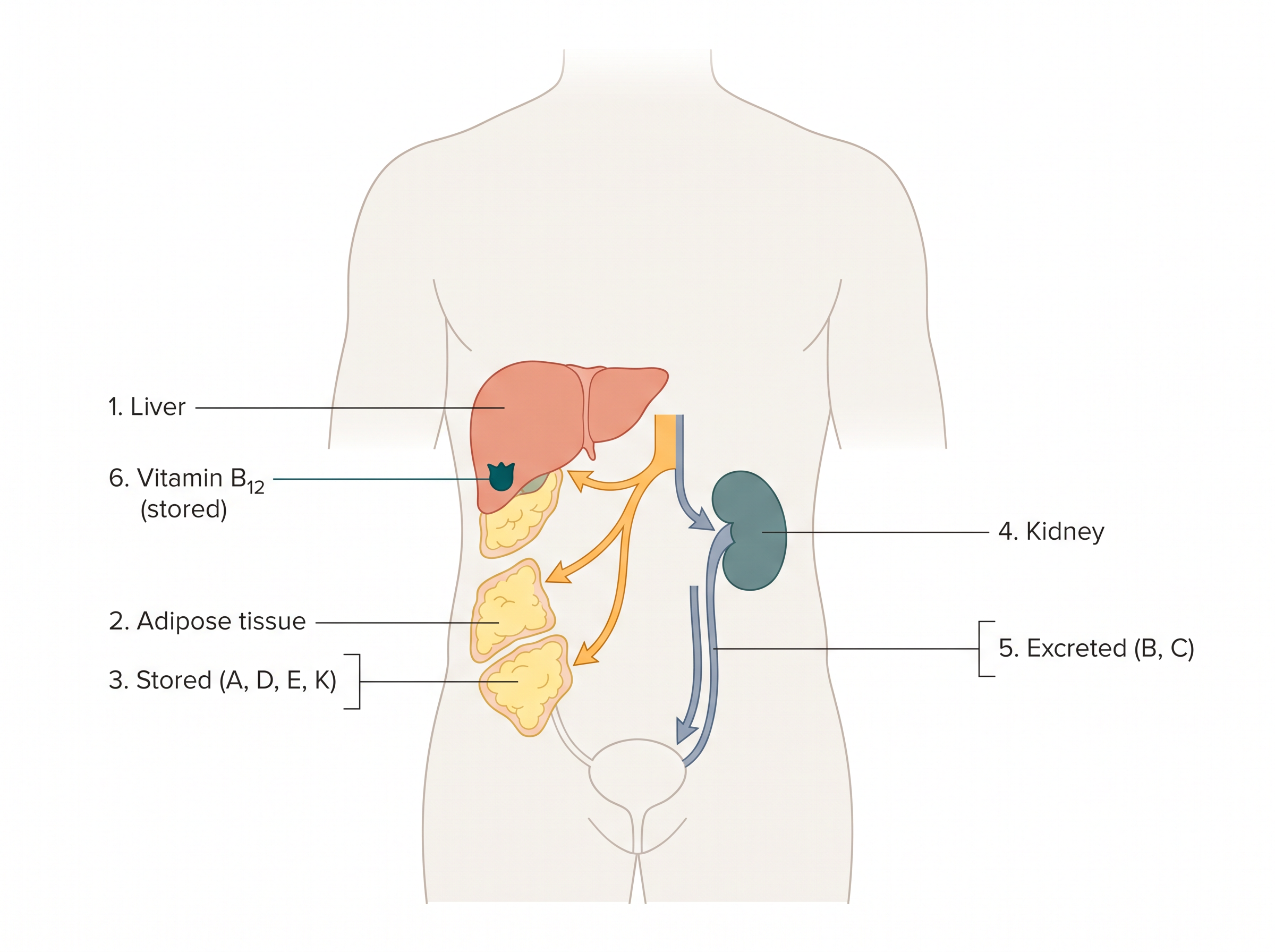
Q4. Vitamin C must be eaten almost every day, while vitamin A can be stored for months. What difference in their molecules decides this?
A. Their solubility. Vitamin A is fat-soluble: it dissolves in fats and oils and is laid down in the liver and adipose tissue, so the body builds up a reserve that can last a long time. Vitamin C is water-soluble: it dissolves in the aqueous fluids of the body and is readily excreted in the urine, and (unlike vitamin \(\\mathrm{B\_{12}}\)) it cannot be stored. With no reserve to fall back on, the supply must be renewed from the diet more or less daily — which is precisely why scurvy appears on long voyages, and why oranges, amla and green leafy vegetables have to be regular items on the plate rather than occasional ones.
Fat-soluble vitamins vs Water-soluble vitamins
| Members |
A, D, E, K |
B group vitamins and C |
| Solubility |
Soluble in fats and oils; insoluble in water |
Soluble in water |
| Storage in the body |
Stored in the liver and adipose (fat-storing) tissue |
Not stored (except \(\\mathrm{B\_{12}}\)) |
| Excretion |
Not readily excreted |
Readily excreted in the urine |
| Frequency of supply needed |
Need not be supplied every day |
Must be supplied regularly in the diet |
| Risk on excess intake |
Can accumulate, so excess is more likely to be harmful |
Excess is largely excreted |
The fat-soluble/water-soluble split is also a safety rule for supplements, and it explains NCERT’s warning that vitamin pills should not be taken without a doctor’s advice. Swallow more vitamin C than you need and most of the excess leaves in the urine within hours. Swallow far more vitamin A or vitamin D than you need, day after day, and there is nowhere for it to go: it dissolves in the fat of the liver and adipose tissue and accumulates, and a large enough accumulation is toxic. “It is only a vitamin, so more must be better” is therefore false chemistry — and dangerously so for exactly four vitamins, A, D, E and K.
Some important vitamins, their sources, and the diseases caused by their deficiency are listed in Table 10.3.
Table 10.3: Some important Vitamins, their Sources and their Deficiency Diseases
| 1. |
Vitamin A |
Fish liver oil, carrots, butter and milk |
Xerophthalmia (hardening of cornea of eye); Night blindness |
| 2. |
Vitamin \(\\mathrm{B\_1}\) (Thiamine) |
Yeast, milk, green vegetables and cereals |
Beri beri (loss of appetite, retarded growth) |
| 3. |
Vitamin \(\\mathrm{B\_2}\) (Riboflavin) |
Milk, eggwhite, liver, kidney |
Cheilosis (fissuring at corners of mouth and lips), digestive disorders and burning sensation of the skin |
| 4. |
Vitamin \(\\mathrm{B\_6}\) (Pyridoxine) |
Yeast, milk, egg yolk, cereals and grams |
Convulsions |
| 5. |
Vitamin \(\\mathrm{B\_{12}}\) |
Meat, fish, egg and curd |
Pernicious anaemia (RBC deficient in haemoglobin) |
| 6. |
Vitamin C (Ascorbic acid) |
Citrus fruits, amla and green leafy vegetables |
Scurvy (bleeding gums) |
| 7. |
Vitamin D |
Exposure to sunlight, fish and egg yolk |
Rickets (bone deformities in children) and osteomalacia (soft bones and joint pain in adults) |
| 8. |
Vitamin E |
Vegetable oils like wheat germ oil, sunflower oil, etc. |
Increased fragility of RBCs and muscular weakness |
| 9. |
Vitamin K |
Green leafy vegetables |
Increased blood clotting time |
Read the last row carefully. A deficiency of vitamin K does not cause a dramatic disease with a name of its own — it simply means that blood takes longer than it should to clot. That single line is the reason vitamin K is given before certain surgeries and to newborn infants, and it is a good illustration of the general principle of this section: a vitamin is needed in a minute amount, for one specific biological function, and its absence shows up as the failure of exactly that function.
Two patients report to a clinic. Patient P has bleeding gums. Patient Q cannot see properly in dim light. For each patient: (i) name the vitamin most likely to be deficient, (ii) state whether it is fat-soluble or water-soluble, and (iii) suggest two dietary sources.
Work backwards from the symptom to the deficiency disease listed in Table 10.3, and then from the vitamin to its class.
Patient P — bleeding gums. Table 10.3 lists bleeding gums as the sign of scurvy, which is the deficiency disease of vitamin C (ascorbic acid). Vitamin C belongs to the water-soluble group, along with the B vitamins. Since it cannot be stored, the supply must be renewed regularly. Sources: citrus fruits and amla (green leafy vegetables are also listed).
Patient Q — poor vision in dim light. This is night blindness, listed against vitamin A. Vitamin A is fat-soluble, and is stored in the liver and adipose tissue. Sources: carrots and fish liver oil (butter and milk are also listed).
Answer: P is deficient in vitamin C — water-soluble; sources: citrus fruits, amla. Q is deficient in vitamin A — fat-soluble; sources: carrots, fish liver oil.
(question only — no solution) A child living in a crowded area with very little exposure to sunlight develops bone deformities. Name the vitamin most likely to be deficient, name the deficiency disease, state the class (fat-soluble or water-soluble) to which the vitamin belongs, and name the corresponding condition seen in adults.
(question only — no solution) Vegetables lose a part of their vitamin content when they are boiled and the cooking water is thrown away, but very little of it when they are lightly fried in oil. Using the classification in this section, explain which class of vitamins is lost in each case and why.
10.5 Nucleic Acids
Every generation of each and every species resembles its ancestors in many ways. How are these characteristics transmitted from one generation to the next?
It has been observed that the nucleus of a living cell is responsible for this transmission of inherent characters, which is also called heredity. The particles in the nucleus of the cell responsible for heredity are called chromosomes, and these are made up of proteins and another type of biomolecules called nucleic acids.
Nucleic acids are mainly of two types: deoxyribonucleic acid (DNA) and ribonucleic acid (RNA). Since nucleic acids are long-chain polymers of nucleotides, they are also called polynucleotides.
Nucleic acids are long-chain polymers of nucleotides, found in the nucleus of the cell, which are responsible for the transmission of hereditary characters from one generation to the next. The two types are DNA and RNA.
James Dewey Watson. Born in Chicago, Illinois, in 1928, Dr Watson received his Ph.D. (1950) from Indiana University in Zoology. He is best known for his discovery of the structure of DNA, for which he shared the 1962 Nobel Prize in Physiology and Medicine with Francis Crick and Maurice Wilkins. They proposed that the DNA molecule takes the shape of a double helix — an elegantly simple structure that resembles a gently twisted ladder. The rails of the ladder are made of alternating units of phosphate and the sugar deoxyribose; the rungs are each composed of a pair of purine/pyrimidine bases. This research laid the foundation for the emerging field of molecular biology. The complementary pairing of nucleotide bases explains how identical copies of parental DNA pass on to two daughter cells. This research launched a revolution in biology that led to modern recombinant DNA techniques.
10.5.1 Chemical Composition of Nucleic Acids
The simplest way to find out what a nucleic acid is made of is to take it apart. Complete hydrolysis of DNA (or RNA) yields three kinds of products:
- a pentose sugar (a five-carbon sugar),
- phosphoric acid, and
- nitrogen-containing heterocyclic compounds, called bases.
The identity of the sugar is the first point of difference between the two nucleic acids. In DNA molecules, the sugar moiety is \(\\beta\)-D-2-deoxyribose, whereas in the RNA molecule it is \(\\beta\)-D-ribose. Both are drawn in Fig. 10.36.
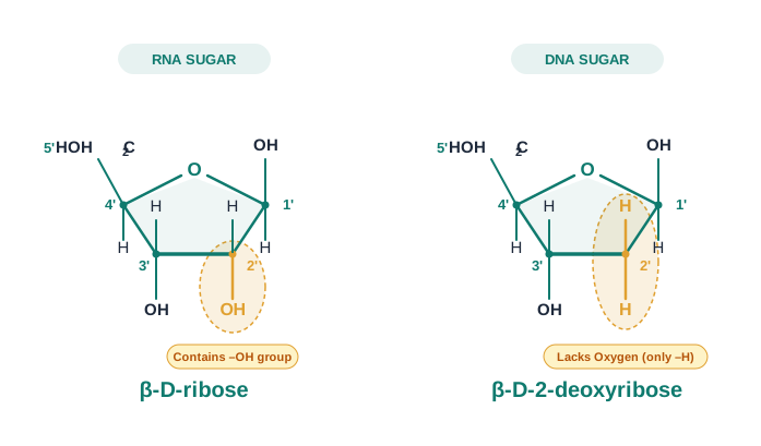
The names carry the difference. “Deoxy” means “minus one oxygen”: 2-deoxyribose is simply ribose with the –OH group at C-2 replaced by –H. One oxygen atom, on one carbon, is what separates the sugar of heredity from the sugar of protein synthesis. Both are aldopentoses, the two sugars mentioned at the end of Section 10.1.
The nitrogenous bases
DNA contains four bases, viz. adenine (A), guanine (G), cytosine (C) and thymine (T). RNA also contains four bases: the first three bases are the same as in DNA, but the fourth one is uracil (U) in place of thymine. The structures of all five are shown in Fig. 10.37.
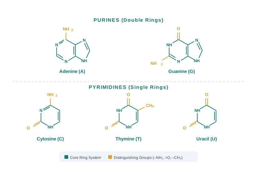
Look at the shapes rather than the details and a pattern appears: adenine and guanine are built on a double ring system (these are the purine bases), while cytosine, thymine and uracil are built on a single ring (the pyrimidine bases). Notice also that thymine and uracil differ only by a –\(\\mathrm{CH\_3}\) group — which is why RNA can use one in place of the other.
10.5.2 Structure of Nucleic Acids
Nucleic acids are built up in a strict order, one piece at a time, and it is worth assembling them in your mind in that order.
A unit formed by the attachment of a base to the 1′ position of the sugar is known as a nucleoside. In nucleosides, the sugar carbons are numbered as 1′, 2′, 3′, etc. (read as “one prime”, “two prime”), in order to distinguish these from the numbering of the atoms in the bases. A nucleoside is shown in Fig. 10.38(a).
When a nucleoside is linked to phosphoric acid at the 5′-position of the sugar moiety, we get a nucleotide, shown in Fig. 10.38(b).
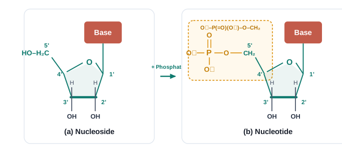
A nucleoside is a unit formed by joining a nitrogenous base to the 1′ position of a pentose sugar.
A nucleotide is a nucleoside joined to phosphoric acid at the 5′ position of its sugar; nucleic acids are polymers of nucleotides.
Nucleoside vs Nucleotide
| Components |
Base + sugar |
Base + sugar + phosphoric acid |
| Point of attachment |
Base at the 1′ position of the sugar |
Base at 1′; phosphate at the 5′ position of the sugar |
| Relationship |
The parent unit |
Nucleoside + phosphate |
| Role |
Building block of a nucleotide |
The repeating monomer unit of nucleic acids |
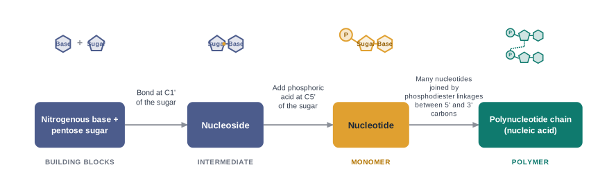
The phosphodiester linkage
Nucleotides are joined together by a phosphodiester linkage between the 5′ and 3′ carbon atoms of the pentose sugar. In other words, one phosphate group forms a bridge: its one end is fixed at the 5′ carbon of one sugar, and its other end at the 3′ carbon of the next sugar. The formation of a typical dinucleotide is shown in Fig. 10.40.

Repeat that linkage many times and you obtain a long chain in which sugar and phosphate alternate along the backbone, with a base hanging off every sugar. A simplified version of the nucleic acid chain is shown in Fig. 10.41.
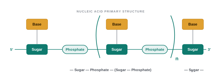
Because the backbone is the same everywhere — sugar, phosphate, sugar, phosphate — the only thing that can vary from one nucleic acid to another is the order of the bases hanging from it. That is where all the information lies.
Primary and secondary structure
Information regarding the sequence of nucleotides in the chain of a nucleic acid is called its primary structure. (The idea is exactly the same as the primary structure of a protein, which is the sequence of amino acids in the chain.)
Nucleic acids have a secondary structure also. James Watson and Francis Crick gave a double strand helix structure for DNA, shown in Fig. 10.42. Two nucleic acid chains are wound about each other and held together by hydrogen bonds between pairs of bases.
The two strands are complementary to each other, because the hydrogen bonds are formed only between specific pairs of bases: adenine forms hydrogen bonds with thymine, whereas cytosine forms hydrogen bonds with guanine.
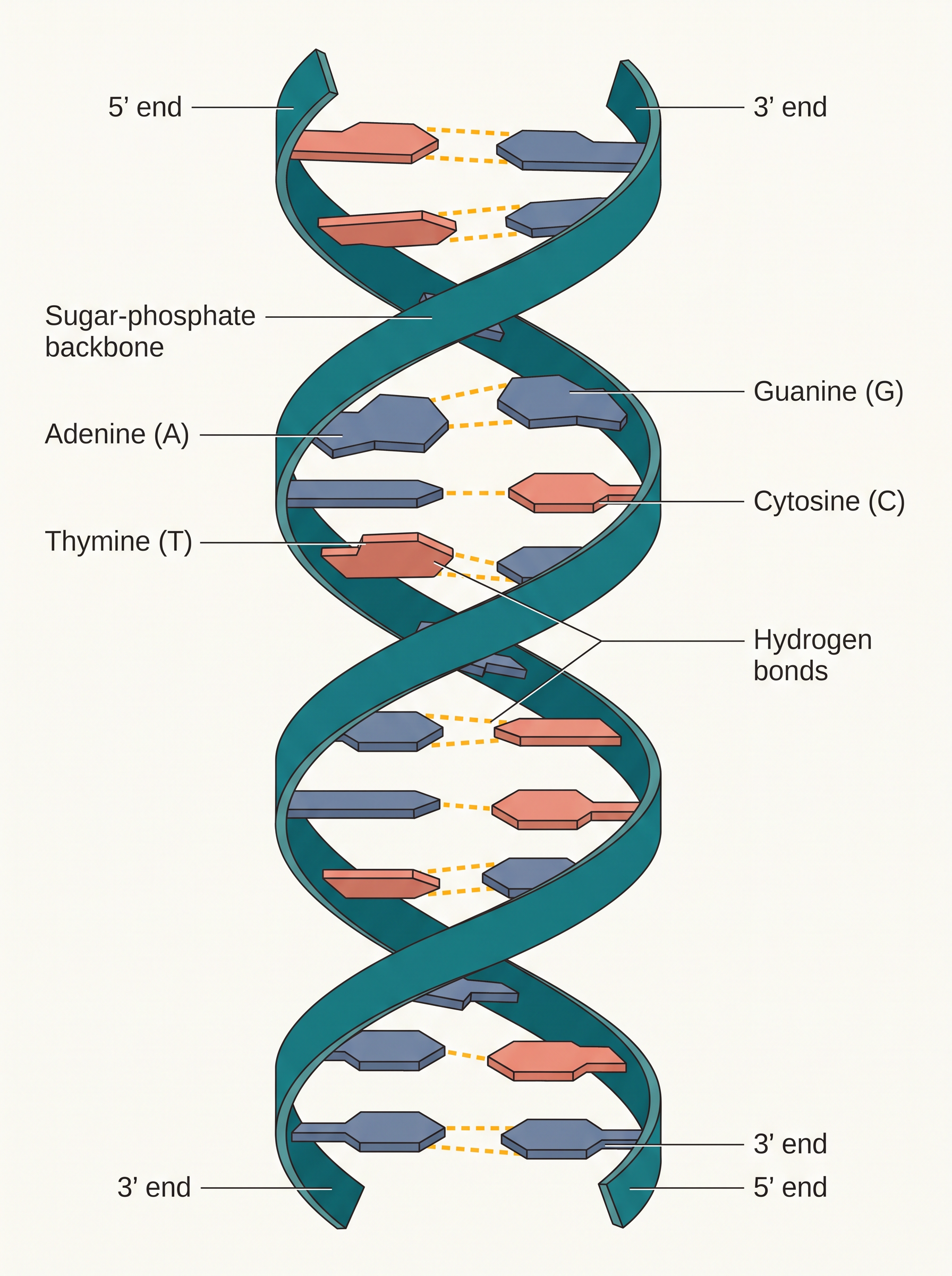
Complementary base pairing in DNA: in the double helix, adenine (A) always pairs with thymine (T), and cytosine (C) always pairs with guanine (G), through hydrogen bonds. The base sequence of one strand therefore fixes the base sequence of the other.
Q5. The two strands of DNA have different base sequences, yet they are called complementary. How can two unlike strands each carry the same message?
A. “Complementary” does not mean identical — it means that each base on one strand fixes the base facing it on the other. Because hydrogen bonds form only between A and T, and only between C and G, a strand reading A–T–G–C can be facing only T–A–C–G. The two strands are therefore unlike in sequence but locked to each other by a strict rule. This is why either strand contains the whole message: given one, the other can be written out with certainty. And it is exactly this that makes self-duplication possible — when the two strands separate during cell division, each one acts as a pattern on which its partner is rebuilt, so both daughter cells receive an identical DNA molecule.
One strand of a DNA segment has the base sequence
\[\\text{A} - \\text{T} - \\text{G} - \\text{C} - \\text{C} - \\text{G} - \\text{T} - \\text{A}\]
Write the base sequence of the complementary strand, and state the type of interaction that holds the two strands together.
Apply the base-pairing rule to each base in turn: A pairs with T, T pairs with A, G pairs with C, and C pairs with G.
|Given strand|A|T|G|C|C|G|T|A|
|-|-|-|-|-|-|-|-|-|
|Complementary strand|T|A|C|G|G|C|A|T|
Answer: The complementary strand reads \(\\text{T} - \\text{A} - \\text{C} - \\text{G} - \\text{G} - \\text{C} - \\text{A} - \\text{T}\). The two strands are held together by hydrogen bonds between the specific base pairs (A with T, C with G).
In a sample of double-stranded DNA, adenine accounts for 30% of the total bases. Calculate the percentage of thymine, guanine and cytosine present.
Use the complementary base-pairing rule. In a double-stranded DNA molecule, every adenine on one strand is paired with a thymine on the other, and every cytosine is paired with a guanine. So, counted over the whole molecule:
\[%,\\mathrm{A} = %,\\mathrm{T} \\qquad \\text{and} \\qquad %,\\mathrm{C} = %,\\mathrm{G}\]
Given \(%,\\mathrm{A} = 30\), it follows at once that \(%,\\mathrm{T} = 30\).
The four bases together must account for all the bases present:
\[%,\\mathrm{A} + %,\\mathrm{T} + %,\\mathrm{G} + %,\\mathrm{C} = 100\]
\[30 + 30 + %,\\mathrm{G} + %,\\mathrm{C} = 100\]
\[%,\\mathrm{G} + %,\\mathrm{C} = 40\]
Since \(%,\\mathrm{G} = %,\\mathrm{C}\), each must be half of 40:
\[%,\\mathrm{G} = %,\\mathrm{C} = 20\]
Answer: Thymine 30%, guanine 20%, cytosine 20%.
Note carefully that this reasoning works only because the DNA is double-stranded. In a single-stranded molecule there is no partner base to pair with, and no such relationship exists among the amounts of the different bases.
The structure of RNA
In the secondary structure of RNA, a single-stranded helix is present, which sometimes folds back on itself. There is no second strand running alongside it, and therefore no requirement that the bases be present in matched amounts.
RNA molecules are of three types, and they perform different functions. They are named as messenger RNA (m-RNA), ribosomal RNA (r-RNA) and transfer RNA (t-RNA).
DNA vs RNA
| Full name |
Deoxyribonucleic acid |
Ribonucleic acid |
| Pentose sugar |
\(\\beta\)-D-2-deoxyribose |
\(\\beta\)-D-ribose |
| Bases present |
Adenine, guanine, cytosine, thymine |
Adenine, guanine, cytosine, uracil |
| Strands |
Double strand, wound into a double helix |
Single strand, which may fold back on itself |
| Base-pairing relationship |
A pairs with T, C pairs with G; amounts of paired bases are equal |
No fixed relationship among the amounts of the bases |
| Types |
One kind |
Three types: m-RNA, r-RNA, t-RNA |
| Chief function |
Chemical basis of heredity; stores the coded message |
Carries out protein synthesis in the cell |
(question only — no solution) A nucleotide is hydrolysed completely. Name the three products obtained, and state which carbon atom of the sugar each of the other two components was attached to.
Har Gobind Khorana was born in 1922. He obtained his M.Sc. degree from Punjab University in Lahore. He worked with Professor Vladimir Prelog, who moulded Khorana’s thought and philosophy towards science, work and effort. After a brief stay in India in 1949, Khorana went back to England and worked with Professor G. W. Kenner and Professor A. R. Todd. It was at Cambridge, U.K., that he became interested in both proteins and nucleic acids. Dr Khorana shared the Nobel Prize for Medicine and Physiology in 1968 with Marshall Nirenberg and Robert Holley for cracking the genetic code — that is, for working out how the sequence of bases in a nucleic acid is read as instructions for building a protein.
DNA fingerprinting. It is known that every individual has unique fingerprints. These occur at the tips of the fingers and have been used for identification for a long time, but they can be altered by surgery. The sequence of bases on DNA is also unique for a person, and information regarding this is called DNA fingerprinting. It is the same for every cell of that person and cannot be altered by any known treatment. DNA fingerprinting is now used:
- in forensic laboratories, for identification of criminals;
- to determine the paternity of an individual;
- to identify dead bodies in an accident, by comparing the DNA with that of parents or children;
- to identify racial groups, to rewrite biological evolution.
10.5.3 Biological Functions of Nucleic Acids
DNA is the chemical basis of heredity and may be regarded as the reserve of genetic information. DNA is exclusively responsible for maintaining the identity of different species of organisms over millions of years.
A DNA molecule is capable of self-duplication during cell division, and identical DNA strands are transferred to the daughter cells. This is the structural consequence of complementary base pairing that you reasoned out above: because each strand carries the full message, each can rebuild its partner.
Another important function of nucleic acids is protein synthesis in the cell. Actually, the proteins are synthesised by the various RNA molecules in the cell, but the message for the synthesis of a particular protein is present in DNA. DNA holds the instructions; RNA carries them out.
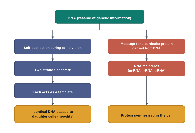
(question only — no solution) Explain why a change in the sequence of bases in a DNA molecule can change the protein that the cell makes. Link your answer to the primary structure of proteins, studied in Section 10.2.3.
10.6 Hormones
Hormones are molecules that act as intercellular messengers — chemical signals sent from one group of cells to another. They are produced by the endocrine glands in the body and are poured directly into the blood stream, which transports them to the site of action.
Notice how different this is from the enzymes of Section 10.3. An enzyme works where it is made, on a substrate it meets there. A hormone is made in one organ, travels in the blood, and acts somewhere else entirely — the thyroid gland does not use thyroxine on itself. This is what “intercellular messenger” means, and it is why the body needs a transport system, the blood, to make the hormone system work at all.
Hormones are chemical messengers produced by the endocrine glands and released directly into the blood stream, which carries them to their site of action elsewhere in the body.
Enzymes vs Hormones
| Chemical nature |
Almost all are globular proteins |
Varied — steroids, polypeptides, or amino acid derivatives |
| Function |
Biocatalysts — they speed up a specific reaction |
Messengers — they signal cells to change their activity |
| Site of action |
Act where they are produced, on their substrate |
Produced in an endocrine gland; act at a distant site |
| How they travel |
Not transported for their action |
Carried to the site of action by the blood stream |
| Quantity needed |
Very small |
Very small |
Chemical nature of hormones
In terms of chemical nature, hormones fall into three families:
- some are steroids, e.g. estrogens and androgens;
- some are polypeptides, for example insulin and endorphins;
- and some others are amino acid derivatives, such as epinephrine and norepinephrine.
The middle entry is a useful reminder that these chapters are connected. Insulin is a polypeptide — a chain of \(\\alpha\)-amino acids joined by peptide linkages, exactly as described in Section 10.2.3 — and it is precisely because it has a well-defined conformation that we call it a protein even though it contains only 51 amino acids.
The three chemical classes of hormones
| Steroids |
Compounds built on the steroid ring system |
Estrogens and androgens; testosterone, estradiol, progesterone; glucocorticoids and mineralocorticoids |
| Polypeptides |
Chains of \(\\alpha\)-amino acids joined by peptide linkages |
Insulin, endorphins |
| Amino acid derivatives |
Molecules derived from a single amino acid |
Epinephrine and norepinephrine; thyroxine (an iodinated derivative of tyrosine) |
Classify the following hormones according to their chemical nature, giving a reason in each case: (i) insulin, (ii) estradiol, (iii) thyroxine.
Ask, in each case, what kind of molecule the hormone actually is.
Insulin. It is a chain of \(\\alpha\)-amino acids (51 of them) joined by peptide linkages. Therefore it is a polypeptide hormone.
Estradiol. It is the main female sex hormone, produced by the gonads. Sex hormones such as the estrogens and androgens are built on the steroid ring system, so estradiol is a steroid hormone.
Thyroxine. It is produced in the thyroid gland and is an iodinated derivative of the amino acid tyrosine. It is therefore an amino acid derivative.
Answer: insulin — polypeptide; estradiol — steroid; thyroxine — amino acid derivative.
Functions of hormones
Hormones have several functions in the body. Above all, they help to maintain the balance of biological activities in the body — they keep the internal conditions of the body steady while the outside world changes.
Insulin, glucagon and blood glucose
The role of insulin in keeping the blood glucose level within a narrow limit is an example of this balancing function. Insulin is released in response to a rapid rise in blood glucose level. On the other hand, the hormone glucagon tends to increase the glucose level in the blood. The two hormones together regulate the glucose level in the blood.
This is a proper two-sided control system, and it is worth seeing why one hormone would not be enough. A single hormone that only lowered glucose could not correct a fall; a single hormone that only raised it could not correct a rise. With a pair of opposing hormones, a deviation in either direction can be pulled back. The loop is drawn in FC 10.44.
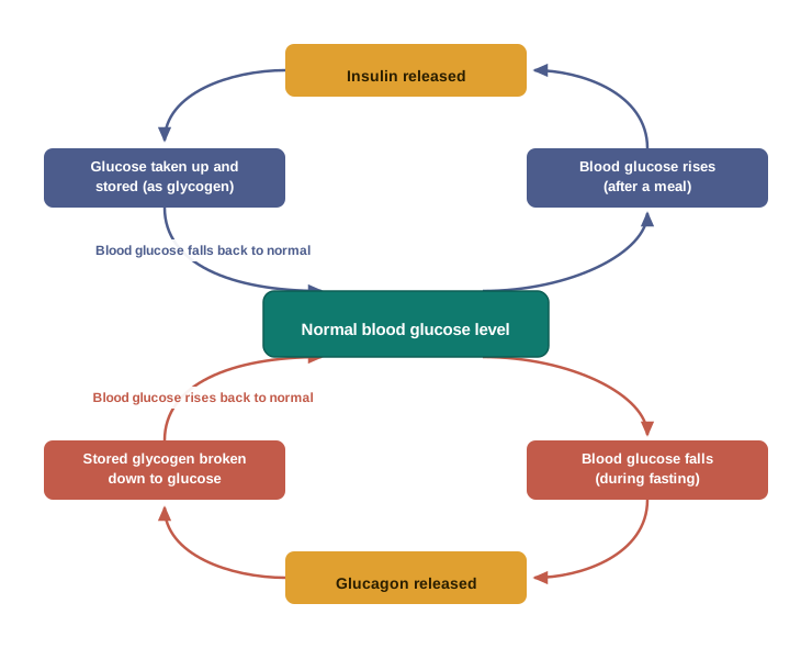
Note the direct link to Section 10.1: the “store” that this system draws on is glycogen, the branched polysaccharide kept in the liver and muscles. Hormones do the signalling; carbohydrates do the storing.
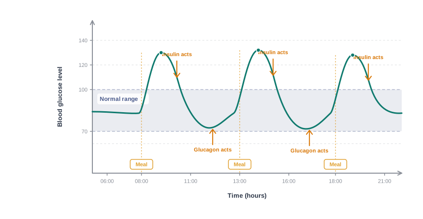
the blood-glucose-versus-time curve of Fig. 10.45 (Study the figure and answer: (a) At which points on the curve would you expect insulin to be released, and at which points glucagon? (b) The curve rises after a meal but does not keep rising — what does this tell you about the kind of control being exercised? (c) Sketch, alongside, the shape you would expect this curve to take for a person whose body cannot produce enough insulin, and state which part of the curve changes most.)
Before the 1920s, a diagnosis of severe diabetes was a death sentence, and the only treatment doctors could offer was near-starvation. In Toronto, Frederick Banting and Charles Best set out to isolate the substance from the pancreas that controlled blood sugar — a substance others had suspected but nobody had obtained in usable form. Working through a summer with borrowed laboratory space, they succeeded, and the extract they prepared was purified and given to a dying teenage patient in early 1922. His blood glucose fell, and he recovered. Insulin did not cure diabetes, and it still does not; but it turned a fatal disease into a manageable one, and it remains one of the clearest demonstrations that a single biomolecule, present in milligram amounts, can hold a whole body’s chemistry in balance.
Epinephrine, growth and sex hormones
Epinephrine and norepinephrine mediate responses to external stimuli — these are the hormones released when the body must react quickly to a sudden situation.
Growth hormones and sex hormones play a role in growth and development.
Thyroxine and iodine
Thyroxine, produced in the thyroid gland, is an iodinated derivative of the amino acid tyrosine. Here again the chapter closes a loop: tyrosine is entry 17 in Table 10.2, and the body takes that ordinary amino acid, attaches iodine to it, and turns it into a hormone.
An abnormally low level of thyroxine leads to hypothyroidism, which is characterised by lethargy and obesity. An increased level of thyroxine causes hyperthyroidism.
Now follow the chemistry one step further. Thyroxine cannot be made without iodine, so a low level of iodine in the diet may lead to hypothyroidism and to enlargement of the thyroid gland. This condition is largely being controlled by adding sodium iodide to commercial table salt — the “iodised” salt sold everywhere.
Iodised salt is one of the most successful public-health interventions ever built on a single chemical fact. Iodine is not evenly spread over the earth’s surface — soils far from the sea, and especially in hill regions, are poor in it, so the food grown there is poor in it too, and whole populations once suffered from goitre and from the developmental damage that iodine deficiency causes in children. Fortifying an item that virtually every household already buys, in an amount too small to taste, delivers the missing element without asking anyone to change what they eat. When you next look at a salt packet marked “iodised”, you are looking at a hormone-precursor supply problem being solved at the level of the grocery shop.
(question only — no solution) A person living in a hill region develops an enlarged thyroid gland, lethargy and weight gain. Name the hormone whose level is likely to be low, name the amino acid from which it is derived, name the element whose dietary shortage is responsible, and state the simple remedy adopted at a national scale.
Steroid hormones
Steroid hormones are produced by the adrenal cortex and by the gonads (testes in males and ovaries in females).
Hormones of the adrenal cortex
Hormones released by the adrenal cortex play a very important role in the functions of the body. For example, glucocorticoids control carbohydrate metabolism, modulate inflammatory reactions, and are involved in reactions to stress. The mineralocorticoids control the level of excretion of water and salt by the kidney.
If the adrenal cortex does not function properly, then one of the results may be Addison’s disease, characterised by hypoglycemia (an abnormally low blood glucose level), weakness, and an increased susceptibility to stress. The disease is fatal unless it is treated by glucocorticoids and mineralocorticoids.
Hormones of the gonads
Hormones released by the gonads are responsible for the development of secondary sex characters.
Testosterone is the major sex hormone produced in males. It is responsible for the development of secondary male characteristics (deep voice, facial hair, general physical constitution).
Estradiol is the main female sex hormone. It is responsible for the development of secondary female characteristics and participates in the control of the menstrual cycle.
Progesterone is responsible for preparing the uterus for the implantation of the fertilised egg.
(question only — no solution) Insulin and glucagon are described as acting “together” to regulate blood glucose, although their effects are opposite. Explain why a control system needs both of them, and name the storage polysaccharide (from Section 10.1) that this system builds up and breaks down.
10.6 Why cannot vitamin C be stored in our body?
10.7 What products would be formed when a nucleotide from DNA containing thymine is hydrolysed?
10.8 When RNA is hydrolysed, there is no relationship among the quantities of the different bases obtained. What does this fact suggest about the structure of RNA?
10.7 Summary
Carbohydrates
- Carbohydrates are optically active polyhydroxy aldehydes or ketones, or molecules which provide such units on hydrolysis.
- They are broadly classified into three groups — monosaccharides, disaccharides (the commonest oligosaccharides) and polysaccharides — on the basis of what they give on hydrolysis.
- Glucose, the most important source of energy for mammals, is obtained by the digestion of starch.
- Monosaccharides are held together by glycosidic linkages to form disaccharides or polysaccharides.
Proteins
- Proteins are the polymers of about twenty different \(\\alpha\)-amino acids, which are linked by peptide bonds (–CO–NH–).
- Ten amino acids are called essential amino acids, because they cannot be synthesised by our body and hence must be provided through the diet.
- Proteins perform various structural and dynamic functions in the organisms. Proteins which contain only \(\\alpha\)-amino acids are called simple proteins.
- The secondary or tertiary structure of proteins gets disturbed on a change of pH or temperature, and the protein is then unable to perform its functions. This is called denaturation of proteins; the primary structure survives it.
Enzymes
- Enzymes are biocatalysts which speed up the reactions in biosystems by lowering the activation energy.
- They are very specific and selective in their action, and chemically the majority of enzymes are proteins.
Vitamins
- Vitamins are accessory food factors required in the diet in small amounts.
- They are classified as fat soluble (A, D, E and K) and water soluble (B group and C).
- Deficiency of vitamins leads to many diseases.
Nucleic acids
- Nucleic acids are the polymers of nucleotides, which in turn consist of a base, a pentose sugar and a phosphate moiety.
- Nucleic acids are responsible for the transfer of characters from parents to offspring.
- There are two types of nucleic acids — DNA and RNA. DNA contains a five-carbon sugar molecule called 2-deoxyribose, whereas RNA contains ribose.
- Both DNA and RNA contain adenine, guanine and cytosine. The fourth base is thymine in DNA and uracil in RNA.
- The structure of DNA is a double strand, whereas RNA is a single strand molecule.
- DNA is the chemical basis of heredity and carries the coded message for the proteins to be synthesised in the cell.
- There are three types of RNA — mRNA, rRNA and tRNA — which actually carry out the protein synthesis in the cell.
Hormones
- Hormones are chemical messengers produced by the endocrine glands and carried by the blood to their site of action.
- Chemically they are steroids (e.g. estradiol, testosterone), polypeptides (e.g. insulin) or amino acid derivatives (e.g. thyroxine, epinephrine), and they keep the biological activities of the body in balance.
The chapter in one line
- Living systems are built from non-living molecules: carbohydrates supply and store energy, proteins build and act, enzymes catalyse, vitamins enable, nucleic acids carry the information, and hormones coordinate the whole.
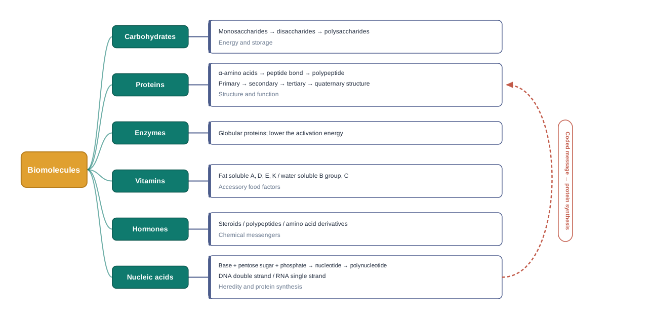
10.8 NCERT Questions
- What are monosaccharides?
- What are reducing sugars?
- Write two main functions of carbohydrates in plants.
- Classify the following into monosaccharides and disaccharides: ribose, 2-deoxyribose, maltose, galactose, fructose and lactose.
- What do you understand by the term glycosidic linkage?
- What is glycogen? How is it different from starch?
- What are the hydrolysis products of (i) sucrose and (ii) lactose?
- What is the basic structural difference between starch and cellulose?
- What happens when D-glucose is treated with the following reagents? (i) HI (ii) Bromine water (iii) \(\\mathrm{HNO\_3}\)
- Enumerate the reactions of D-glucose which cannot be explained by its open chain structure.
- What are essential and non-essential amino acids? Give two examples of each type.
- Define the following as related to proteins: (i) Peptide linkage (ii) Primary structure (iii) Denaturation.
- What are the common types of secondary structure of proteins?
- What type of bonding helps in stabilising the \(\\alpha\)-helix structure of proteins?
- Differentiate between globular and fibrous proteins.
- How do you explain the amphoteric behaviour of amino acids?
- What are enzymes?
- What is the effect of denaturation on the structure of proteins?
- How are vitamins classified? Name the vitamin responsible for the coagulation of blood.
- Why are vitamin A and vitamin C essential to us? Give their important sources.
- What are nucleic acids? Mention their two important functions.
- What is the difference between a nucleoside and a nucleotide?
- The two strands in DNA are not identical but are complementary. Explain.
- Write the important structural and functional differences between DNA and RNA.
- What are the different types of RNA found in the cell?
10.9 Check Your Concepts
- Acetic acid, \(\\mathrm{CH\_3COOH}\), fits the general formula \(\\mathrm{C\_x(H\_2O)*y}\), yet it is not a carbohydrate; rhamnose, \(\\mathrm{C\_6H*{12}O\_5}\), does not fit the formula, yet it is a carbohydrate. Using these two facts, explain why chemists abandoned the formula-based definition of carbohydrates.
- Assertion (A): Glucose does not give Schiff’s test and does not form the hydrogensulphite addition product with \(\\mathrm{NaHSO\_3}\). Reason (R): In solution, glucose exists almost entirely in a cyclic hemiacetal form in which the –CHO group is not free. Choose the correct option and justify: (a) both A and R are true and R is the correct explanation of A; (b) both A and R are true but R is not the correct explanation of A; (c) A is true but R is false; (d) A is false but R is true.
- Bromine water oxidises D-glucose to gluconic acid, while nitric acid oxidises it to saccharic acid. Write both products, and explain what this difference in behaviour tells you about the two reagents and about the two ends of the glucose chain.
- The pentaacetate of D-glucose does not react with hydroxylamine, whereas D-glucose itself does. Explain this observation with reference to the cyclic structure of glucose.
- Arrange the following in order of increasing number of monosaccharide units obtained on hydrolysis, and name the units obtained in each case: cellulose, sucrose, glucose, maltose.
- Sucrose is non-reducing, but both maltose and lactose are reducing sugars, even though all three are disaccharides. Explain the difference in terms of the carbon atoms involved in the glycosidic linkage.
- Explain why the hydrolysis of sucrose is accompanied by a change in the sign of optical rotation, and state the name given to the product mixture.
- Both glucose and fructose have the molecular formula \(\\mathrm{C\_6H\_{12}O\_6}\), yet glucose forms a six-membered ring on cyclisation and fructose a five-membered one. Account for this difference, and name the two ring systems.
- Match the following:
| a. Amylose |
i. branched polymer of \(\\alpha\)-D-glucose, stored in the liver |
| b. Amylopectin |
ii. straight-chain polymer of \(\\beta\)-D-glucose |
| c. Cellulose |
iii. unbranched chain of \(\\alpha\)-D-(+)-glucose, C1–C4 linked, water soluble |
| d. Glycogen |
iv. branched polymer of \(\\alpha\)-D-glucose with C1–C6 branching, insoluble in water |
- Herbivorous animals can obtain energy from grass, but human beings cannot digest cellulose at all. Explain this in terms of the type of glycosidic linkage present and the specificity of enzymes.
- Glycine is the only naturally occurring \(\\alpha\)-amino acid that is optically inactive. Explain why, referring to the groups attached to the \(\\alpha\)-carbon atom.
- Amino acids are high-melting, water-soluble crystalline solids that behave like salts. What structural feature accounts for this, and how does the same feature explain their amphoteric behaviour?
- Write the structures of the two dipeptides that can be formed from glycine and alanine. Name each, mark the peptide linkage, and state why the two are not the same compound.
- Assertion (A): During denaturation of a protein, the primary structure remains intact. Reason (R): The primary structure is held together by covalent peptide bonds, whereas the secondary and tertiary structures depend mainly on hydrogen bonds. State whether both statements are true, and whether R correctly explains A.
- Distinguish between the \(\\alpha\)-helix and the \(\\beta\)-pleated sheet structures of proteins in terms of (i) the shape of the chain and (ii) whether the hydrogen bonding is within a chain or between chains.
- A protein solution is warmed gently and then cooled back to room temperature, but its biological activity is not restored. Explain what has happened, name the process, and state which level of protein structure is lost and which is retained.
- Enzymes are said to be biocatalysts. Using the activation energy values given in the chapter for the acid hydrolysis and the enzyme-catalysed hydrolysis of sucrose, explain what an enzyme does to the reaction path and what it does not change.
- The active site of an enzyme owes its shape to the folding of a globular protein. Use this fact to explain (i) the high specificity of enzymes and (ii) the loss of enzyme activity at high temperature or extreme pH.
- Name the enzymes that would catalyse the hydrolysis of (i) maltose, (ii) sucrose and (iii) urea, and state the rule of nomenclature you have used.
- Why must water-soluble vitamins be supplied regularly in the diet, while fat-soluble vitamins need not be? Name the one water-soluble vitamin that is an exception.
- A person with a poor intake of citrus fruits and green leafy vegetables develops bleeding gums, while another with almost no exposure to sunlight develops soft, deformed bones. Identify the vitamin deficient in each case, name the disease, and classify the vitamin as fat soluble or water soluble.
- Explain why an excess of vitamin A or vitamin D taken as pills over a long period can be harmful, whereas an excess of vitamin C is largely harmless.
- Complete the following: complete hydrolysis of a nucleotide from RNA gives ____, ____ and ____. Name the sugar and the extra base that would have been obtained instead if the nucleotide had come from DNA.
- One strand of a DNA segment reads \(\\text{A} - \\text{G} - \\text{C} - \\text{T} - \\text{T} - \\text{G} - \\text{C} - \\text{A}\). Write the sequence of the complementary strand, and name the interaction that holds the two strands together.
- In a double-stranded DNA sample, cytosine forms 22% of the total bases. Deduce the percentage of guanine, adenine and thymine, and state the rule you have used.
- When RNA is hydrolysed, the amounts of the four bases obtained bear no fixed relationship to one another, whereas in DNA they do. What does this tell you about the difference in the structures of the two nucleic acids?
- Explain how the complementary base-pairing rule allows a DNA molecule to duplicate itself exactly during cell division.
- Classify the following hormones according to their chemical nature — steroid, polypeptide or amino acid derivative: insulin, testosterone, epinephrine, estradiol, thyroxine, endorphins.
- Insulin and glucagon are described as acting together, although their effects on blood glucose are opposite. Explain why a control system needs both, and name the polysaccharide that is built up and broken down in the process.
- Thyroxine is an iodinated derivative of an amino acid. Name that amino acid, explain how a low level of iodine in the diet can lead to hypothyroidism, and state the measure adopted to prevent this at a national scale.
- Both enzymes and hormones are required by the body in very small amounts, yet they are entirely different kinds of molecule with entirely different roles. Bring out three points of difference between them.
- Insulin cannot be taken as a tablet and must be injected. Explain this in terms of the chemical nature of insulin and the action of digestive enzymes.