Chapter 9: Biotechnology: Principles and Processes
In the early 1970s, two scientists sat talking over a late-night snack at a scientific meeting in Hawaii. One of them, Stanley Cohen of Stanford University, had spent years studying tiny rings of DNA called plasmids that live inside bacteria and copy themselves independently. The other, Herbert Boyer of the University of California, San Francisco, had been working with special bacterial proteins that could cut DNA at precise points, leaving neatly matched ends behind.
As they compared notes, a simple but powerful idea took shape. What if Boyer’s molecular “scissors” were used to cut open one of Cohen’s plasmids, slip a chosen piece of DNA inside, seal it shut, and then place that rebuilt plasmid back into a living bacterium? The bacterium, unaware of the trick, would treat the new DNA as its own and copy it every time it divided.
In 1972, they did exactly that. They joined a gene carrying antibiotic resistance to a bacterial plasmid and put it into the gut bacterium Escherichia coli, which then multiplied the gene faithfully. That single experiment gave the world its first artificial recombinant DNA molecule and opened the door to modern biotechnology — the ability to move a gene from one organism into another and put living cells to work as tiny factories.
By the end of this chapter, you will be able to answer each of these questions and understand the tools and step-by-step processes that make genetic engineering possible.
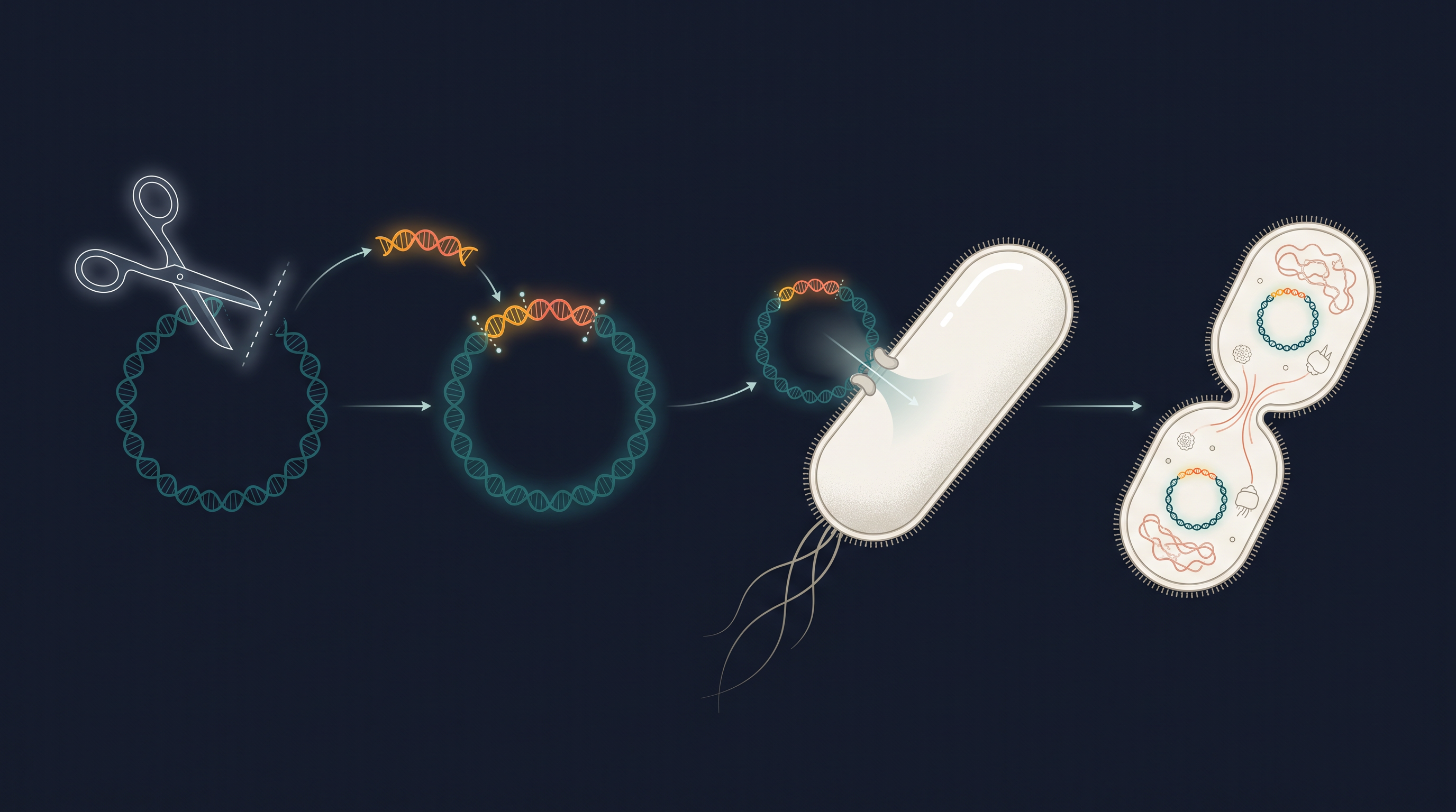
Fig. 9.0 – Building and cloning a recombinant plasmid.
9.1 Principles of Biotechnology
What Biotechnology Means
Biotechnology deals with the techniques of using living organisms, or enzymes taken from organisms, to make products and carry out processes that are useful to humans. Seen in this broad way, some very old and familiar activities already count as biotechnology. Making curd from milk, raising bread with yeast, or fermenting fruit juice into wine are all processes driven by microbes, and each turns a raw material into something more useful. These are examples of traditional biotechnology.
Today, however, the word is used in a more restricted sense. Modern biotechnology refers to those processes that use genetically modified organisms — organisms whose genetic material has been deliberately changed — to achieve such results on a much larger and more controlled scale. In addition, several other advanced techniques are grouped under biotechnology. For example, in vitro fertilisation leading to a “test-tube” baby, chemically synthesising a gene and putting it to use, developing a DNA vaccine, or correcting a defective gene in a patient are all considered part of biotechnology.
Here, in vitro simply means “outside the living body,” in a glass vessel or dish in the laboratory, as opposed to in vivo, which means inside a living organism.
To capture both the old and the new views, the European Federation of Biotechnology (EFB) has offered a single, wide-ranging definition of the subject.
The Two Core Techniques
Among the many advances that made modern biotechnology possible, two techniques stand out as its true foundations. Almost everything in this chapter rests on these two ideas working together.
The first is genetic engineering. This is the set of techniques used to alter the chemistry of the genetic material — DNA and RNA — and then to introduce this altered material into a host organism, thereby changing the phenotype of that host. The phenotype is simply the set of observable traits of an organism, and it changes because the new gene brings with it a new instruction for the cell to follow.
The second is bioprocess engineering. This is the maintenance of a sterile, or microbial contamination-free, environment during chemical engineering processes, so that only the desired microbe or eukaryotic cell is allowed to grow, and to grow in large quantities. This large-scale, clean growth is what makes it possible to manufacture biotechnological products such as antibiotics, vaccines and enzymes.
Why Genetic Engineering Was Needed
To see why genetic engineering became so valuable, it helps to recall the difference between sexual and asexual reproduction. Sexual reproduction provides opportunities for variation and for forming unique new combinations of genes, some of which may benefit the individual organism and even the whole population. Asexual reproduction, by contrast, simply preserves the existing genetic information and passes on an identical copy, so it permits little or no variation.
Traditional breeders have long used this variation through hybridisation — crossing selected plants or animals to bring desirable traits together. But hybridisation has an important weakness. Along with the desired genes, undesirable genes very often get carried across as well, and both are then multiplied together in the offspring. The breeder cannot easily separate the good from the unwanted.
Genetic engineering overcomes exactly this limitation. Using techniques that include the creation of recombinant DNA, gene cloning and gene transfer, it allows us to isolate and introduce only one desired gene, or a chosen set of desired genes, without dragging any undesirable genes into the target organism. This precision is the central advantage of the whole approach.
How Alien DNA Survives in a New Cell
Suppose a piece of DNA is somehow introduced into an unrelated (“alien”) organism. What is its likely fate? In most cases, this stray piece of DNA cannot multiply on its own in the progeny cells, so it is simply lost as the cells divide. It survives and spreads only if it becomes integrated into the genome — the complete set of DNA — of the recipient, because then it is copied and inherited along with the host’s own DNA.
The reason lies in a special sequence found on every chromosome. This sequence, called the origin of replication, is the site responsible for starting the copying (replication) of that DNA. For any alien piece of DNA to be multiplied inside an organism, it must therefore become part of a chromosome, or be joined to a piece of DNA, that carries this origin of replication.
When an alien piece of DNA is linked to an origin of replication in this way, it can now replicate and multiply itself inside the host organism. Producing many identical copies of a template DNA in this manner is called cloning.
The First Recombinant DNA Molecule
We can now follow how the very first artificial recombinant DNA molecule was built. The idea arose from the possibility of linking a gene that encodes antibiotic resistance to a native plasmid taken from the bacterium Salmonella typhimurium.
A plasmid is a small, circular piece of DNA that lies outside the main chromosome of a bacterium (it is extra-chromosomal) and that can replicate on its own, independently of the chromosome. Because it copies itself and can carry passenger DNA, a plasmid is extremely useful in genetic engineering.
Stanley Cohen and Herbert Boyer put this idea into practice in 1972. They first isolated the antibiotic-resistance gene by cutting out the relevant piece of DNA from a plasmid that gave the bacterium its resistance. Cutting DNA at such specific locations only became possible because of the discovery of a group of enzymes nicknamed “molecular scissors” — the restriction enzymes, which recognise particular DNA sequences and cut there. (We will study these enzymes in detail in the next section.)
The cut-out piece of DNA was then linked to a plasmid. A plasmid used in this way acts as a vector — a carrier that transfers an attached piece of DNA into a host cell. A familiar parallel makes this clear.
Joining the antibiotic-resistance gene to the plasmid vector became possible with the enzyme DNA ligase, which acts on cut DNA molecules and seals their ends together. The result is a brand-new combination of circular, self-replicating DNA, put together in the test tube (in vitro). This is what we call recombinant DNA.
When this recombinant DNA was transferred into Escherichia coli — a bacterium closely related to Salmonella — it could replicate using the new host’s own DNA polymerase enzyme and so make many copies of itself. This ability to multiply copies of the antibiotic-resistance gene inside E. coli was described as cloning of that gene in E. coli.
The whole conceptual sequence of building and multiplying that first recombinant DNA is summarised in FC 9.1.
Flowchart
FC 9.1 – Building the first recombinant DNA: antibiotic-resistance gene cut out with restriction enzyme → cut gene joined to plasmid vector using DNA ligase → recombinant DNA formed in vitro → transferred into E. coli host → replicates using host DNA polymerase → many identical copies (cloning)
The Three Basic Steps of Genetic Modification
From all of the above, we can draw out the general logic of genetically modifying any organism. Whatever the details, the process always comes down to three basic steps, shown in FC 9.2.
First, identification of the DNA that carries the desirable gene. Second, introduction of this identified DNA into the host organism. Third, maintenance of the introduced DNA inside the host, and its transfer to the host’s progeny so that it is not lost.
Flowchart
FC 9.2 – Three basic steps of genetic modification: (i) identify DNA with the desirable gene → (ii) introduce the identified DNA into the host → (iii) maintain the DNA in the host and pass it on to the progeny
Keeping these three steps in mind will make the rest of the chapter easier to follow, because the tools described in Section 9.2 and the step-by-step processes in Section 9.3 are simply the practical ways of carrying out these three basic steps.
9.2 Tools of Recombinant DNA Technology
We now know from the previous section that genetic engineering, or recombinant DNA technology, can be carried out only if we have a set of key tools. These tools are the restriction enzymes, the polymerase enzymes, the ligases, the vectors, and finally the host organism into which the recombinant DNA is placed. Each tool does one specific job in the overall task of moving a gene from one organism into another. Let us examine several of these in detail.
9.2.1 Restriction Enzymes
The story of restriction enzymes begins in 1963, when two enzymes responsible for restricting the growth of bacteriophages (viruses that infect bacteria) inside Escherichia coli were isolated. One of these enzymes added methyl groups to DNA, while the other cut DNA. It was this second, DNA-cutting enzyme that came to be called a restriction endonuclease.
The first restriction endonuclease whose action depended on a specific DNA nucleotide sequence was Hind II, isolated and characterised about five years after that initial discovery. It was found that Hind II always cuts a DNA molecule at one particular point, because it recognises a specific sequence of six base pairs. This specific base sequence that the enzyme looks for is called its recognition sequence.
Besides Hind II, we today know more than 900 restriction enzymes that have been isolated from over 230 strains of bacteria, and each of these recognises a different recognition sequence. This large “toolkit” of scissors, each cutting at its own sequence, is what gives genetic engineers such precise control over DNA.
How Restriction Enzymes Are Named
There is a fixed convention for naming these enzymes. The first letter of the name comes from the genus of the bacterium, and the next two letters come from its species. For example, EcoRI comes from Escherichia coli RY 13. In EcoRI, the letter “R” is taken from the name of the strain. The Roman numerals that follow the name indicate the order in which the enzymes were isolated from that particular strain of bacteria.
Restriction enzymes belong to a larger class of enzymes called nucleases. Nucleases are of two kinds. Exonucleases remove nucleotides from the ends of a DNA molecule, whereas endonucleases make cuts at specific positions within the DNA. Restriction enzymes are therefore endonucleases, because they cut inside the molecule rather than nibbling from the ends.
How Restriction Enzymes Cut DNA
Each restriction endonuclease works by first “inspecting” the length of a DNA sequence. Once it finds its own specific recognition sequence, it binds to the DNA and then cuts each of the two strands of the double helix at specific points within their sugar–phosphate backbones, as shown in Fig. 9.1. Every restriction endonuclease recognises a specific palindromic nucleotide sequence in the DNA.
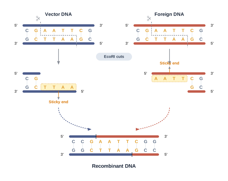
Fig. 9.1 – Action of the restriction enzyme EcoRI.
To understand these sequences we first need to know what a palindrome is. A palindrome is a group of letters that reads the same forward and backward, such as the word “MALAYALAM.” In DNA, a palindrome is a sequence of base pairs that reads the same on the two strands when the orientation of reading is kept the same.
Consider the following example, which reads the same on both strands in the 5′→3′ direction (and is equally true when read in the 3′→5′ direction):
5′—— G A A T T C ——3′ 3′—— C T T A A G ——5′
Reading the top strand left to right in the 5′→3′ direction gives GAATTC; reading the bottom strand in its own 5′→3′ direction also gives GAATTC. That mirror symmetry is what makes it a DNA palindrome.
Restriction enzymes cut the DNA strand a little away from the centre of the palindrome, but between the same two bases on the opposite strands. This staggered cut leaves short single-stranded portions at the ends. These overhanging stretches are called sticky ends, as marked in Fig. 9.1.
They are called sticky ends because they can form hydrogen bonds with their complementary cut counterparts. This stickiness of the ends is exactly what facilitates the action of the enzyme DNA ligase, which then seals the joint permanently.
Restriction endonucleases are used in genetic engineering to form “recombinant” molecules of DNA, which are made up of DNA drawn from different sources or genomes. The crucial point is this: when two different pieces of DNA are cut by the same restriction enzyme, both fragments have the same kind of sticky ends. Because their overhangs are complementary, these fragments can then be joined together end-to-end using DNA ligases, as illustrated in Fig. 9.2.
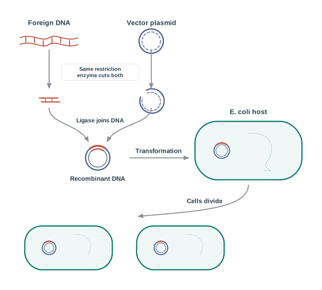
Fig. 9.2 – Overview of recombinant DNA technology.
You may have realised an important practical rule from all this: unless one cuts both the vector and the source DNA with the same restriction enzyme, the recombinant vector molecule cannot be created — because only then will their sticky ends match.
Separation and Isolation of DNA Fragments
The cutting of DNA by restriction endonucleases produces a mixture of DNA fragments of different sizes. To use a particular fragment, we must first separate it from the rest. This separation is done by a technique known as gel electrophoresis.
Since DNA fragments are negatively charged molecules, they can be separated by forcing them to move towards the anode (the positive electrode) under an electric field, through a suitable medium or matrix. The negative charge comes from the phosphate groups in the DNA backbone, so every fragment is pulled towards the positive electrode.
Nowadays the most commonly used matrix is agarose, a natural polymer extracted from sea weeds. The gel acts like a molecular sieve. The DNA fragments resolve (separate out) according to their size through this sieving effect, so that the smaller a fragment is, the farther it moves through the gel. Larger fragments get held back near the wells where the sample was loaded, as seen in Fig. 9.3.
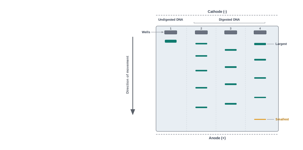
Fig. 9.3 – DNA separation by agarose gel electrophoresis.
The separated DNA fragments cannot be seen directly, because pure DNA is invisible in visible light. They can be visualised only after staining the DNA with a compound known as ethidium bromide, followed by exposure to ultraviolet (UV) radiation. Under UV light, the stained DNA shows up as bright orange-coloured bands (Fig. 9.3).
Once the bands are visible, the separated band of interest is cut out from the agarose gel and the DNA is extracted from that gel piece. This step of recovering DNA from the gel is called elution. The DNA fragments purified in this way are then used to construct recombinant DNA by joining them with cloning vectors.
Flowchart
FC 9.3 – Isolating a DNA fragment: cut DNA with restriction enzyme → load fragments in agarose gel wells → apply electric field (fragments move towards anode) → fragments separate by size (smaller travel farther) → stain with ethidium bromide and view under UV → cut out the required band → elute (extract) the DNA from the gel
9.2.2 Cloning Vectors
We already know that plasmids and bacteriophages can replicate within bacterial cells independently of the control of the chromosomal DNA. Bacteriophages, because of their high numbers per cell, carry very high copy numbers of their genome inside the bacterial cell. Among plasmids, some may have only one or two copies per cell, whereas others may have 15–100 copies per cell, and the number can go even higher.
This copy number matters a great deal. If we are able to link an alien piece of DNA with bacteriophage or plasmid DNA, we can multiply the alien DNA to a number equal to the copy number of that plasmid or bacteriophage. Vectors used at present are engineered specially so that they make it easy both to link foreign DNA into them and to select the recombinants from the non-recombinants afterwards.
The following are the features that a good cloning vector needs in order to make cloning possible.
(i) Origin of Replication (ori)
The origin of replication is a sequence from where replication starts, so that any piece of DNA linked to it can be made to replicate within the host cells. (We met this idea in Section 9.1.) This sequence is also responsible for controlling the copy number of the linked DNA. Therefore, if one wants to recover many copies of the target DNA, it should be cloned in a vector whose origin supports a high copy number.
(ii) Selectable Marker
In addition to the ori, a vector requires a selectable marker. A selectable marker helps in identifying and eliminating the non-transformants, while selectively permitting the growth of the transformants. Here, transformation is the procedure through which a piece of DNA is introduced into a host bacterium (its practical steps are covered in Section 9.2.3).
Normally, the genes that encode resistance to antibiotics such as ampicillin, chloramphenicol, tetracycline or kanamycin are used as convenient selectable markers for E. coli. This works because normal E. coli cells do not carry resistance against any of these antibiotics. So if a cell has become resistant, it must have taken up the vector carrying the resistance gene.
(iii) Cloning Sites
In order to link the alien DNA, the vector needs to have very few — preferably single — recognition sites for the commonly used restriction enzymes. If there is more than one recognition site within the vector, the enzyme will cut the vector into several fragments, which complicates gene cloning. The restriction map of the well-known E. coli cloning vector pBR322 is shown in Fig. 9.4.
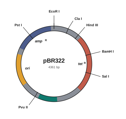
Fig. 9.4 – Restriction map of the cloning vector pBR322.
The ligation of alien DNA is carried out at a restriction site present within one of the two antibiotic-resistance genes. For example, one can ligate a foreign DNA at the BamH I site of the tetracycline-resistance gene in the vector pBR322. When foreign DNA is inserted there, the recombinant plasmids will lose their tetracycline resistance because the insertion breaks that gene. However, these recombinants can still be selected apart from the non-recombinant plasmids.
The selection works in two stages. The transformants are first plated on a medium containing ampicillin; the recombinants grow because their ampicillin-resistance gene is still intact. These colonies are then transferred onto a medium containing tetracycline. The recombinants will grow on ampicillin but not on tetracycline (their tetracycline gene is broken), whereas the non-recombinants — which still have both intact genes — grow on medium containing either antibiotic. In this scheme, one antibiotic-resistance gene helps in selecting the transformants, while the other antibiotic-resistance gene gets inactivated by the insertion of alien DNA and thereby helps in selecting the recombinants.
Flowchart
FC 9.4 – Two-antibiotic selection with pBR322: insert gene at BamH I in tet\(^R\) → plate on ampicillin (only transformants grow) → replica onto tetracycline → grows on tet = non-recombinant (tet\(^R\) intact); does not grow on tet = recombinant (tet\(^R\) disrupted)
A Simpler Way to Select Recombinants
Selecting recombinants by the inactivation of antibiotic resistance is a cumbersome procedure, because it requires simultaneous plating on two plates carrying different antibiotics. Therefore, alternative selectable markers have been developed that tell recombinants and non-recombinants apart by their ability to produce colour in the presence of a chromogenic substrate (a colourless substance that gives a coloured product when acted upon by an enzyme).
In this method, the recombinant DNA is inserted within the coding sequence of an enzyme called β-galactosidase. This insertion inactivates the gene for making that enzyme, an effect referred to as insertional inactivation. If the plasmid in the bacterium has no insert, the β-galactosidase gene is intact, and the chromogenic substrate is converted into a blue product, giving blue colonies. If an insert is present, the β-galactosidase gene is inactivated, no colour is produced, and these colourless colonies are identified as the recombinant colonies.
(iv) Vectors for Cloning Genes in Plants and Animals
It may be surprising, but we have learnt the very trick of transferring genes into plants and animals from bacteria and viruses, which have “known” how to do this for ages — that is, how to deliver genes into eukaryotic cells and force those cells to do what the microbe wants.
For example, Agrobacterium tumifaciens, a pathogen of several dicot plants, is able to deliver a piece of DNA known as “T-DNA” that transforms normal plant cells into a tumour, and then directs these tumour cells to produce the chemicals that the pathogen needs. Similarly, retroviruses in animals have the ability to transform normal cells into cancerous cells.
A better understanding of this natural art of delivering genes has allowed scientists to convert these tools of pathogens into useful vectors for delivering genes of our own interest. The tumour-inducing (Ti) plasmid of Agrobacterium tumifaciens has now been modified into a cloning vector that is no longer pathogenic to plants, yet still uses the same mechanisms to deliver our chosen genes into a variety of plants. In the same way, retroviruses have been “disarmed” and are now used to deliver desirable genes into animal cells.
So, once a gene or a DNA fragment has been ligated into a suitable vector, it is transferred into a bacterial, plant or animal host, where it multiplies.
9.2.3 Competent Host (For Transformation with Recombinant DNA)
Having a recombinant vector is not enough; the host cell must be persuaded to take it up. Since DNA is a hydrophilic (water-loving) molecule, it cannot on its own pass through the cell membrane, which has an oily, water-repelling core. So, in order to force bacteria to take up the plasmid, the bacterial cells must first be made “competent” to take up DNA.
This is done by treating the cells with a specific concentration of a divalent cation, such as calcium, which increases the efficiency with which DNA can enter the bacterium through pores in its cell wall. Once the cells are competent, recombinant DNA can be forced into them by first incubating the cells together with the recombinant DNA on ice, then placing them briefly at 42 °C (a step called heat shock), and then putting them back on ice. This sudden temperature change enables the bacteria to take up the recombinant DNA.
Chemical transformation with calcium is not the only way to introduce alien DNA into host cells. There are several other methods, and the best choice often depends on whether the host is a bacterium, a plant or an animal cell.
In a method known as micro-injection, recombinant DNA is directly injected into the nucleus of an animal cell using a fine needle. In another method, suitable for plant cells, the cells are bombarded with high-velocity micro-particles of gold or tungsten that are coated with DNA; this method is known as biolistics, or the gene gun. Finally, the “disarmed pathogen” vectors described above act as a delivery method in their own right: when they are allowed to infect the cell, they transfer the recombinant DNA into the host.
Flowchart
FC 9.5 – Ways to introduce recombinant DNA into a host: if host is a bacterium → make competent with Ca²⁺ then heat shock; if host is an animal cell → micro-injection into the nucleus; if host is a plant cell → biolistics (gene gun); for many cell types → infect with a disarmed pathogen vector
Now that we have learnt about the tools for constructing recombinant DNA — the restriction enzymes, ligases, vectors and competent hosts — we are ready to see how these tools are used, step by step, in the actual processes of recombinant DNA technology described in Section 9.3.
9.3 Processes of Recombinant DNA Technology
Recombinant DNA technology is not a single step but a whole sequence of steps that must be carried out in a specific order. These steps are: the isolation of DNA; the fragmentation of DNA by restriction endonucleases; the isolation of the desired DNA fragment; the ligation of that DNA fragment into a vector; the transfer of the recombinant DNA into a host; the culturing of the host cells in a medium at large scale; and finally the extraction of the desired product.
The overall pipeline is summarised in FC 9.6, and we will now examine each of these steps in some detail.
Flowchart
FC 9.6 – The recombinant DNA technology pipeline: isolate DNA → cut DNA with restriction endonucleases → isolate the desired fragment → ligate the fragment into a vector → transfer recombinant DNA into a host → culture the host cells at large scale → extract and purify the desired product
9.3.1 Isolation of the Genetic Material (DNA)
Recall that nucleic acid is the genetic material of all organisms without exception, and in the majority of organisms this material is deoxyribonucleic acid, or DNA. Before we can cut this DNA with restriction enzymes, it must be available in a pure form, free from the other macromolecules of the cell.
Since the DNA lies enclosed within membranes inside the cell, our first job is to break the cell open so as to release the DNA along with the other macromolecules — the RNA, proteins, polysaccharides and lipids. Breaking open the cell wall is achieved by treating the cells or tissue with specific wall-digesting enzymes: lysozyme is used for bacteria, cellulase for plant cells, and chitinase for fungi. Each of these enzymes attacks the particular material that makes up that organism’s wall.
Once the cell is broken open, we are left with a mixture. The genes we want are located on long molecules of DNA that are intertwined with proteins such as histones (the packaging proteins around which DNA is coiled). To obtain pure DNA, the unwanted molecules are removed one by one. The RNA is removed by treatment with the enzyme ribonuclease, while the proteins are removed by treatment with the enzyme protease. Other molecules are removed by other appropriate treatments.
After these treatments, the purified DNA finally precipitates out when chilled ethanol is added to the mixture. This precipitated DNA can be seen as a collection of fine threads suspended in the liquid, as shown in Fig. 9.5. These threads can be lifted out by spooling them onto a glass rod.
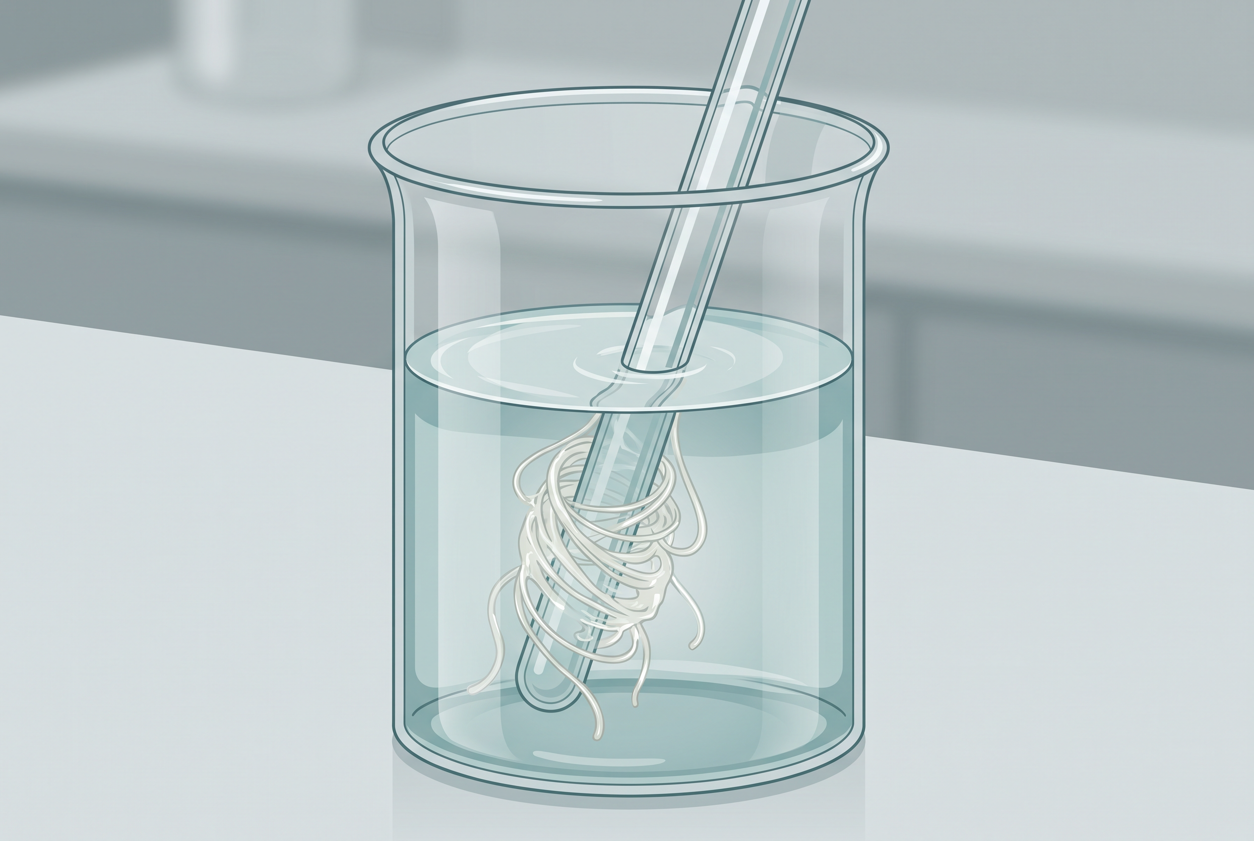
Fig. 9.5 – Spooling precipitated DNA.
Flowchart
FC 9.7 – Isolating pure DNA: break open the cell wall (lysozyme / cellulase / chitinase) → release DNA with other macromolecules → remove RNA with ribonuclease → remove proteins with protease → remove other molecules by suitable treatments → add chilled ethanol → pure DNA precipitates as fine threads → spool out
9.3.2 Cutting of DNA at Specific Locations
With pure DNA in hand, the next step is to cut it at the chosen sites. Restriction enzyme digestions are performed by incubating the purified DNA molecules with the restriction enzyme under the optimal conditions (such as the correct temperature and buffer) for that specific enzyme.
To check how far the digestion has progressed, agarose gel electrophoresis is used. Because DNA is a negatively charged molecule, it moves towards the positive electrode, the anode, during electrophoresis (as we saw in Fig. 9.3). By looking at the pattern of fragments, we can tell whether the enzyme has finished cutting. The same cutting process is then repeated with the vector DNA, using the same restriction enzyme.
The joining of DNA now involves a few coordinated actions. After both the source DNA and the vector DNA have been cut with the same restriction enzyme, the cut-out gene of interest from the source DNA is mixed together with the cut vector (which now has a matching gap). DNA ligase is then added, and it seals the fragment into the vector. This results in the preparation of the recombinant DNA molecule.
9.3.3 Amplification of Gene of Interest using PCR
Often we have only a very small amount of the gene of interest, which is not enough to work with. PCR solves this problem. PCR stands for Polymerase Chain Reaction. In this reaction, multiple copies of the gene (or piece of DNA) of interest are synthesised in vitro using two sets of primers and the enzyme DNA polymerase.
The primers are small, chemically synthesised oligonucleotides (short single strands of nucleotides) that are complementary to the two regions of DNA flanking the segment to be copied. The DNA polymerase then extends each primer, building a new strand using the nucleotides provided in the reaction and the original genomic DNA as the template.
Each round of PCR has three steps, shown in Fig. 9.6. First comes denaturation, in which heat separates the double-stranded DNA into two single strands. Next comes annealing, in which the primers bind to their complementary sequences on each single strand. Last comes extension, in which DNA polymerase adds nucleotides to extend the primers and build the new complementary strands.
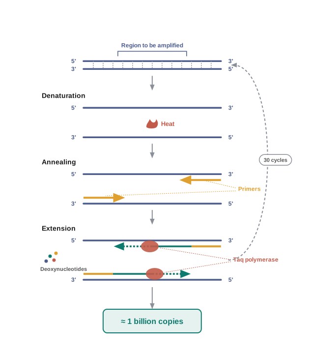
Fig. 9.6 – The steps of the polymerase chain reaction (PCR).
If this process of replication is repeated many times, the segment of DNA can be amplified to approximately a billion times — that is, about 1 billion copies are made from a single starting molecule. This is possible because each cycle roughly doubles the number of copies.
There is one practical obstacle. Ordinary DNA polymerase would be destroyed by the high temperature needed for denaturation in each cycle. This is overcome by using a thermostable DNA polymerase, isolated from the bacterium Thermus aquaticus, which remains active even during the high-temperature-induced denaturation of double-stranded DNA.
The amplified fragment, if desired, can now be used to ligate with a vector for further cloning, as indicated in Fig. 9.6.
9.3.4 Insertion of Recombinant DNA into the Host Cell/Organism
Once the recombinant DNA has been prepared, it must be placed inside a living host so that it can multiply. There are several methods of introducing the ligated DNA into recipient cells, and (as we saw in Section 9.2.3) the recipient cells are first made “competent” so that they can take up DNA present in their surroundings.
The clever part is how we then confirm that the DNA has entered. Suppose a recombinant DNA that carries a gene for resistance to an antibiotic such as ampicillin is transferred into E. coli cells. The host cells that take up this DNA become transformed into ampicillin-resistant cells. If we now spread the treated cells on agar plates containing ampicillin, only the transformants will grow, while the untransformed recipient cells will die.
Because the ampicillin-resistance gene allows us to pick out a transformed cell in the presence of ampicillin, this gene is acting as a selectable marker. In other words, the antibiotic acts as a filter that lets only the successfully transformed cells survive and form colonies.
9.3.5 Obtaining the Foreign Gene Product
When a piece of alien DNA is inserted into a cloning vector and transferred into a bacterial, plant or animal cell, the alien DNA gets multiplied. But multiplying the DNA is rarely the final goal. In almost all recombinant technologies, the ultimate aim is to produce a desirable protein. For this, the recombinant DNA must be expressed — that is, the foreign gene must be switched on so that the cell reads it and manufactures its protein. The foreign gene is made to express under appropriate conditions, and getting this expression right involves understanding many technical details.
After the gene of interest has been cloned and the conditions to induce expression of the target protein have been optimised, one has to think about producing that protein on a large scale. If any protein-encoding gene is expressed in a heterologous host — a host different from the organism the gene originally came from — the protein produced is called a recombinant protein.
The cells that harbour the cloned genes of interest may first be grown on a small scale in the laboratory, and these cultures can be used to extract the desired protein, which is then purified using different separation techniques. To boost the yield, the cells can also be multiplied in a continuous culture system. In such a system, the used medium is drained out from one side while fresh medium is added from the other side, so that the cells are kept in their physiologically most active log (exponential) phase of growth. This produces a larger biomass, and therefore higher yields of the desired protein.
Small-volume cultures cannot yield appreciable quantities of products. To obtain large quantities, the development of bioreactors was required, in which large volumes — 100 to 1000 litres — of culture can be processed at a time.
The most commonly used bioreactors are of the stirring type, shown in Fig. 9.7. A stirred-tank reactor is usually cylindrical, or has a curved base, so as to make it easier to mix the contents of the reactor.
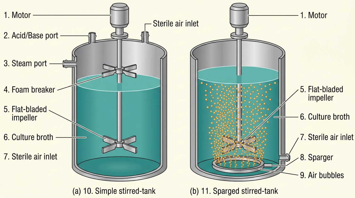
Fig. 9.7 – (a) Simple stirred-tank bioreactor and (b) sparged stirred-tank bioreactor.
The stirrer helps in two ways: it ensures even mixing throughout the vessel, and it keeps oxygen available to all the cells. Alternatively, air can be bubbled through the reactor to supply oxygen. If you examine the figure closely, you will see that the bioreactor also has an agitator system, an oxygen delivery system, a foam control system, a temperature control system and a pH control system, along with sampling ports through which small volumes of the culture can be withdrawn periodically to monitor its progress.
9.3.6 Downstream Processing
Making the product inside the bioreactor is only the biosynthetic stage. After this stage is complete, the product has to pass through a series of further processes before it is ready to be marketed as a finished product.
These processes include separation and purification of the product, and together they are collectively referred to as downstream processing. Once separated and purified, the product then has to be formulated with suitable preservatives so that it stays stable and usable.
Such a formulation must undergo thorough clinical trials, as in the case of drugs, to make sure it is safe and effective in living beings. In addition, strict quality control testing is required for each product. It is important to note that the exact downstream processing and quality control testing vary from one product to another, depending on what the product is and how it will be used.
Flowchart
FC 9.9 – Downstream processing: finished biosynthesis in bioreactor → separation → purification → formulation with preservatives → clinical trials (for drugs) → quality control testing → finished, marketable product
9.4 Summary
- Biotechnology is the use of living organisms, cells or enzymes to make useful products and carry out useful processes, and modern biotechnology does this on a large scale for marketing.
- Modern biotechnology, based on genetically modified organisms, became possible only once scientists learnt how to alter the chemistry of DNA and build recombinant DNA. This central process is called recombinant DNA technology, or genetic engineering.
- The key tools of this technology are restriction endonucleases (which cut DNA at specific recognition sequences to leave sticky ends), DNA ligase (which seals cut DNA ends together), and suitable plasmid or viral vectors (which carry the foreign DNA into a host cell). A competent host cell then takes up this DNA.
- The overall process isolates the foreign gene, ferries it into a host organism using a vector, and gets the foreign gene expressed so that the host makes the desired functional protein.
- The protein product is then purified during downstream processing, and finally made into a suitable formulation for marketing, after quality control and, where needed, clinical trials.
- Large-scale production of the product is carried out using bioreactors, which provide the optimum conditions for the cells to grow and manufacture the product in large quantities.
Flowchart
FC 9.10 – The whole chapter in one sequence: alter DNA chemistry → cut gene and vector with the same restriction enzyme → join with DNA ligase to form recombinant DNA → transfer into a competent host → select transformants/recombinants → express the foreign gene → grow at large scale in a bioreactor → downstream processing (purify, formulate, test) → finished product
9.5 Points to Ponder
- Bacteria evolved restriction enzymes long before humans existed, as a defence against invading viruses. Is it fair to say that genetic engineering was “invented” by bacteria, and that humans have only learned to borrow their tools?
- The same antibiotic-resistance gene that scientists use as a convenient selectable marker in the laboratory is, in nature, a serious problem when it spreads among disease-causing bacteria. Should the routine use of antibiotic-resistance markers in biotechnology be reconsidered? What alternatives might work as well?
- A restriction enzyme cuts a bacterium’s own DNA sequence just as readily as a virus’s. How might a cell protect its own DNA from being destroyed by the very scissors it makes? What could go wrong if that protection failed?
- PCR can turn a single molecule of DNA into a billion copies. This power is used in medical diagnosis and in forensic science. What responsibilities come with a technique so sensitive that a trace of DNA left behind can be amplified and read?
- We call Agrobacterium tumifaciens a “disarmed” pathogen once its disease-causing genes are removed. How confident can we be that such an organism is truly harmless once released into fields, and what should we watch for?
- A recombinant protein made in E. coli is identical in sequence to the natural human protein, yet it is manufactured inside a bacterium. Does the source of a medicine matter, so long as the molecule is the same? Why might some people feel it does?
- Traditional breeding mixes thousands of genes at once, while genetic engineering moves a single chosen gene. Which approach carries the greater risk of an unexpected outcome, and on what grounds would you argue your case?
- Every step of recombinant DNA technology depends on being able to distinguish the cells that worked from the vast majority that did not. Why is selection, rather than the cutting and joining of DNA, often the real bottleneck in an experiment?
9.6 NCERT Questions
- Can you list 10 recombinant proteins that are used in medical practice? Find out where each is used as a therapeutic (use the internet).
- Make a chart (with a diagrammatic representation) showing a restriction enzyme, the substrate DNA on which it acts, the site at which it cuts the DNA, and the product it produces.
- From what you have learnt, can you tell whether enzymes are larger or DNA is larger in molecular size? How did you decide?
- What would be the molar concentration of human DNA in a human cell? Consult your teacher.
- Do eukaryotic cells have restriction endonucleases? Justify your answer.
- Besides better aeration and mixing properties, what other advantages do stirred-tank bioreactors have over shake flasks?
- Collect 5 examples of palindromic DNA sequences by consulting your teacher. Better still, try to create a palindromic sequence yourself by following the base-pairing rules.
- Can you recall meiosis and indicate at what stage a recombinant DNA is made?
- Can you think and answer how a reporter enzyme can be used to monitor the transformation of host cells by foreign DNA, in addition to a selectable marker?
- Describe briefly the following:
- Origin of replication
- Bioreactors
- Downstream processing
- Explain briefly:
- PCR
- Restriction enzymes and DNA
- Chitinase
- Discuss with your teacher and find out how to distinguish between:
- Plasmid DNA and Chromosomal DNA
- RNA and DNA
- Exonuclease and Endonuclease
9.7 Check Your Concepts
- Why is it essential to cut both the vector DNA and the source DNA with the same restriction enzyme when constructing recombinant DNA? What would go wrong if two different enzymes were used?
- A restriction enzyme cuts the sequence shown below. Copy it, mark the palindrome, and show where the enzyme cuts each strand to produce sticky ends. 5′—— G A A T T C ——3′ 3′—— C T T A A G ——5′
- Assertion–Reason. Assertion: Sticky ends make the joining of two DNA fragments easier. Reason: Sticky ends are single-stranded overhangs that form hydrogen bonds with complementary overhangs. State whether each statement is true, and whether the reason correctly explains the assertion.
- Explain how the name EcoRI is derived. What does each part of the name tell you about the enzyme?
- Distinguish between an exonuclease and an endonuclease, and state which of the two is used to cut DNA at chosen internal sites in genetic engineering.
- Arrange the following steps of recombinant DNA technology in the correct sequence: ligation into a vector; transfer into a host; isolation of DNA; extraction of the product; cutting of DNA with restriction enzymes; large-scale culture.
- Why can a stray piece of DNA usually not multiply inside a new host cell unless it is joined to an origin of replication?
- In the vector pBR322, a foreign gene is inserted into the tetracycline-resistance gene. Explain how the recombinant colonies can be distinguished from the non-recombinant colonies using two antibiotic plates.
- What is insertional inactivation? Explain how the blue–white colony method uses this idea to identify recombinant colonies, and state which colour indicates a recombinant.
- Distinguish between a transformant and a recombinant. Can a cell be a transformant without being a recombinant? Explain.
- Match each organism/enzyme in Column I with its correct role in Column II.
| Column I | Column II |
|---|---|
| a. lysozyme | i. removes proteins |
| b. cellulase | ii. breaks bacterial cell wall |
| c. chitinase | iii. removes RNA |
| d. ribonuclease | iv. breaks fungal cell wall |
| e. protease | v. breaks plant cell wall |
- Why must bacterial cells be made “competent” before they can take up recombinant DNA? Describe the role of a divalent cation such as calcium and of the heat-shock step.
- Name and briefly describe two methods, other than chemical (calcium) transformation, that are used to introduce foreign DNA into (a) an animal cell and (b) a plant cell.
- Explain why a thermostable DNA polymerase from Thermus aquaticus is used in PCR instead of an ordinary DNA polymerase. Which step of each PCR cycle makes this necessary?
- Assertion–Reason. Assertion: Agrobacterium tumifaciens is called a natural genetic engineer. Reason: It can transfer a piece of its DNA (T-DNA) into plant cells and change their behaviour. State whether each statement is true, and whether the reason correctly explains the assertion.
- In an agarose gel, one DNA fragment has travelled far from the wells while another remains close to them. Which fragment is smaller, and why does it move farther? At which end was the sample loaded?
- What happens if the used medium is continuously drained and fresh medium continuously added during culturing? How does this “continuous culture” improve the yield of the recombinant protein?
- “The final purified protein is not the end of the process.” List, in order, the steps of downstream processing that a biotechnological product must pass through before it can be marketed.