Chapter 2: Biological Classification
Picture a biology class a few centuries ago. The rule seemed simple: every living thing was either a plant or an animal. Plants stood still and made their own food; animals moved about and ate other organisms. This tidy two-kingdom idea, popular in the time of the great naturalist Linnaeus, worked beautifully for oak trees and tigers.
But nature does not always respect our neat boxes. Consider a tiny pond organism called Euglena. In sunlight it turns green and manufactures its own food, behaving just like a plant. Move it into the dark, and it begins hunting and swallowing smaller organisms — behaving just like an animal. So which kingdom does it belong to? Biologists simply could not agree.
Euglena was not alone. Bacteria, moulds, pond scum, diatoms — thousands of organisms refused to sit neatly on either side of the plant–animal line. Some had no proper nucleus at all. Some had cell walls made of unusual materials. The two-kingdom system was straining at the seams.
In 1969, an American ecologist named R.H. Whittaker offered a bold fix. Instead of two kingdoms, he proposed five — Monera, Protista, Fungi, Plantae and Animalia — sorting organisms not merely by how they looked, but by their cell structure, body organisation, and mode of nutrition. It is this five-kingdom map of the living world that you are about to explore.
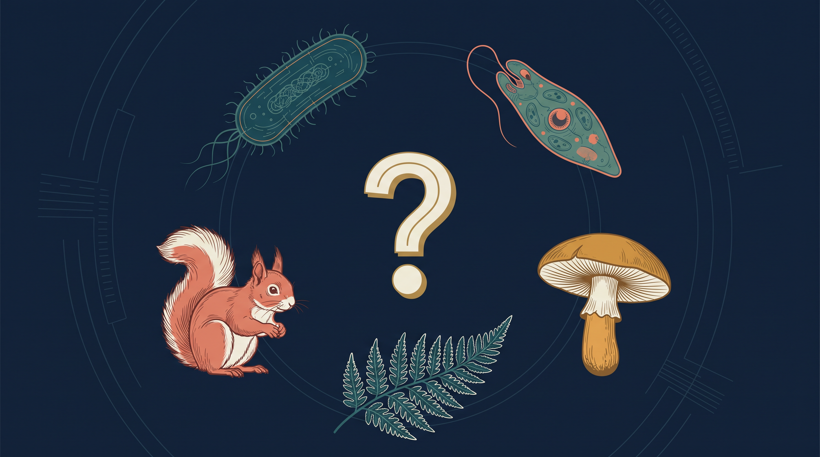
Fig. 2.0 – The diversity of the living world.
As you work through this chapter, keep these questions in mind — by the end, you will be able to answer every one of them.
2.1 Kingdom Monera
Kingdom Monera contains a single, remarkable group of organisms: the bacteria. Bacteria are the sole members of this kingdom, and they are the most abundant micro-organisms on Earth — living things so tiny that they are visible only under a microscope. A single handful of ordinary soil may hold hundreds of them.
What makes bacteria so successful is where they can live. They occur almost everywhere, from the food on your plate to the water you drink. They even thrive in extreme habitats — scalding hot springs, dry deserts, freezing snow and the crushing depths of the oceans — places where very few other life forms can survive. Many bacteria also live in or on other organisms as parasites, drawing their nourishment from a host.
Bacteria belong to Kingdom Monera because they are prokaryotic. A prokaryote is an organism whose cell has no true, membrane-bound nucleus — its genetic material (DNA) lies freely in the cytoplasm instead of being packed inside a nucleus. This simple cell design sets Monera apart from every other kingdom, whose members are eukaryotic (having a proper, membrane-bound nucleus).
Shapes of bacteria
Although bacteria are enormously varied, they take only a few basic shapes. Based on shape, they are grouped under four categories, as shown in Fig. 2.1: the spherical Coccus (plural: cocci), the rod-shaped Bacillus (plural: bacilli), the comma-shaped Vibrium (plural: vibrio) and the spiral Spirillum (plural: spirilla).
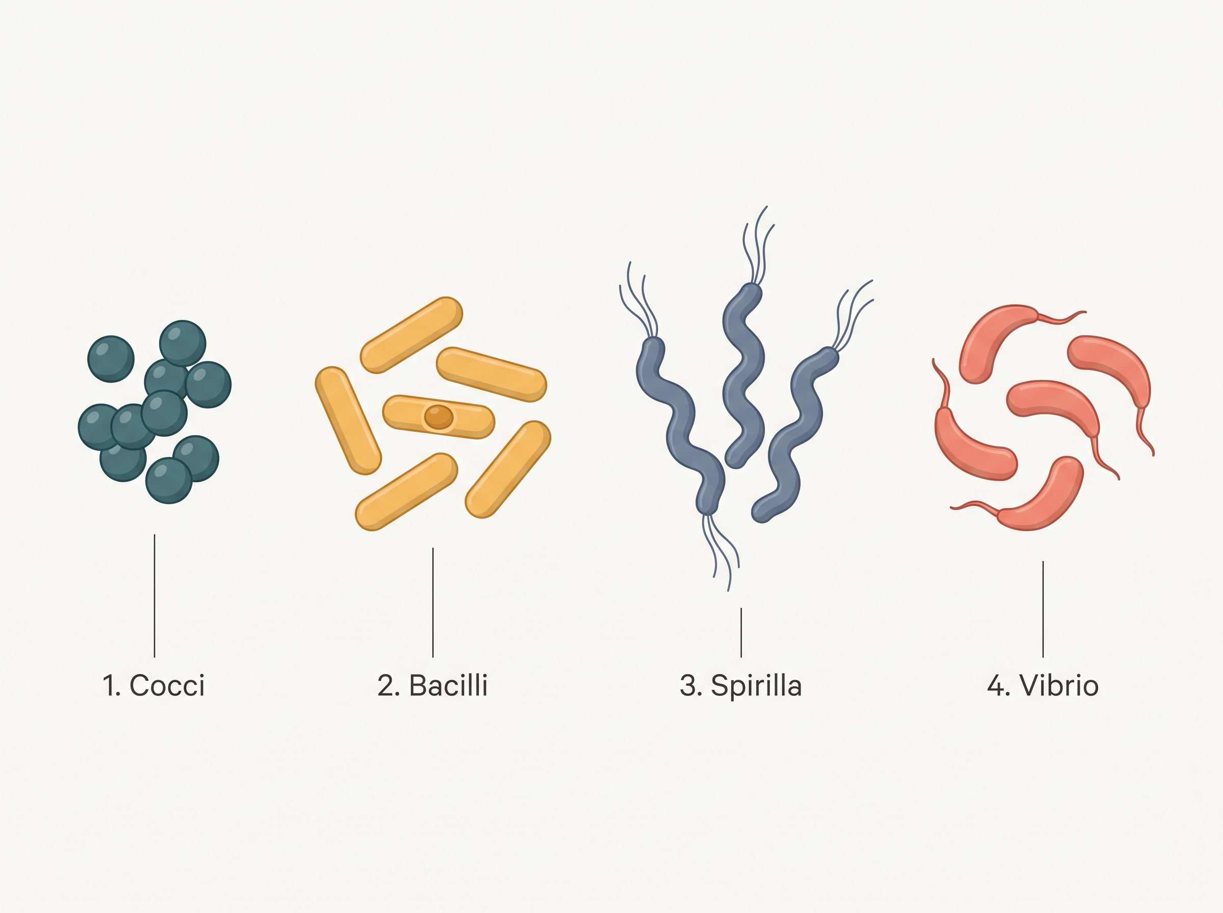
Fig. 2.1 – The four basic shapes of bacteria.
Metabolic diversity and nutrition
The bacterial cell has a very simple structure, yet bacteria are extremely complex in their behaviour. As a group, they show the most extensive metabolic diversity of all living organisms — meaning they can obtain energy and food in a far greater variety of ways than any other group of organisms.
Some bacteria are autotrophic: they build their own food from simple inorganic substances. Autotrophic bacteria are of two kinds — photosynthetic autotrophs, which capture the energy of sunlight, and chemosynthetic autotrophs, which use the energy released by chemical reactions.
The vast majority of bacteria, however, are heterotrophic. Heterotrophs cannot make their own food and must depend on other organisms, or on dead organic matter, for their nourishment.
This branching of bacterial nutrition is summarised in FC 2.1.
Flowchart
FC 2.1 – Nutrition in bacteria: Bacteria → Autotrophic (→ Photosynthetic; → Chemosynthetic) and Heterotrophic (→ Saprophytic, feeding on dead matter; → Parasitic, feeding on a living host).
For convenience, bacteria are placed in two broad divisions — the Archaebacteria and the Eubacteria — separated mainly by differences in their cell wall and the habitats they occupy.
2.1.1 Archaebacteria
Archaebacteria are special because they live in some of the harshest habitats on Earth. Three well-known types are named after the conditions they prefer: halophiles live in extremely salty areas, thermoacidophiles live in hot springs, and methanogens live in marshy, oxygen-poor areas.
What allows archaebacteria to survive such punishing conditions is their unusual cell wall. Their cell wall has a different structure from that of all other bacteria, and it is this feature that shields the cell and keeps it alive where other organisms would perish.
Methanogens are especially interesting. They live in the gut of several ruminant animals such as cows and buffaloes, and they produce methane gas — extreme gas we call biogas. This is why the dung of these animals can be used to generate methane (biogas).
2.1.2 Eubacteria
The eubacteria, or “true bacteria,” form the second great group, and there are thousands of different kinds. They are recognised by two features: a rigid cell wall, and — if the bacterium is able to move — a whip-like flagellum used for swimming.
Cyanobacteria
The cyanobacteria, also called blue-green algae, are photosynthetic eubacteria. They contain chlorophyll a — the same green pigment found in green plants — and are therefore photosynthetic autotrophs, as shown in Fig. 2.2.
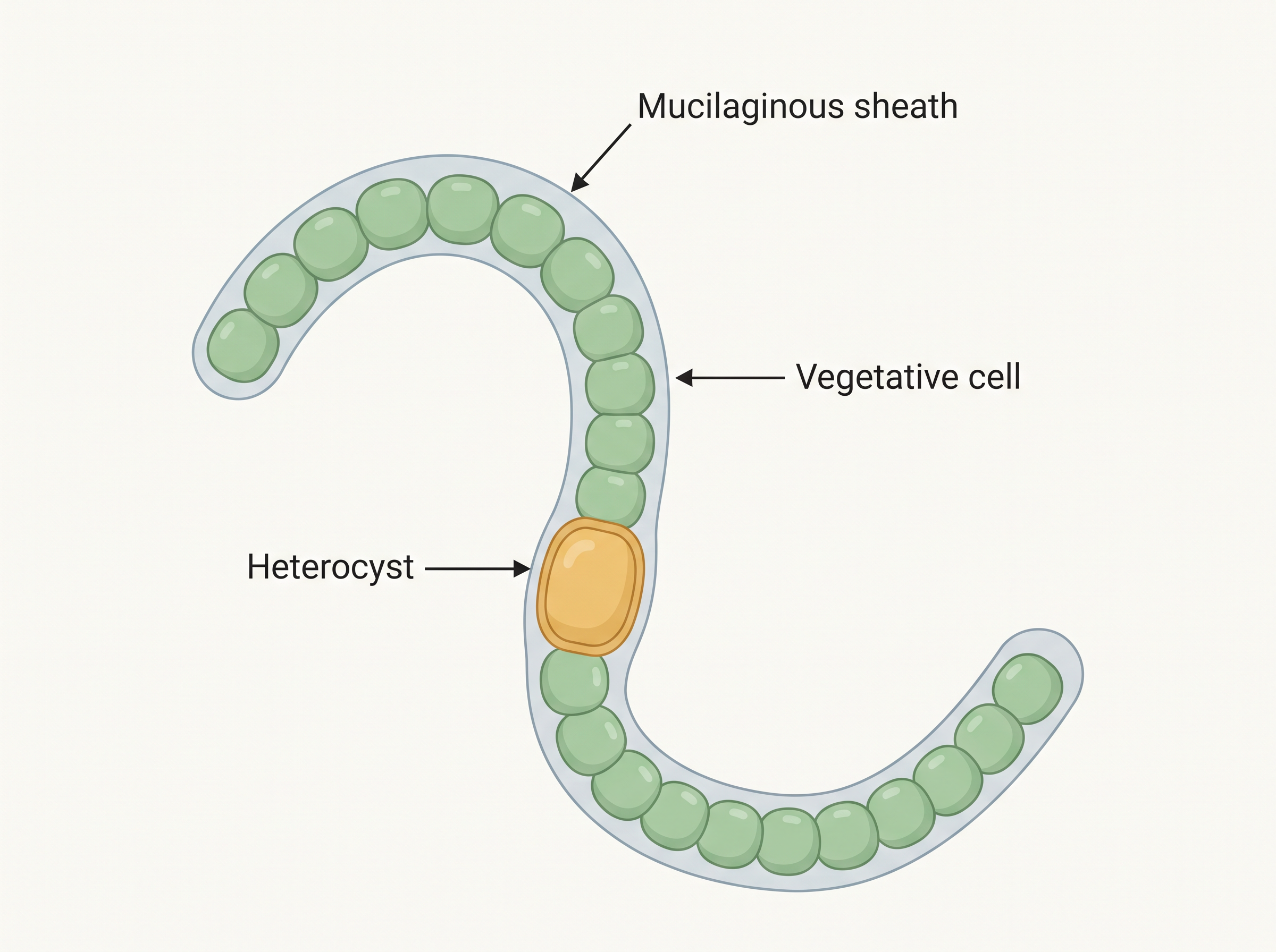
Fig. 2.2 – A filamentous blue-green alga, Nostoc.
Cyanobacteria may be unicellular, colonial or filamentous, and they are found in freshwater, marine or terrestrial habitats. Their colonies are usually surrounded by a gelatinous (jelly-like) sheath. In polluted water bodies that are rich in nutrients, they often multiply rapidly to form visible blooms on the water surface.
Some cyanobacteria can fix atmospheric nitrogen — that is, convert the unusable nitrogen gas of the air into a form that living things can use. They do this inside specialised, thick-walled cells called heterocysts; Nostoc and Anabaena are common examples.
Chemosynthetic bacteria
Chemosynthetic autotrophic bacteria do not depend on sunlight. Instead, they oxidise inorganic substances such as nitrates, nitrites and ammonia, and use the energy released to make ATP (the cell’s energy currency). In doing so, they play a great role in recycling nutrients like nitrogen, phosphorus, iron and sulphur, returning them to forms that other organisms can use.
Heterotrophic bacteria
Heterotrophic bacteria are the most abundant of all bacteria in nature, and the majority are important decomposers — they break down dead matter and return its nutrients to the soil. Many of them affect human affairs deeply, in both helpful and harmful ways.
On the useful side, heterotrophic bacteria help us make curd from milk, produce antibiotics, and fix nitrogen in the root nodules of leguminous plants (such as gram and pea), naturally enriching the soil.
Some heterotrophic bacteria are pathogens — disease-causing organisms — that damage human beings, crops, farm animals and pets.
How bacteria reproduce
Bacteria reproduce mainly by fission — a single cell simply splits into two identical daughter cells, as shown in Fig. 2.3. Under unfavourable conditions, some bacteria protect themselves by producing tough spores that can wait out the bad times and germinate later.
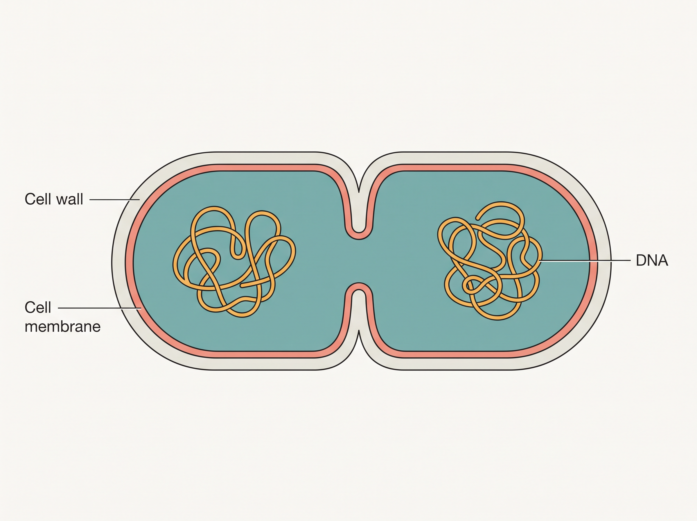
Fig. 2.3 – A dividing bacterium.
Bacteria can also carry out a primitive kind of sexual reproduction, in which a piece of DNA is transferred from one bacterium to another. This is not true sexual reproduction as seen in higher organisms, but it does let bacteria exchange genetic material and gain new characteristics.
Mycoplasma — bacteria without a wall
Finally, the Mycoplasma are unusual organisms that completely lack a cell wall. They are the smallest living cells known, and they can survive without oxygen. Many mycoplasma are pathogenic, causing diseases in both animals and plants.
2.2 Kingdom Protista
All single-celled eukaryotes are placed in Kingdom Protista. (Recall that a eukaryote is an organism whose cells contain a true, membrane-bound nucleus, unlike the prokaryotic bacteria of Monera.) The boundaries of this kingdom, however, are not sharply defined: an organism that one biologist calls “a photosynthetic protistan,” another may prefer to call “a plant.” Because of this fuzziness, different books group protists slightly differently. In this book we include the Chrysophytes, Dinoflagellates, Euglenoids, Slime moulds and Protozoans under Protista.
Most protists are aquatic — they live in water or watery surroundings. This kingdom holds a special place: it forms a link between the simpler Monera and the three “higher” kingdoms of plants, animals and fungi. In other words, protists are the eukaryotic starting point from which more complex organisms are thought to have arisen.
Being eukaryotes, the protistan cell body contains a well-defined nucleus and other membrane-bound organelles — the tiny “compartments” inside a cell that each carry out a specific job. Some protists move about with the help of flagella (whip-like threads) or cilia (many short, hair-like threads). Protists reproduce both asexually and sexually; the sexual process involves the fusion of two cells and the formation of a zygote (the single cell produced when two gametes fuse).
2.2.1 Chrysophytes
This group includes the diatoms and the golden algae (desmids). They live in both fresh water and marine (sea) environments. Chrysophytes are microscopic and float passively in water currents — organisms that drift about in this way are together called plankton. Most of them are photosynthetic.
The diatoms are especially remarkable for their cell walls. In a diatom, the cell wall forms two thin overlapping shells that fit together neatly, much like the two halves of a soap box. These walls are embedded with silica (the same hard mineral found in glass and sand), which makes them practically indestructible.
Because the walls do not decay, dead diatoms leave their glassy shells behind. Over billions of years, these have piled up into enormous deposits known as “diatomaceous earth.”
2.2.2 Dinoflagellates
Dinoflagellates are mostly marine and photosynthetic. They appear yellow, green, brown, blue or red, depending on which pigments dominate in their cells. Their cell wall carries stiff cellulose plates on the outer surface, giving them an armoured look, as seen in Fig. 2.4a.
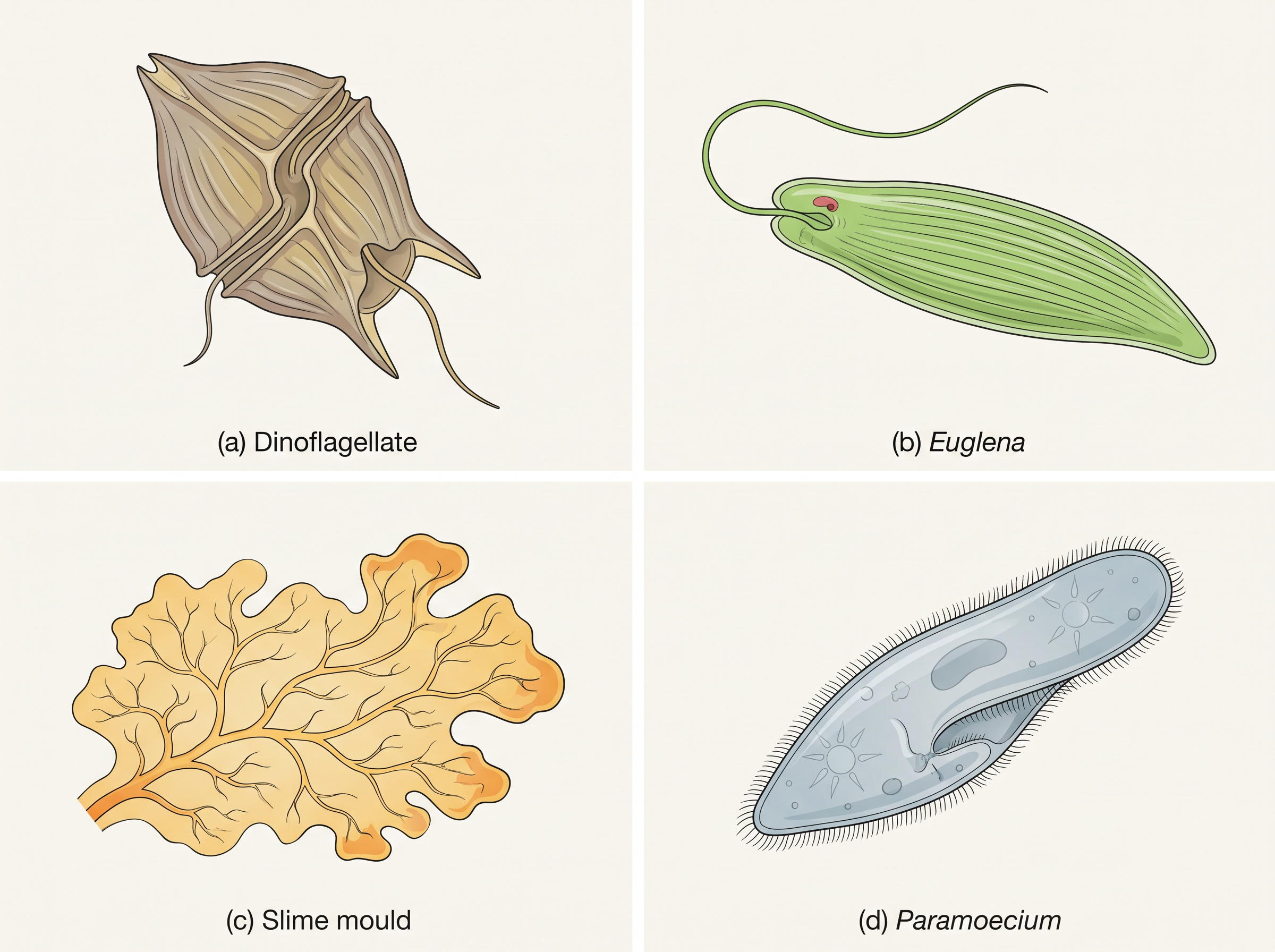
Fig. 2.4 – Representative protists: (a) dinoflagellate, (b) Euglena, (c) slime mould and (d) Paramoecium.
Most dinoflagellates have two flagella: one lies longitudinally (along the length of the body) and the other transversely (across it), running in a furrow between the wall plates. This crossed arrangement makes the organism spin as it swims.
Very often, red dinoflagellates such as Gonyaulax multiply so rapidly that they turn the sea red — an event called a red tide. The toxins released by such vast numbers can even kill other marine animals, such as fishes.
2.2.3 Euglenoids
The majority of euglenoids are freshwater organisms found in stagnant (still) water. Instead of a rigid cell wall, they have a protein-rich layer called a pellicle, which makes the body flexible so that it can change shape. They move using two flagella — a short one and a long one.
Euglenoids have a fascinating double lifestyle. In the presence of sunlight they are photosynthetic, making their own food like a plant. But when deprived of sunlight, they behave like heterotrophs, capturing and feeding on other, smaller organisms — like an animal. Interestingly, the pigments of euglenoids are identical to those found in higher plants. A common example is Euglena (Fig. 2.4b).
2.2.4 Slime Moulds
Slime moulds are saprophytic protists — that is, they feed on dead and decaying organic matter. The body creeps along decaying twigs and leaves, engulfing the organic material it meets on the way.
Under suitable, favourable conditions, the slime mould body forms an aggregation called a plasmodium — a large mass of living material that may grow and spread over several feet. When conditions turn unfavourable, the plasmodium differentiates and forms fruiting bodies that bear spores at their tips.
These spores are special: they possess true walls, are extremely resistant, and can survive for many years even under harsh conditions. Once released, the spores are dispersed by air currents to new places, where they can grow again when conditions improve.
2.2.5 Protozoans
All protozoans are heterotrophs, living either as predators (hunting other organisms) or as parasites (feeding on a host). They are believed to be primitive relatives of animals — an early evolutionary hint of the animal kingdom to come. There are four major groups of protozoans, compared in the table below.
Amoeboid protozoans
These organisms live in fresh water, sea water or moist soil. They move and capture their prey by pushing out pseudopodia — literally “false feet,” temporary bulges of the cell — as seen in Amoeba. Marine (sea-living) forms often have silica shells on their surface. Some amoeboid protozoans, such as Entamoeba, are parasites.
Flagellated protozoans
The members of this group are either free-living or parasitic, and they move using flagella. The parasitic forms cause diseases such as sleeping sickness. A well-known example is Trypanosoma.
Ciliated protozoans
These are aquatic and actively moving, thanks to the thousands of cilia covering their bodies. They possess a cavity called a gullet that opens to the outside of the cell. The coordinated, wave-like beating of the rows of cilia sweeps water — laden with food particles — into the gullet. A classic example is Paramoecium (Fig. 2.4d).
Sporozoans
This group includes diverse organisms that all share one feature: an infectious, spore-like stage in their life cycle. The most notorious sporozoan is Plasmodium, the malarial parasite, which causes malaria — a disease that has had a staggering effect on human populations.
2.3 Kingdom Fungi
The fungi make up a unique kingdom of heterotrophic organisms — living things that cannot make their own food and must absorb it from their surroundings. Fungi show great diversity in both their form (morphology) and the places they live (habitat).
You have almost certainly met fungi already. The fuzzy growth on a slice of moist bread, the soft patches on rotten fruit, the common mushroom you eat and the poisonous toadstool are all fungi. The white spots that appear on mustard leaves are caused by a parasitic fungus. Some unicellular fungi, such as yeast, are used to make bread and beer, while others cause diseases in plants and animals — the wheat rust-causing Puccinia is an important example. A few, like Penicillium, are valuable sources of antibiotics.
Fungi are cosmopolitan — they occur almost everywhere: in air, water and soil, and on animals and plants. They prefer to grow in warm and humid places. This is precisely why we store food in the refrigerator.
The body of a fungus
With the exception of the unicellular yeasts, fungi are filamentous — their bodies are built from long, slender, thread-like structures called hyphae (singular: hypha). A whole network of hyphae together is called a mycelium.
Not all hyphae are alike. Some are continuous tubes filled with cytoplasm that contains many nuclei — these are called coenocytic hyphae. Others are divided by septae, or cross walls, into separate compartments. The cell walls of fungi are made of chitin (a tough material also found in the outer skeletons of insects) together with polysaccharides.
How fungi feed
Most fungi are heterotrophic and absorb soluble organic matter from dead substrates; such fungi are called saprophytes. (Recall from the slime moulds that a saprophyte lives on dead, decaying matter.) Those that depend on living plants and animals for food are called parasites.
Fungi can also live as symbionts — partners in a close, mutually helpful relationship with another organism. In association with algae they form lichens, and in association with the roots of higher plants they form mycorrhiza.
Reproduction in fungi
Fungi reproduce in several ways. By vegetative means, they multiply through fragmentation (breaking into pieces), fission (splitting) and budding (small outgrowths pinching off). Asexual reproduction is by spores called conidia, sporangiospores or zoospores. Sexual reproduction is by oospores, ascospores and basidiospores. All these various spores are produced in distinct structures called fruiting bodies.
The sexual cycle of fungi involves three steps: (i) Plasmogamy — the fusion of the protoplasms (cell contents) of two motile or non-motile gametes. (ii) Karyogamy — the fusion of the two nuclei that plasmogamy brought together. (iii) Meiosis in the zygote, which produces haploid spores. (Meiosis is a special cell division that halves the chromosome number; a haploid cell, written \(n\), has a single set of chromosomes, while a diploid cell, written \(2n\), has two sets.)
Flowchart
FC 2.2 – Fungal sexual cycle: two compatible haploid hyphae meet → Plasmogamy (protoplasms fuse) → in some fungi nuclei fuse at once to give diploid (\(2n\)); in ascomycetes and basidiomycetes → a dikaryotic stage (\(n + n\), two nuclei per cell) forms first → Karyogamy (parental nuclei fuse, cell becomes diploid \(2n\)) → Meiosis in the fruiting body → haploid (\(n\)) spores.
When a fungus reproduces sexually, two haploid hyphae of compatible mating types come together and fuse. In some fungi, the fusion of the two haploid cells immediately produces diploid cells (\(2n\)). In other fungi — the ascomycetes and basidiomycetes — there is an in-between dikaryotic stage (\(n + n\), meaning two nuclei per cell) before fusion. This two-nuclei condition is called a dikaryon, and the phase is called the dikaryophase. Later, the parental nuclei fuse and the cells become diploid. The fungi then form fruiting bodies in which reduction division (meiosis) takes place, leading to the formation of haploid spores.
Basis of classification
The morphology of the mycelium, the mode of spore formation, and the type of fruiting bodies together form the basis for dividing Kingdom Fungi into its various classes.
2.3.1 Phycomycetes
Members of the Phycomycetes are found in aquatic habitats and on decaying wood in moist, damp places, or as obligate parasites on plants (parasites that cannot survive without their host). The mycelium is aseptate and coenocytic — a continuous tube without cross walls.
Asexual reproduction takes place by zoospores, which are motile (able to swim), or by aplanospores, which are non-motile. These spores are produced endogenously (inside) a structure called the sporangium.
In sexual reproduction, a zygospore is formed by the fusion of two gametes. These gametes may be similar in shape and size (isogamous) or dissimilar (anisogamous, or oogamous when one is a large, non-motile egg). Some common examples are Mucor (Fig. 2.5a), Rhizopus (the bread mould mentioned earlier) and Albugo (the parasitic fungus on mustard).
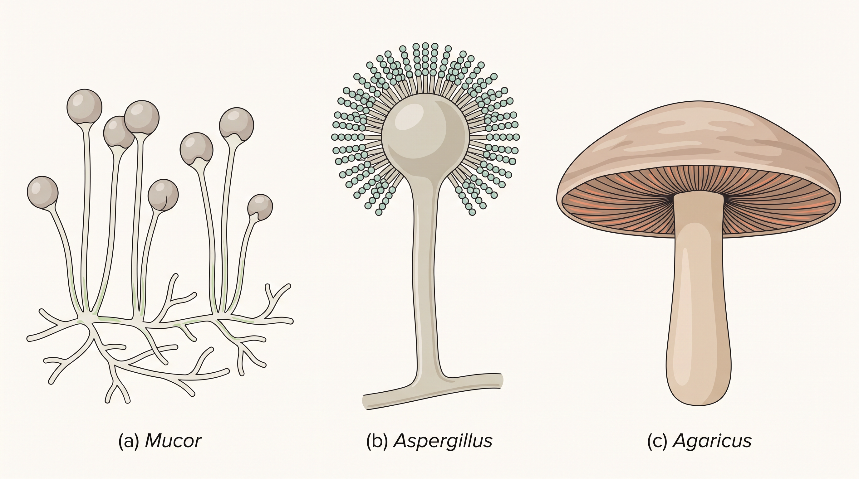
Fig. 2.5 – Representative fungi: (a) Mucor, (b) Aspergillus and (c) Agaricus.
2.3.2 Ascomycetes
Commonly known as sac-fungi, the ascomycetes are mostly multicellular, such as Penicillium, or rarely unicellular, such as yeast (Saccharomyces). They may be saprophytic, decomposers, parasitic or coprophilous (growing on dung). The mycelium is branched and septate.
The asexual spores are conidia, produced exogenously (on the outside) on a special mycelium called the conidiophore. On germination, conidia produce a new mycelium. The sexual spores are ascospores, produced endogenously inside sac-like structures called asci (singular: ascus). These asci are arranged in various types of fruiting bodies called ascocarps.
Some examples are Aspergillus (Fig. 2.5b), Claviceps and Neurospora. Neurospora is used extensively in biochemical and genetic research. Many members, like morels and truffles, are edible and are prized as delicacies.
2.3.3 Basidiomycetes
The commonly known forms of basidiomycetes are mushrooms, bracket fungi and puffballs. They grow in soil, on logs and tree stumps, and inside living plant bodies as parasites — for example, the rusts and smuts. The mycelium is branched and septate.
Asexual spores are generally not found in this group, but vegetative reproduction by fragmentation is common. The sex organs are absent; instead, plasmogamy is brought about by the fusion of two vegetative or somatic cells of different strains or genotypes. The resulting structure is dikaryotic, and it ultimately gives rise to the basidium. Karyogamy and meiosis take place inside the basidium, producing four basidiospores. The basidiospores are produced exogenously on the basidium (plural: basidia). The basidia are arranged in fruiting bodies called basidiocarps.
Some common members are Agaricus (the mushroom, Fig. 2.5c), Ustilago (smut) and Puccinia (rust fungus).
2.3.4 Deuteromycetes
The deuteromycetes are commonly known as the imperfect fungi, because only the asexual or vegetative phases of these fungi are known. “Imperfect” here simply means that their sexual (perfect) stage had not been observed.
There is an interesting history behind this class. When the sexual forms of these fungi were later discovered, the fungi were moved into the classes they truly belong to. Sometimes the asexual and vegetative stage of a fungus had been given one name (and placed under deuteromycetes), while its sexual stage had been given a different name (and placed in another class). Later, when the links between the two were established, the fungi were correctly identified and moved out of the deuteromycetes — usually into the ascomycetes or basidiomycetes, once their perfect (sexual) stages were found.
The deuteromycetes reproduce only by asexual spores known as conidia. Their mycelium is septate and branched. Some members are saprophytes or parasites, while a large number are decomposers of litter that help in mineral cycling — returning nutrients to the soil. Some examples are Alternaria, Colletotrichum and Trichoderma.
2.4 Kingdom Plantae
Kingdom Plantae includes all eukaryotic, chlorophyll-containing organisms that we commonly call plants. Chlorophyll is the green pigment that captures sunlight for photosynthesis, and it is housed in cell organelles called chloroplasts — which is what makes plant cells green and self-feeding (autotrophic).
A few plants, however, are only partially heterotrophic. These include the insectivorous plants, which trap and digest insects, and parasites, which draw food from a host. Bladderwort and the Venus flytrap are examples of insectivorous plants, while Cuscuta is a parasite.
The plant body
Plant cells have a typical eukaryotic structure with prominent chloroplasts and a cell wall made mainly of cellulose (a strong structural carbohydrate). You will study the eukaryotic cell structure in detail in Chapter 8.
Kingdom Plantae is a large and varied group. It includes the algae, bryophytes, pteridophytes, gymnosperms and angiosperms — a series that ranges from simple water-dwelling forms to complex flowering plants.
Flowchart
FC 2.3 – Major groups of Plantae, arranged from simplest to most advanced: Algae → Bryophytes → Pteridophytes → Gymnosperms → Angiosperms.
Life cycle: alternation of generations
The life cycle of plants has two distinct phases — a diploid sporophytic phase and a haploid gametophytic phase — that alternate with each other. (Recall that a diploid cell, \(2n\), carries two sets of chromosomes, while a haploid cell, \(n\), carries only one.)
The lengths of these two phases, and whether each phase is free-living or dependent on the other, vary among the different groups of plants. This alternation between a diploid and a haploid generation is called alternation of generations.
Flowchart
FC 2.4 – Alternation of generations: diploid sporophyte (\(2n\)) → meiosis → haploid spores → grow into a haploid gametophyte (\(n\)) → produces gametes → fertilisation (gametes fuse) → diploid zygote (\(2n\)) → grows into the sporophyte again (cycle repeats).
You will study further details of this kingdom in Chapter 3.
2.5 Kingdom Animalia
Kingdom Animalia is characterised by heterotrophic, eukaryotic organisms that are multicellular and whose cells lack cell walls. Not having a rigid cell wall around each cell is one key feature that sets animals apart from plants and fungi.
Animals cannot make their own food, so they depend — directly or indirectly — on plants for it. (An animal that eats grass depends on plants directly; one that eats other animals depends on plants indirectly, through its prey.)
Nutrition and body plan
Animals digest their food inside an internal cavity within the body, and store their food reserves as glycogen or fat. Their mode of nutrition is holozoic — food is taken in by ingestion, that is, by swallowing it whole or in pieces and then digesting it internally.
Growth, response and reproduction
Animals follow a definite growth pattern: they grow into adults that have a definite shape and size, rather than growing endlessly. Higher animals show an elaborate sensory and neuromotor mechanism — a well-developed system of sense organs, nerves and muscles that lets them detect changes and respond quickly. Most animals are also capable of locomotion, moving actively from place to place.
Sexual reproduction in animals takes place by the copulation of a male and a female, followed by embryological development (the growth of the fertilised egg into an embryo and then into a new individual).
Flowchart
FC 2.5 – Levels of body organisation in animals: Cells → Tissues → Organs → Organ systems → Organism.
The salient features of the various animal phyla are described in Chapter 4.
2.6 Viruses, Viroids, Prions and Lichens
In Whittaker’s five kingdom classification, there is no mention of lichens or of certain acellular organisms such as viruses, viroids and prions. “Acellular” means not made of cells — and since these agents lack a proper cell structure, they did not fit neatly into any of the five kingdoms. They are briefly introduced here.
Viruses
All of us who have suffered the ill effects of the common cold or ‘flu’ know what viruses can do to us, even if we did not connect our illness to them at the time.
Viruses did not find a place in the classification because they are not considered truly ‘living’ — at least, not if we define living things as those that have a cell structure. Viruses are non-cellular, and outside a living cell they exist as an inert crystalline structure, showing no signs of life at all.
Everything changes once a virus gets inside a host cell. There, it takes over the machinery of the host cell to make copies of itself, usually killing the host in the process. So — would you call viruses living or non-living?
Discovery of viruses
The word “virus” means venom or poisonous fluid — a fitting name for something that makes us ill.
Because they are inert outside their own specific host cell and can multiply only inside it, viruses are obligate parasites — they cannot reproduce anywhere except within a living host.
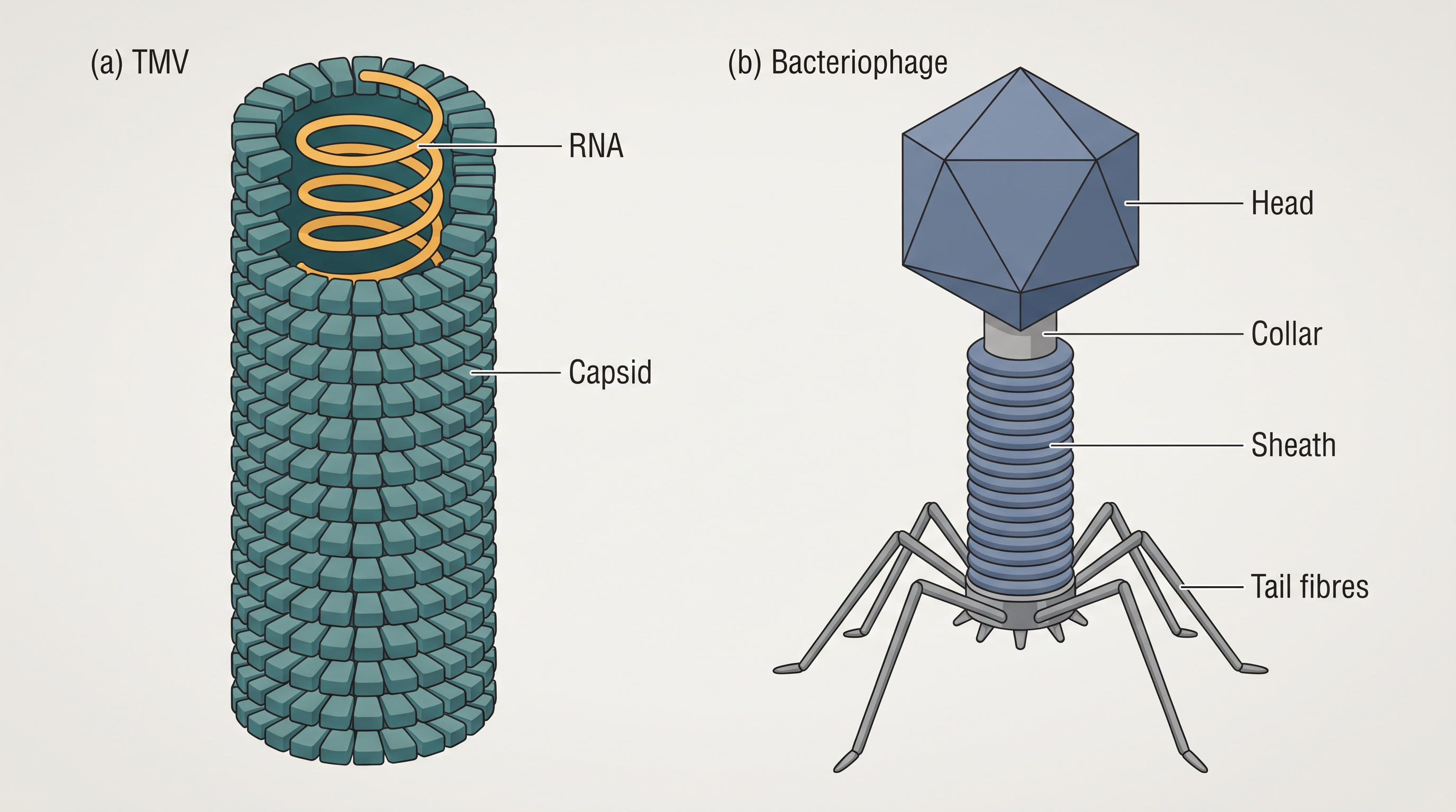
Fig. 2.6 – Viruses: (a) Tobacco Mosaic Virus and (b) Bacteriophage.
Structure and genetic material of viruses
Besides protein, every virus also contains genetic material, which may be either RNA or DNA — but never both. No virus contains both RNA and DNA together. A virus is therefore a nucleoprotein (nucleic acid plus protein), and its genetic material is infectious.
The type of genetic material tends to match the kind of host it attacks. In general, viruses that infect plants have single-stranded RNA, while viruses that infect animals have either single- or double-stranded RNA, or double-stranded DNA. Bacterial viruses, or bacteriophages — viruses that infect bacteria — are usually double-stranded DNA viruses, as shown in Fig. 2.6b.
The nucleic acid of a virus is protected by an outer protein coat called the capsid. The capsid is built from many small subunits called capsomeres, which are arranged in either helical or polyhedral (many-sided) geometric shapes.
Flowchart
FC 2.6 – How a virus multiplies: virus reaches a host cell → its nucleic acid enters the cell → takes over the host cell’s machinery → the machinery makes many copies of viral nucleic acid and protein → new virus particles assemble → the host cell is killed and new viruses are released.
Diseases caused by viruses
Viruses cause many familiar diseases in humans, including mumps, small pox, herpes and influenza. AIDS in humans is also caused by a virus. In plants, viral infection can show up as mosaic formation on the leaves, leaf rolling and curling, yellowing and vein clearing, dwarfing and stunted growth.
Viroids
In 1971, T.O. Diener discovered a new infectious agent that was even smaller than a virus and caused potato spindle tuber disease. It turned out to be a free RNA molecule that lacked the protein coat found in viruses — and it was this missing coat that gave it the name viroid. The RNA of the viroid was of low molecular weight (that is, it was a very small RNA).
Prions
In modern medicine, certain infectious neurological diseases (diseases of the nervous system) were found to be transmitted by an agent made of abnormally folded protein. This agent was similar in size to viruses, and such agents were named prions.
The most notable diseases caused by prions are bovine spongiform encephalopathy (BSE) — commonly called mad cow disease in cattle — and its human counterpart, Creutzfeldt–Jacob disease (CJD).
Lichens
Lichens are symbiotic associations — mutually useful partnerships — between algae and fungi. (You met lichens briefly with the fungi; here we look at how the partnership actually works.) The algal component is called the phycobiont and the fungal component is called the mycobiont; these are autotrophic and heterotrophic, respectively.
Each partner does a different job. The alga, being autotrophic, prepares food for the fungus, while the fungus provides shelter and absorbs mineral nutrients and water for its partner. So close is their association that, on seeing a lichen in nature, one would never imagine that it was made of two entirely different organisms living together.
Lichens are also very good pollution indicators — they do not grow in polluted areas. Where the air is dirty, lichens vanish; where they flourish, the air is clean.
2.7 Summary
Biological classification is the way we sort the huge variety of living organisms into orderly groups, and our ideas about how to do this have changed greatly over time.
- Aristotle was the first to attempt a scientific classification, using simple morphological (external form) characters.
- Linnaeus later classified all living organisms into two kingdoms — Plantae and Animalia.
- Whittaker proposed a more elaborate five kingdom classification — Monera, Protista, Fungi, Plantae and Animalia. Its main criteria were cell structure, body organisation, mode of nutrition, mode of reproduction, and phylogenetic (evolutionary) relationships.
Flowchart
FC 2.7 – How classification developed over time: Aristotle (morphological groups) → Linnaeus (two kingdoms: Plantae and Animalia) → Whittaker (five kingdoms: Monera, Protista, Fungi, Plantae, Animalia).
In the five kingdom classification, the bacteria are placed in Kingdom Monera.
- Bacteria are cosmopolitan — found almost everywhere.
- As a group they show the most extensive metabolic diversity of all organisms.
- In their mode of nutrition, bacteria may be autotrophic or heterotrophic.
Kingdom Protista includes all single-celled eukaryotes.
- Its members are the Chrysophytes, Dinoflagellates, Euglenoids, Slime-moulds and Protozoans.
- Being eukaryotes, they have a well-defined nucleus and other membrane-bound organelles.
- They reproduce both asexually and sexually.
Members of Kingdom Fungi show great diversity in structure and habitat.
- Most fungi are saprophytic in their mode of nutrition.
- They show both asexual and sexual reproduction.
- The kingdom has four classes: Phycomycetes, Ascomycetes, Basidiomycetes and Deuteromycetes.
Kingdom Plantae includes all eukaryotic, chlorophyll-containing organisms.
- It covers the algae, bryophytes, pteridophytes, gymnosperms and angiosperms.
- The plant life cycle shows alternation of generations, between a gametophytic and a sporophytic generation.
Kingdom Animalia includes the heterotrophic, eukaryotic, multicellular organisms that lack a cell wall.
- Their mode of nutrition is holozoic (food is taken in by ingestion).
- They reproduce mainly by the sexual mode.
Finally, some organisms are not included in the five kingdom system at all.
- The acellular agents — viruses, viroids and prions — and the lichens fall outside the five kingdoms.
- Viruses are non-cellular and become active only inside a host cell; viroids are free RNA without a protein coat; prions are infectious, abnormally folded proteins.
- Lichens are symbiotic partnerships between an alga (phycobiont) and a fungus (mycobiont), and are good indicators of pollution.
2.8 NCERT Questions
Discuss how classification systems have undergone several changes over a period of time.
State two economically important uses of:
- heterotrophic bacteria
- archaebacteria
What is the nature of cell-walls in diatoms?
Find out what the terms ‘algal bloom’ and ‘red-tides’ signify.
How are viroids different from viruses?
Describe briefly the four major groups of Protozoa.
Plants are autotrophic. Can you think of some plants that are partially heterotrophic?
What do the terms phycobiont and mycobiont signify?
Give a comparative account of the classes of Kingdom Fungi under the following:
- mode of nutrition
- mode of reproduction
What are the characteristic features of Euglenoids?
Give a brief account of viruses with respect to their structure and nature of genetic material. Also name four common viral diseases.
2.9 Check Your Concepts
Why was the two-kingdom system of classification (Plantae and Animalia) eventually found to be inadequate? Give at least two reasons.
Assertion (A): Cyanobacteria are placed in Kingdom Monera and not in Kingdom Plantae, even though they are called “blue-green algae.” Reason (R): Cyanobacteria are prokaryotic and lack a true, membrane-bound nucleus. State whether both A and R are true, and whether R correctly explains A.
Bacteria have a very simple structure, yet as a group they show the most extensive metabolic diversity of all organisms. Explain what “metabolic diversity” means here, using examples of autotrophic and heterotrophic bacteria.
What is a heterocyst? Explain how its structure is suited to its function of fixing atmospheric nitrogen.
Explain why archaebacteria are able to survive in extreme habitats such as hot springs, very salty areas and marshy soils, where most other organisms cannot.
Match each organism in Column I with its correct group in Column II.
| Column I | Column II |
|---|---|
| a. Nostoc | i. Dinoflagellate |
| b. Gonyaulax | ii. Sporozoan |
| c. Euglena | iii. Cyanobacterium |
| d. Plasmodium | iv. Ciliated protozoan |
| e. Paramoecium | v. Euglenoid |
Arrange the following steps of the fungal sexual cycle in their correct sequence: karyogamy, formation of haploid spores, plasmogamy, meiosis.
Distinguish between septate and coenocytic (aseptate) hyphae, and give one example of a fungal class showing each type.
Assertion (A): In ascomycetes and basidiomycetes, a cell may contain two nuclei (a dikaryon). Reason (R): In these fungi, plasmogamy is not immediately followed by karyogamy. State whether both A and R are true, and whether R correctly explains A.
Why are diatoms called the chief ‘producers’ of the oceans? What property of the diatom cell wall makes diatomaceous earth useful in polishing and filtration?
The Venus flytrap is a green plant, yet it is described as partially heterotrophic. Explain this apparent contradiction.
Define alternation of generations. Which of the two generations is haploid and which is diploid, and what does each produce?
“A virus behaves as non-living outside a host cell but as living inside one.” Explain this statement, and use it to describe why viruses are hard to classify.
How do (i) viroids and (ii) prions each differ in their infectious material from a typical virus?
A lichen is often described as “two organisms behaving as one.” Name the two partners, give the technical term for each, and state what each partner contributes to the association.
Draw a labelled diagram of a bacteriophage, labelling its head, collar, sheath and tail fibres.
State one useful role for each of the following: (i) methanogens, (ii) Neurospora, (iii) lichens as pollution indicators.