Chapter 18: Neural Control and Coordination
In the 1780s, in the Italian city of Bologna, an anatomist named Luigi Galvani was dissecting a frog on a table where an electrical machine also happened to be running. When his metal scalpel touched a nerve in the frog’s leg at the moment a spark jumped nearby, the dead frog’s leg suddenly twitched — as if it had come back to life.
Galvani was astonished. He repeated the experiment again and again, hanging frog legs on metal hooks, and each time the muscles kicked. He became convinced that living tissue carried its own kind of electricity, which he called “animal electricity.” His frog-leg experiments started a fierce scientific debate and opened up an entirely new question that would occupy biologists for the next two centuries: do our nerves really run on electricity?
Today we know Galvani was on to something profound. Every thought you think, every step you take, and every beat of your heart depends on tiny electrical signals racing along your nerves — signals your body generates itself, without any outside spark. In this chapter you will meet the cells that make this possible, follow a nerve signal from the moment it is born to the moment it jumps to the next cell, and tour the remarkable organ that sits in command of it all: the brain.
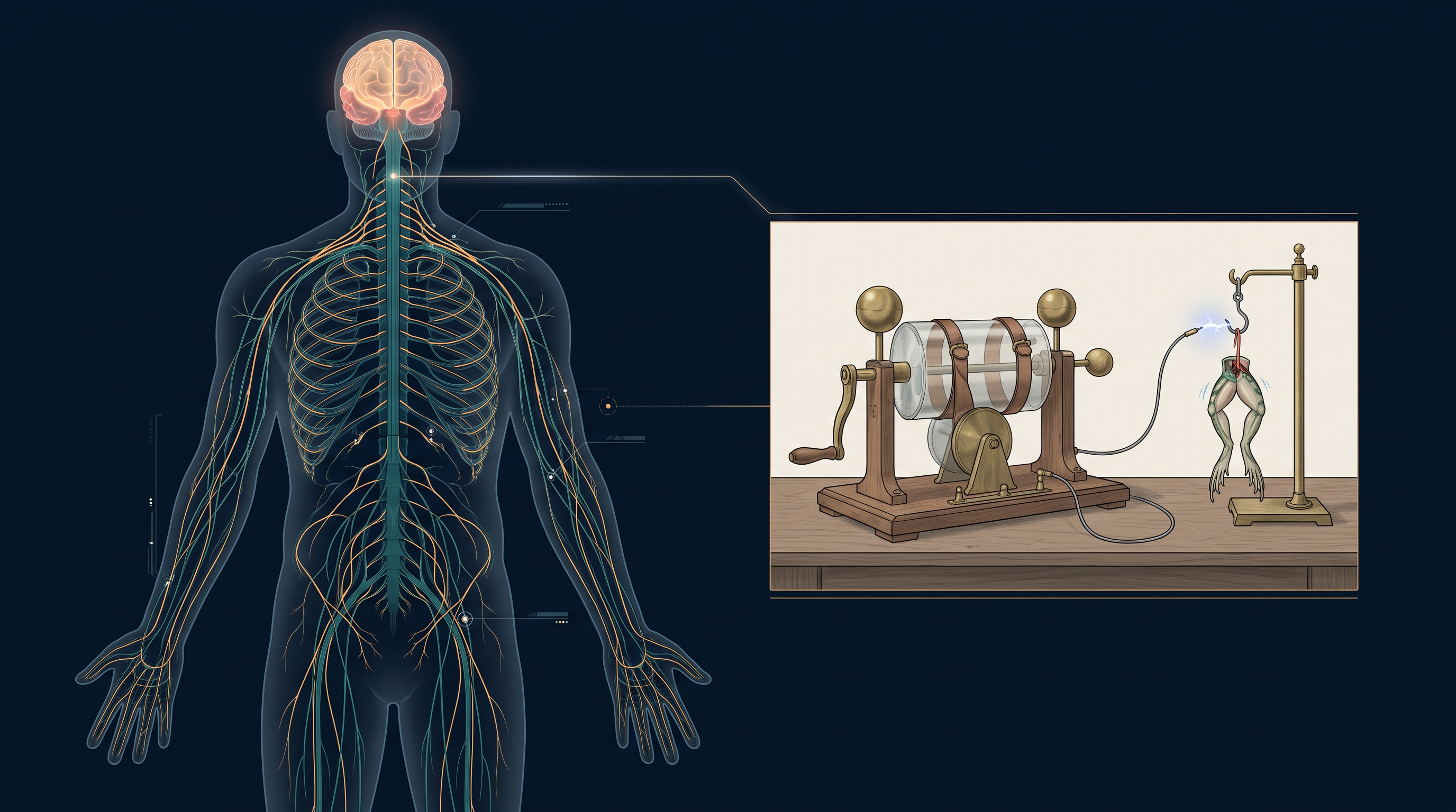
Fig. 18.0 – The human nervous system and Galvani’s frog experiment.
By the end of this chapter, you will be able to answer each of these questions yourself.
18.1 Neural System
Every organ in your body has its own job to do, yet none of them works alone. Your muscles, lungs, heart, blood vessels and kidneys must all act together, second by second, if the body is to stay healthy. This teamwork among organs is called coordination.
Think about what happens when you exercise. Your working muscles suddenly demand more energy, so they need more oxygen. To supply that oxygen, your breathing rate rises, your heart beats faster, and blood flows more quickly through the vessels. The moment you stop exercising, all of these — nerves, lungs, heart and kidneys — gradually settle back to their resting state.
This smooth rise and fall, with every organ adjusting in step, is coordination in action. It keeps the body’s internal conditions stable — a balanced state known as homeostasis, in which temperature, water content and the chemistry of the blood are all held within safe limits.
Cells that carry the signals
The neural system of all animals is built from highly specialised cells called neurons. A neuron can detect a stimulus (any change it is able to respond to), receive it, and transmit it onward as a signal. This signalling ability is exactly what makes the whole neural system work.
How complex is a neural system?
Neural systems are not equally developed in all animals. In lower invertebrates the arrangement is very simple — in Hydra, for example, the neurons are spread out as a loose network, with no brain to direct them. Insects have a better-organised system, with a definite brain together with a number of ganglia (clusters of nerve cell bodies) and neural tissues. Vertebrates, including humans, have the most developed neural system of all. This increasing complexity is summarised in FC 18.1.
Flowchart
FC 18.1 – Increasing complexity of the neural system across animals: lower invertebrates (e.g., Hydra) — simple nerve net → insects — brain + ganglia + neural tissues → vertebrates — highly developed neural system
18.2 Human Neural System
The human neural system is a vast communication network, but its overall plan is surprisingly simple. It is divided into two parts:
- the central neural system (CNS)
- the peripheral neural system (PNS)
The command centre and its wiring
The CNS is made up of the brain and the spinal cord. This is where information is received, processed and acted upon — the site of control and decision-making for the whole body.
The PNS is everything else: all the nerves of the body that connect to the CNS. If the CNS is the control room, the PNS is the wiring that runs out to every corner of the body and back again. The overall organisation of the human neural system is shown in FC 18.2.
Flowchart
FC 18.2 – Organisation of the human neural system: Human neural system → CNS (brain + spinal cord) and PNS; PNS fibres → afferent fibres + efferent fibres; PNS divisions → somatic neural system + autonomic neural system; autonomic neural system → sympathetic + parasympathetic
Fibres that carry traffic both ways
The nerve fibres of the PNS are of two types, depending on the direction in which they carry signals:
- afferent fibres
- efferent fibres
Afferent nerve fibres carry impulses from the tissues and organs towards the CNS — they bring in reports about what is happening in the body. Efferent fibres carry regulatory impulses from the CNS out to the concerned tissues and organs — they carry the commands. Together they allow two-way communication between the CNS and the rest of the body.
Somatic and autonomic divisions
The PNS is further divided into two divisions based on what it controls:
- the somatic neural system
- the autonomic neural system
The somatic neural system relays impulses from the CNS to the skeletal muscles — these are the muscles you move on purpose, such as when you lift a bag or write a sentence. The autonomic neural system transmits impulses from the CNS to the involuntary organs and the smooth muscles of the body — the ones that work without your conscious control.
The autonomic neural system is itself divided into two parts: the sympathetic neural system and the parasympathetic neural system.
Visceral nervous system
Finally, one more term describes the nerves that serve the internal organs. The viscera are the soft internal organs of the body cavity, such as the heart, lungs and intestines.
Here a plexus is a network where nerves branch and rejoin, and ganglia (introduced earlier) are clusters of nerve cell bodies lying outside the CNS.
18.3 Neuron as Structural and Functional Unit of Neural System
The neuron is not just any cell — it is the basic structural and functional unit of the entire neural system. Everything the nervous system does, from feeling a pinprick to solving a problem, is ultimately the work of neurons signalling to one another. A neuron is a microscopic cell built from three major parts: the cell body, the dendrites and the axon, as shown in Fig. 18.1.
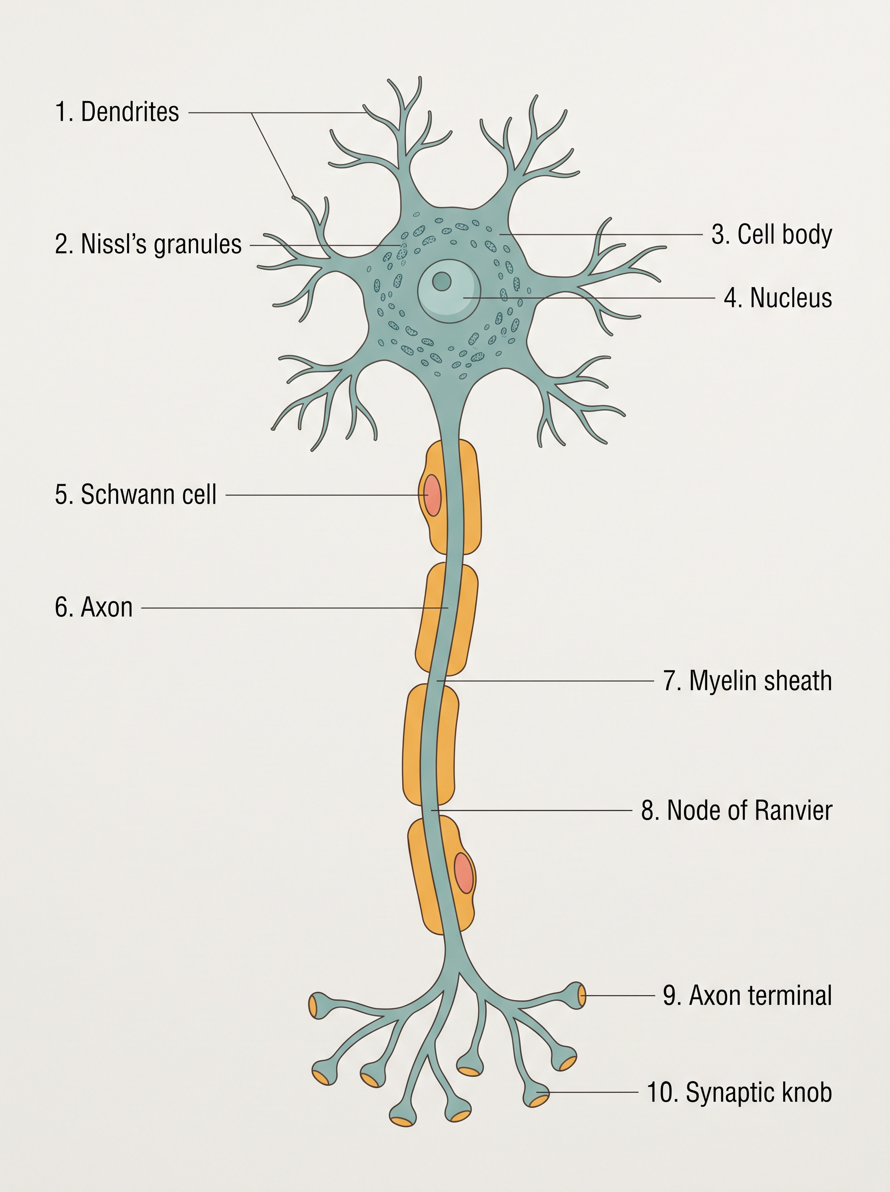
Fig. 18.1 – Structure of a neuron.
The three parts of a neuron
The cell body contains cytoplasm with the usual cell organelles, along with certain granular bodies unique to neurons called Nissl’s granules. These granules are also found in the dendrites.
The dendrites are short fibres that branch repeatedly and project out from the cell body. Their job is to receive signals and carry impulses towards the cell body.
The axon is a single long fibre whose far (distal) end is branched. Each of these branches ends in a bulb-like swelling called a synaptic knob. Inside each synaptic knob are synaptic vesicles holding chemicals called neurotransmitters. The axon carries nerve impulses away from the cell body — either to a synapse (the junction with the next neuron) or to a neuro-muscular junction (the junction with a muscle).
Types of neurons by shape
Neurons can be sorted into three types based on how many axons and dendrites arise from the cell body.
Myelinated and non-myelinated axons
Axons themselves come in two kinds, and the difference lies in a covering made by helper cells called Schwann cells.
In a myelinated nerve fibre, Schwann cells wrap around the axon and form an insulating layer called the myelin sheath. This sheath is not continuous — the small gaps between two adjacent stretches of myelin are called the nodes of Ranvier. Myelinated nerve fibres are found in the spinal and cranial nerves.
In an unmyelinated (non-myelinated) nerve fibre, a Schwann cell still encloses the axon but does not form a myelin sheath around it. Such fibres are commonly found in the autonomic and the somatic neural systems.
18.3.1 Generation and Conduction of Nerve Impulse
Neurons are described as excitable cells, and the reason is that their membranes are kept in a polarised state — that is, the two sides of the membrane carry opposite electrical charges, ready to change the instant the neuron is stimulated.
The resting membrane
The neuron’s membrane carries different kinds of ion channels, and each channel is selectively permeable, meaning it lets some ions through but not others.
When a neuron is not conducting an impulse — that is, when it is resting — its membrane is comparatively more permeable to potassium ions (\(\text{K}^+\)) and nearly impermeable to sodium ions (\(\text{Na}^+\)). It is also impermeable to the large, negatively charged proteins present in the axoplasm (the cytoplasm inside the axon).
As a result, the axoplasm inside the axon holds a high concentration of \(\text{K}^+\) and negatively charged proteins, together with a low concentration of \(\text{Na}^+\). Outside the axon it is the reverse: a low concentration of \(\text{K}^+\) and a high concentration of \(\text{Na}^+\). This difference in concentration across the membrane is a concentration gradient.
These ionic gradients do not maintain themselves. They are kept up by the active transport of ions by the sodium–potassium pump, which pushes out \(3\ \text{Na}^+\) for every \(2\ \text{K}^+\) it brings in (\(3\,\text{Na}^+ : 2\,\text{K}^+\)). Because of all this, the outer surface of the axonal membrane ends up positively charged while its inner surface is negatively charged — the membrane is polarised. The electrical potential difference across the resting plasma membrane is called the resting potential.
Generating the action potential
Now suppose a stimulus is applied at a particular point on the polarised membrane — call it site A, as shown in Fig. 18.2.
At site A, the membrane suddenly becomes freely permeable to \(\text{Na}^+\). Sodium ions rush inward in a rapid influx, and this reverses the polarity at that spot: the outer surface now becomes negative and the inner surface becomes positive. Because the membrane’s normal polarity has been reversed, the membrane at site A is said to be depolarised.
The electrical potential difference across the membrane at this depolarised site is called the action potential — and this action potential is exactly what we mean by a nerve impulse.
Conducting the impulse along the axon
A nerve impulse is only useful if it travels. Here is how it moves along the axon, as traced in Fig. 18.2.
At the sites immediately ahead of A — say, site B — the membrane is still in its resting condition: positive on the outer surface and negative on the inner surface. So the moment A depolarises, there is an electrical difference between A and B. A current now flows along the inner surface from A to B, while along the outer surface it flows from B to A, completing the circuit.
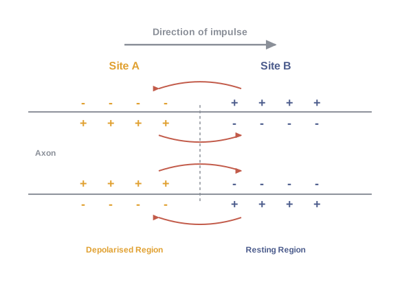
Fig. 18.2 – Impulse conduction through an axon.
This local current reverses the polarity at site B, so an action potential is generated at B as well. In other words, the impulse born at site A now arrives at site B. This same sequence repeats point after point all the way along the axon, and in this way the impulse is conducted from one end of the fibre to the other.
Returning to rest
The surge in \(\text{Na}^+\) permeability is extremely short-lived. It is quickly followed by a rise in the membrane’s permeability to \(\text{K}^+\). Within a fraction of a second, \(\text{K}^+\) diffuses outward across the membrane, which restores the resting potential at the site that was just excited. This recovery step is called repolarisation, and once it is complete the fibre is ready to respond to a fresh stimulus.
The whole cycle, from rest through depolarisation to recovery, is summarised in FC 18.3.
Flowchart
FC 18.3 – Generation and conduction of a nerve impulse: resting membrane (polarised) → stimulus at site A → membrane becomes permeable to Na⁺ → rapid Na⁺ influx → polarity reverses → depolarisation (action potential) at A → local current sets up depolarisation at site B → impulse propagates point-by-point along the axon → K⁺ permeability rises → K⁺ efflux → repolarisation → resting potential restored → membrane ready for the next stimulus
18.3.2 Transmission of Impulses
A single axon can carry an impulse from one end to the other, but the body needs neurons to pass their signals on to other neurons. This handover happens at special junctions called synapses.
The pre-synaptic neuron is the one delivering the impulse; the post-synaptic neuron is the one receiving it. The narrow space that may lie between them is the synaptic cleft.
Two kinds of synapses
Synapses come in two types — electrical synapses and chemical synapses — and they pass on the signal in very different ways.
Across an electrical synapse
At an electrical synapse, the membranes of the two neurons sit so close together that electrical current can flow straight from one neuron into the next. Because of this, transmission here is very similar to ordinary impulse conduction along a single axon, and it is always faster than transmission at a chemical synapse. Electrical synapses, however, are rare in our system.
Across a chemical synapse
At a chemical synapse, the two membranes are separated by the fluid-filled synaptic cleft, so the impulse cannot simply jump across as an electric current. Instead, the neuron uses chemical messengers — the neurotransmitters stored in the axon terminals — to carry the message over, as shown in Fig. 18.3.
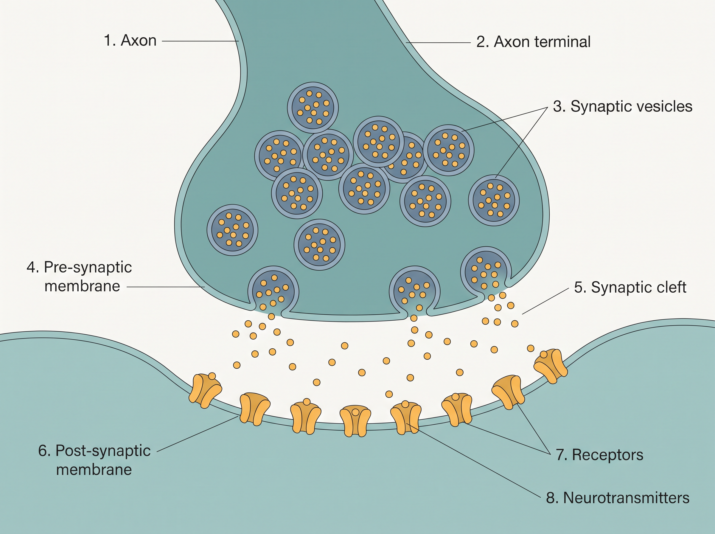
Fig. 18.3 – An axon terminal and synapse.
The axon terminals contain vesicles filled with neurotransmitters. When an impulse (action potential) arrives at the axon terminal, it makes these synaptic vesicles move towards the membrane and fuse with it. As they fuse, they release their neurotransmitters into the synaptic cleft.
The released neurotransmitters cross the cleft and bind to their specific receptors on the post-synaptic membrane. This binding opens ion channels on the post-synaptic neuron, letting ions enter and generating a new potential in that neuron. The signal has now been passed on.
The sequence of events at a chemical synapse is summarised in FC 18.4.
Flowchart
FC 18.4 – Transmission across a chemical synapse: impulse arrives at axon terminal → synaptic vesicles move to the pre-synaptic membrane → vesicles fuse and release neurotransmitters into the synaptic cleft → neurotransmitters bind to specific receptors on the post-synaptic membrane → ion channels open → ions enter the post-synaptic neuron → a new potential is generated (excitatory or inhibitory)
An excitatory or an inhibitory message
The new potential created in the post-synaptic neuron is not always the same. It may be either excitatory or inhibitory.
18.4 Central Neural System
You already know that the central neural system (CNS) is made up of the brain and the spinal cord. This section focuses on its most complex part — the brain.
The command and control organ
The brain is the central information-processing organ of the body — its “command and control system”. It carries out an enormous range of jobs. It controls voluntary movements, the balance of the body, and the working of vital involuntary organs such as the lungs, heart and kidneys. It manages thermoregulation (keeping body temperature steady), hunger and thirst, and our circadian rhythms — the roughly 24-hour internal cycles that govern sleep and wakefulness. It also oversees the activity of several endocrine glands and shapes human behaviour.
On top of all this, the brain is the site where vision, hearing, speech, memory, intelligence, emotions and thoughts are processed. In short, almost everything you sense, feel, decide and do is handled here.
How the brain is protected
Such a vital organ needs strong protection, and it has two layers of it. On the outside, the brain is enclosed by the bony skull. Inside the skull, it is wrapped in three protective membranes together called the cranial meninges:
- an outer layer, the dura mater
- a very thin middle layer, the arachnoid
- an inner layer in contact with the brain tissue, the pia mater
The three major parts of the brain
For study, the brain is divided into three major parts, as shown in Fig. 18.4:
- the forebrain
- the midbrain
- the hindbrain
Each of these is taken up in detail in the sections that follow; their overall division is shown in FC 18.5.
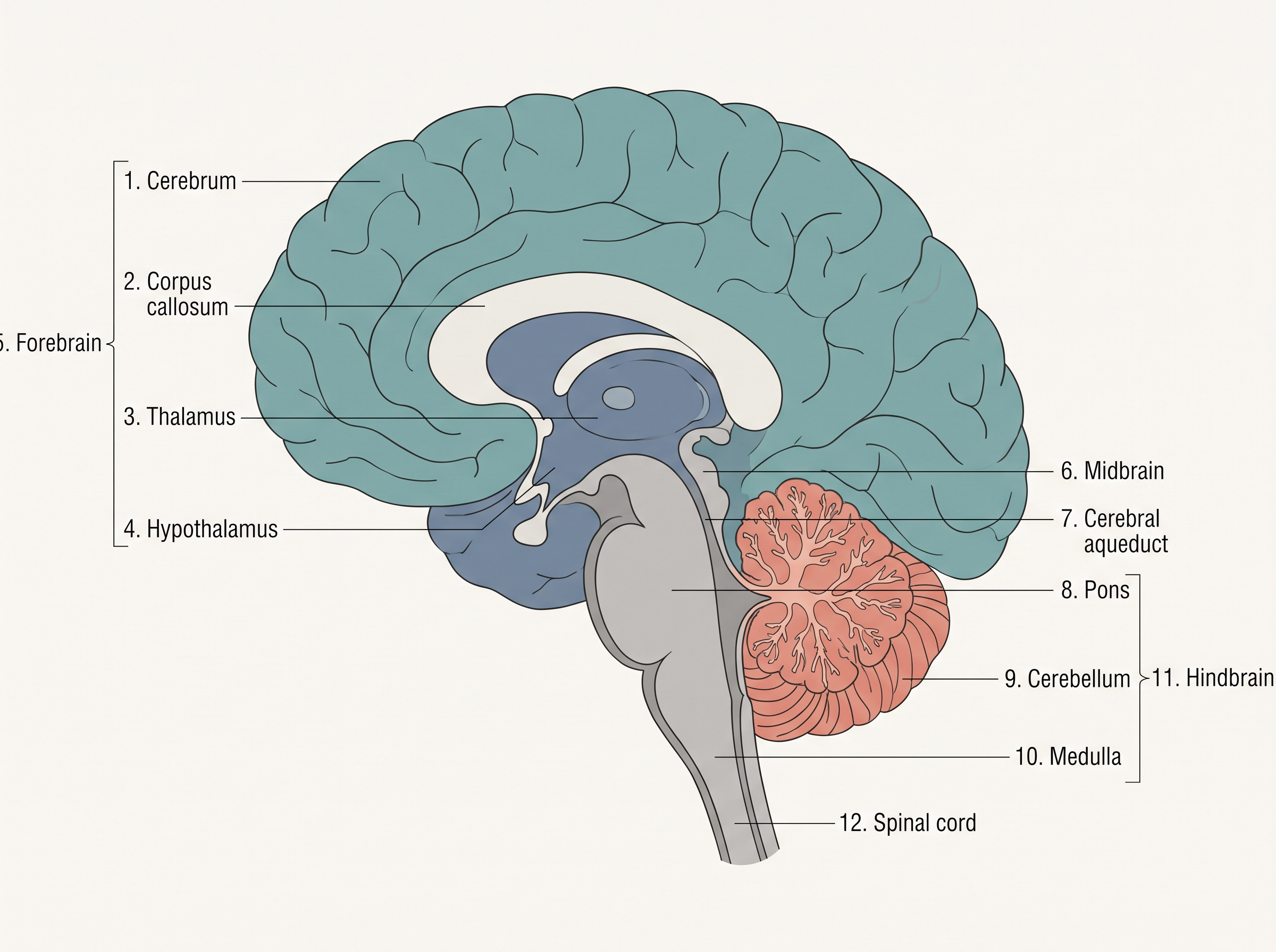
Fig. 18.4 – Sagittal section of the human brain.
Flowchart
FC 18.5 – Major divisions of the brain: Brain → forebrain + midbrain + hindbrain
18.4.1 Forebrain
The forebrain is the largest and most advanced region of the brain. It is made up of three parts: the cerebrum, the thalamus and the hypothalamus (see Fig. 18.4).
The cerebrum
The cerebrum forms the major part of the human brain. A deep cleft runs down its middle, dividing it lengthwise into two halves called the left and right cerebral hemispheres. These two hemispheres do not work in isolation — they are joined by a thick band of nerve fibres called the corpus callosum, which lets the two sides share information.
Grey matter and white matter
The outer layer of cells covering each cerebral hemisphere is the cerebral cortex, and it is thrown into prominent folds. Because these folds pack a large sheet of cortex into a limited space, the brain can hold far more neurons than a smooth surface would allow.
The cortex looks greyish and is therefore called the grey matter; the colour comes from the neuron cell bodies concentrated there. The cortex contains motor areas (which direct movement), sensory areas (which receive sensations), and large regions that are neither clearly motor nor sensory. These in-between regions are the association areas, responsible for complex functions such as linking different senses together (intersensory associations), memory and communication.
Beneath the cortex lie tracts of nerve fibres wrapped in myelin sheath. These make up the inner part of the hemisphere and, because myelin looks opaque and pale, this region is called the white matter.
The thalamus
The cerebrum wraps around a structure called the thalamus. The thalamus is a major coordinating centre for sensory and motor signalling — sensory information heading towards the cortex and motor commands heading outward are both routed through it.
The hypothalamus
Lying at the base of the thalamus is another very important part, the hypothalamus. Small though it is, it contains a number of control centres — for body temperature and for the urge to eat and drink. It also holds several groups of neurosecretory cells, which secrete hormones known as hypothalamic hormones.
The limbic system
The inner parts of the cerebral hemispheres, together with a group of associated deep structures such as the amygdala and hippocampus, form a complex structure called the limbic lobe or limbic system. Working along with the hypothalamus, the limbic system is involved in regulating sexual behaviour, in the expression of emotions (such as excitement, pleasure, rage and fear), and in motivation.
18.4.2 Midbrain
The midbrain sits in the middle of the brain, exactly where its name suggests. It is located between the thalamus and hypothalamus of the forebrain above and the pons of the hindbrain below, forming a link between the two (see Fig. 18.4).
Running through the midbrain is a narrow canal called the cerebral aqueduct. On its dorsal (back) portion, the midbrain carries mainly four rounded swellings, or lobes, together called the corpora quadrigemina.
In terms of function, the midbrain receives and integrates visual, tactile and auditory inputs — that is, information from sight, touch and hearing is brought together here.
18.4.3 Hindbrain
The hindbrain is the lowest part of the brain and comprises three structures: the pons, the cerebellum and the medulla (also called the medulla oblongata). Their positions can be seen in Fig. 18.4.
Pons
The pons is made up of fibre tracts that interconnect different regions of the brain, allowing these regions to communicate with one another.
Cerebellum
The cerebellum has a very convoluted (deeply folded) surface. These folds create additional space so that the cerebellum can hold many more neurons than a smooth surface would. Functionally, it integrates the information it receives from the semicircular canals of the ear and from the auditory system, which is why it is closely linked with balance.
Medulla oblongata
The medulla is connected to the spinal cord and marks the point where the brain meets it. It contains vital centres that control respiration, cardiovascular reflexes (the automatic adjustments of the heart and blood vessels), and gastric secretions.
The brain stem
Three regions of the brain together make up the brain stem: the midbrain, the pons and the medulla oblongata.
18.5 Summary
The neural system coordinates and integrates the functions, along with the metabolic and homeostatic activities, of all the organs of the body.
Neurons are the functional units of the neural system. They are excitable cells, and this excitability comes from a difference in the concentration of ions across their membrane.
The electrical potential difference across the membrane of a resting neuron is called the resting potential.
A nerve impulse travels along the axon membrane as a wave of depolarisation followed by repolarisation.
A synapse is formed by the membranes of a pre-synaptic neuron and a post-synaptic neuron, which may or may not be separated by a gap called the synaptic cleft. The chemicals that carry the impulse across a chemical synapse are called neurotransmitters.
The human neural system has two parts: the central neural system (CNS) and the peripheral neural system. The CNS is made up of the brain and the spinal cord.
The brain is divided into three major parts: the forebrain, the midbrain and the hindbrain. This overall organisation is summarised in FC 18.6.
Flowchart
FC 18.6 – Organisation of the central neural system: CNS → brain + spinal cord; brain → forebrain (cerebrum, thalamus, hypothalamus) + midbrain + hindbrain (pons, cerebellum, medulla)
The forebrain consists of the cerebrum, thalamus and hypothalamus. The cerebrum is divided lengthwise into two halves that are connected by the corpus callosum.
The hypothalamus, an important part of the forebrain, controls body temperature and the urge for eating and drinking.
The inner parts of the cerebral hemispheres, together with certain associated deep structures, form the limbic system, which is concerned with olfaction, autonomic responses, the regulation of sexual behaviour, the expression of emotional reactions, and motivation.
The midbrain receives and integrates visual, tactile and auditory inputs.
The hindbrain comprises the pons, cerebellum and medulla. The pons consists of fibre tracts that interconnect different regions of the brain.
The cerebellum integrates the information received from the semicircular canals of the ear and the auditory system.
The medulla contains centres that control respiration, cardiovascular reflexes and gastric secretions.
18.6 Points to Ponder
The body coordinates its organs using two systems — a fast, electrical neural system and a slower, chemical endocrine system. Why might it be useful to have both, instead of relying on just one? Think of situations where speed matters most, and others where a longer-lasting effect matters more.
A resting neuron spends energy continuously, using its sodium–potassium pump to keep ions unevenly distributed even when it is not sending any signal. Why might it be worth spending energy just to stay ready, rather than building up the charge only at the moment a signal is needed?
Some nerve fibres are wrapped in a myelin sheath while others are not. What might be the advantage of insulating only certain fibres rather than all of them?
Transmission across a chemical synapse is slower than across an electrical synapse, yet chemical synapses are far more common in our body. What might a chemical synapse be able to do — that an electrical one cannot — to make this slower route worthwhile?
The cerebral cortex is thrown into deep folds so that more neurons can fit within the skull. If the human skull could somehow be made much larger, how might the need for these folds change?
Your medulla keeps you breathing automatically, without your having to think about it — and yet you can also choose to hold your breath. What does this tell you about how voluntary control and involuntary control share the same body?
The corpus callosum allows the left and right cerebral hemispheres to exchange information. What do you imagine everyday life might be like if the two halves of the brain could no longer communicate with each other?
18.7 NCERT Questions
Briefly describe the structure of the brain.
Compare the following:
- Central neural system (CNS) and Peripheral neural system (PNS)
- Resting potential and action potential
Explain the following processes:
- Polarisation of the membrane of a nerve fibre
- Depolarisation of the membrane of a nerve fibre
- Transmission of a nerve impulse across a chemical synapse
Draw labelled diagrams of the following:
- Neuron
- Brain
Write short notes on the following:
- Neural coordination
- Forebrain
- Midbrain
- Hindbrain
- Synapse
Give a brief account of the mechanism of synaptic transmission.
Explain the role of \(\text{Na}^+\) in the generation of action potential.
Differentiate between:
- Myelinated and non-myelinated axons
- Dendrites and axons
- Thalamus and Hypothalamus
- Cerebrum and Cerebellum
Answer the following:
- Which part of the human brain is the most developed?
- Which part of our central neural system acts as a master clock?
Distinguish between:
- afferent neurons and efferent neurons
- impulse conduction in a myelinated nerve fibre and an unmyelinated nerve fibre
- cranial nerves and spinal nerves
18.8 Check Your Concepts
Assertion–Reason. Read the two statements and decide whether each is true and whether the reason correctly explains the assertion. Assertion (A): The outer surface of a resting axonal membrane carries a positive charge. Reason (R): The sodium–potassium pump transports \(3\ \text{Na}^+\) out of the cell for every \(2\ \text{K}^+\) it brings in.
Why is the membrane of a resting neuron described as “polarised”? Refer to the distribution of ions on the two sides of the membrane in your answer.
What would happen to the resting potential of a neuron if its sodium–potassium pump suddenly stopped working? Explain your reasoning.
Arrange the following events of chemical synaptic transmission in the correct sequence:
- Neurotransmitters bind to specific receptors on the post-synaptic membrane.
- An impulse (action potential) reaches the axon terminal.
- Synaptic vesicles fuse with the pre-synaptic membrane.
- Ion channels open and a new potential is generated in the post-synaptic neuron.
- Neurotransmitters are released into the synaptic cleft.
Match the brain part in Column I with its function in Column II.
| Column I | Column II |
|---|---|
| a. Cerebellum | i. Controls body temperature, eating and drinking |
| b. Medulla | ii. Connects the two cerebral hemispheres |
| c. Hypothalamus | iii. Controls respiration and cardiovascular reflexes |
| d. Corpus callosum | iv. Integrates information from the semicircular canals of the ear |
Looking at a labelled diagram of a neuron, name the part that:
- contains the nucleus and Nissl’s granules,
- carries impulses away from the cell body,
- stores neurotransmitters in vesicles.
Assertion–Reason. Read the two statements and decide whether each is true and whether the reason correctly explains the assertion. Assertion (A): Myelinated nerve fibres are found in the spinal and cranial nerves. Reason (R): In a myelinated fibre, Schwann cells form a myelin sheath around the axon, with gaps called nodes of Ranvier.
Distinguish between the grey matter and the white matter of the cerebrum, and state what gives each of them its characteristic colour.
What would happen to communication between the two cerebral hemispheres if the corpus callosum were cut? Explain why.
Name the division of the peripheral neural system that controls (a) the skeletal (voluntary) muscles and (b) the involuntary organs and smooth muscles. Then name the two further subdivisions of the division you identified in (b).
Odd one out. Identify the term that does not belong with the others and give the reason for your choice: dura mater, arachnoid, pia mater, corpus callosum.
The cerebral cortex is thrown into folds and the cerebellum has a convoluted surface. Explain how these two features serve a similar purpose in the brain.