Chapter 8: Cell: The Unit of Life
In the year 1665, an English scientist named Robert Hooke sat hunched over one of the earliest microscopes, examining a wafer-thin slice of cork — the same soft bark used to seal wine bottles. What he saw amazed him. The cork was not solid at all. It was built from thousands of tiny, empty, box-like compartments packed neatly side by side, like a honeycomb.
Hooke needed a name for these little rooms. They reminded him of the small, bare chambers that monks lived in inside a monastery, which were called cells. So he borrowed the word, and in his famous book Micrographia he called these compartments “cells.” The name stuck, and biology has used it ever since.
What Hooke did not realise was how important his little empty boxes truly were. He was actually looking at the dead outer walls of once-living plant cells. He had no way of knowing that every living thing — a blade of grass, an elephant, a mushroom, and Hooke himself — is built from cells, and that inside a living cell lies a busy, crowded world of activity that no wine-cork could reveal.
That crowded world is what this chapter is about.
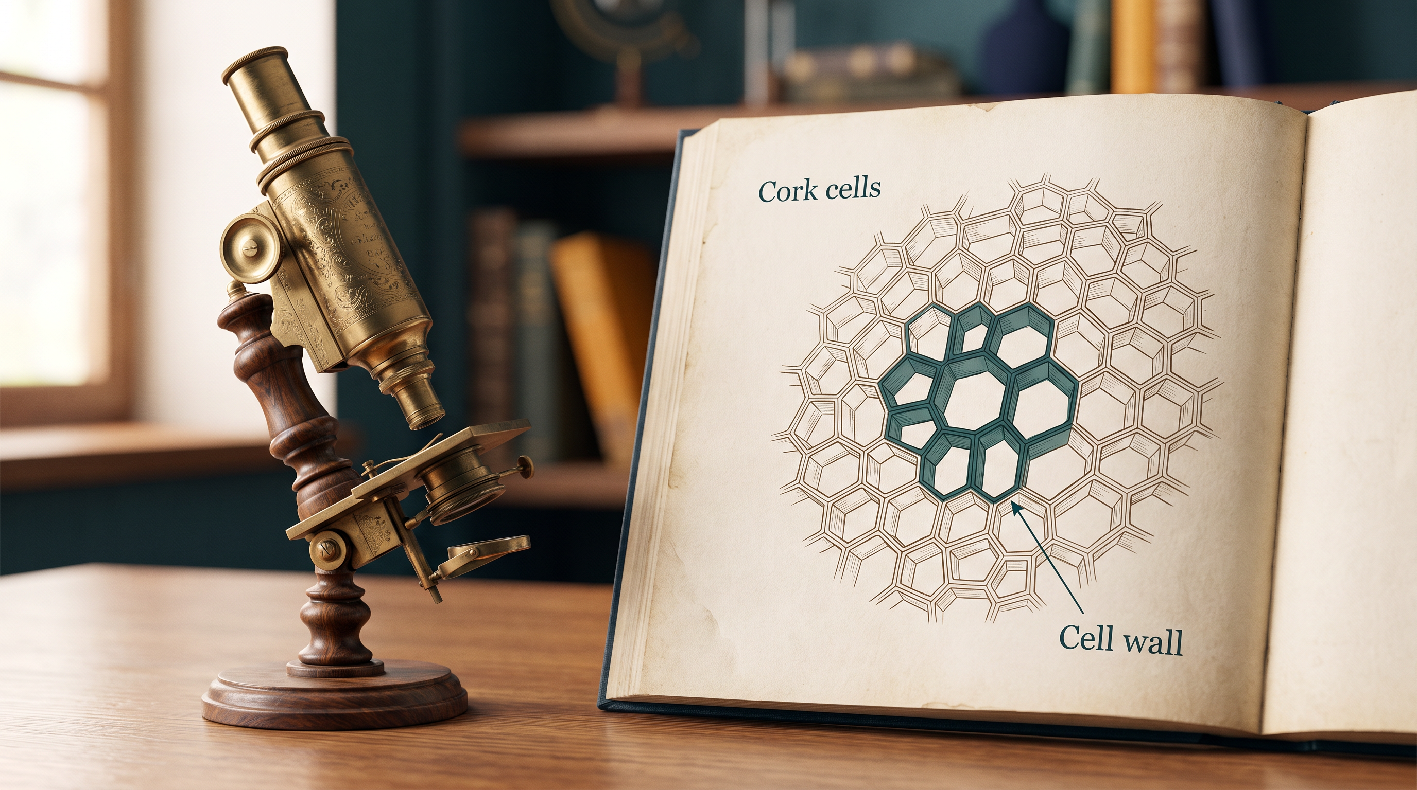
Fig. 8.0 – Robert Hooke’s microscope and cork cells.
By the end of this chapter, you will be able to answer each of these questions for yourself.
8.1 What is a Cell?
When you look around, you notice two kinds of things: living and non-living. A stone, a piece of chalk, and a drop of water are non-living, while a plant, a dog, and you yourself are living. This makes us ask a simple but deep question — what is it that a living thing has, which a non-living thing does not?
The answer lies in a single structure that every living organism shares: the cell. A cell is the basic unit of life, present in every living organism from the tiniest bacterium to the largest whale.
Some organisms are made of just one cell and can complete their entire life within it. These are called unicellular organisms, such as Amoeba and bacteria. Others, like plants and animals (including us), are made of many cells working together, and are called multicellular organisms.
Why the Cell Is the Unit of Life
A single-celled organism is remarkable because that one cell can do two things entirely on its own: it can exist independently, and it can carry out all the essential functions of life — taking in food, releasing energy, growing, responding to its surroundings, and reproducing.
This tells us something important. Anything smaller or less complete than a whole cell cannot live on its own. A fragment of a cell — a stray piece of membrane or a single loose organelle — has no independent life. Only when all the parts are assembled into a complete cell does life become possible.
For this reason, the cell is called the fundamental structural and functional unit of all living organisms: structural because every body is built out of cells, and functional because the activities of life actually happen inside them. In board and NEET papers, this idea is frequently tested as an assertion–reason statement, so it is worth understanding rather than merely memorising.
Seeing the First Cells
For most of history, no one knew that cells existed, simply because they are far too small for the naked eye to see. Cells became visible only after the microscope was invented, and each improvement in the microscope revealed a little more of what lay inside.
8.2 Cell Theory
We now know that the cell is the unit of life, but this was not always obvious. It took the careful work of several scientists, spread across many years, to build the idea into a proper theory. That idea is called the cell theory.
How the Idea Took Shape
In 1838, a German botanist named Matthias Schleiden examined a large number of plants under his microscope. He observed that all plants are built from different kinds of cells, and that these cells group together to form the tissues of the plant.
At about the same time, in 1839, a German zoologist named Theodore Schwann studied many different types of animal cells. He reported that animal cells are covered by a thin outer layer — the structure we today call the plasma membrane, the boundary that separates the inside of a cell from its surroundings. Comparing plants and animals, Schwann also concluded that the presence of a cell wall is a unique feature of plant cells, one that animal cells lack.
From these observations, Schwann proposed a bold hypothesis: that the bodies of both animals and plants are composed of cells and the products of cells. Working together, Schleiden and Schwann combined their findings into the first version of the cell theory.
The Missing Piece: Where Do New Cells Come From?
The early cell theory had one important gap: it did not explain how new cells are actually formed. Schleiden and Schwann had shown what organisms are made of, but not how their cells arise.
This missing piece was supplied by Rudolf Virchow in 1855. He explained that cells divide, and that every new cell is produced from a pre-existing cell — a idea captured in his famous Latin phrase Omnis cellula-e cellula, meaning “every cell from a cell.” Virchow’s addition modified the hypothesis of Schleiden and Schwann and gave the cell theory its final shape.
Flowchart
FC 8.1 – Development of the cell theory: Schleiden (1838, all plants made of cells) → Schwann (1839, animal cells and the plasma membrane; bodies made of cells and cell products) → Schleiden + Schwann formulate the first cell theory → gap: how are new cells formed? → Virchow (1855, cells arise from pre-existing cells, Omnis cellula-e cellula) → modern two-point cell theory.
In board and NEET questions, this topic is a favourite for match-the-following and assertion–reason items — for example, correctly pairing each scientist with his exact contribution, or recognising that the original Schleiden–Schwann theory did not explain the origin of new cells.
8.3 An Overview of Cell
Before we study each part of the cell in detail, let us take a bird’s-eye view of what a cell looks like from the outside in. You have probably already observed two familiar cells under the microscope — the cells of an onion peel and the cells scraped from the inside of your own cheek. Recalling them is a good place to start.
The Outer Boundary and the Nucleus
The onion cell is a typical plant cell. Its outermost boundary is a distinct cell wall, and just inside the wall lies the cell membrane (also called the plasma membrane). A human cheek cell, being an animal cell, has no cell wall — here the cell membrane itself is the outer, delimiting boundary of the cell.
Inside every one of these cells sits a dense, rounded, membrane-bound structure: the nucleus. The nucleus contains thread-like bodies called chromosomes, and the chromosomes carry the cell’s genetic material, DNA — the chemical that stores the instructions for building and running the cell.
Two Great Types of Cells
The nucleus gives us the most fundamental way of sorting all cells into two groups. Cells that keep their DNA inside a proper membrane-bound nucleus are called eukaryotic cells. Cells that have no membrane-bound nucleus — their genetic material lies free in the cell — are called prokaryotic cells.
What Fills the Cell
Whether a cell is prokaryotic or eukaryotic, most of its volume is taken up by a semi-fluid, jelly-like matrix called the cytoplasm. The cytoplasm is the main arena of cellular activity in both plant and animal cells. It is here that countless chemical reactions take place continuously — reactions that keep the cell in what we call the “living state.”
Organelles: The Cell’s Working Parts
Besides the nucleus, a eukaryotic cell contains several other distinct, membrane-bound structures called organelles — tiny “organs” of the cell, each doing a specific job. These include the endoplasmic reticulum (ER), the golgi complex, lysosomes, mitochondria, microbodies, and vacuoles. Prokaryotic cells lack all of these membrane-bound organelles. (You will study each organelle in detail later in the chapter.)
Not every organelle is wrapped in a membrane, however. Ribosomes are organelles that have no membrane, and they are found in all cells — both prokaryotic and eukaryotic. Inside the cell, ribosomes occur not only free in the cytoplasm but also within two organelles — the chloroplasts (in plant cells) and the mitochondria — as well as attached to the surface of rough ER.
Animal cells contain one more non-membrane-bound organelle that plant cells generally lack: the centrosome, which plays a role in cell division.
How Cells Differ in Size and Shape
Cells vary enormously in size, shape, and activity, as shown in Fig. 8.1.
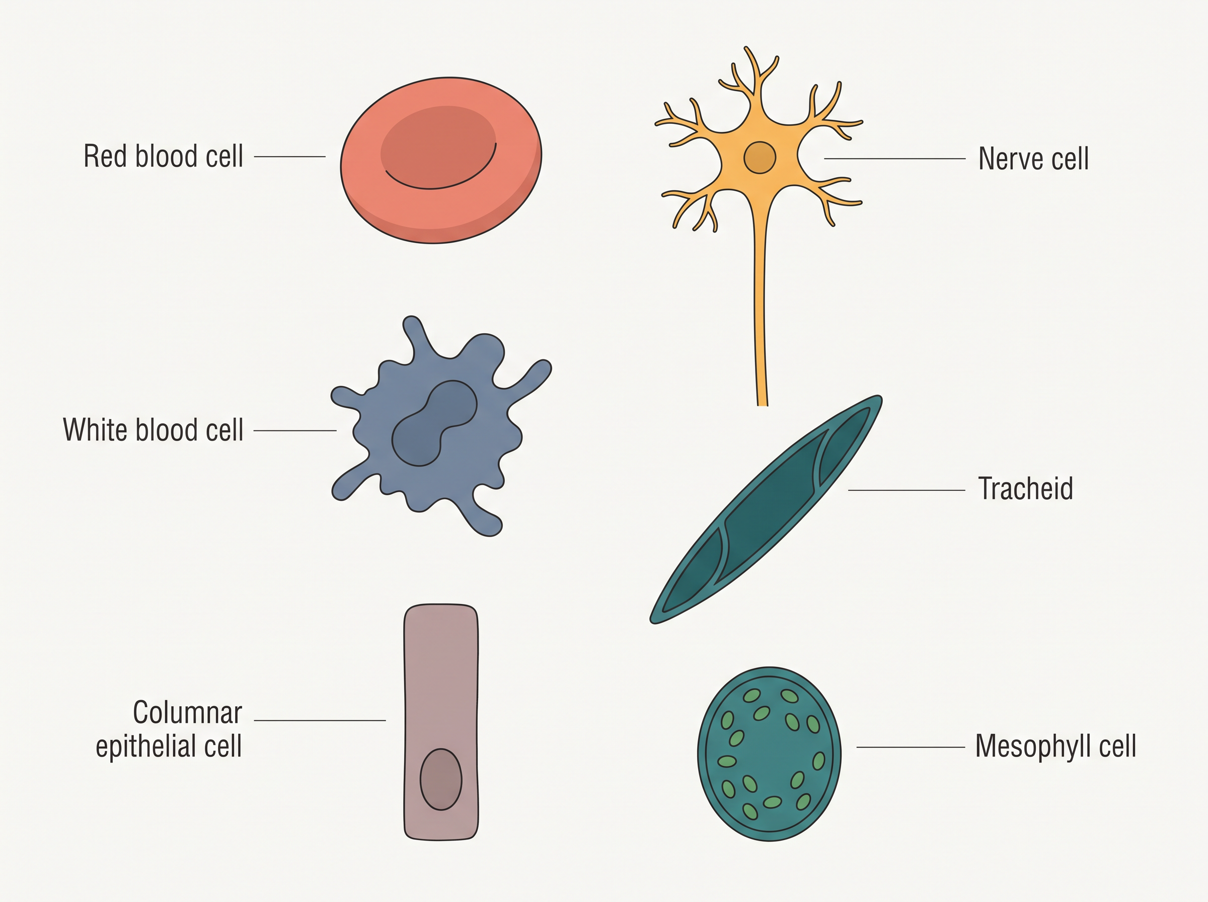
Fig. 8.1 – Different shapes of cells.
In size, the smallest cells are the Mycoplasmas, only about 0.3 µm long, while bacteria may be 3 to 5 µm. The largest isolated single cell is the egg of an ostrich. Among the cells of multicellular organisms, human red blood cells are about 7.0 µm in diameter, and nerve cells are among the longest cells of all.
Cells also differ greatly in shape. They may be disc-like, polygonal, columnar, cuboid, thread-like, or even irregular. Very often, the shape of a cell is matched to the job it performs.
8.4 Prokaryotic Cells
Having seen that all cells fall into two broad types, let us now look closely at the simpler of the two — the prokaryotic cell.
Who the Prokaryotes Are
Prokaryotic cells are found in bacteria, in blue-green algae (also called cyanobacteria), in mycoplasma, and in PPLO (Pleuro Pneumonia Like Organisms). Compared with eukaryotic cells, prokaryotic cells are generally smaller and multiply much more rapidly. You can see this size difference in Fig. 8.2, where a whole bacterium is dwarfed by a typical eukaryotic cell.
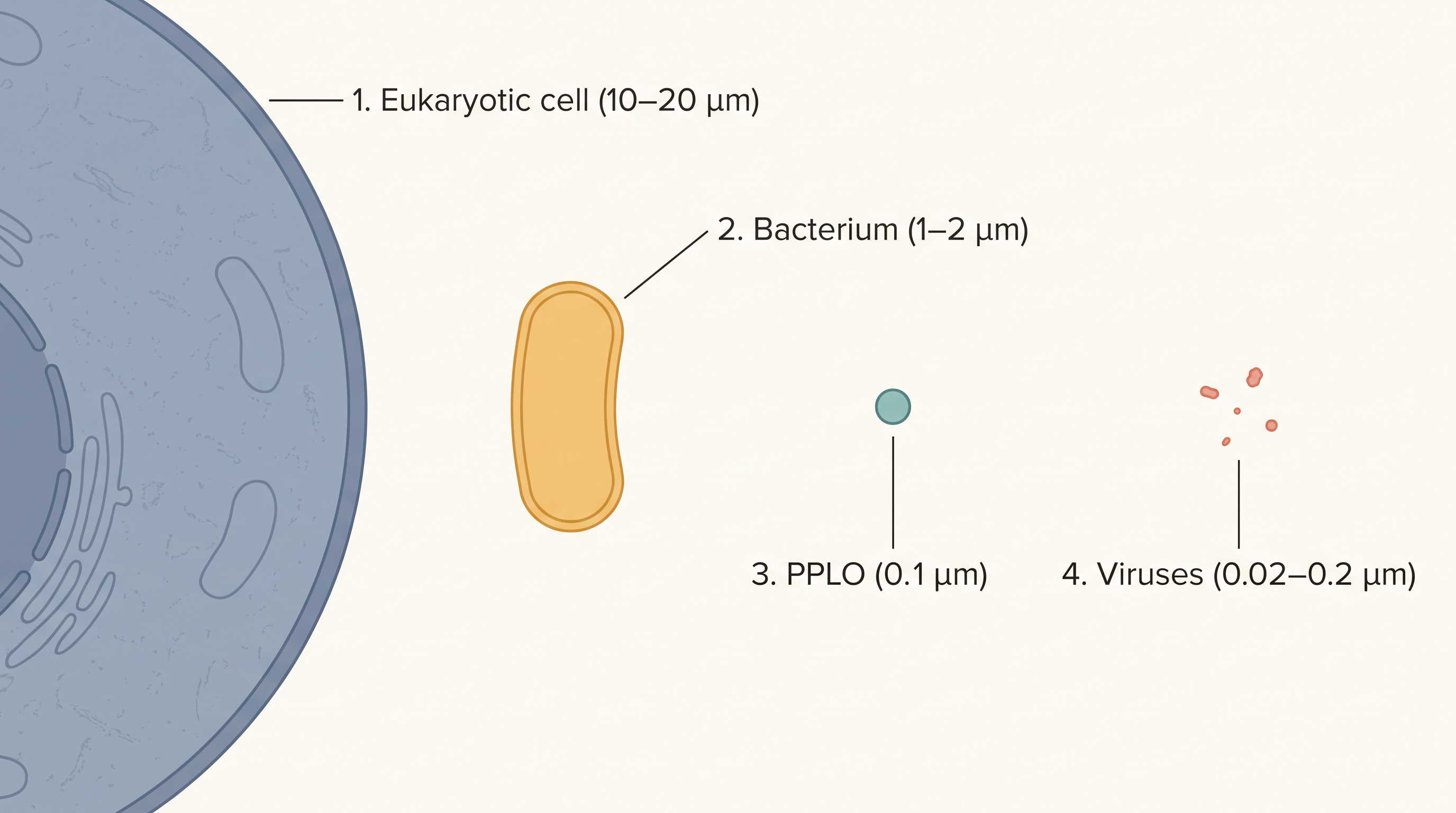
Fig. 8.2 – Relative sizes of microbes and cells.
Prokaryotes vary greatly in shape and size, but bacteria in particular come in four basic shapes: bacillus (rod-like), coccus (spherical), vibrio (comma-shaped), and spirillum (spiral). Recognising these shapes is a common requirement in board and NEET diagram-based questions.
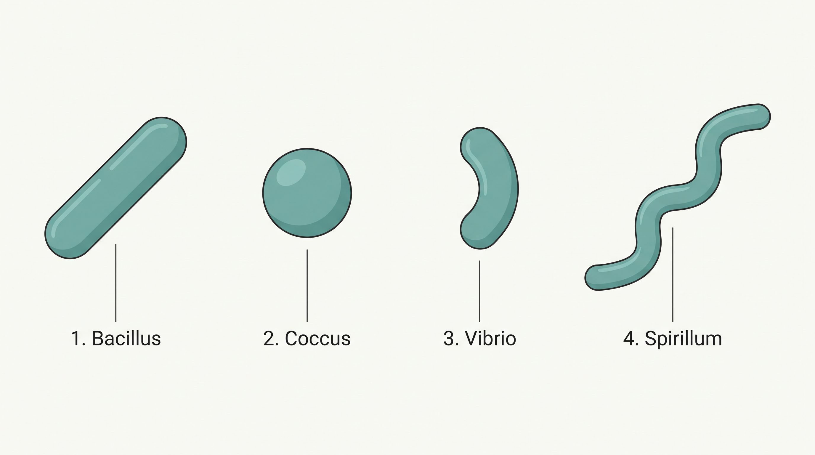
Fig. 8.3 – The four basic shapes of bacteria.
The Basic Plan of a Prokaryotic Cell
Even though prokaryotes show a wide variety of shapes and functions, the internal organisation of a prokaryotic cell is fundamentally the same in all of them.
On the outside, almost all prokaryotes have a cell wall surrounding the cell membrane. The one important exception is mycoplasma, which has no cell wall at all. Filling the cell is the semi-fluid cytoplasm.
The defining feature of a prokaryote is what it lacks: there is no well-defined nucleus. Its genetic material lies “naked” in the cytoplasm — that is, the DNA is not enveloped by a nuclear membrane. This single main piece of DNA, the genomic DNA, is usually one circular chromosome.
Plasmids: Extra Rings of DNA
In addition to the genomic DNA, many bacteria carry small, separate rings of circular DNA lying outside the main chromosome. These are called plasmids.
Plasmid DNA gives such bacteria certain extra characters that the main chromosome alone would not provide. One important example is resistance to antibiotics — a plasmid can carry the genes that let a bacterium survive a drug that would otherwise kill it. In higher classes you will also learn that plasmid DNA is used as a tool to carry foreign DNA into bacteria, a technique used to monitor bacterial transformation.
No Organelles, But Some Unique Features
Since prokaryotes have no nuclear membrane, it is no surprise that they also lack the membrane-bound organelles found in eukaryotes. The one organelle they do possess is the ribosome, needed for making proteins.
What prokaryotes have instead are some features of their own. They store reserve materials in structures called inclusions. They also possess a specialised, differentiated form of the cell membrane called the mesosome, which is a characteristic feature of prokaryotes and is essentially an infolding of the cell membrane. We will look at the cell envelope, the mesosome, the ribosomes, and the inclusion bodies more closely in the subsections that follow.
8.4.1 Cell Envelope and its Modifications
The Three-Layered Cell Envelope
Most prokaryotic cells, and bacterial cells in particular, are wrapped in a chemically complex covering called the cell envelope. This envelope is a tightly bound, three-layered structure. From the outside inward, the layers are: the glycocalyx (outermost), then the cell wall, and finally the plasma membrane. Each layer has its own distinct function, yet the three act together as a single protective unit.
The nature of this envelope also gives us a way to classify bacteria. When bacteria are treated with a staining procedure developed by the scientist Gram, some bacteria take up the stain and some do not. Those that take up the Gram stain are called Gram positive bacteria, and those that do not are called Gram negative bacteria. The two groups differ in the make-up of their cell envelopes.
Glycocalyx, Cell Wall, and Plasma Membrane
The outermost layer, the glycocalyx, differs in composition and thickness from one bacterium to another. In some bacteria it is a loose sheath called the slime layer, while in others it is thick and tough and is called the capsule.
Beneath the glycocalyx lies the cell wall. It determines the shape of the cell and provides strong structural support, preventing the bacterium from bursting or collapsing.
The innermost layer is the plasma membrane. It is selectively permeable, meaning it controls what enters and leaves, and it is the part that interacts with the outside world. Structurally, this membrane is similar to the plasma membrane of eukaryotic cells.
Mesosome and Chromatophores
A special membranous structure found in prokaryotes is the mesosome. It is formed when the plasma membrane extends and folds inward into the cell, taking the form of vesicles, tubules, and lamellae.
Because these infoldings add a great deal of extra membrane, the mesosome takes on many jobs. It helps in the formation of the cell wall, in the replication of DNA and its distribution to daughter cells, and in respiration and secretion. It also increases the surface area of the plasma membrane and raises its enzyme content.
In some prokaryotes, such as the cyanobacteria (blue-green algae), the cytoplasm also contains other membranous extensions called chromatophores, which carry pigments used in photosynthesis.
Surface Structures: Flagella, Pili, and Fimbriae
Bacterial cells may be motile (able to move) or non-motile. A motile bacterium has thin, filamentous extensions arising from its cell wall called flagella. Different bacteria show a range in the number and arrangement of their flagella.
A bacterial flagellum is built from three parts: the filament, the hook, and the basal body. The filament is the longest part and extends from the cell surface out into the surroundings.
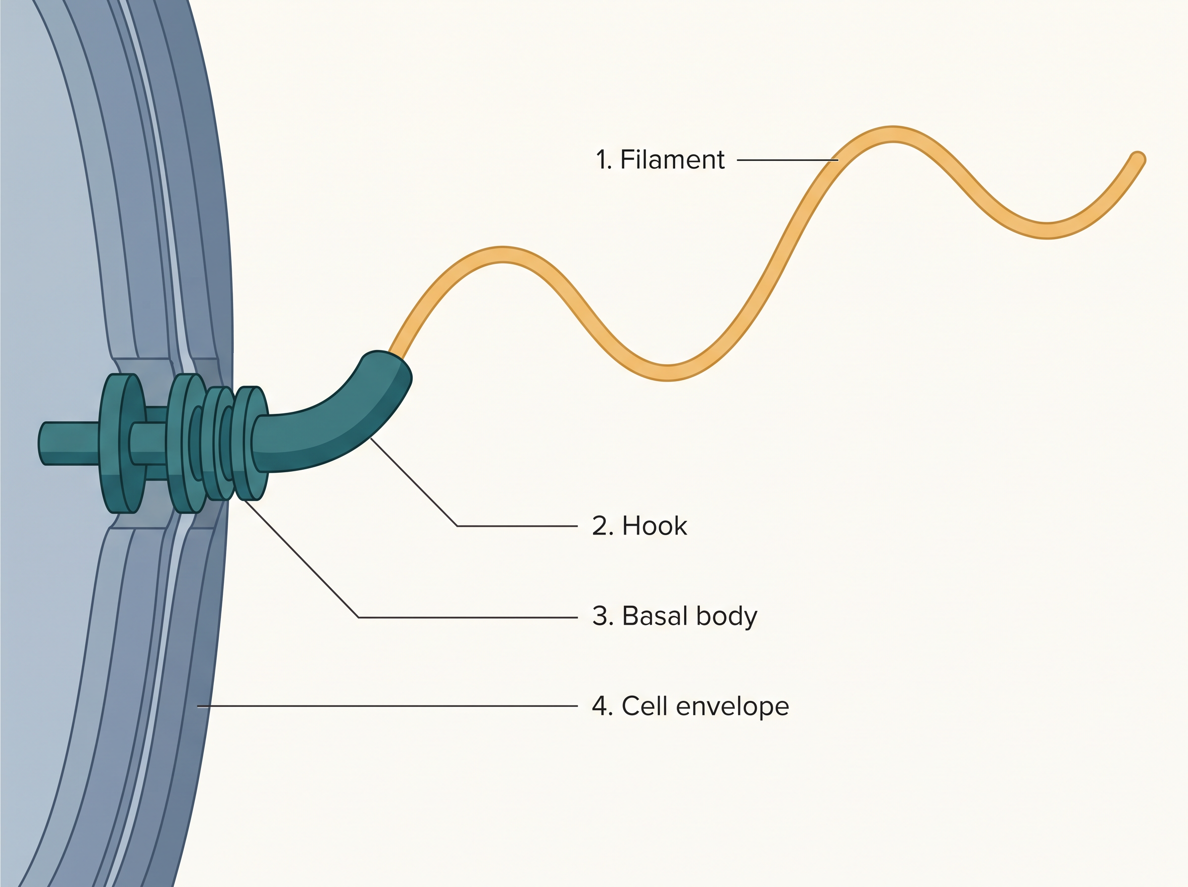
Fig. 8.4 – Parts of a bacterial flagellum.
Not every surface projection is for movement. Besides flagella, bacteria may also bear pili and fimbriae, which are surface structures that do not take part in motility. The pili are elongated tubular structures made of a special protein. The fimbriae are small, bristle-like fibres that sprout from the cell surface; in some bacteria they help the cell attach to rocks in flowing streams and, importantly, to the tissues of a host.
8.4.2 Ribosomes and Inclusion Bodies
Ribosomes: The Protein Factories
In prokaryotes, the ribosomes are associated with the plasma membrane of the cell. Each ribosome is about 15 nm by 20 nm in size and is built from two subunits — a larger 50S subunit and a smaller 30S subunit. When these two come together, they form the complete 70S ribosome that is characteristic of prokaryotes.
Ribosomes are the site of protein synthesis — the place where the cell builds its proteins. During this process, several ribosomes may attach to a single strand of mRNA (messenger RNA, the molecule that carries the genetic instructions for a protein). Such a chain of ribosomes on one mRNA is called a polyribosome or polysome. All the ribosomes of a polysome work together to translate the mRNA into proteins.
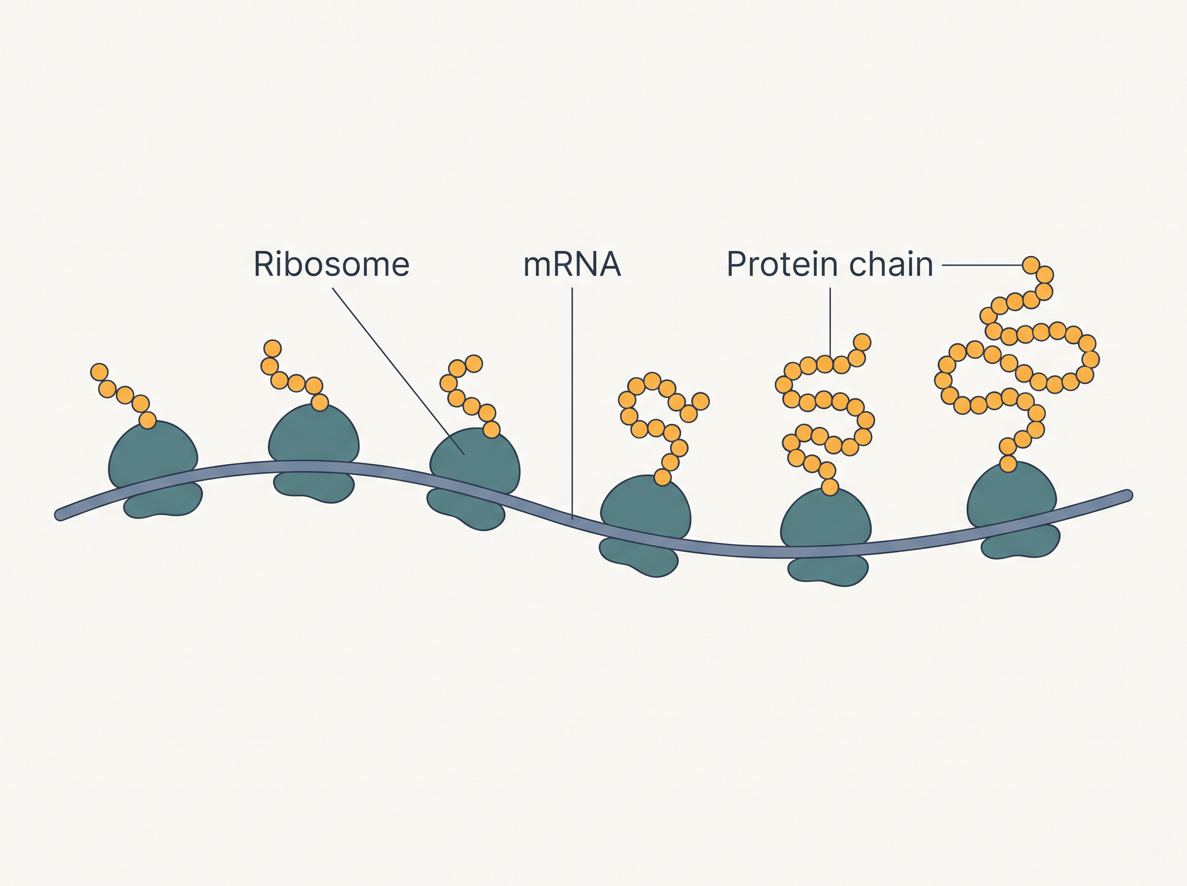
Fig. 8.5 – A polysome.
Inclusion Bodies
Prokaryotic cells store their reserve materials in the cytoplasm in the form of inclusion bodies. Unlike organelles, these are not bound by any membrane and simply lie free in the cytoplasm. Examples include phosphate granules, cyanophycean granules, and glycogen granules.
Certain photosynthetic prokaryotes have another kind of inclusion — the gas vacuoles — found in blue-green, purple, and green photosynthetic bacteria.
8.5 Eukaryotic Cells
We now turn to the more complex of the two cell types — the eukaryotic cell.
What Makes a Cell Eukaryotic
The eukaryotes include all the protists, plants, animals, and fungi. What sets a eukaryotic cell apart is the way its interior is divided up. Through the presence of many membrane-bound organelles, the cytoplasm of a eukaryotic cell is extensively compartmentalised — split into separate enclosed spaces, each specialised for particular tasks.
A eukaryotic cell also possesses a properly organised nucleus surrounded by a nuclear envelope. In addition, eukaryotic cells have a variety of complex locomotory structures and an internal supporting framework called the cytoskeleton. Their genetic material is neatly organised into chromosomes.
Plant Cells vs Animal Cells
Not all eukaryotic cells are identical. The clearest example is the difference between plant and animal cells, shown in Fig. 8.6. A plant cell has a cell wall, plastids, and a large central vacuole — all of which are absent in animal cells. An animal cell, in turn, has centrioles, which are absent in almost all plant cells.
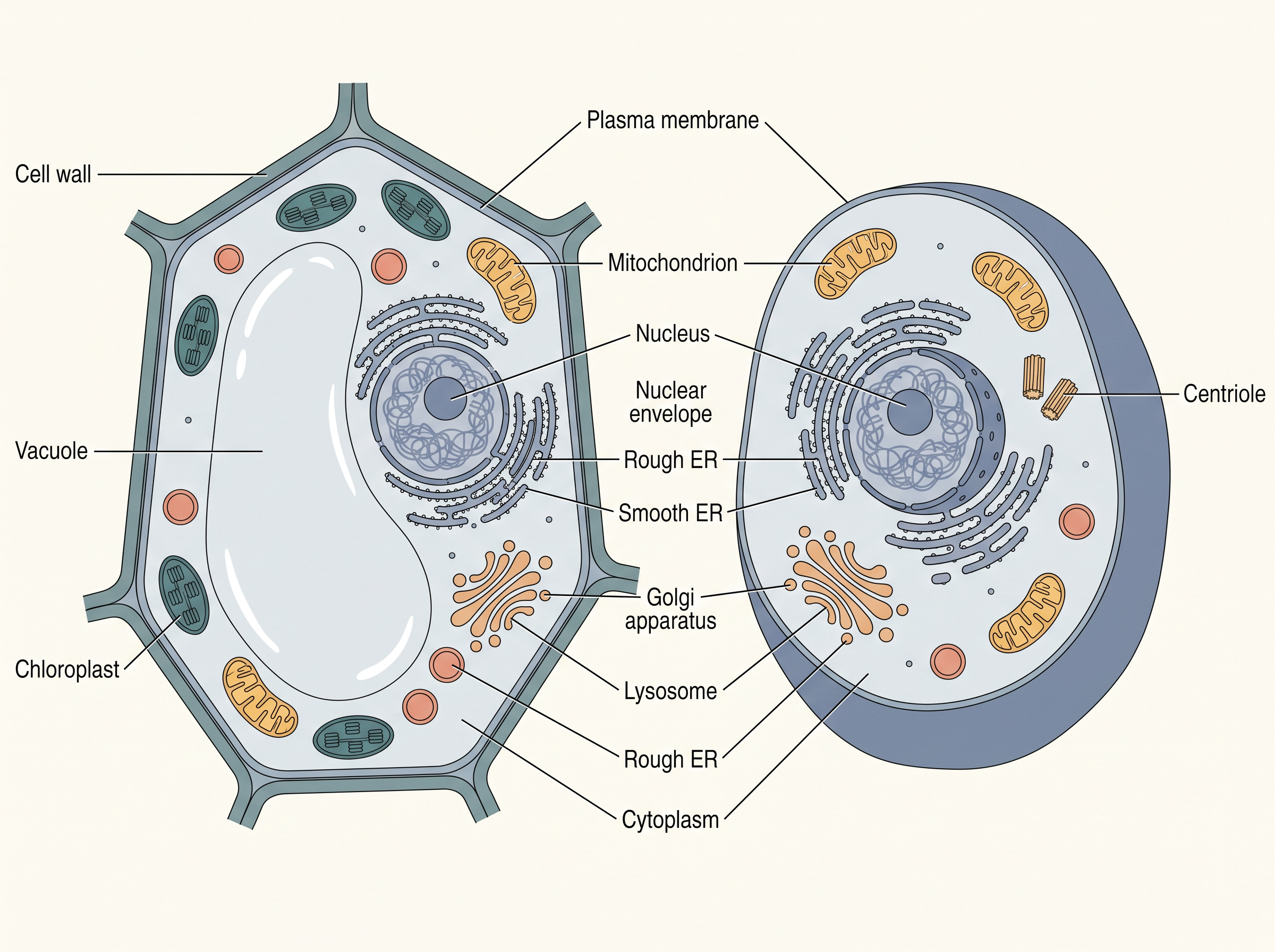
Fig. 8.6 – (a) Plant cell and (b) animal cell.
With this overview in hand, let us now examine the individual cell organelles one by one to understand each one’s structure and functions.
8.5.1 Cell Membrane
The cell membrane, or plasma membrane, is the boundary that separates the living contents of the cell from its surroundings. Simple as it sounds, working out exactly how this membrane is built took scientists a long time.
How the Membrane’s Structure Was Worked Out
The fine, detailed structure of the membrane could be seen directly only after the electron microscope came into use in the 1950s. Even before that, however, chemical studies of the cell membrane — especially studies on human red blood cells (RBCs) — allowed scientists to work out its likely structure indirectly.
What the Membrane Is Made Of
These studies showed that the cell membrane is composed mainly of lipids (fats) and proteins. The major lipids present are phospholipids, and they are arranged in two layers, forming a bilayer.
Each phospholipid molecule has two ends with opposite personalities: a water-loving polar head and a water-fearing (hydrophobic) tail. Within the membrane, the lipids arrange themselves with the polar heads facing outward, towards the watery environment on both sides, and the hydrophobic tails tucked away in the inner part of the membrane, as shown in Fig. 8.7. This arrangement ensures that the nonpolar tails of these saturated hydrocarbons are kept protected from the surrounding water.
In addition to phospholipids, the membrane also contains cholesterol. Later biochemical work revealed that cell membranes carry proteins and carbohydrates as well. The ratio of protein to lipid varies considerably from one cell type to another. In humans, for instance, the membrane of the erythrocyte (red blood cell) is made of approximately 52% protein and 40% lipids.
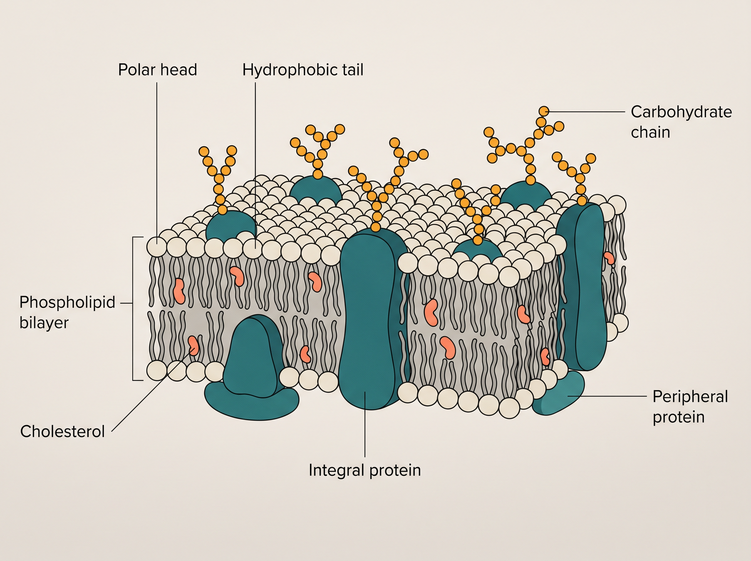
Fig. 8.7 – Fluid mosaic model of plasma membrane.
The proteins of the membrane can be sorted into two kinds, based on how easily they can be extracted from it. Peripheral proteins lie on the surface of the membrane, while integral proteins are partially or wholly buried within it.
The Fluid Mosaic Model
Because the lipid is fluid, the proteins embedded in it are able to drift sideways within the bilayer. This ability of the membrane’s components to move about within it is measured as its fluidity.
The fluid nature of the membrane matters for far more than just movement of proteins. It is essential for many activities of the cell, including cell growth, the formation of junctions between neighbouring cells, secretion, endocytosis (taking material into the cell), and cell division.
Transport Across the Membrane
One of the most important functions of the plasma membrane is the transport of molecules across it. The membrane is selectively permeable, letting only certain molecules pass while holding others back.
Some molecules can cross the membrane without the cell spending any energy. This is called passive transport. Neutral solutes cross by simple diffusion, moving along their concentration gradient — that is, from a region of higher concentration to one of lower concentration. Water, too, can move across the membrane from higher to lower concentration; this diffusion of water is given a special name, osmosis.
Not everything can slip through so easily. Because polar molecules cannot pass through the nonpolar lipid bilayer, they need the help of a carrier protein in the membrane to ferry them across.
Finally, a few ions or molecules are moved in the opposite, “uphill” direction — against their concentration gradient, from lower to higher concentration. This cannot happen for free: it is an energy-dependent process in which ATP (the cell’s energy currency) is used. This is called active transport, and a well-known example is the \(\text{Na}^+/\text{K}^+\) pump.
8.5.2 Cell Wall
As you may recall, the cell wall is a non-living, rigid structure that forms an outer covering over the plasma membrane in fungi and plants. Animal cells, by contrast, have no cell wall.
What the Cell Wall Does
The cell wall does several jobs at once. It gives the cell its shape and protects it from mechanical damage and from infection. It also helps in cell-to-cell interaction and acts as a barrier that keeps undesirable large molecules (macromolecules) from entering the cell.
The chemical make-up of the wall varies. In algae, the cell wall is made of cellulose, galactans, mannans, and minerals such as calcium carbonate. In other plants, it consists of cellulose, hemicellulose, pectins, and proteins.
Layers of the Wall
The wall of a young plant cell is called the primary wall, and it is capable of growth. As the cell matures, this ability to grow gradually diminishes, and a secondary wall is laid down on the inner side of the cell — that is, on the side towards the plasma membrane.
Between the walls of neighbouring cells lies the middle lamella, a layer made mainly of calcium pectate that holds or glues adjacent cells together. Running through the cell wall and middle lamella are fine channels called plasmodesmata, which connect the cytoplasm of one cell to that of its neighbours.
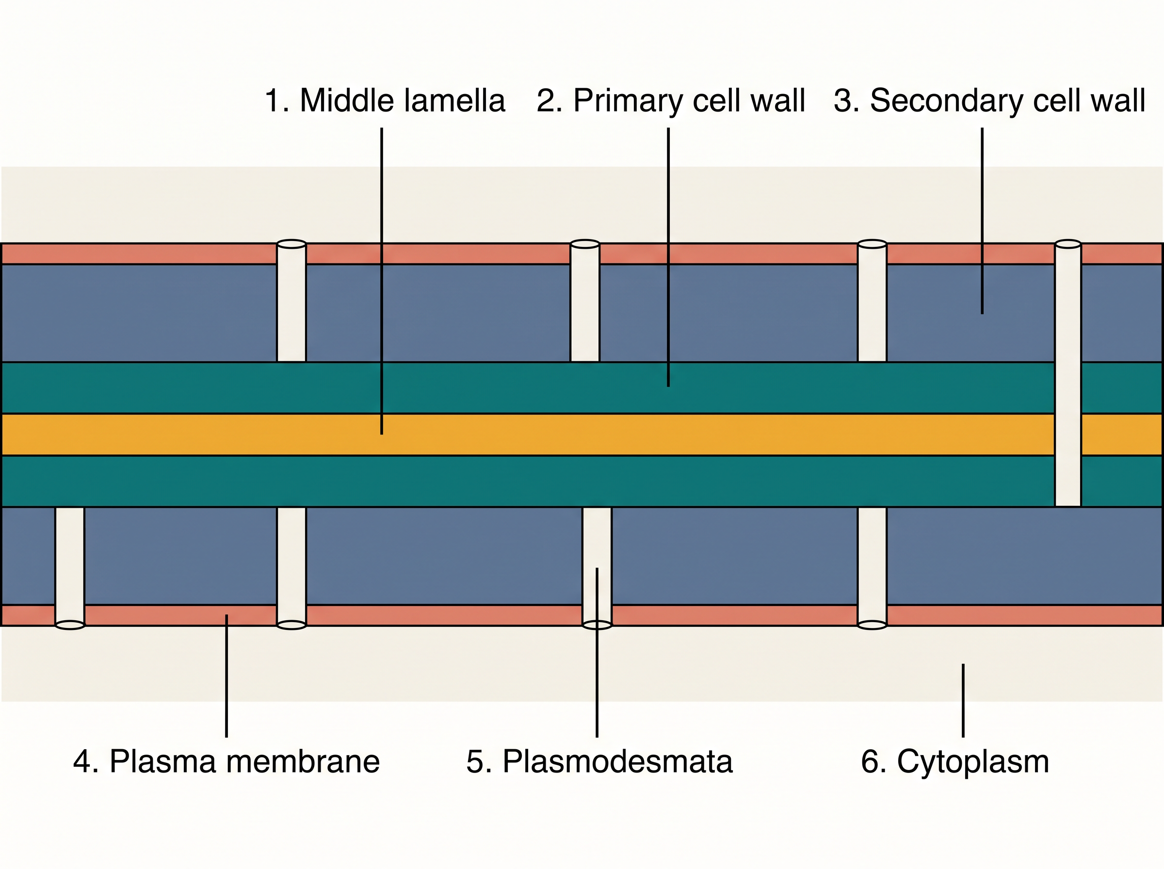
Fig. 8.8 – Section through adjacent plant cell walls.
8.5.3 Endomembrane System
Each of the membrane-bound organelles inside a eukaryotic cell is distinct in its own structure and function. Yet several of them work so closely together that biologists group them into a single team called the endomembrane system.
The organelles are placed in this group not because they look alike, but because their functions are coordinated — the output of one feeds into the work of the next. The endomembrane system includes the endoplasmic reticulum (ER), the golgi complex, the lysosomes, and the vacuoles.
Some organelles are deliberately left out of this system. The mitochondria, the chloroplast, and the peroxisomes are not considered part of the endomembrane system, because their functions are not coordinated with the four members listed above.
Flowchart
FC 8.2 – Coordinated working of the endomembrane system: ER (makes proteins and lipids) → golgi complex (modifies, sorts, and packages them into vesicles) → vesicles delivered to lysosomes, vacuoles, or secreted out of the cell.
8.5.3.1 The Endoplasmic Reticulum (ER)
When eukaryotic cells are examined under the electron microscope, they reveal a network — a reticulum — of tiny tubular structures scattered throughout the cytoplasm. This network is called the endoplasmic reticulum, or ER, and it is shown in Fig. 8.9.
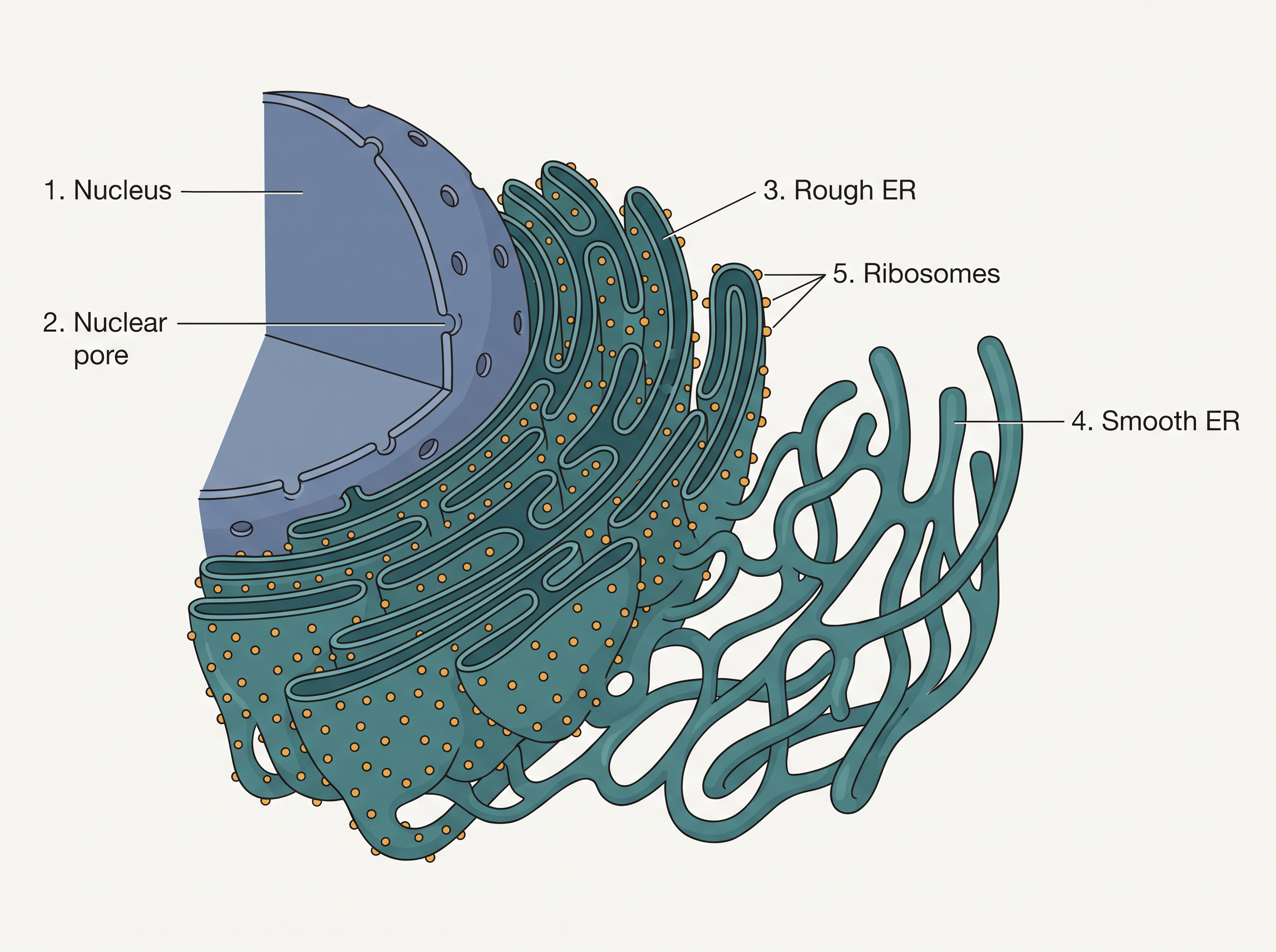
Fig. 8.9 – The endoplasmic reticulum.
Because it forms an interconnected system of tubes and sacs, the ER divides the space inside the cell into two distinct compartments: the luminal compartment (the space inside the ER) and the extra-luminal compartment (the cytoplasm outside it).
The ER often carries ribosomes attached to its outer surface. Where ribosomes are present, the surface looks rough, and this type is called the rough endoplasmic reticulum (RER). Where ribosomes are absent, the surface appears smooth, and this type is called the smooth endoplasmic reticulum (SER).
The two types do different jobs. The RER is frequently seen in cells that are actively engaged in protein synthesis and secretion. It is extensive and lies continuous with the outer membrane of the nucleus, so that proteins can be made right beside the nucleus. The SER, on the other hand, is the major site for the synthesis of lipids. In animal cells, lipid-like steroidal hormones are also made in the SER.
8.5.3.2 Golgi apparatus
The Golgi apparatus is built from many flat, disc-shaped sacs called cisternae, each about 0.5 µm to 1.0 µm in diameter, as shown in Fig. 8.10. These cisternae are stacked parallel to one another, and the number of cisternae in a single Golgi complex can vary.
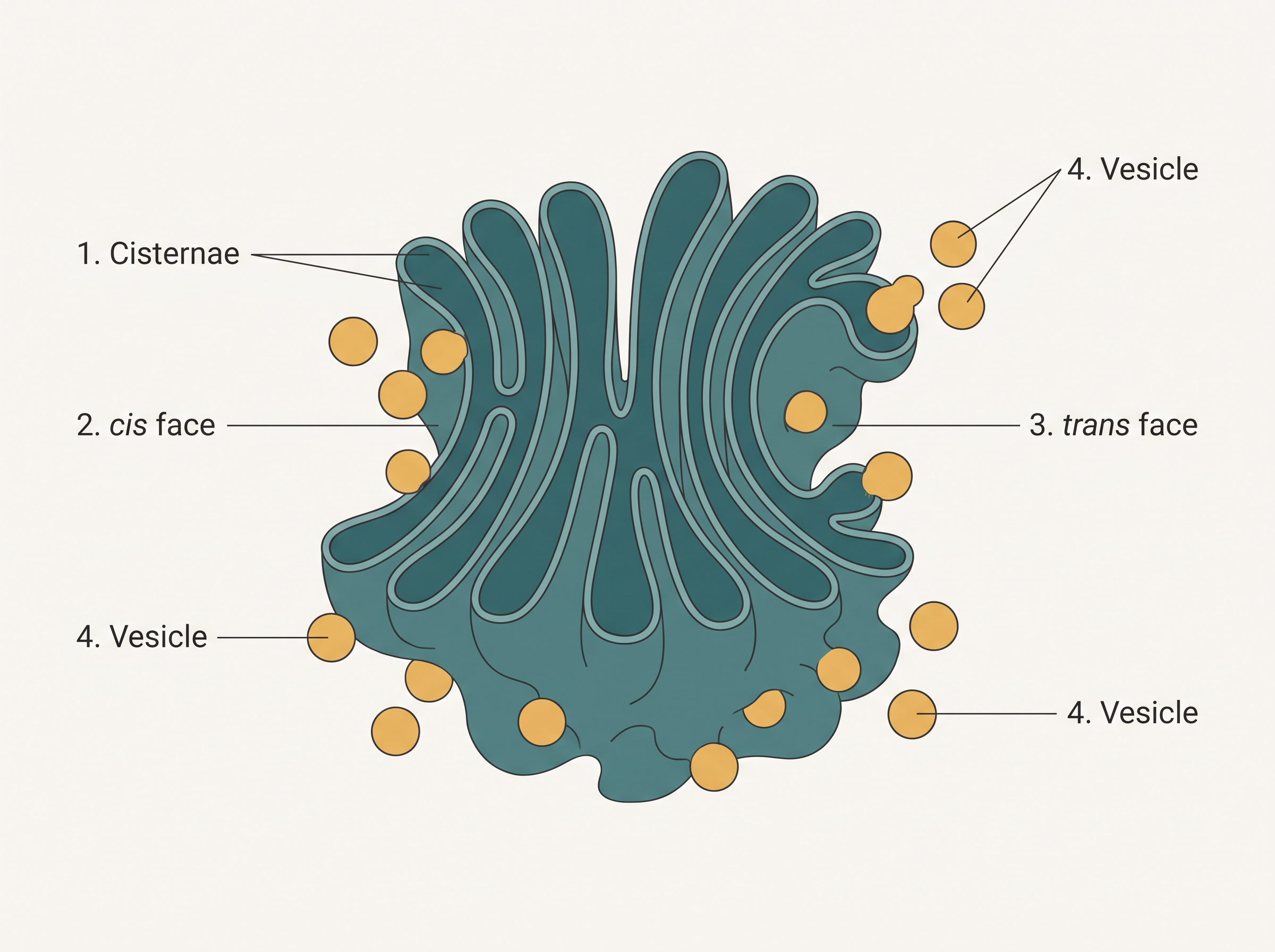
Fig. 8.10 – The Golgi apparatus.
The stack is arranged concentrically near the nucleus and has two clearly different sides. The convex side is the cis face, also called the forming face, and the concave side is the trans face, also called the maturing face. These two faces are entirely different from each other, yet they remain interconnected.
The Golgi apparatus mainly works as the cell’s packaging and dispatch centre. It packages materials to be delivered either to targets inside the cell or to be secreted outside it. Materials arrive from the ER as small vesicles, which fuse with the cis face and then move through the cisternae towards the maturing face. This constant traffic from the ER is exactly why the Golgi stays in close association with the endoplasmic reticulum.
As the materials pass through, they are worked on. Many proteins made by ribosomes on the ER are modified inside the Golgi cisternae before being released from the trans face. The Golgi is also the important site where glycoproteins (protein plus sugar) and glycolipids (lipid plus sugar) are formed.
Flowchart
FC 8.3 – Golgi packaging pathway: ER makes material → vesicle bud off ER → fuses with cis (forming) face → modified while moving through cisternae (glycoproteins and glycolipids formed) → released from trans (maturing) face in vesicles → delivered to intracellular targets or secreted out of the cell.
8.5.3.3 Lysosomes
Lysosomes are membrane-bound, vesicle-like structures that are formed by the packaging activity of the golgi apparatus. In other words, they are made by the same organelle we have just studied.
When lysosomal vesicles are isolated and examined, they are found to be very rich in almost all kinds of hydrolytic enzymes, also called hydrolases — these include lipases, proteases, and carbohydrases. A special feature of these enzymes is that they are optimally active at an acidic pH. Together, they are capable of digesting all four major types of biological molecules: carbohydrates, proteins, lipids, and nucleic acids.
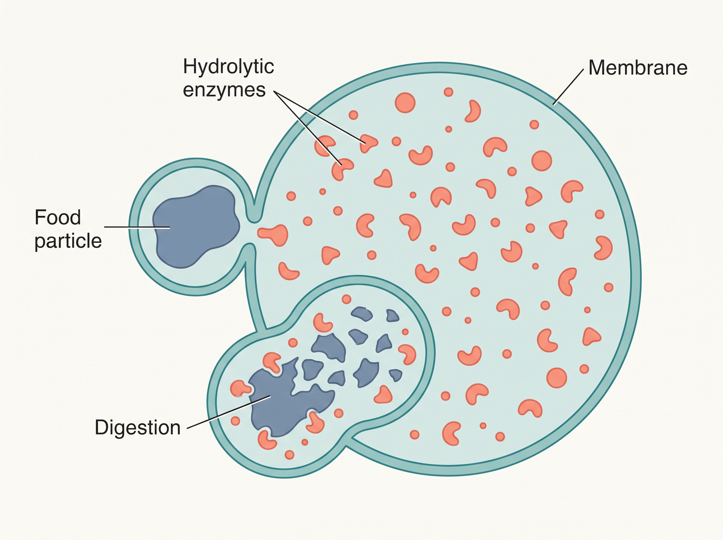
Fig. 8.11 – A lysosome breaking down material.
8.5.3.4 Vacuoles
The vacuole is a membrane-bound space found in the cytoplasm. It is not empty — it contains water, cell sap, excretory products, and other materials that are not immediately useful to the cell. The single membrane that encloses the vacuole has its own name: the tonoplast.
In plant cells, the vacuole can be remarkably large, occupying up to 90% of the total volume of the cell.
The tonoplast is not just a passive boundary. In plants, it actively transports a number of ions and other materials against their concentration gradients into the vacuole — an example of the active transport we met earlier. As a result, the concentration of these substances is significantly higher inside the vacuole than in the surrounding cytoplasm.
Some organisms have specialised vacuoles. In Amoeba, the contractile vacuole is important for osmoregulation (balancing water content) and for excretion. In many cells, especially in protists, food vacuoles are formed by engulfing food particles so that they can be digested inside the cell.
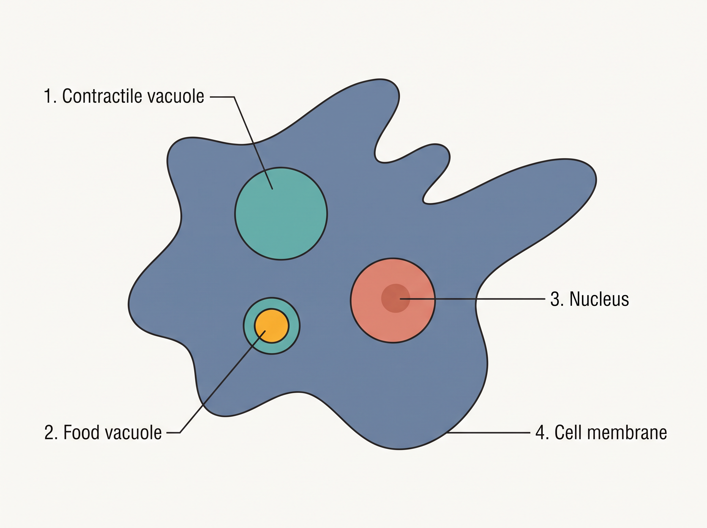
Fig. 8.12 – Vacuoles in Amoeba.
8.5.4 Mitochondria
The mitochondria (singular: mitochondrion) are among the most important organelles in the cell, yet unless they are specifically stained, they are not easily visible under an ordinary microscope.
Shape, Size, and Number
The number of mitochondria in a cell is not fixed — it varies depending on how much physiological activity the cell carries out. Their shape and size also vary considerably. A typical mitochondrion is sausage-shaped or cylindrical, with a diameter of 0.2–1.0 µm (on average about 0.5 µm) and a length of 1.0–4.1 µm.
The Double-Membrane Structure
Each mitochondrion is a double membrane-bound structure, having both an outer membrane and an inner membrane. These two membranes divide the interior distinctly into two aqueous (watery) compartments — the outer compartment and the inner compartment.
The inner compartment is filled with a dense, homogeneous substance called the matrix. The outer membrane forms the smooth, continuous limiting boundary of the whole organelle. The inner membrane, however, is thrown into a number of infoldings called the cristae (singular: crista) that project towards the matrix, as shown in Fig. 8.13. Each of the two membranes carries its own specific enzymes linked to the work of the mitochondrion.
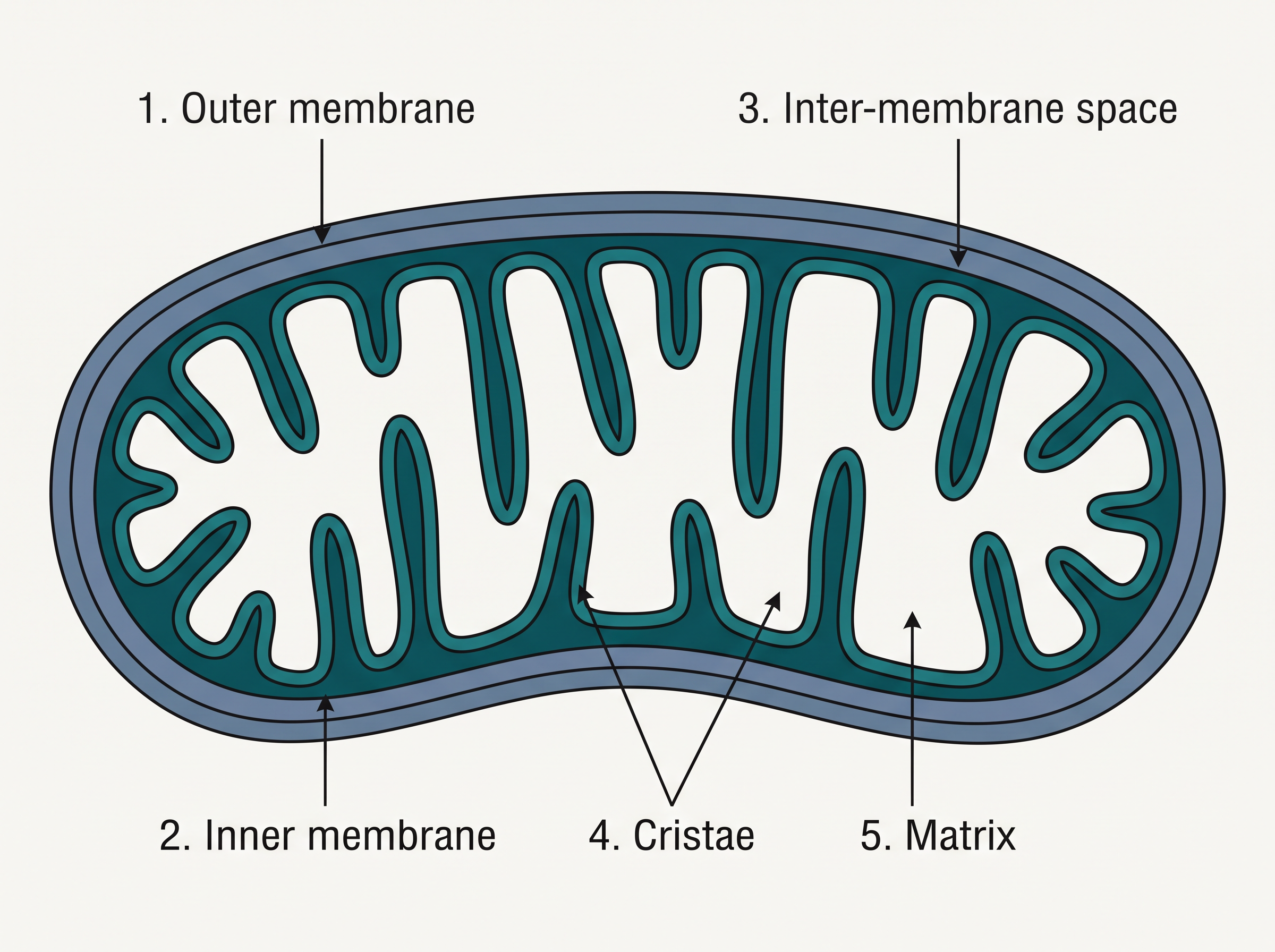
Fig. 8.13 – Longitudinal section of a mitochondrion.
The Power House with Its Own DNA
Mitochondria are the sites of aerobic respiration — respiration that uses oxygen. Here they produce the cell’s usable energy in the form of ATP, which is exactly why mitochondria are famously called the “power houses” of the cell.
Remarkably, the matrix also contains a single circular DNA molecule, a few RNA molecules, ribosomes (of the 70S type), and the other components needed to make proteins. Because of this, a mitochondrion can synthesise some of its own proteins, and it multiplies on its own by fission (splitting into two).
8.5.5 Plastids
Plastids are organelles found in all plant cells and in euglenoides. Unlike most organelles, they are large enough to be observed easily under an ordinary microscope. Plastids carry specific pigments, and it is these pigments that give different parts of a plant their characteristic colours.
Three Kinds of Plastids
Based on the type of pigment they carry, plastids are classified into three types: chloroplasts, chromoplasts, and leucoplasts.
The chloroplasts contain chlorophyll and carotenoid pigments. These pigments are responsible for trapping the light energy that is essential for photosynthesis — the process by which plants make their food.
The chromoplasts contain fat-soluble carotenoid pigments such as carotene, xanthophylls, and others. These give the part of the plant a yellow, orange, or red colour.
The leucoplasts are colourless plastids of varied shapes and sizes, and their job is to store nutrients. There are three kinds: amyloplasts store carbohydrates in the form of starch (for example, in the potato); elaioplasts store oils and fats; and aleuroplasts store proteins.
Inside the Chloroplast
The majority of a green plant’s chloroplasts are found in the mesophyll cells of its leaves. They come in several shapes — lens-shaped, oval, spherical, discoid, or even ribbon-like — with a variable length of 5–10 µm and a width of 2–4 µm. Their number varies too, from just 1 per cell in the green alga Chlamydomonas to 20–40 per cell in the mesophyll.
Like the mitochondria, chloroplasts are double membrane bound. Of the two membranes, the inner chloroplast membrane is relatively less permeable. The space enclosed by the inner membrane is called the stroma.
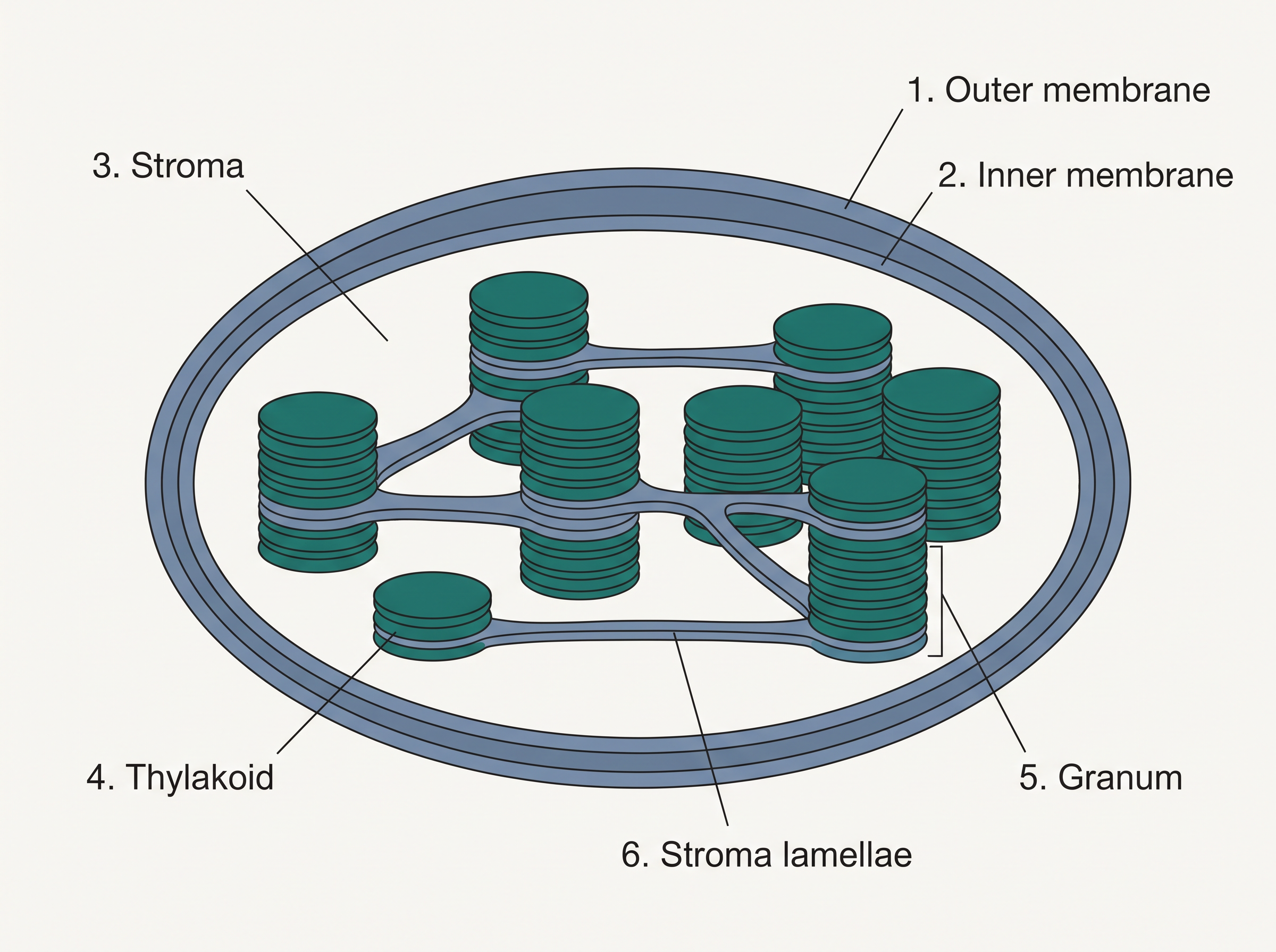
Fig. 8.14 – Sectional view of a chloroplast.
Within the stroma lie a number of organised, flattened membranous sacs called thylakoids, shown in Fig. 8.14. The thylakoids are arranged in stacks, like piles of coins, and each stack is called a granum (plural: grana); such stacked thylakoids are the intergranal thylakoids. Connecting the thylakoids of different grana are flat membranous tubules called the stroma lamellae. The membrane of each thylakoid encloses an inner space called the lumen.
The stroma itself is not just empty fluid. It contains the enzymes required for the synthesis of carbohydrates and proteins, and it also holds small, double-stranded circular DNA molecules and ribosomes — which is why the chloroplast, like the mitochondrion, can make some of its own proteins. The chlorophyll pigments are located in the thylakoids. As in mitochondria, the ribosomes of the chloroplast are the smaller 70S type, compared with the 80S ribosomes of the cytoplasm.
8.5.6 Ribosomes
We have already met ribosomes as the sites of protein synthesis. Here we look more closely at what they are made of and how their sizes are described.
Ribosomes are composed of ribonucleic acid (RNA) and proteins, and, unlike the organelles we have studied so far, they are not surrounded by any membrane.
Two Subunits and the Svedberg Unit
Ribosomes come in two main sizes. Eukaryotic ribosomes are of the 80S type, while prokaryotic ribosomes are of the 70S type. Every ribosome, of either size, is made of two subunits — one larger and one smaller — as seen in Fig. 8.15.
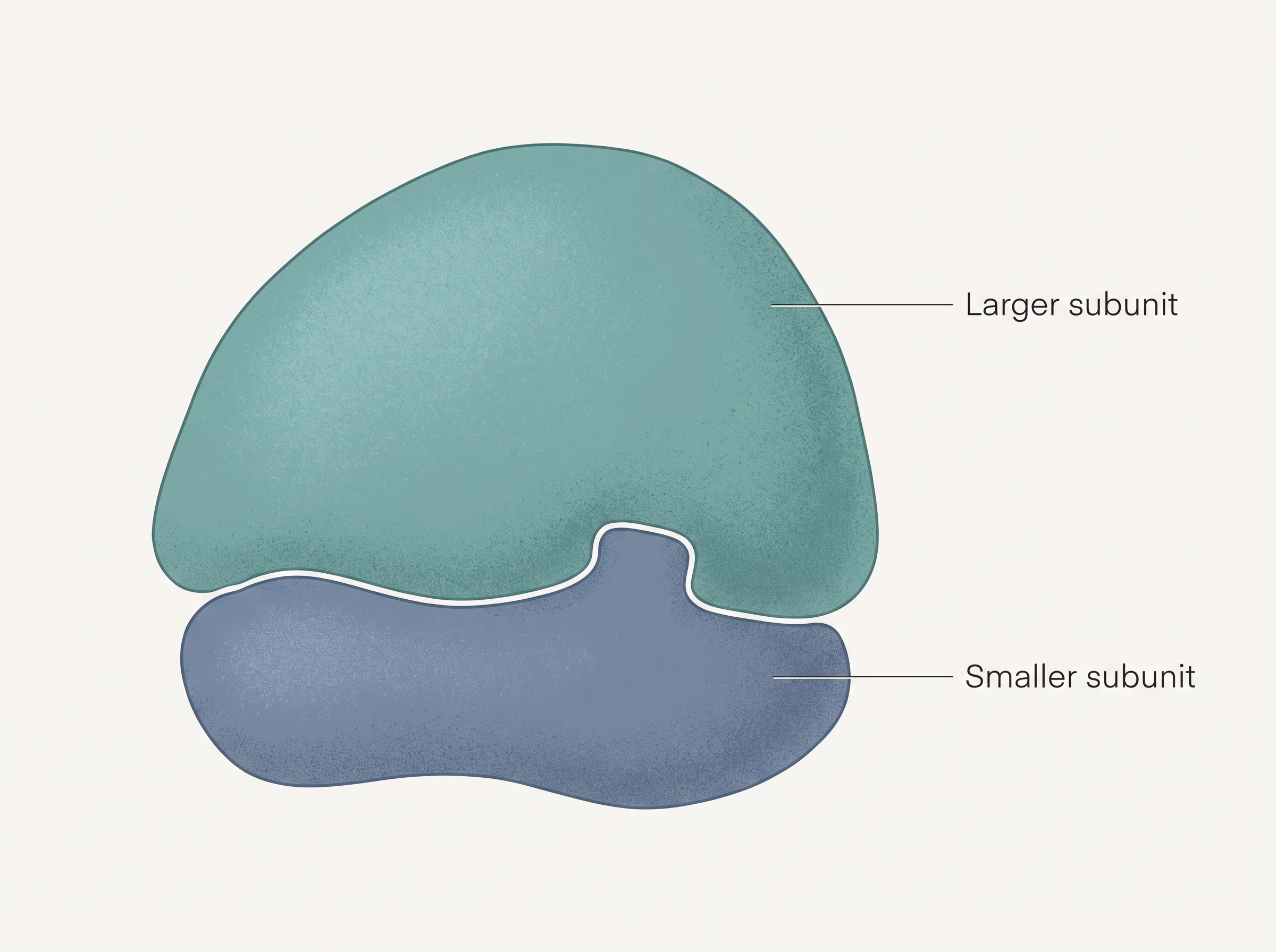
Fig. 8.15 – The two subunits of a ribosome.
In the 80S ribosome, the two subunits are 60S and 40S; in the 70S ribosome, they are 50S and 30S. In every case, the complete ribosome is built from just these two subunits.
The letter “S” in these names stands for the Svedberg’s Unit, which measures the sedimentation coefficient — how fast a particle settles when spun at high speed. Since settling speed depends on a particle’s density and size, “S” is an indirect measure of both.
Because S depends on size, shape, and density together — and not on mass alone — the S values of the subunits do not add up arithmetically. This is why 50S and 30S combine to give 70S (not 80S), and 60S and 40S combine to give 80S (not 100S), finally explaining the puzzle noted earlier.
8.5.7 Cytoskeleton
Running throughout the cytoplasm of a eukaryotic cell is an elaborate network of thread-like, protein (proteinaceous) structures. This network is made up of three kinds of filaments — microtubules, microfilaments, and intermediate filaments — and taken together it is called the cytoskeleton.
Just as a skeleton supports and shapes an animal’s body from within, the cytoskeleton supports and organises the cell from the inside. It is involved in several important functions: it provides mechanical support, it enables motility (movement), and it maintains the shape of the cell.
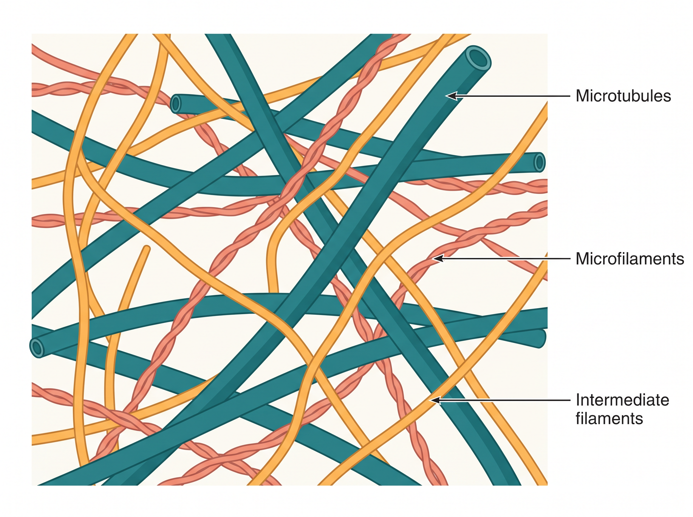
Fig. 8.16 – The cytoskeleton in a eukaryotic cell.
8.5.8 Cilia and Flagella
Cilia (singular: cilium) and flagella (singular: flagellum) are hair-like outgrowths that project from the cell membrane. Though they look similar, they differ in size and in the exact job they do.
What Cilia and Flagella Do
Cilia are small structures that work like tiny oars. By beating back and forth, they cause the movement of either the cell itself or the fluid surrounding the cell. Flagella are comparatively longer and are chiefly responsible for the movement of the cell.
It is important to note that prokaryotic bacteria also possess flagella, but the bacterial flagellum is structurally quite different from the eukaryotic one.
The Internal Structure: The 9+2 Array
When a cilium or flagellum is studied under the electron microscope, it is seen to be covered by the plasma membrane. Inside, its core is called the axoneme, which contains a set of microtubules running parallel to its long axis.
In a typical axoneme, there are nine doublets (pairs) of peripheral microtubules arranged in a ring, plus a single pair of microtubules in the centre. This characteristic pattern — nine outer doublets around two central tubules — is called the 9+2 array, shown in Fig. 8.17.
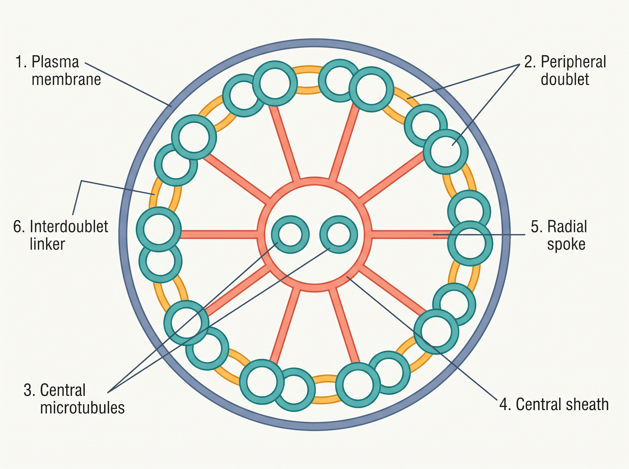
Fig. 8.17 – Cross-section of a cilium or flagellum.
The two central tubules are joined by bridges and are enclosed within a central sheath. This central sheath is connected to one tubule of each peripheral doublet by a radial spoke — and since there are nine peripheral doublets, there are nine radial spokes in all. The peripheral doublets themselves are interconnected by linkers.
Both the cilium and the flagellum arise from a centriole-like structure at their base called the basal body.
8.5.9 Centrosome and Centrioles
The centrosome is an organelle that usually contains two cylindrical structures called centrioles. These centrioles are surrounded by a shapeless (amorphous) material known as the pericentriolar material. Within a centrosome, the two centrioles lie perpendicular to each other, that is, at right angles.
Structure of the Centriole
Each centriole has an internal organisation that looks like a cartwheel. It is made up of nine evenly spaced peripheral fibrils, and these fibrils are built from a protein called tubulin. Each of these nine peripheral fibrils is not single but a triplet (a set of three), and adjacent triplets are linked to one another.
At the centre of the proximal region of the centriole is a proteinaceous core called the hub. The hub is connected to the tubules of the peripheral triplets by radial spokes made of protein — much like the spokes of a wheel joining the axle to the rim.
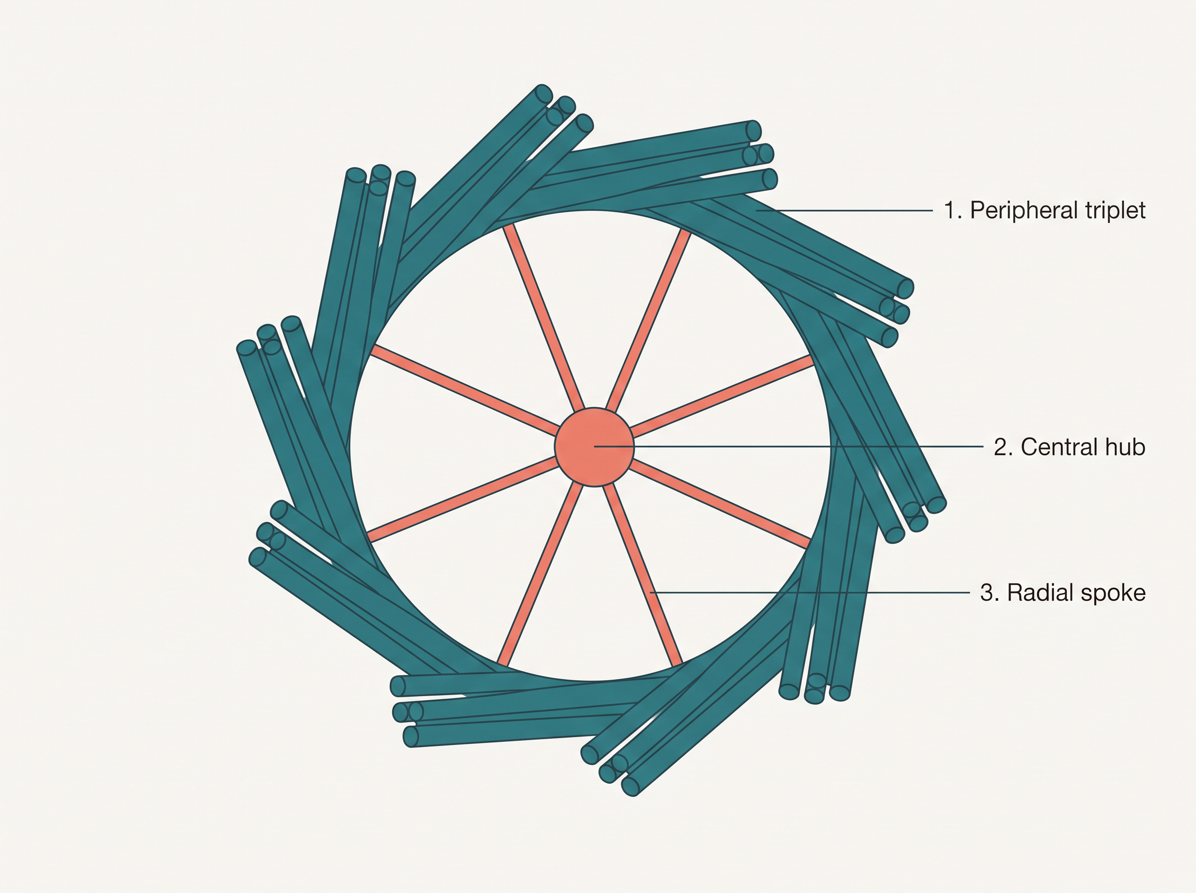
Fig. 8.18 – Cross-section of a centriole.
What Centrioles Do
Centrioles have two important roles. First, they form the basal body of cilia or flagella — the very structures we saw those hair-like outgrowths emerging from. Second, in animal cells they give rise to the spindle fibres, which build the spindle apparatus used during cell division.
8.5.10 Nucleus
The nucleus is the control centre of the eukaryotic cell. As we noted earlier, it was first described by Robert Brown, as far back as 1831. Later, when scientists stained the material inside the nucleus with basic dyes, the scientist Flemming gave this stained material the name chromatin.
When a cell is not dividing, its nucleus is called the interphase nucleus. Inside it, we find highly extended and elaborate nucleoprotein fibres (the chromatin), a supporting nuclear matrix, and one or more spherical bodies called nucleoli (singular: nucleolus), as shown in Fig. 8.19.
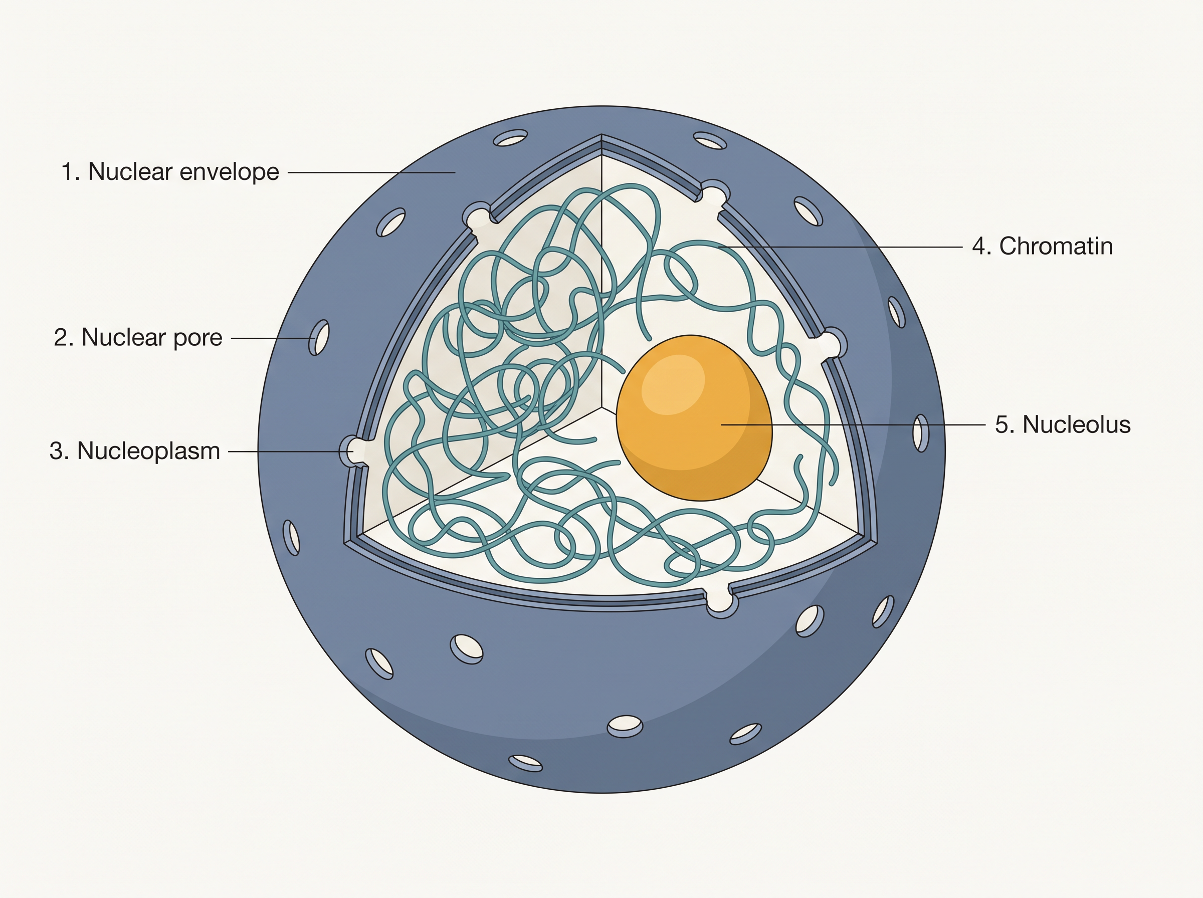
Fig. 8.19 – Structure of the nucleus.
The Nuclear Envelope and Pores
Electron microscopy has revealed that the nucleus is enclosed by a nuclear envelope made of two parallel membranes. Between these two membranes lies a gap of 10 to 50 nm called the perinuclear space. This envelope forms a barrier that separates the materials inside the nucleus from those in the cytoplasm.
The outer membrane of the envelope usually stays continuous with the endoplasmic reticulum and even bears ribosomes on its surface. At many places, the envelope is interrupted by minute openings called nuclear pores, which are formed where the two membranes fuse together.
These nuclear pores are far from being simple holes — they are controlled passages through which RNA and protein molecules move in both directions between the nucleus and the cytoplasm.
Usually there is only one nucleus per cell, though variations in the number of nuclei are frequently seen. Interestingly, some mature cells even lack a nucleus altogether — for example, the erythrocytes (red blood cells) of many mammals and the sieve tube cells of vascular plants.
Nucleoplasm and Nucleolus
The nuclear matrix, also called the nucleoplasm, is the fluid that contains both the nucleolus and the chromatin. The nucleoli are spherical structures floating in this nucleoplasm. A nucleolus is not bounded by any membrane, so its content is simply continuous with the surrounding nucleoplasm.
The nucleolus has a specific and important job: it is the site of active ribosomal RNA synthesis — the making of the RNA that goes into ribosomes. This is why cells that are busily carrying out protein synthesis tend to have larger and more numerous nucleoli.
Chromatin and Chromosomes
Recall that in the interphase nucleus, the genetic material exists as a loose, indistinct network of nucleoprotein fibres — the chromatin. But during the different stages of cell division, this loose network is packed into neat, structured bodies called chromosomes, which appear in place of the nucleus.
Chromatin is not DNA alone. It contains DNA together with some basic proteins called histones, along with some non-histone proteins and also RNA. A single human cell contains an astonishing amount of DNA — a thread roughly two metres long — distributed among its 46 chromosomes (that is, 23 pairs). (The detailed way in which this long thread is packaged into a chromosome is a topic you will study in Class XII.)
Types of Chromosomes
Every chromosome — remember, these are clearly visible only in dividing cells — has a primary constriction called the centromere. On either side of the centromere lie disc-shaped structures called kinetochores, shown in Fig. 8.20. The centromere is important because it holds together the two chromatids of a chromosome.
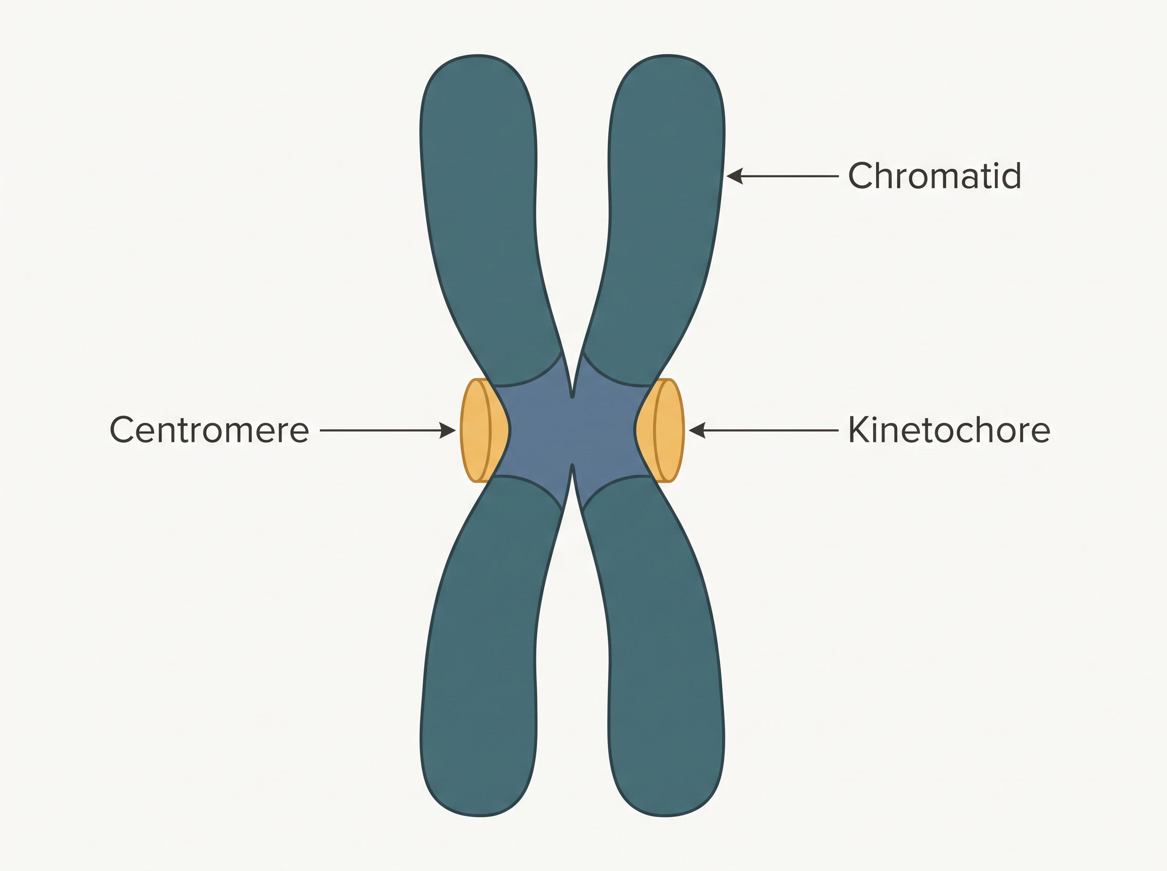
Fig. 8.20 – A chromosome with kinetochores.
Based on where the centromere is placed, chromosomes are classified into four types, shown in Fig. 8.21. In a metacentric chromosome the centromere is in the middle, forming two equal arms. In a sub-metacentric chromosome it is shifted slightly away from the middle, giving one shorter arm and one longer arm. In an acrocentric chromosome the centromere lies close to one end, forming one extremely short arm and one very long arm. In a telocentric chromosome the centromere is right at the end (terminal).
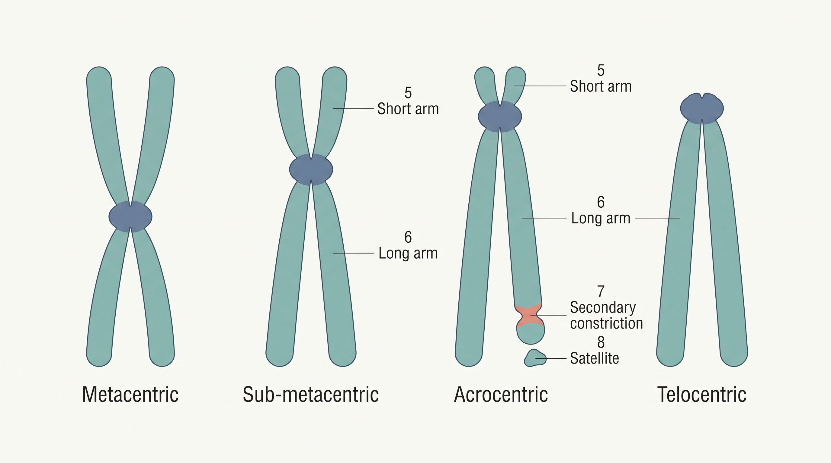
Fig. 8.21 – Types of chromosomes based on centromere position.
Finally, a few chromosomes carry a non-staining secondary constriction at a fixed location. The small piece of chromosome beyond this secondary constriction appears as a little fragment called the satellite.
8.5.11 Microbodies
The last of the organelles we meet in this chapter are the microbodies. These are many tiny, membrane-bound vesicles that contain various enzymes. Unlike some organelles that occur in only one kind of cell, microbodies are present in both plant and animal cells.
8.6 Summary
All organisms are made of cells or aggregates of cells. Cells vary greatly in their shape, size, and activities or functions.
Based on the presence or absence of a membrane-bound nucleus and other organelles, cells — and hence the organisms built from them — are named either prokaryotic or eukaryotic.
A typical eukaryotic cell consists of a cell membrane, a nucleus, and cytoplasm. Plant cells have an additional cell wall lying outside the cell membrane.
The plasma membrane is selectively permeable and helps in the transport of several molecules across it.
The endomembrane system — the ER, golgi complex, lysosomes, and vacuoles — is a group of organelles whose functions are coordinated. All the cell organelles perform different but specific functions.
The endoplasmic reticulum contains tubules or cisternae and is of two types, rough and smooth. It helps in the transport of substances and in the synthesis of proteins, lipoproteins, and glycogen.
The golgi body is a membranous organelle made of flattened sacs. The secretions of the cell are packed in it and transported out of the cell.
Lysosomes are single-membrane structures containing enzymes that can digest all types of macromolecules.
Ribosomes are involved in protein synthesis. They occur freely in the cytoplasm or remain associated with the ER.
Mitochondria help in oxidative phosphorylation and in the generation of adenosine triphosphate (ATP). They are bound by a double membrane — the outer membrane is smooth, while the inner membrane folds into several cristae.
Plastids are pigment-containing organelles found only in plant cells. In plant cells, chloroplasts trap the light energy essential for photosynthesis; within the plastid, the grana are the site of the light reactions and the stroma of the dark reactions. Green plastids that contain chlorophyll are chloroplasts, whereas the other coloured plastids are chromoplasts, which may contain pigments like carotene and xanthophyll.
Centrosome and centriole form the basal body of cilia and flagella, which facilitate locomotion. In animal cells, centrioles also form the spindle apparatus during cell division.
The nucleus is enclosed by a nuclear envelope — a double-membrane structure bearing nuclear pores. The inner membrane encloses the nucleoplasm and the chromatin material. The nucleus contains nucleoli and a chromatin network; it not only controls the activities of the organelles but also plays a major role in heredity.
Thus, the cell is the structural and functional unit of life.
Flowchart
FC 8.4 – Overview of the cell: Cell → (branch) Prokaryotic cell (no membrane-bound nucleus or organelles; e.g., bacteria) OR Eukaryotic cell (membrane-bound nucleus + organelles). Eukaryotic cell → cell membrane + cytoplasm + nucleus; cytoplasm holds the organelles (ER, golgi complex, lysosomes, vacuoles, mitochondria, plastids, ribosomes, cytoskeleton, centrosome, microbodies); plant cells additionally have a cell wall and plastids, animal cells additionally have centrioles.
8.7 Points to Ponder
When a cell is torn open, its separated organelles cannot survive on their own, yet the intact whole cell is clearly alive. What does this tell us about why “life” seems to belong to the complete cell rather than to any single part of it?
Mitochondria and chloroplasts carry their own circular DNA and 70S ribosomes and multiply by fission — features they share with bacteria. What might this resemblance suggest about how these two organelles could have originated?
Some mature cells, such as the red blood cells of many mammals, lose their nucleus completely. In what sense can such cells still be called “living,” and what limitations must losing the nucleus place upon them?
Prokaryotes carry out every function of life without any membrane-bound organelles. Why, then, might eukaryotic cells have “invested” in dividing their interior into so many separate compartments?
Viruses are far smaller than the smallest known cells and are not made of cells at all. Should the cell theory be extended to account for them, or are viruses better regarded as lying outside the usual definition of a living cell?
The shape of a cell is often matched to the job it performs. If you were shown an unfamiliar cell under the microscope, what might its shape alone allow you to guess about the function it carries out?
8.8 NCERT Questions
- Which of the following is not correct?
- Robert Brown discovered the cell.
- Schleiden and Schwann formulated the cell theory.
- Virchow explained that cells are formed from pre-existing cells.
- A unicellular organism carries out its life activities within a single cell.
- New cells generate from
- bacterial fermentation
- regeneration of old cells
- pre-existing cells
- abiotic materials
- Match the following.
| Column I | Column II |
|---|---|
| a. Cristae | i. Flat membranous sacs in stroma |
| b. Cisternae | ii. Infoldings in mitochondria |
| c. Thylakoids | iii. Disc-shaped sacs in Golgi apparatus |
Which of the following is correct?
- Cells of all living organisms have a nucleus.
- Both animal and plant cells have a well-defined cell wall.
- In prokaryotes, there are no membrane-bound organelles.
- Cells are formed de novo from abiotic materials.
What is a mesosome in a prokaryotic cell? Mention the functions that it performs.
How do neutral solutes move across the plasma membrane? Can the polar molecules also move across it in the same way? If not, then how are these transported across the membrane?
Name two cell-organelles that are double membrane bound. What are the characteristics of these two organelles? State their functions and draw labelled diagrams of both.
What are the characteristics of prokaryotic cells?
Multicellular organisms have division of labour. Explain.
Cell is the basic unit of life. Discuss in brief.
What are nuclear pores? State their function.
Both lysosomes and vacuoles are endomembrane structures, yet they differ in terms of their functions. Comment.
Describe the structure of the following with the help of labelled diagrams.
- Nucleus
- Centrosome
What is a centromere? How does the position of the centromere form the basis of classification of chromosomes? Support your answer with a diagram showing the position of the centromere on different types of chromosomes.
8.9 Check Your Concepts
Assertion–Reason. Assertion: Mycoplasma lacks a cell wall. Reason: All prokaryotes lack a cell wall. State whether the assertion and the reason are true, and whether the reason correctly explains the assertion.
Why is the cell described as the fundamental structural as well as functional unit of all living organisms? Explain both parts of the statement.
Assertion–Reason. Assertion: Antibiotics such as streptomycin can kill bacteria without harming human cells. Reason: Bacterial ribosomes (70S) differ from the ribosomes of human cells (80S). Decide whether the reason correctly explains the assertion.
What would happen to a plant cell if its large central vacuole lost most of its water? Explain your answer in terms of the cell and the plant.
Distinguish between rough and smooth endoplasmic reticulum in terms of both structure and function.
Sequence-ordering. Arrange the three layers of a bacterial cell envelope in order from the outermost to the innermost.
Sequence-ordering. Trace the path of a secretory protein from the site where it is made to the point where it is released outside the cell, naming the organelles involved in the correct order.
Match-the-following. Match each structure in Column I with its function in Column II.
| Column I | Column II |
|---|---|
| a. Ribosome | i. Photosynthesis |
| b. Nucleolus | ii. Digestion of macromolecules |
| c. Lysosome | iii. Protein synthesis |
| d. Chloroplast | iv. Ribosomal RNA synthesis |
Diagram-labelling. Draw a labelled diagram of a mitochondrion in longitudinal section and label the outer membrane, inner membrane, cristae, and matrix.
Odd-one-out. From the group — endoplasmic reticulum, golgi complex, lysosome, mitochondrion — pick the odd one out with respect to the endomembrane system, and justify your choice.
Why do the sedimentation (S) values of the two ribosomal subunits not add up arithmetically — for example, why do a 50S and a 30S subunit together form a 70S ribosome and not an 80S one?
Differentiate between the arrangement of microtubules in the axoneme of a cilium (the 9+2 array) and the arrangement of fibrils in a centriole.
Explain how the folding of the inner membrane of the mitochondrion into cristae suits the organelle’s function.
What happens if… Suppose all the nuclear pores of a cell were sealed shut. How would this affect the working of the cell? Explain.
Assertion–Reason. Assertion: Chromatin and chromosomes are made of the same material. Reason: Chromatin condenses and coils to form chromosomes during cell division. Decide whether the reason correctly explains the assertion.
Name and briefly describe the four types of chromosomes classified on the basis of the position of the centromere.
Why are the majority of a green plant’s chloroplasts located in the mesophyll cells of its leaves?
Distinguish between passive transport and active transport across the plasma membrane, giving one example of each.