In the year 1828, a young German chemist named Friedrich Wöhler was working with a simple, lifeless salt called ammonium cyanate. He heated it in his laboratory, expecting one ordinary chemical to turn into another. Instead, he found himself staring at crystals of urea — the very compound that our kidneys pack into urine, a substance that until then everyone believed only a living body could make.
This was startling. At the time, most scientists were convinced that the chemicals of life were special. They thought a mysterious “vital force,” present only inside living creatures, was needed to build compounds like urea, sugar or blood. Non-living matter, they believed, could never produce them. Wöhler had just made a “living” chemical from a “non-living” one, on a laboratory bench, with no organism in sight.
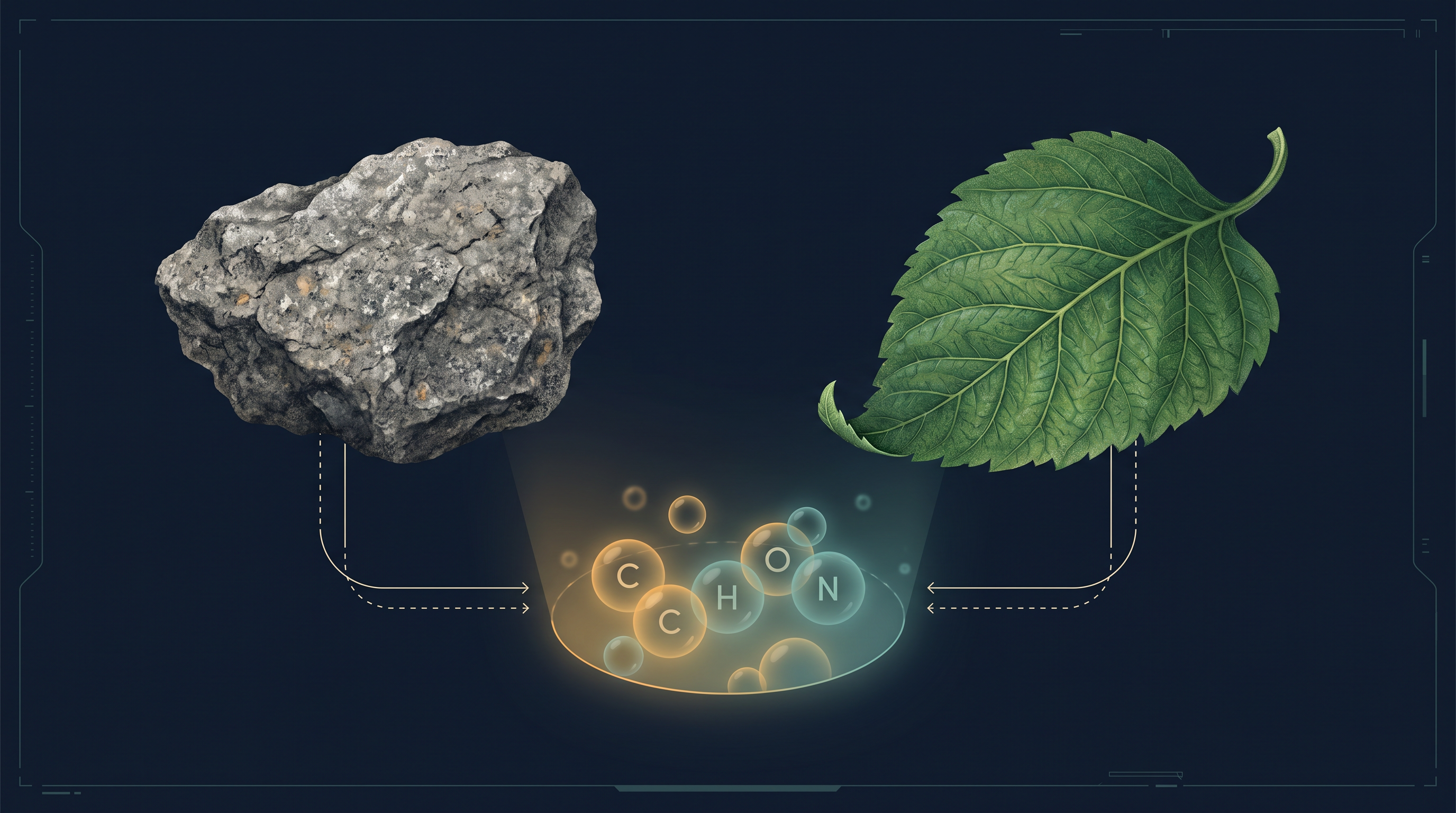
His quiet experiment cracked open a huge idea: the molecules of life are made of the same ordinary elements — carbon, hydrogen, oxygen, nitrogen — that build rocks, air and water. There is no magic ingredient. What makes life special is not a secret element, but the way these common atoms are arranged into remarkable molecules called biomolecules, and how those molecules work together.
In this chapter, we will meet these molecules of life — sugars, fats, amino acids, proteins, nucleic acids and enzymes — and learn how the same handful of elements can be organised into the astonishing chemistry of a living cell.
Keep these questions in mind as you read. By the end of the chapter, you will be able to answer every one of them.
9.1 How to Analyse Chemical Composition?
Same Elements, Living and Non-living
Our biosphere is filled with an astonishing variety of living organisms. A natural question follows: are all these living things built from the same chemicals — the same elements and compounds? To answer this, we can borrow a method you have already met in chemistry, called elemental analysis. An element is a pure substance made of only one kind of atom (carbon, hydrogen, oxygen, and so on), while a compound is formed when two or more elements join chemically.
If we perform elemental analysis on a plant tissue, an animal tissue, or a paste of microbes, we get a list of the elements present — carbon, hydrogen, oxygen and several others — together with how much of each is present per unit mass of the tissue. A tissue, remember, is simply a group of similar cells working together.
Now suppose we run the very same analysis on a piece of the earth’s crust, chosen as an example of non-living matter. We get a strikingly similar list. In fact, in absolute terms no clear difference can be drawn between the two: every element found in the earth’s crust is also found in living tissue. There is no “special” element that appears only in life.
A closer look, however, reveals one meaningful difference. The relative abundance of carbon and hydrogen — how much of them there is compared with the other elements — is distinctly higher in any living organism than in the earth’s crust. Life is, chemically speaking, richer in carbon and hydrogen. This is clear from Table 9.1.
Table 9.1 A Comparison of Elements Present in Non-living and Living Matter
| Hydrogen (H) |
0.14 |
9.5 |
| Carbon (C) |
0.03 |
18.5 |
| Oxygen (O) |
46.6 |
65.0 |
| Nitrogen (N) |
very little |
3.3 |
| Sulphur (S) |
0.03 |
0.3 |
| Sodium (Na) |
2.8 |
0.2 |
| Calcium (Ca) |
3.6 |
1.5 |
| Magnesium (Mg) |
2.1 |
0.1 |
| Silicon (Si) |
27.7 |
negligible |
(Adapted from C. N. R. Rao, Understanding Chemistry, Universities Press, Hyderabad.)
Notice how carbon jumps from a mere \(0.03\%\) in the crust to \(18.5\%\) in the human body, and hydrogen from \(0.14\%\) to \(9.5\%\), while silicon does the opposite — abundant in rock, negligible in us. Life has clearly built itself around carbon and hydrogen.
Q1. If a rock and a living leaf are made of the very same elements, what actually makes the chemistry of a living thing different?
A. It is not a secret ingredient. Living and non-living matter draw on exactly the same set of elements. The difference is one of proportion and organisation: living tissue contains a much higher relative abundance of carbon and hydrogen, and these carbon-rich atoms are assembled into the special carbon-based molecules of life — the biomolecules we will study through this chapter.
Pulling the Molecules Out of a Tissue
We can keep asking questions in the same spirit. What kinds of carbon compounds are actually found in living organisms? To find out, we must do a chemical analysis of the tissue itself.
Take any living tissue — a vegetable, or a piece of liver, for example — and grind it thoroughly with a mortar and pestle in trichloroacetic acid, \(\text{Cl}_3\text{CCOOH}\). This gives a thick slurry. If we strain this slurry through a cheesecloth or cotton, it separates into two parts, as summarised in FC 9.1.
One part is the liquid that passes through, called the filtrate, or more technically the acid-soluble pool. The other is the material left behind on the cloth, called the retentate, or the acid-insoluble fraction. When scientists examined the acid-soluble pool, they found it contained thousands of different organic compounds. An organic compound, in this context, simply means a carbon-containing compound of the kind that living tissues make.
Identifying and Purifying a Compound
How does one go from “thousands of compounds mixed together” to knowing a single compound? In higher classes you will study this in detail, but the idea is straightforward. One first extracts the compounds, then puts the extract through a series of separation techniques, repeating them until a single compound has been separated from all the others. In short, the compound is isolated and purified.
Once a compound is pure, analytical techniques can be applied to it. These reveal its molecular formula — the actual count of each kind of atom in one molecule — and give a good idea of its probable structure, that is, how those atoms are arranged.
All the carbon compounds we obtain from living tissues in this way can be given a single collective name: biomolecules.
Biomolecules are the carbon-based (organic) compounds found in living tissues.
Wet Weight, Dry Weight and Ash
Living organisms are not made only of carbon compounds — they also contain inorganic elements and compounds. How can we show this? For that, a different and rather destructive experiment is needed, summarised in FC 9.2.
First, weigh a small amount of living tissue, say a leaf or a bit of liver. This is its wet weight. Now dry it completely so that all the water evaporates; the material that remains gives the dry weight. Finally, burn the tissue fully. On burning, all the carbon compounds are oxidised into gases — carbon dioxide (\(\text{CO}_2\)) and water vapour — which escape. What is left behind is a residue called ash.
This ash contains the inorganic elements of the tissue, such as calcium and magnesium. Inorganic compounds like sulphate and phosphate, meanwhile, appear in the acid-soluble fraction we met earlier.
Wet weight vs Dry weight vs Ash Wet weight includes the tissue’s water. Dry weight is what remains after only the water has evaporated (all organic and inorganic matter is still there). Ash is what remains after the tissue has been fully burnt — the organic carbon compounds are gone as gas, so ash is essentially the inorganic mineral content.
Putting these experiments together, elemental analysis tells us the elemental make-up of a tissue — how much hydrogen, oxygen, chlorine, carbon and so on it contains. Analysis of the compounds, on the other hand, tells us about the kinds of organic constituents (shown in Fig. 9.1) and the inorganic constituents (listed in Table 9.2) present in living tissue.
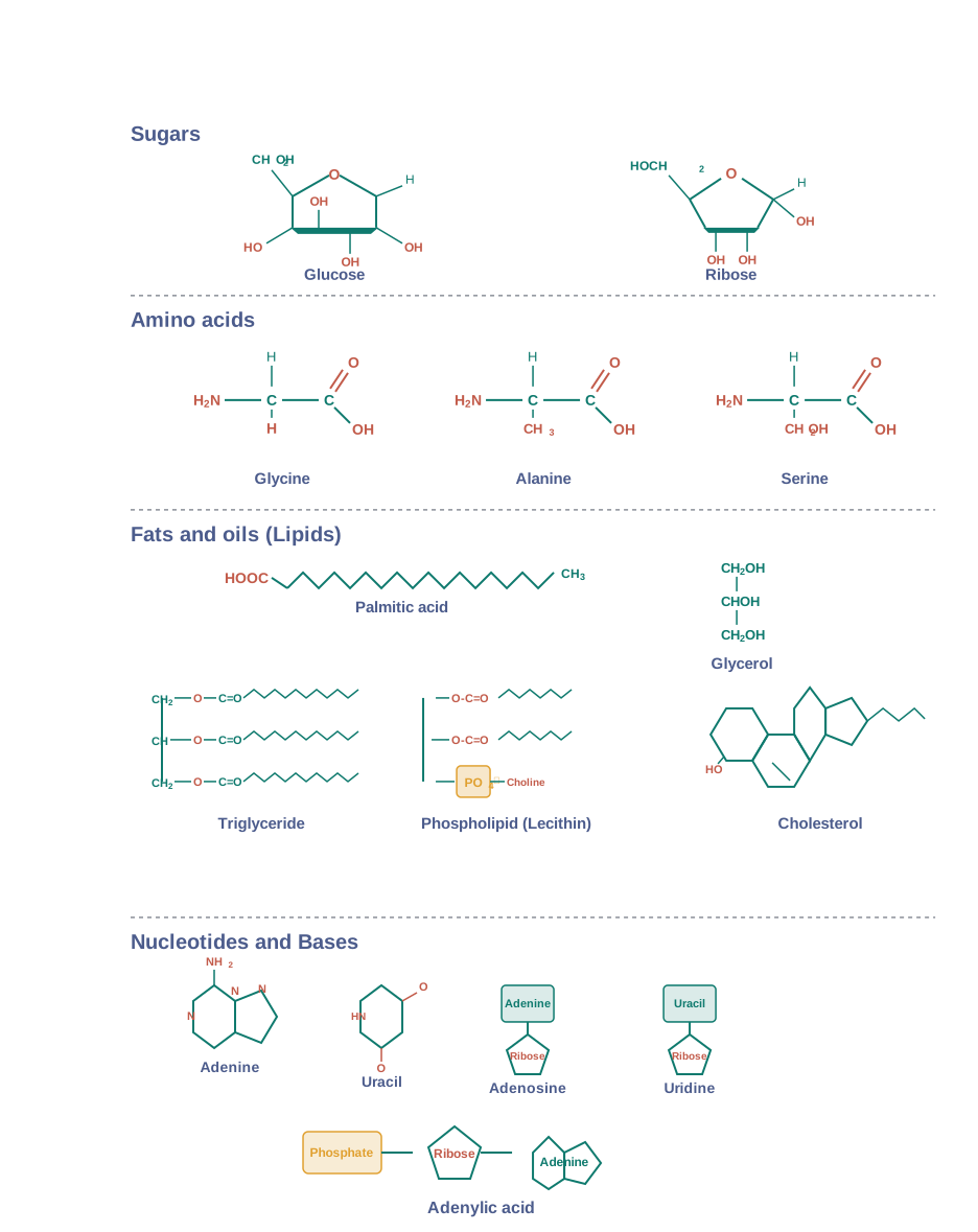
Table 9.2 A List of Representative Inorganic Constituents of Living Tissues
| Sodium |
\(\text{Na}^+\) |
| Potassium |
\(\text{K}^+\) |
| Calcium |
\(\text{Ca}^{++}\) |
| Magnesium |
\(\text{Mg}^{++}\) |
| Water |
\(\text{H}_2\text{O}\) |
| Compounds |
\(\text{NaCl},\ \text{CaCO}_3,\ \text{PO}_4^{3-},\ \text{SO}_4^{2-}\) |
Two Ways to Read the Same Molecules
The organic constituents of a tissue can be looked at from two points of view. From a chemistry point of view, we identify the functional groups present — aldehydes, ketones, aromatic rings, and so on. A functional group is a small cluster of atoms that gives a molecule its characteristic chemical behaviour.
From a biology point of view, we are more interested in what the molecules do in the cell, so we sort them into biological categories: amino acids, nucleotide bases, fatty acids, and the like. The next few paragraphs introduce these small biomolecules one family at a time.
Amino Acids
Amino acids are organic compounds that carry two key functional groups on the same carbon atom: an amino group (\(-\text{NH}_2\)) and an acidic carboxyl group (\(-\text{COOH}\)). That shared central carbon is called the α-carbon (alpha-carbon), which is why these molecules are called α-amino acids.
An α-amino acid is an organic compound carrying both an amino group (\(-\text{NH}_2\)) and a carboxyl group (\(-\text{COOH}\)) as substituents on the same (α) carbon.
You can think of an amino acid as a substituted methane: a central carbon with four groups attached at its four valency positions. These four groups are a hydrogen atom, a carboxyl group, an amino group, and a fourth variable group written as R. Three examples are shown in Fig. 9.1.
the α-carbon of an amino acid (central α-carbon holding four different groups — H, \(-\text{COOH}\), \(-\text{NH}_2\) and a variable R → the fixed \(-\text{NH}_2\) and \(-\text{COOH}\) let any two amino acids link together in the same way, while the variable R makes each amino acid chemically distinct → this “same backbone, different side” design lets 20 amino acids join in any order to build endlessly varied proteins.)
It is the R group that makes one amino acid differ from another. If R is just a hydrogen atom, the amino acid is glycine; if R is a methyl group, it is alanine; if R is a hydroxy-methyl group, it is serine, and so on. In principle many amino acids are possible, but the ones that occur in proteins are of only twenty types.
The chemical and physical behaviour of an amino acid comes essentially from its amino, carboxyl and R functional groups. Based on the number of amino and carboxyl groups, amino acids may be acidic (for example glutamic acid), basic (for example lysine), or neutral (for example valine). Some also carry aromatic rings and are called aromatic amino acids (tyrosine, phenylalanine, tryptophan).
A special property of amino acids is that both the \(-\text{NH}_2\) and \(-\text{COOH}\) groups are ionizable — they can gain or lose a hydrogen ion (\(\text{H}^+\)). Because of this, the very structure of an amino acid changes as the pH of the surrounding solution changes:
\[\text{H}_3\overset{+}{\text{N}}-\underset{\text{R}}{\text{CH}}-\text{COOH} \;\rightleftharpoons\; \text{H}_3\overset{+}{\text{N}}-\underset{\text{R}}{\text{CH}}-\text{COO}^- \;\rightleftharpoons\; \text{H}_2\text{N}-\underset{\text{R}}{\text{CH}}-\text{COO}^-\] \[\text{(A)} \qquad\qquad\quad \text{(B)} \qquad\qquad\quad \text{(C)}\]
Here form (A) exists in acidic (low pH) conditions with both groups protonated, and form (C) in basic (high pH) conditions with both groups deprotonated. The middle form (B), which carries a positive charge on the amino end and a negative charge on the carboxyl end at the same time, is called the zwitterionic form.
Lipids
Lipids are a family of biomolecules that are generally water insoluble. The simplest lipids are fatty acids. A fatty acid is built from a carboxyl group (\(-\text{COOH}\)) attached to an R group. Here the R group is a carbon chain — it could be as short as a methyl group (\(-\text{CH}_3\)) or an ethyl group (\(-\text{C}_2\text{H}_5\)), or it could be a longer run of \(-\text{CH}_2-\) units, ranging from 1 carbon up to 19 carbons. For example, palmitic acid has 16 carbons including the carboxyl carbon, while arachidonic acid has 20 carbon atoms including the carboxyl carbon.
Fatty acids come in two kinds. A saturated fatty acid has no carbon–carbon double bonds. An unsaturated fatty acid has one or more \(\text{C}=\text{C}\) double bonds in its chain.
Saturated vs Unsaturated fatty acids | Feature | Saturated fatty acid | Unsaturated fatty acid | |—|—|—| | C=C double bonds | None | One or more | | Carbon chain | “Full” of hydrogen | Has “missing” hydrogens at each double bond | | Typical physical state | Tends to be more solid | Tends to be more oil-like |
Another simple lipid is glycerol, which is trihydroxy propane (a three-carbon molecule carrying three \(-\text{OH}\) groups). Many lipids are built by combining glycerol with fatty acids. In these, the fatty acids become chemically joined to glycerol through esterification — the reaction between an acid group and an alcohol (\(-\text{OH}\)) group that links them by an ester bond.
Depending on how many fatty acids are attached to one glycerol, we get monoglycerides (one fatty acid), diglycerides (two), or triglycerides (three). These same molecules are also called fats and oils, and we tell fats and oils apart by their melting point. Oils have a lower melting point — gingelly oil, for instance, stays liquid even in the cold of winter.
The kitchen difference between “oil” and “solid fat” comes straight from this chemistry. Cooking oils, rich in unsaturated fatty acids with their kinked \(\text{C}=\text{C}\) bonds, cannot pack tightly and so stay liquid at room temperature. Fats like ghee or butter, richer in straight saturated fatty acid chains, pack closely and set solid. This is why sesame (gingelly) oil pours freely in winter while ghee turns firm.
Some lipids contain phosphorus, in the form of a phosphorylated organic group attached to the molecule. These are called phospholipids, and they are a major building material of the cell membrane. Lecithin is one well-known example. Certain tissues, especially the neural (nerve) tissues, contain lipids with even more complex structures.
Nitrogen Bases, Nucleosides and Nucleotides
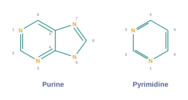
Living organisms also contain many carbon compounds built around heterocyclic rings — ring structures in which the ring contains not only carbon but at least one other kind of atom (here, nitrogen). Among the most important of these are the nitrogen bases: adenine, guanine, cytosine, uracil and thymine.
These bases rarely float about alone. When a nitrogen base is joined to a sugar, the combination is called a nucleoside. Adenosine, guanosine, thymidine, uridine and cytidine are all nucleosides.
If, in addition, a phosphate group is joined (esterified) to the sugar of a nucleoside, the molecule becomes a nucleotide. Adenylic acid, thymidylic acid, guanylic acid, uridylic acid and cytidylic acid are nucleotides.
Nucleoside vs Nucleotide A nucleoside = nitrogen base + sugar. A nucleotide = nitrogen base + sugar + phosphate. The single difference is the phosphate group.
telling nucleoside from nucleotide apart “NucleoTide has a phosphaTe.” The letter T links the tide to the phosphate; a nucleoside (no T-word) has no phosphate.
Nucleotides matter enormously, because the nucleic acids DNA and RNA are made of nucleotides and nothing else. DNA and RNA are the molecules that function as the genetic material of the cell — the chemical carriers of hereditary information, a role we will return to in later chapters.
small biomolecules Zoom in: each of these families — amino acids, fatty acids, nitrogen bases, sugars — is a small “building-block” molecule. Zoom out: joined together in large numbers, these blocks build the giant molecules of life (proteins, lipids, nucleic acids, polysaccharides) that the rest of this chapter explores.
9.3 Biomacromolecules
In the last two sections we separated a tissue into an acid-soluble pool and an acid-insoluble fraction. If we now look carefully at the sizes of the molecules in each, a clear pattern emerges — and it gives us a natural way to sort all biomolecules into two size classes.
Two Size Classes of Biomolecules
The compounds found in the acid-soluble pool share one common feature: they are small. Their molecular weights range from about 18 to around 800 daltons (Da). A dalton is simply the unit used to measure the mass of a molecule; one dalton is roughly the mass of a single hydrogen atom, so these are genuinely lightweight molecules.
The acid-insoluble fraction tells a different story. It contains only four types of organic compounds — proteins, nucleic acids, polysaccharides and lipids. Apart from the lipids, these classes have very large molecular weights, in the range of ten thousand daltons and above.
For this reason, the biomolecules of any living organism fall neatly into two groups:
Micromolecules (also simply called biomolecules) are the small biomolecules with molecular weights less than about one thousand daltons, found in the acid-soluble pool.
Macromolecules, or biomacromolecules, are the large biomolecules found in the acid-insoluble fraction — proteins, nucleic acids, polysaccharides and (by association) lipids.
Most of these large molecules are polymers: long chains built by joining many small repeating units together, rather like a chain built from many identical links. The small unit is called a monomer, and the long chain a polymer.
Why Lipids Behave Like Macromolecules
Here is a puzzle. The three true macromolecules — proteins, nucleic acids and polysaccharides — really are giant polymers. But lipids have molecular weights that do not exceed about 800 Da, which is small. So why do lipids end up in the acid-insoluble, macromolecular fraction at all?
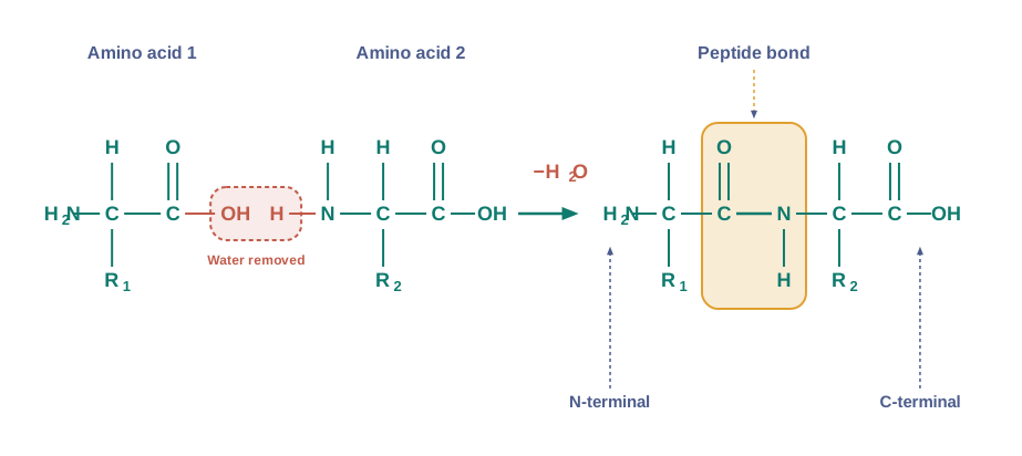
The chemical reaction that joins them is illustrated in Fig. 9.2 later in the text, but the structural explanation sits here in the cell’s physical organisation.
The answer lies in how lipids are arranged inside the cell. Lipids are not present only as free single molecules; they are also organised into larger structures such as the cell membrane and other internal membranes. When we grind a tissue to analyse it, we tear apart the cell’s structure. The membranes break into pieces, and these pieces reseal into tiny closed sacs called vesicles, which are not water-soluble.
Because these membrane vesicles will not dissolve, they get separated along with the acid-insoluble pool during the experiment, and so they land in the macromolecular fraction. In other words, lipids are swept into the macromolecular fraction because of the water-insoluble membrane structures they form — not because each lipid molecule is truly large. Strictly speaking, then, lipids are not macromolecules.
Q2. A drop of fat is small and light, yet biologists file it under “big molecules” (macromolecules). How can something so small be counted as big?
A. A single lipid molecule really is small (under about 800 Da). But in a cell, lipids are packed into membranes. When the tissue is ground up, these membranes break into water-insoluble vesicles that settle into the acid-insoluble (macromolecular) fraction. So lipids get grouped with the macromolecules by the way the experiment separates things — even though, molecule for molecule, they are not truly large.
The Two Fractions, Read Together
These two fractions each correspond to a part of the cell. The acid-soluble pool represents, roughly, the composition of the cytoplasm — the fluid material that fills the cell. The macromolecules from the cytoplasm and the organelles (the tiny working structures inside a cell) make up the acid-insoluble fraction. Put together, the two fractions represent the entire chemical composition of a living tissue or organism.
Water: the Most Abundant Molecule
Finally, if we set aside size for a moment and simply ask which chemicals are most abundant in a living tissue, arranging them class by class, one result stands out clearly: water is by far the most abundant chemical in living organisms, as Table 9.4 shows.
Table 9.4 Average Composition of Cells
| Water |
70–90 |
| Proteins |
10–15 |
| Carbohydrates |
3 |
| Lipids |
2 |
| Nucleic acids |
5–7 |
| Ions |
1 |
what Table 9.4 tells us about a cell The right-hand column gives the chemical composition breakdown. The single biggest entry, by a wide margin, is water at 70–90% — so most of a living cell is, by mass, simply water. Of the biomolecules, proteins lead (10–15%), followed by nucleic acids and then carbohydrates and lipids, with ions making up the smallest share. Reading the table this way reminds us that “the chemistry of life” happens overwhelmingly in a watery medium.
9.5 Polysaccharides
Polysaccharides: Long Chains of Sugars
Besides proteins and nucleic acids, the acid-insoluble pellet holds another class of macromolecules — the polysaccharides, which are a kind of carbohydrate. Polysaccharides are long chains of sugars. It helps to picture them literally as threads, like a cotton thread, in which the individual “fibres” of the thread are single sugar units.
Each single sugar unit is called a monosaccharide — the simplest kind of carbohydrate, such as glucose or fructose. A polysaccharide is built by joining many of these monosaccharide building blocks into a long chain.
A polysaccharide is a macromolecule made of many monosaccharide (single-sugar) units joined together into a long chain.
Storage and Structural Polysaccharides
Different polysaccharides are built from different monosaccharides, and this gives them very different roles.
Cellulose is a polymer made of only one type of monosaccharide — glucose. Because it is built from a single kind of monomer, cellulose is a homopolymer. Starch is a variant of the same idea; it too is built from glucose, but it serves as a store house of energy in plant tissues. Animals keep a similar energy store in the form of glycogen. And inulin is a polymer of a different sugar altogether — fructose.
Storage vs Structural polysaccharides | Feature | Cellulose | Starch | Glycogen | Inulin | |—|—|—|—|—| | Monosaccharide unit | Glucose | Glucose | Glucose | Fructose | | Found in | Plant cell walls | Plant tissues | Animal tissues | Some plants | | Main role | Structural | Energy storage (plants) | Energy storage (animals) | Storage (fructose) | | Polymer type | Homopolymer | — | Branched | — |
Reading a Polysaccharide Chain
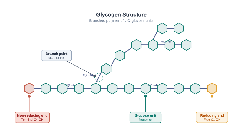
A polysaccharide chain has two chemically different ends. Taking glycogen as an example, the right end of the chain is called the reducing end and the left end is called the non-reducing end. The chain is also not always a simple straight line — glycogen, for instance, carries branches, shown as a cartoon in Fig. 9.4.
The shape a polysaccharide takes matters too. Starch coils up into helical secondary structures — the chain twists like a spring. This coiling has a striking consequence: within the hollow of the helix, starch can trap iodine (\(\text{I}_2\)) molecules, and the resulting starch–iodine complex is deep blue in colour. Cellulose, by contrast, does not form these complex helices, so it cannot hold \(\text{I}_2\) and gives no blue colour.
the starch helix (starch chain coils into a helix → the coil forms a hollow tube → \(\text{I}_2\) molecules slip inside and are held → a blue-coloured starch–iodine complex forms; cellulose, having no such helix, has nowhere to trap \(\text{I}_2\) and stays colourless.)
This is the basis of the everyday iodine test for starch. Add a drop of iodine solution to a food and a blue-black colour means starch is present — which is why a slice of potato or bread turns blue-black with iodine, but a strip of filter paper (cellulose) does not. The same test is used in school laboratories and in food testing to detect starch quickly.
Complex Polysaccharides
Cellulose is not just a laboratory curiosity — it is everywhere around us. Plant cell walls are made of cellulose, and the paper made from plant pulp, as well as cotton fibre, is cellulosic (made of cellulose).
Nature also builds more complex polysaccharides. Instead of plain glucose, these use amino-sugars and chemically modified sugars as their building blocks, such as glucosamine and N-acetyl galactosamine. A well-known example is chitin, the tough polysaccharide that forms the exoskeleton (the hard external skeleton) of arthropods such as insects and crabs. Despite being “complex,” these polysaccharides are mostly homopolymers — long chains built from one repeating modified-sugar unit.
9.7 Structure of Proteins
What “Structure” Means for a Protein
Proteins, as we saw in Section 9.4, are heteropolymers — strings of amino acids. But the word “structure” means different things to different scientists. In inorganic chemistry, “structure” usually means just the molecular formula (for example \(\text{NaCl}\) or \(\text{MgCl}_2\)). Organic chemists draw a two-dimensional picture of the molecule (as for benzene or naphthalene). Physicists imagine the full three-dimensional shape.
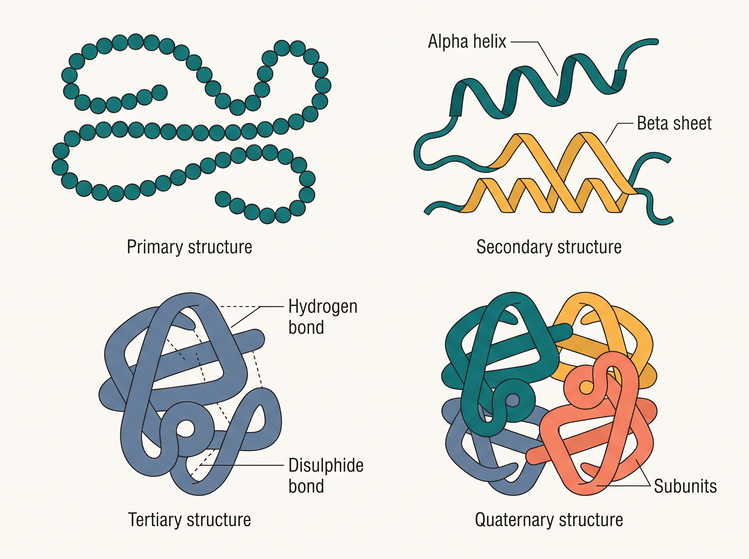
Biologists go one step further: they describe a protein’s structure at four levels — primary, secondary, tertiary and quaternary. These four levels, shown together in Fig. 9.3, describe the same protein from its simplest sequence up to its complete assembled shape.
Primary Structure: the Sequence
The primary structure of a protein is its sequence of amino acids — the positional information telling us which amino acid comes first, which is second, and so on along the chain (Fig. 9.3a). We imagine the protein as a line: the left end is the first amino acid and the right end is the last.
The primary structure of a protein is the sequence (positional order) of amino acids in its polypeptide chain.
The two ends of the chain have special names. The first amino acid, at the start, is the N-terminal amino acid (it keeps a free amino group), and the last amino acid, at the end, is the C-terminal amino acid (it keeps a free carboxyl group).
N-terminal vs C-terminal The N-terminal is the first amino acid of the chain and carries a free amino (\(-\text{NH}_2\), hence “N”) group. The C-terminal is the last amino acid and carries a free carboxyl (\(-\text{COOH}\), hence “C”) group. Sequences are written and read from N-terminal to C-terminal.
Secondary Structure: Local Folding
A protein thread does not remain an extended, rigid rod. Parts of it fold up. In some regions the thread twists into a helix — a spiral shape, much like a revolving staircase. Only certain portions of the thread take up this helical form, and in proteins only right-handed helices are found. Other regions fold into different repeating shapes. All of this local folding of the thread is called the secondary structure (Fig. 9.3b).
The secondary structure of a protein is the local folding of the polypeptide chain into regular shapes such as the right-handed helix and the pleated sheet.
Tertiary Structure: the 3-D Shape
Beyond this local folding, the long protein chain also folds back upon itself as a whole, packing together like a hollow woollen ball. This overall folding gives rise to the tertiary structure (Fig. 9.3c), which provides the complete three-dimensional view of the protein.
The tertiary structure of a protein is the overall three-dimensional shape produced when the whole chain folds upon itself.
The tertiary structure is not just a matter of appearance — it is absolutely necessary for the many biological activities of proteins. A protein can only do its job if it is folded into its correct 3-D shape.
the tertiary fold of a protein (the whole chain folds into a specific 3-D shape → this shape creates precisely arranged pockets and surfaces → these give the protein its ability to bind other molecules and act, for example as an enzyme’s active site → so the folded shape is the function; unfold it, and the protein stops working.)
Quaternary Structure: Assembling Subunits
Some proteins are not a single chain at all, but an assembly of more than one polypeptide, each called a subunit. The way these separate folded subunits are arranged with respect to one another — for example as a linear string of spheres, or spheres stacked into a cube or plate — is the protein’s architecture, called its quaternary structure (Fig. 9.3d).
The quaternary structure of a protein is the arrangement of two or more folded polypeptide subunits with respect to each other in a multi-subunit protein.
A classic example is adult human haemoglobin (Hb), the oxygen-carrying protein of our blood. It consists of 4 subunits, of which two are identical to each other: two subunits of the α (alpha) type and two of the β (beta) type come together to form one haemoglobin molecule.
Just how much the sequence matters is shown by sickle-cell anaemia. A change in a single amino acid in the β subunit of haemoglobin alters the protein’s shape, making red blood cells collapse into a sickle (crescent) form that cannot carry oxygen properly. One small edit in the primary structure ripples all the way up to change the whole molecule’s behaviour — a striking reminder that a protein’s higher structure, and its function, all rest on its amino-acid sequence.
where protein structure sits Zoom in: the primary structure is just a sequence of amino-acid molecules. Zoom out: fold that sequence and it becomes a working 3-D protein; assemble several such proteins and you get a multi-subunit machine like haemoglobin — which in turn is packed inside red blood cells, tissues and, ultimately, the whole organism.
9.8 Enzymes
Enzymes Are (Mostly) Proteins
Almost all enzymes are proteins. An enzyme is a biological catalyst — a substance that greatly speeds up a chemical reaction in a living cell without itself being used up in the process. Since enzymes are proteins, everything we learned about protein structure applies to them.
An enzyme is a biological catalyst (in most cases a protein) that speeds up a specific chemical reaction in a living organism without being consumed by it.
There is one interesting exception. A few nucleic acids can also behave like enzymes and catalyse reactions. These catalytic nucleic acids are called ribozymes.
A ribozyme is a nucleic acid (RNA) that can act as an enzyme, catalysing a chemical reaction.
The Active Site
An enzyme, being a protein, can be drawn as a simple line diagram, and like any protein it has a primary structure (its amino-acid sequence), as well as secondary and tertiary structures.
The tertiary structure is the key to how an enzyme works. When the chain folds upon itself, it criss-crosses so that many crevices and pockets are formed on the enzyme’s surface. One such pocket is very special: it is the active site.
The active site of an enzyme is a crevice or pocket into which the substrate fits.
The substrate is simply the molecule on which the enzyme acts. Because the substrate fits neatly into the active site, the enzyme can, through this site, catalyse its reaction at a very high rate.
the active site (the enzyme’s chain folds into a specific tertiary shape → this fold creates a pocket, the active site, shaped to fit one kind of substrate → the substrate slots in and the reaction is speeded up enormously → so the enzyme’s folded shape directly enables its catalytic power.)
Enzymes versus Inorganic Catalysts
Enzyme catalysts differ from ordinary inorganic catalysts in many ways, but one difference stands out. Inorganic catalysts work efficiently at high temperatures and high pressures. Enzymes, being proteins, are the opposite — they are damaged at high temperatures (say, above about 40°C), because heat destroys their delicate folded shape.
Enzyme (biological) catalyst vs Inorganic catalyst | Feature | Enzyme catalyst | Inorganic catalyst | |—|—|—| | Chemical nature | Usually a protein | Non-living chemical | | Favoured conditions | Mild (around body temperature) | High temperature and pressure | | Effect of high heat | Gets damaged (above ~40°C) | Works efficiently |
Enzymes from Extreme Heat
There is a remarkable exception here too. Some organisms normally live in extremely hot places — such as deep-sea hot vents and sulphur springs. The enzymes isolated from these organisms are unusually stable and keep their catalytic power even at high temperatures, up to about 80°–90°C. This thermal stability is a prized quality of enzymes taken from such heat-loving, or thermophilic, organisms.
This thermal stability turned out to be extraordinarily useful. From a bacterium living in a hot spring, scientists obtained a heat-stable enzyme (Taq polymerase) that does not fall apart at high temperature. This enzyme became the engine of the polymerase chain reaction (PCR) — the technique used to copy DNA millions of times over, which today drives everything from disease diagnosis to forensic testing. An enzyme evolved for life in scalding water became a cornerstone of modern biotechnology.
9.8.1 Chemical Reactions
To understand how enzymes work, we must first be clear about what a chemical reaction actually is.
Physical Changes and Chemical Reactions
Chemical compounds can undergo two broad kinds of change. A physical change is a change in shape without any breaking of chemical bonds — this is a physical process. A change in the state of matter is another physical process: when ice melts into water, or water turns into vapour, no bonds are broken, so these too are physical changes.
A chemical reaction is different. Here, old bonds are broken and new bonds are formed as one set of substances is transformed into another.
A chemical reaction is a change in which chemical bonds are broken and new bonds are formed, transforming the starting substances into new ones.
Physical change vs Chemical reaction In a physical change (melting, boiling, change of shape), no chemical bonds are broken — the substance is still the same substance. In a chemical reaction, bonds are broken and new ones formed, producing a genuinely new substance.
An example of an inorganic chemical reaction is: \[\text{Ba(OH)}_2 + \text{H}_2\text{SO}_4 \rightarrow \text{BaSO}_4 + 2\text{H}_2\text{O}\]
An organic chemical reaction is the hydrolysis of starch into glucose — hydrolysis meaning the breaking of a molecule by adding water.
The Rate of a Reaction
The rate of any physical or chemical process is the amount of product formed per unit time. It can be written as: \[\text{rate} = \dfrac{\delta P}{\delta t}\]
Here \(\delta P\) is the small amount of product formed and \(\delta t\) is the small interval of time taken. When the direction of the process is also specified, the rate may be called the velocity.
The rate of a reaction is the amount of product formed per unit time.
Rates are influenced by several factors, temperature among them. A useful rule of thumb is that the rate roughly doubles, or falls to half, for every 10°C change in temperature, in either direction.
The Power of a Catalyst
Reactions that are catalysed proceed at rates vastly higher than the same reactions left uncatalysed. This is especially dramatic for enzyme-catalysed reactions. Consider the reaction in which carbon dioxide and water form carbonic acid: \[\text{CO}_2 + \text{H}_2\text{O} \; \overset{\text{carbonic anhydrase}}{\rightleftharpoons} \; \text{H}_2\text{CO}_3\]
Written out, this is: carbon dioxide + water \(\rightleftharpoons\) carbonic acid.
Without any enzyme, this reaction is painfully slow — only about 200 molecules of \(\text{H}_2\text{CO}_3\) are formed in an hour. But with the enzyme carbonic anhydrase, present in the cell’s cytoplasm, the reaction races along, forming about 600,000 molecules every second. The enzyme has speeded the reaction up by roughly 10 million times. The power of enzymes is incredible indeed.
comparing the reaction with and without the enzyme Compare the two figures carefully: 200 molecules per hour without the enzyme, against 600,000 molecules per second with it. Because one is measured per hour and the other per second, the true difference is even larger than the raw numbers suggest — it works out to an acceleration of about ten-million-fold. The data make vivid just how enormously a single enzyme can boost a reaction.
9.8.2 How do Enzymes bring about such High Rates of Chemical Conversions?
To answer this, we need to look at enzymes a little more closely. We already understand the idea of the active site. A chemical or metabolic conversion is simply a reaction, and the chemical that gets converted into a product is called the substrate.
Substrate, Product and the ES Complex
An enzyme, then, is a protein with a three-dimensional structure (including an active site) that converts a substrate, S, into a product, P. We can write this simply as: \[\text{S} \rightarrow \text{P}\]
A substrate is the chemical (reactant) on which an enzyme acts and which it converts into a product.
For this to happen, the substrate S must first bind to the enzyme at its active site, inside the cleft or pocket. The substrate has to diffuse (move) towards the active site and settle into it. This binding produces, without exception, an enzyme–substrate complex, written as the ES complex (here E stands for enzyme). This complex is transient — it forms only briefly.
The Transition State
While the substrate is held in the active site, its structure is pushed into a new, short-lived form called the transition state structure. Very soon after the expected breaking and making of bonds is completed, the product is released from the active site. In other words, the structure of the substrate is transformed, by way of the transition state, into the structure of the product. The whole trip is easiest to follow step by step, as in FC 9.5.
a substrate becoming a product 1. The substrate S diffuses towards the enzyme and binds at the active site. 2. An enzyme–substrate (ES) complex forms — this is transient. 3. Held in the active site, the substrate is bent into the high-energy transition state structure. 4. Bonds are broken and new bonds made. 5. The finished product P is released, and the enzyme is free again.
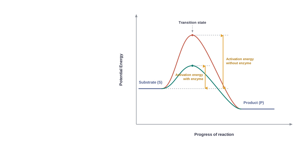
Between the stable substrate and the stable product there may be many “altered structural states.” All these in-between states are unstable. Stability here is tied to the energy of a structure: unstable states sit at higher energy. When we draw this out as a graph, it looks like Fig. 9.6.
Activation Energy
On this graph, the y-axis shows the potential energy content, and the x-axis shows the progression of the structural change through the transition state. Two things stand out.
First, the energy difference between S and P. If P lies at a lower energy level than S, the reaction is exothermic — one need not supply energy (by heating) to form the product.
Exothermic vs Endothermic reaction In an exothermic (spontaneous) reaction, the product sits at lower energy than the substrate, so energy is released and no heating is needed. In an endothermic reaction, the product sits at higher energy, so energy must be supplied. Either way, the substrate must still climb over the transition-state peak.
Second — and this is the crucial point — whether the reaction is exothermic and spontaneous or endothermic and energy-requiring, the substrate S must always pass through a much higher energy state, the transition state. The difference in average energy content between S and this transition state is called the activation energy.
Activation energy is the extra energy a substrate must gain to reach the transition state — the energy “hill” that must be climbed before a reaction can proceed.
reading the activation-energy graph (Fig. 9.6) Follow the curve from left to right: the substrate must first go up to the transition-state peak before coming down to the product. The height of that peak above the substrate is the activation energy — the barrier to reaction. Notice the two curves: the peak for the enzyme-catalysed reaction is lower than the peak for the uncatalysed one. A lower peak means a smaller barrier, so the reaction can happen far more easily.
How the Enzyme Helps
The enzyme’s trick is now clear: enzymes bring down this energy barrier, lowering the activation energy and so making the transition of S to P much easier.
Q4. Some enzymes make a reaction happen millions of times faster than it would on its own. How can a single protein speed things up by so much?
A. Every reaction must climb an energy hill — the activation energy — to reach the transition state. An enzyme binds the substrate in its active site and lowers that hill, so the substrate reaches the transition state far more easily. With a smaller barrier to cross, vastly more molecules make it over per second — which is how an enzyme like carbonic anhydrase can accelerate a reaction millions of times.
9.8.3 Nature of Enzyme Action
The Enzyme–Substrate Complex
Every enzyme (E) carries a substrate-binding site — the active site — within its molecule. When the substrate (S) binds there, a highly reactive enzyme–substrate complex (ES) is produced. This complex is short-lived. It breaks apart into the product(s) P and the unchanged enzyme, passing through an intermediate stage called the enzyme–product complex (EP).
The formation of the ES complex is essential for catalysis; without it, the enzyme cannot act. The whole event can be written as: \[\text{E} + \text{S} \rightleftharpoons \text{ES} \rightarrow \text{EP} \rightarrow \text{E} + \text{P}\]
Notice that the enzyme (E) appears on both sides of the equation — it goes in and comes out unchanged, ready to be used again. This is exactly what we expect of a catalyst.
The enzyme–substrate (ES) complex is the short-lived, reactive combination formed when a substrate binds to an enzyme’s active site; its formation is essential for catalysis.
The Catalytic Cycle
The action of an enzyme can be described as a repeating catalytic cycle with four steps:
- First, the substrate binds to the active site of the enzyme, fitting into it.
- This binding induces the enzyme to alter its shape, so that it fits more tightly around the substrate.
- The active site, now held close around the substrate, breaks the chemical bonds of the substrate, and a new enzyme–product complex is formed.
- The enzyme releases the products of the reaction, and the now-free enzyme is ready to bind another substrate molecule and run through the cycle once again.
the “induced fit” of the active site (step 2) (the substrate enters the active site → its binding induces the enzyme to change shape → the active site closes more snugly around the substrate → this tight grip strains the substrate’s bonds and helps break them → so the enzyme is not a rigid lock but flexes to grip and act on its substrate.)
9.8.4 Factors Affecting Enzyme Activity
The activity of an enzyme depends on its delicate three-dimensional shape, so anything that alters the tertiary structure of the protein will affect how well the enzyme works. The main such factors are temperature, pH, a change in substrate concentration, and the binding of specific chemicals that regulate the enzyme’s activity.
Temperature and pH
Enzymes generally work only within a narrow range of temperature and pH, as Fig. 9.7 shows. Each enzyme has a particular temperature and a particular pH at which it is most active — its optimum temperature and optimum pH. On either side of this optimum — both below it and above it — the enzyme’s activity declines.
The optimum temperature (or optimum pH) of an enzyme is the temperature (or pH) at which it shows its highest activity.
But low and high temperatures affect an enzyme in very different ways. A low temperature simply preserves the enzyme in a temporarily inactive state — warm it again and it recovers. A high temperature, however, destroys enzyme activity, because the heat denatures the protein — that is, it unravels the enzyme’s folded structure so that it can no longer function.
Low temperature vs High temperature on enzymes Low temperature makes an enzyme temporarily inactive — the enzyme is preserved and can work again on warming. High temperature denatures the enzyme — its folded shape is destroyed and the loss of activity is not recovered by cooling.
Q5. Why does a high fever, or a very hot day, threaten to damage the proteins and enzymes inside our body?
A. Enzymes work only near their optimum temperature, because their function depends on a precisely folded (tertiary) shape. Excess heat denatures proteins — it unravels that fold. Once denatured, an enzyme can no longer bind its substrate, so its activity is lost. This is why a very high fever is dangerous: it risks denaturing the body’s own enzymes and proteins.
Concentration of Substrate
If we steadily increase the substrate concentration, the velocity of the enzyme-catalysed reaction rises at first. But it does not rise forever. The reaction eventually reaches a maximum velocity, written \(V_{max}\), beyond which adding more substrate makes no difference (Fig. 9.7).
Why does the velocity level off? Because the enzyme molecules are far fewer than the substrate molecules. Once every enzyme’s active site is occupied — a state called saturation — there are simply no free enzyme molecules left to bind the extra substrate, so the rate cannot climb any higher.
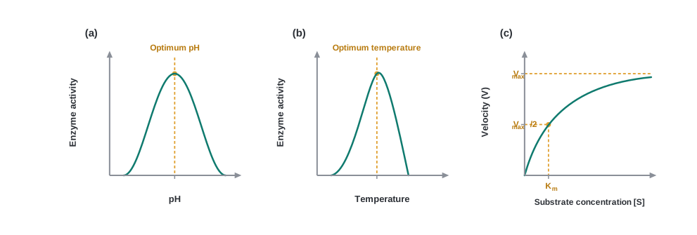
reading the three graphs of Fig. 9.7 Graphs (a) and (b) are bell-shaped: activity climbs to a peak at the optimum pH or temperature and falls away on both sides — telling us each enzyme has one “best” condition. Graph (c) is different: velocity rises steeply with substrate concentration, then bends over and flattens into a plateau at \(V_{max}\), the sign that the enzyme is saturated. The point on the [S] axis where velocity is exactly half of \(V_{max}\) is marked \(K_m\). Together the graphs show that enzyme activity is not limitless — it is bounded both by conditions and by the amount of enzyme available.
Inhibition of Enzymes
An enzyme’s activity is also sensitive to specific chemicals that bind to it. When the binding of such a chemical shuts off enzyme activity, the process is called inhibition, and the chemical responsible is an inhibitor.
Inhibition is the shutting off of enzyme activity by a chemical (an inhibitor) that binds to the enzyme.
One important type is the competitive inhibitor. Here the inhibitor closely resembles the substrate in its molecular structure. Because it looks so much like the real substrate, it competes with the substrate for the enzyme’s substrate-binding (active) site. As a result the substrate cannot bind, and the enzyme’s action declines.
A competitive inhibitor is a molecule structurally similar to the substrate that competes with it for the active site, thereby reducing enzyme activity.
how a competitive inhibitor works (the inhibitor’s shape closely mimics the substrate → it fits into the same active site → while the inhibitor sits there, the real substrate is locked out → fewer substrate molecules are converted → so a molecule that merely looks like the substrate can throttle the reaction.)
A classic example is the inhibition of the enzyme succinic dehydrogenase by malonate, which closely resembles the enzyme’s true substrate, succinate, in structure.
Because competitive inhibitors can selectively jam a specific enzyme, they are often used to control bacterial pathogens. The sulpha drugs, for instance, work as competitive inhibitors: they mimic a molecule that bacteria need to make an essential vitamin, so they block a key bacterial enzyme and halt the microbe’s growth — an early and famous class of antibacterial medicines built on exactly this principle.
9.8.5 Classification and Nomenclature of Enzymes
Thousands of enzymes have been discovered, isolated and studied. To make sense of so many, most of them have been sorted into groups based on the type of reaction they catalyse. Under this scheme, enzymes are divided into 6 classes, each with 4 to 13 subclasses, and each enzyme is then named by a four-digit number.
The four-digit number is the enzyme’s “EC number” (Enzyme Commission number). Reading left to right, the digits narrow it down step by step — the first digit gives the main class (1 to 6), and the following digits give the subclass, the sub-subclass, and finally the individual enzyme. It is rather like a postal code that pinpoints one enzyme among thousands.
The Six Classes of Enzymes
Oxidoreductases / dehydrogenases catalyse oxidoreduction — that is, oxidation–reduction reactions — between two substrates, S and S′: \[\text{S reduced} + \text{S' oxidised} \rightarrow \text{S oxidised} + \text{S' reduced}\]
Transferases catalyse the transfer of a group G (other than hydrogen) from one substrate to another, between a pair of substrates S and S′: \[\text{S-G} + \text{S'} \rightarrow \text{S} + \text{S'-G}\]
Hydrolases catalyse the hydrolysis (breaking with the addition of water) of ester, ether, peptide, glycosidic, C–C, C–halide or P–N bonds.
Lyases catalyse the removal of groups from substrates by mechanisms other than hydrolysis, leaving behind a double bond: \[\text{X-C-C-Y} \rightarrow \text{X-Y} + \text{C}=\text{C}\]
Isomerases include all enzymes that catalyse the inter-conversion of isomers — optical, geometric or positional. (Isomers are molecules with the same formula but different arrangements of atoms.)
Ligases catalyse the linking together (joining) of two compounds — for example, enzymes that catalyse the joining of C–O, C–S, C–N or P–O bonds.
The six classes of enzymes | Class | What it catalyses | Example / bonds | |—|—|—| | Oxidoreductases | Oxidation–reduction between two substrates | S reduced + S′ oxidised → S oxidised + S′ reduced | | Transferases | Transfer of a group (not H) between substrates | S–G + S′ → S + S′–G | | Hydrolases | Hydrolysis of bonds using water | Ester, ether, peptide, glycosidic, C–C, C–halide, P–N | | Lyases | Removal of groups (not by hydrolysis), leaving a double bond | X–C–C–Y → X–Y + C=C | | Isomerases | Inter-conversion of isomers | Optical, geometric, positional | | Ligases | Joining two compounds together | C–O, C–S, C–N, P–O bonds |
Hydrolase vs Lyase Both split a substrate, but the method differs. A hydrolase breaks a bond by adding water (hydrolysis). A lyase removes a group without using water, by a different mechanism, and typically leaves a double bond behind.
the six classes in order Take the first letters — Oxidoreductases, Transferases, Hydrolases, Lyases, Isomerases, Ligases — and remember: “Over The Hill Lies India’s Land” (O-T-H-L-I-L).
9.8.6 Co-factors
When an Enzyme Needs a Helper
Enzymes are made of one or several polypeptide chains. In many cases, though, the protein part alone is not enough: a non-protein constituent, called a co-factor, must be bound to the enzyme to make it catalytically active.
A co-factor is a non-protein constituent bound to an enzyme that is needed to make the enzyme catalytically active.
When cofactors are involved, the protein portion of the enzyme is given a special name — the apoenzyme. On its own the apoenzyme is inactive; only when its cofactor is in place does the complete, working enzyme (apoenzyme + cofactor) exist.
The apoenzyme is the protein part of an enzyme, which becomes catalytically active only when its cofactor is bound.
The complete active enzyme — apoenzyme together with its cofactor — is commonly called the holoenzyme. Strip the cofactor away and only the inactive apoenzyme remains.
Three kinds of cofactors can be identified, as shown in FC 9.7: prosthetic groups, co-enzymes and metal ions.
The Three Kinds of Cofactors
Prosthetic groups are organic compounds. What sets them apart from the other cofactors is that they are tightly bound to the apoenzyme. For example, in the enzymes peroxidase and catalase, which catalyse the breakdown of hydrogen peroxide into water and oxygen, the prosthetic group is haem, and it forms part of the active site: \[2\text{H}_2\text{O}_2 \rightarrow 2\text{H}_2\text{O} + \text{O}_2\]
Co-enzymes are also organic compounds, but their association with the apoenzyme is only transient, usually happening just during the course of catalysis. A single co-enzyme can serve as a cofactor for a number of different enzyme-catalysed reactions. Importantly, the essential chemical components of many co-enzymes are vitamins — for example, the co-enzymes nicotinamide adenine dinucleotide (NAD) and NADP contain the vitamin niacin.
Metal ions are the third kind of cofactor. A number of enzymes need a metal ion to work. The metal ion forms coordination bonds with side chains at the active site and, at the same time, one or more coordination bonds with the substrate — so it acts as a bridge holding the substrate in place. For example, zinc is a cofactor for the protein-digesting enzyme carboxypeptidase.
The three kinds of cofactors | Cofactor type | Nature | Binding to apoenzyme | Example | |—|—|—|—| | Prosthetic group | Organic | Tightly (permanently) bound | Haem in catalase / peroxidase | | Co-enzyme | Organic | Transient (during catalysis) | NAD, NADP (contain niacin) | | Metal ion | Inorganic | Coordination bonds | Zinc in carboxypeptidase |
Prosthetic group vs Co-enzyme Both are organic cofactors, so they are easily confused. The difference is how firmly they stick: a prosthetic group stays tightly bound to the enzyme all the time, whereas a co-enzyme binds only transiently, joining in just for the moment of catalysis and then leaving.
a metal-ion cofactor (the metal ion forms coordination bonds with the enzyme’s active site and with the substrate at the same time → it locks the substrate into the correct position against the active site → correctly positioned, the substrate’s bonds can be acted upon → so the metal ion works as a bridge that makes catalysis possible.)
Since many co-enzymes are built from vitamins, a shortage of a vitamin in the diet cripples the enzymes that depend on it. A lack of niacin, the vitamin in NAD and NADP, leads to the deficiency disease pellagra. This is one reason vitamins are essential in our food — they are not fuel, but the raw material for co-enzymes that keep our enzymes working.
Why Cofactors Matter
There is a simple, telling test of how important cofactors are: catalytic activity is lost the moment the cofactor is removed from the enzyme. This shows that cofactors play a crucial role in an enzyme’s catalytic activity — the apoenzyme simply cannot do the job alone.