Chapter 12: Respiration in Plants
In the late 1890s, a German chemist named Eduard Buchner set out to test an idea that most scientists of his time thought was impossible. Everyone knew that living yeast could turn sugar into alcohol — this is fermentation. But many believed the change needed some mysterious “life force” that only a whole, living cell could provide. Buchner disagreed.
He ground yeast cells with fine sand until they were completely crushed, then squeezed out a clear, lifeless juice. To keep the juice from spoiling, he mixed in a large amount of sugar. To his astonishment, the dead extract began to bubble: it was fermenting the sugar into alcohol and carbon dioxide, exactly as living yeast would have done.
No living cell was needed — only the molecular machinery from inside it. Buchner had shown that the breakdown of food to release energy is not magic but ordinary chemistry, carried out by proteins called enzymes. For this insight he was awarded a Nobel Prize in 1907, and biology gained one of its central ideas: respiration is a set of carefully controlled chemical reactions happening inside every living cell — in yeast, in animals, and, as you will see, in every non-green cell of a plant.
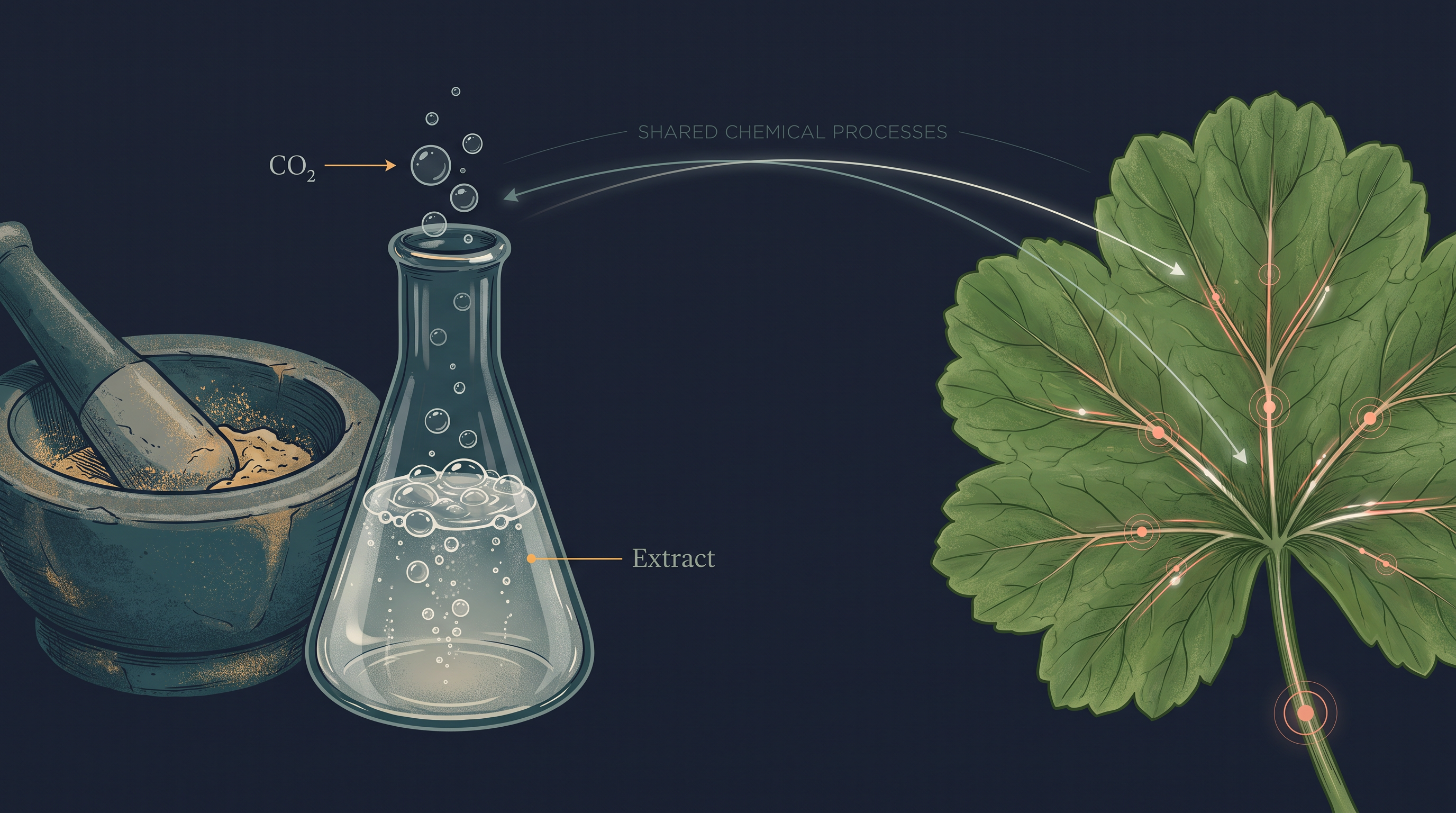
Fig. 12.0 – Cell-free fermentation and plant respiration.
By the end of this chapter, you will be able to answer each of these questions for yourself.
12.1 Do Plants Breathe?
Why Every Living Cell Needs Energy
Every living organism needs a constant supply of energy. Absorbing nutrients, transporting materials, moving, growing, reproducing — even the moment-to-moment maintenance of being alive — all of it runs on energy. So a natural question is: where does this energy come from, and how does a cell actually get it out of food?
All the energy used for life processes is released by the oxidation of certain large molecules that we loosely call “food”. Oxidation here means the controlled breaking down of a molecule with the removal of electrons (and usually hydrogen), a process that releases the chemical energy locked in its bonds.
Green plants and cyanobacteria make their own food. Through photosynthesis they capture light energy and store it as chemical energy in the bonds of carbohydrates such as glucose, sucrose and starch. But it is important to notice that not every cell in a green plant photosynthesises. Only cells that contain chloroplasts — usually those in the outer, light-exposed layers — can trap light. Every other cell, tissue and organ that is non-green cannot make its own food, yet still needs energy. That is why food made in the green parts has to be transported (translocated) to all the non-green parts, where it can be oxidised for energy.
Animals cannot make their own food at all; they are heterotrophic, obtaining food either directly from plants (herbivores) or indirectly (carnivores). Saprophytes such as many fungi feed on dead and decaying matter. Whichever route the food takes, one fact stays true: ultimately, all the food that living things respire traces back to photosynthesis.
What Exactly Is Respiration?
This chapter is about cellular respiration — the way food materials are broken down within the cell to release energy, and how that energy is then trapped to make ATP.
There is a neat division of labour inside a eukaryotic cell. Photosynthesis takes place inside the chloroplasts, while the breakdown of complex molecules to yield energy takes place in the cytoplasm and in the mitochondria. Both the mitochondria and this energy-releasing machinery are found only in eukaryotes.
When the carbon–carbon (C–C) bonds of complex compounds are broken by oxidation inside cells, a considerable amount of energy is released. This is what we call respiration.
The compounds that are oxidised during respiration are called respiratory substrates. Carbohydrates are the usual substrate, but proteins, fats and even organic acids can serve as respiratory substrates in some plants under certain conditions.
A key point is that a cell does not release all this energy at once. If it did, most of the energy would escape uselessly as heat. Instead, the energy is released in a series of slow, step-wise reactions, each controlled by a specific enzyme, and it is captured as chemical energy in the form of ATP. So the energy from oxidation is not used directly; it is first used to make ATP, and ATP is then broken down whenever and wherever energy is needed. For this reason ATP is called the energy currency of the cell — just as money can be “spent” on many different things, ATP can power many different energy-requiring reactions. Meanwhile, the carbon skeletons left over during respiration are not wasted either: they serve as raw material (precursors) for building other molecules the cell needs.
Do Plants Breathe? The Honest Answer
The answer to the chapter’s title question is not quite straightforward. Yes — plants do require O₂ for respiration to occur, and they do give out CO₂. So plants must have some arrangement that keeps O₂ available to their cells.
Unlike animals, plants have no specialised organs for gaseous exchange. Instead they use two kinds of natural openings: stomata (tiny adjustable pores, mainly in leaves and young stems) and lenticels (loose openings in the bark of woody stems and roots). We can see how plants manage without breathing organs by looking at three simple reasons, illustrated in Fig. 12.1a.
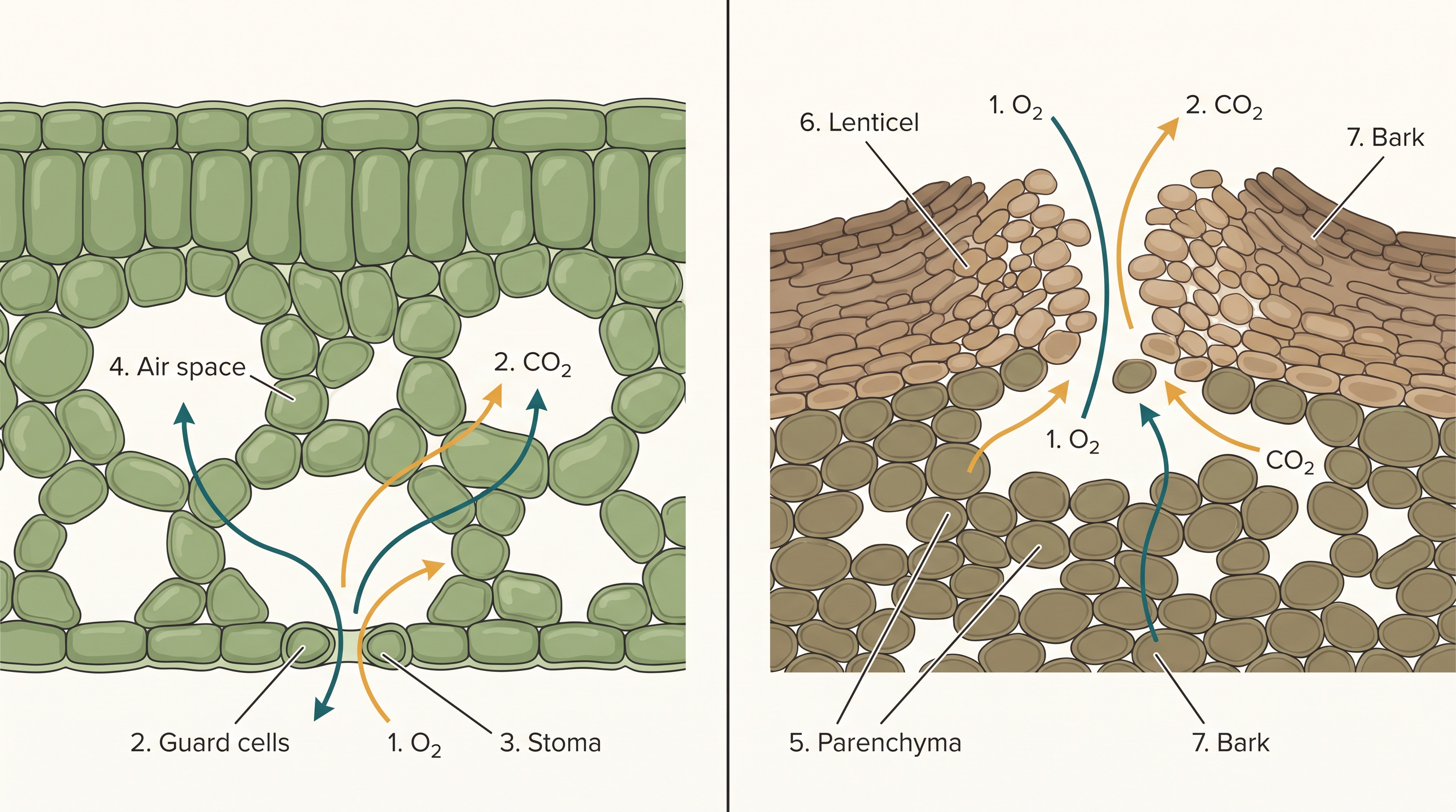
Fig. 12.1a – Gas exchange structures in a leaf and stem.
First, each plant part looks after its own gas-exchange needs. There is very little transport of gases from one part of a plant to another; a leaf handles its own exchange, a root handles its own, and so on.
Second, plants do not make heavy demands for gas exchange. Roots, stems and leaves respire far more slowly than an animal’s tissues do, so their oxygen needs are modest. Large volumes of gas move only during photosynthesis, and each leaf is well adapted to manage its own exchange during those periods. In fact, when a cell is photosynthesising, getting O₂ is no problem at all — the cell is releasing O₂ within itself.
Third, gases never have to travel far inside a plant. Even in a large, bulky plant, every living cell lies quite close to the surface. This is easy to accept for a thin leaf, but what about a thick, woody stem or root? Inside such stems, the living cells are arranged in thin layers just inside and beneath the bark, and the bark carries lenticels for gas exchange. The cells deep in the interior are dead and serve only for mechanical support — so they place no demand on gas supply. As a result, most cells of a plant have at least part of their surface in contact with air. This is further helped by the loose packing of parenchyma cells in leaves, stems and roots, which leaves an interconnected network of air spaces running through the tissue.
Burning vs. Controlled Release
We can now see why respiration is more subtle than simply “burning” food. The complete combustion of glucose gives carbon dioxide and water as end products, releasing energy — but most of that energy escapes as heat:
\[C_6H_{12}O_6 + 6O_2 \rightarrow 6CO_2 + 6H_2O + \text{Energy}\]
Here glucose (\(C_6H_{12}O_6\)) reacts with oxygen to yield six molecules of carbon dioxide, six of water, and energy. If this energy is to be useful, the cell must be able to capture it and use it to build the molecules it needs. Heat alone cannot do that.
So the cell uses a clever strategy: it catabolises (breaks down) glucose in such a way that not all the energy is lost as heat. The trick is to oxidise glucose not in one violent step but in several small steps. Each small step releases a packet of energy that is “just large enough” to be coupled to the making of ATP. How exactly this is achieved is, essentially, the whole story of respiration.
When Oxygen Is Scarce
During respiration, oxygen is used up while carbon dioxide, water and energy are released. The combustion of glucose clearly needs oxygen — but not every cell lives where oxygen is available.
There are good reasons to believe that the very first cells on Earth lived in an atmosphere that had no oxygen at all. Even today, many organisms are adapted to anaerobic conditions (life without oxygen). Some of these are facultative anaerobes, which can manage with or without oxygen; in others the requirement for anaerobic conditions is obligate, meaning they must live without it. Whatever the case, every living organism keeps the enzyme machinery needed to partially oxidise glucose without the help of oxygen.
This partial, oxygen-independent breakdown of glucose to pyruvic acid is called glycolysis — the common starting point of respiration in all living cells, and the subject of the next section.
Flowchart
FC 12.1 – The branching fate of glucose introduced here: Glucose → (glycolysis, no O₂ needed) → Pyruvic acid → if O₂ absent → fermentation (anaerobic); if O₂ present → aerobic respiration (complete oxidation to CO₂ + H₂O).
12.2 Glycolysis
What the Word Means
The word glycolysis comes from two Greek words — glycos, meaning sugar, and lysis, meaning splitting. So the name itself tells us what happens: a sugar molecule is split apart.
Glycolysis is remarkable for being universal. It occurs in the cytoplasm of the cell and is present in all living organisms, from bacteria to human beings. In organisms that live without oxygen (anaerobic organisms), it is the only respiratory process available. In this pathway, glucose undergoes partial oxidation to form two molecules of pyruvic acid.
Where the Glucose Comes From
In plants, the glucose fed into glycolysis is derived from sucrose — the end product of photosynthesis — or from stored carbohydrates such as starch. Sucrose is first broken down into glucose and fructose by the enzyme invertase, and both of these monosaccharides can readily enter the glycolytic pathway.
Once inside, glucose and fructose are phosphorylated — that is, a phosphate group is attached to them. This produces glucose-6-phosphate, a reaction driven by the enzyme hexokinase. Glucose-6-phosphate then rearranges its atoms (a change called isomerisation) to form fructose-6-phosphate. From this point on, the steps for glucose and fructose are identical, so we can simply follow one pathway.
The Ten Reactions, Step by Step
The various steps of glycolysis are shown in Fig. 12.2, and the same sequence is condensed with its energy events in FC 12.2. Altogether, a chain of ten reactions, each under the control of a different enzyme, converts glucose into pyruvate. As you study the pathway, watch carefully for the exact steps where ATP is used up, where ATP is made, and where NADH + H⁺ is formed.
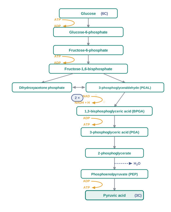
Fig. 12.2 – The steps of glycolysis.
Flowchart
FC 12.2 – Glycolysis with energy events: Glucose →(ATP used, hexokinase) Glucose-6-phosphate → Fructose-6-phosphate →(ATP used) Fructose-1,6-bisphosphate → splits into PGAL + DHAP (DHAP → PGAL) → 2×PGAL →(NAD⁺ → NADH+H⁺) 2×BPGA →(ATP made) 2×PGA → 2×2-phosphoglycerate →(−H₂O) 2×PEP →(ATP made) 2×Pyruvic acid.
Two of the early steps consume energy. ATP is used at two points: first, in the conversion of glucose into glucose-6-phosphate, and second, in the conversion of fructose-6-phosphate to fructose-1,6-bisphosphate. So the cell begins by investing two ATP molecules.
Next comes the “splitting” that gives glycolysis its name. Fructose-1,6-bisphosphate, a six-carbon molecule, is split into two three-carbon molecules: dihydroxyacetone phosphate and 3-phosphoglyceraldehyde (PGAL). These two are interconvertible, and the dihydroxyacetone phosphate is steadily converted into PGAL, so in effect two molecules of PGAL move forward — which is why the later steps are counted as “2×”.
There is a single step where NADH + H⁺ is formed. Here, PGAL is oxidised and, with the addition of an inorganic phosphate, is converted into 1,3-bisphosphoglycerate (BPGA). During this oxidation, two hydrogen atoms (called redox equivalents) are removed from PGAL and handed to a molecule of NAD⁺. NAD⁺ is a coenzyme that acts as a hydrogen and electron carrier: when it accepts these two hydrogens it becomes NADH + H⁺, and it stores that captured energy for later use.
Counting the Energy
The pathway now begins to pay back the cell. The conversion of BPGA to 3-phosphoglyceric acid (PGA) is an energy-yielding step, and this energy is captured by forming ATP. After PGA is rearranged to 2-phosphoglycerate and then (by loss of a water molecule) to phosphoenolpyruvate (PEP), a second ATP is synthesised as PEP is converted to pyruvic acid.
The Fate of Pyruvate
Pyruvic acid is the key end product of glycolysis. But what happens to it next? That depends entirely on the needs and circumstances of the cell.
There are three major ways in which different cells handle the pyruvic acid produced by glycolysis: lactic acid fermentation, alcoholic fermentation, and aerobic respiration. The two kinds of fermentation take place under anaerobic conditions in many prokaryotes and unicellular eukaryotes. For the complete oxidation of glucose all the way to CO₂ and H₂O, however, organisms take a different route — the Krebs’ cycle of aerobic respiration, which requires a supply of O₂. The next sections follow each of these fates in turn.
12.3 Fermentation
We ended the last section with three possible fates for pyruvate. Two of them — alcoholic fermentation and lactic acid fermentation — are the anaerobic routes, and both are grouped under the single name fermentation.
Two Roads for Pyruvate
In some organisms, such as yeast, the incomplete oxidation of glucose is carried out anaerobically by a set of reactions in which pyruvic acid is converted to CO₂ and ethanol (an alcohol). Two enzymes drive this — pyruvic acid decarboxylase and alcohol dehydrogenase.
Other organisms take a different road. Some bacteria convert pyruvic acid into lactic acid instead. This same lactic-acid route also operates in our own muscle cells during vigorous exercise: when oxygen cannot be supplied fast enough for normal cellular respiration, pyruvic acid is reduced to lactic acid by the enzyme lactate dehydrogenase. The steps of both pathways are shown in Fig. 12.3, and the branch point is summarised in FC 12.3.
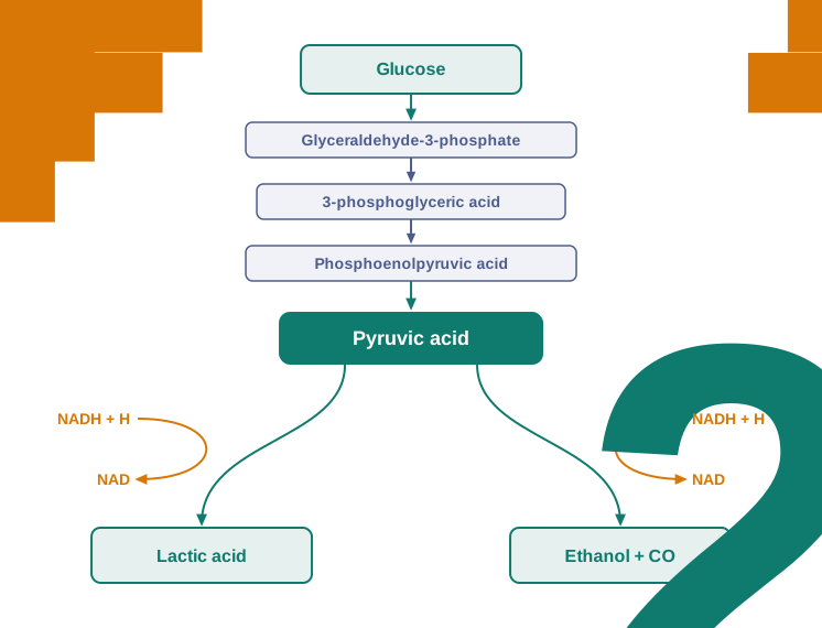
Fig. 12.3 – Major pathways of anaerobic respiration.
Flowchart
FC 12.3 – Fate of pyruvate under anaerobic conditions: Glucose → glycolysis → Pyruvic acid → if yeast/similar microbes → (pyruvic acid decarboxylase, alcohol dehydrogenase) → Ethanol + CO₂; if certain bacteria or exercising muscle → (lactate dehydrogenase) → Lactic acid. In both branches NADH+H⁺ is reoxidised to NAD⁺.
Why NAD⁺ Must Be Recycled
In both routes, the reducing agent is NADH + H⁺, which is reoxidised back to NAD⁺. This detail is easy to overlook but is actually the whole point of fermentation.
A Wasteful, Risky Bargain
Fermentation keeps a cell alive without oxygen, but it is a poor way to extract energy. In both lactic acid and alcohol fermentation, not much energy is released — less than seven per cent of the energy stored in glucose is set free, and not even all of that is trapped in the high-energy bonds of ATP. Most of the glucose’s energy stays locked up in the ethanol or lactic acid that is left behind.
The processes are also hazardous to the organism, because either an acid or an alcohol accumulates. This has a striking limit in yeast: yeasts poison themselves to death when the concentration of alcohol reaches about 13 per cent.
Toward Complete Oxidation
Fermentation leaves most of glucose’s energy untapped. So what is the process by which organisms can completely oxidise glucose and extract the far larger amount of energy needed to make all the ATP that cellular metabolism demands?
In eukaryotes, these steps take place inside the mitochondria and require O₂. This is aerobic respiration — the complete oxidation of organic substances in the presence of oxygen, releasing CO₂, water, and a large amount of the energy stored in the substrate. It is the commonest form of respiration in higher organisms, and it is the subject of the next section.
12.4 Aerobic Respiration
Into the Mitochondria
Aerobic respiration is where the cell finally cashes in the large energy reserve still locked inside glucose. For it to happen, the final product of glycolysis — pyruvate — must first be transported out of the cytoplasm and into the mitochondria, the organelles where the rest of respiration takes place.
The Two Crucial Events
Aerobic respiration comes down to two crucial events. First, pyruvate is completely oxidised by the stepwise removal of all its hydrogen atoms, leaving three molecules of CO₂ (per pyruvate). Second, the electrons carried by those removed hydrogen atoms are passed on to molecular O₂, and this transfer is coupled to the synthesis of ATP.
What is interesting is that these two events happen in two different places. The first — the removal of hydrogen and release of CO₂ — takes place in the matrix (the fluid-filled interior) of the mitochondrion. The second — the handing of electrons to O₂ with ATP synthesis — is located on the inner membrane of the mitochondrion. Fig. 12.4a shows this division of labour.
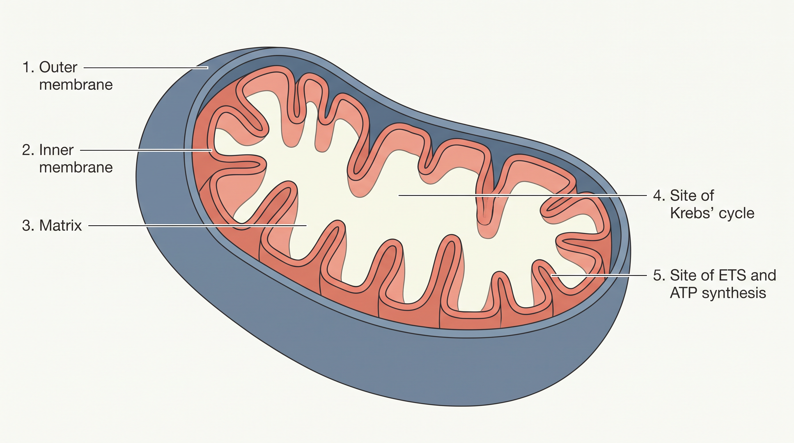
Fig. 12.4a – Structure of a mitochondrion and sites of respiration.
Flowchart
FC 12.4 – Overview of aerobic respiration: Pyruvate (cytosol) → enters mitochondrial matrix → oxidative decarboxylation (pyruvate dehydrogenase) → Acetyl CoA + CO₂ + NADH → Krebs’ cycle (matrix) → NADH + FADH₂ + CO₂ → electron transport system (inner membrane) → electrons to O₂ → H₂O + ATP.
Preparing the Fuel: Pyruvate to Acetyl CoA
Before the cyclic pathway can begin, pyruvate must be “prepared”. Pyruvate — formed by the breakdown of carbohydrates in the cytosol — enters the mitochondrial matrix and there undergoes oxidative decarboxylation. This simply means it is oxidised and, at the same time, loses one carbon as a molecule of CO₂. The reaction is carried out by a complex enzyme called pyruvic dehydrogenase, which needs the help of several coenzymes, including NAD⁺ and Coenzyme A (CoA) — a carrier molecule that picks up the two-carbon fragment that remains:
\[\text{Pyruvic acid} + \text{CoA} + NAD^+ \xrightarrow[\text{pyruvate dehydrogenase}]{Mg^{2+}} \text{Acetyl CoA} + CO_2 + NADH + H^+\]
In this reaction, three-carbon pyruvic acid loses one carbon as CO₂ and the remaining two-carbon acetyl group is attached to Coenzyme A to form acetyl CoA; at the same time NAD⁺ is reduced to NADH + H⁺. During this process, two molecules of NADH are produced from the two molecules of pyruvic acid that came from a single glucose molecule during glycolysis.
The Cycle Named After a Scientist
The acetyl CoA now enters a cyclic pathway — the tricarboxylic acid cycle, more commonly called the Krebs’ cycle.
12.4.1 Tricarboxylic Acid Cycle
How the Cycle Begins
The Krebs’ cycle — also called the tricarboxylic acid (TCA) cycle or citric acid cycle — begins when the two-carbon acetyl group of acetyl CoA joins with a four-carbon molecule called oxaloacetic acid (OAA), together with a molecule of water. This condensation yields the six-carbon citric acid, and is catalysed by the enzyme citrate synthase; the Coenzyme A that carried the acetyl group is released to be used again. The full cycle is shown in Fig. 12.5 and traced as an ordered loop in FC 12.5.
Citric acid is then rearranged (isomerised) into isocitrate, setting the stage for the energy-yielding steps that follow.
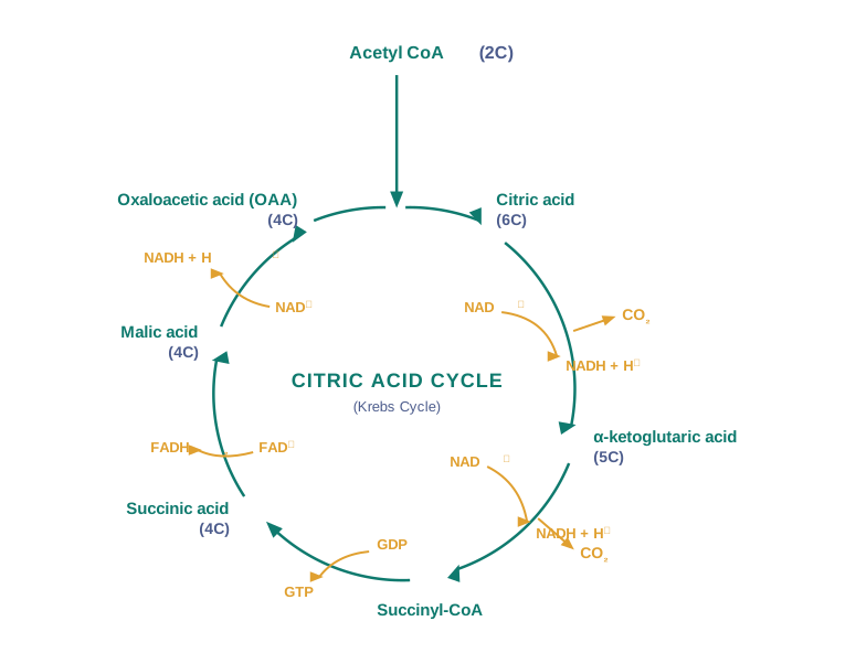
Fig. 12.5 – The citric acid (Krebs’) cycle.
Two Carbons Leave as CO₂
The cycle now passes through two successive steps of decarboxylation — each removing a carbon atom as CO₂. Isocitrate is first converted into the five-carbon α-ketoglutaric acid, and then into succinyl-CoA. With these two CO₂ molecules gone, the carbons that entered as acetyl CoA have been fully accounted for.
In the remaining steps, succinyl-CoA is progressively oxidised back to OAA, allowing the cycle to turn again. During the conversion of succinyl-CoA to succinic acid, a molecule of GTP is synthesised directly.
The GTP formed here is then used in a coupled reaction: GTP is converted to GDP while an ATP is simultaneously made from ADP. This is the cycle’s own small, direct ATP contribution.
Harvesting Electrons
The real prize of the cycle is not this single ATP but the hydrogen atoms (and the electrons they carry) that are stripped from the intermediates. There are three points in the cycle where NAD⁺ is reduced to NADH + H⁺, and one point where FAD⁺ is reduced to FADH₂. Like NAD⁺, FAD⁺ is a coenzyme that acts as a hydrogen and electron carrier; when it accepts two hydrogens it becomes FADH₂. (The single FAD⁺ step occurs as succinic acid is converted, by way of fumaric acid, into malic acid; malic acid is then oxidised to regenerate OAA.)
The Balance So Far
The whole of this phase of respiration, starting from one pyruvate, can be summarised in a single equation:
\[\text{Pyruvic acid} + 4NAD^+ + FAD^+ + 2H_2O + ADP + Pi \xrightarrow{\text{Mitochondrial Matrix}} 3CO_2 + 4NADH + 4H^+ + FADH_2 + ATP\]
Taking stock, we have now broken glucose down completely to CO₂ and have made eight molecules of NADH + H⁺ (two from the oxidation of pyruvate plus six from the two turns of the cycle) and two of FADH₂, besides just two molecules of ATP from the TCA cycle itself.
At this point you might reasonably wonder why respiration has been worth discussing at all — oxygen has not yet entered the picture, and the promised large yield of ATP has not appeared. What, then, is the role of all this NADH + H⁺ and FADH₂? The answer, and the entry of O₂, comes in the next section.
12.4.2 Electron Transport System (ETS) and Oxidative Phosphorylation
Passing Electrons Down the Chain
We now come to the step that finally brings in oxygen and unlocks the bulk of glucose’s energy. The task at this stage is to release and use the energy stored in all that NADH + H⁺ and FADH₂ made earlier. This is done by oxidising them through a chain of carriers and passing the electrons, step by step, to O₂ — which combines with hydrogen to form H₂O.
The pathway through which an electron is handed from one carrier to the next is called the electron transport system (ETS), shown in Fig. 12.6, and it sits in the inner mitochondrial membrane. The ordered path of the electrons is summarised in FC 12.6.
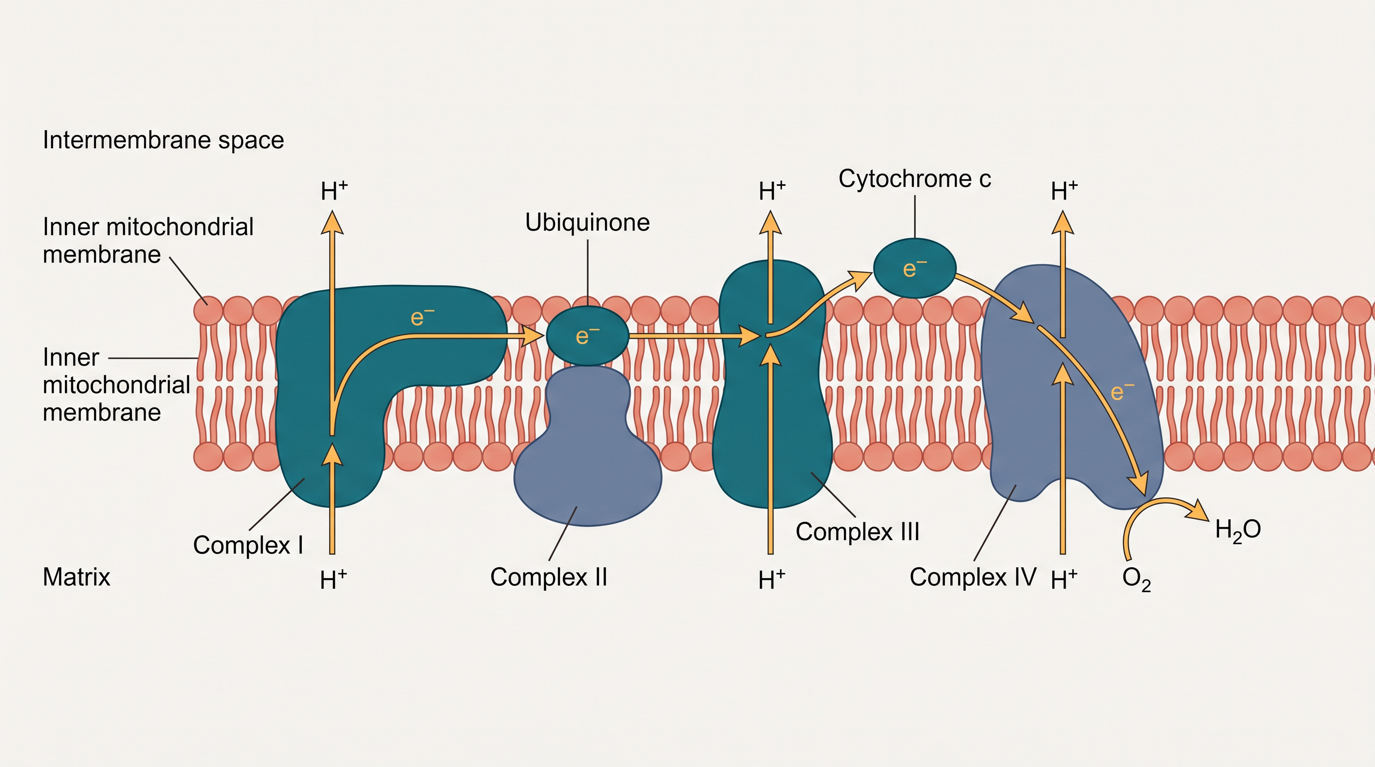
Fig. 12.6 – The electron transport system (ETS).
Let us follow the electrons. Those from NADH — produced in the matrix during the citric acid cycle — are first passed to NADH dehydrogenase (complex I), which transfers them to a small mobile molecule called ubiquinone within the membrane. Ubiquinone also collects electrons from a second source: the FADH₂ generated when succinate is oxidised in the cycle feeds in through complex II.
The now-reduced ubiquinone (ubiquinol) is next oxidised as it hands its electrons to cytochrome c, by way of the cytochrome bc₁ complex (complex III). Cytochrome c is a small protein attached to the outer surface of the inner membrane, and it acts as a mobile carrier ferrying electrons between complex III and complex IV. Finally, complex IV — the cytochrome c oxidase complex, which contains cytochromes a and a₃ and two copper centres — receives the electrons and delivers them to oxygen.
Flowchart
FC 12.6 – Electron path through the ETS: NADH → Complex I → ubiquinone → Complex III → cytochrome c → Complex IV → O₂ → H₂O; FADH₂ enters at Complex II → ubiquinone (joining the same route). Electron flow through Complexes I–IV is coupled to ATP synthase (Complex V) → ATP.
Oxygen: The Final Acceptor
When electrons pass along complex I to complex IV, their flow is coupled to ATP synthase (complex V) for the production of ATP from ADP and inorganic phosphate. The number of ATP made depends on where the electrons entered: the oxidation of one molecule of NADH gives rise to 3 molecules of ATP, while that of one molecule of FADH₂ produces 2 molecules of ATP.
Although aerobic respiration happens only when oxygen is present, oxygen’s own role is confined to the very last step. Yet its presence is absolutely vital, because it drives the whole process by removing hydrogen from the system: oxygen acts as the final hydrogen acceptor. If oxygen were absent, electrons would have nowhere to go, the carriers would stay reduced, and the entire chain — and with it the cycle that feeds it — would grind to a halt.
Coupling Electron Flow to ATP: Oxidative Phosphorylation
Unlike photophosphorylation — studied earlier, where it is light energy that builds the proton gradient needed for phosphorylation — here it is the energy of oxidation–reduction (electrons falling through the carriers) that builds the gradient. For this reason the process is called oxidative phosphorylation.
Inside ATP Synthase
You have already met the chemiosmotic hypothesis, which explains how ATP synthesis is linked to a membrane. The energy released during electron transport is used to make ATP with the help of ATP synthase (complex V), and Fig. 12.7 shows how this remarkable machine is built.
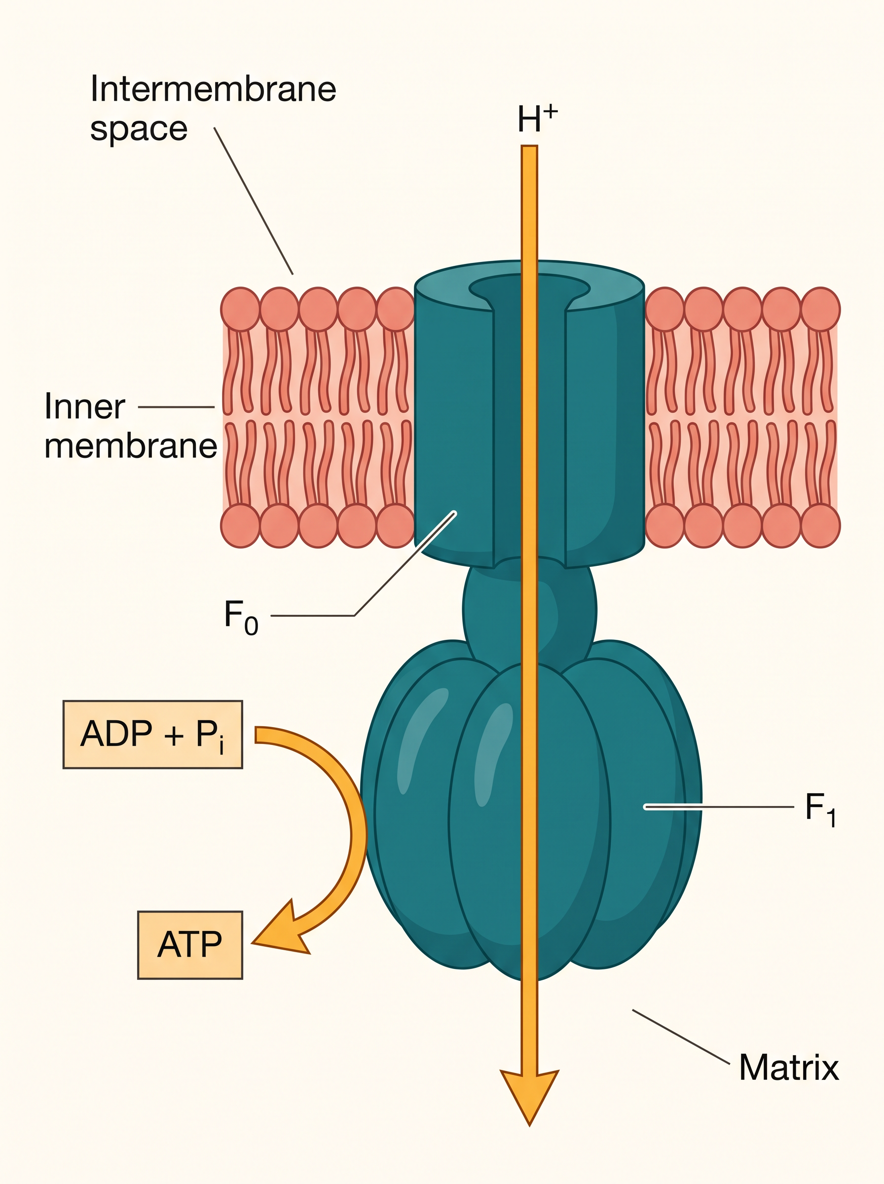
Fig. 12.7 – ATP synthase (Complex V).
ATP synthase has two major components, F₁ and F₀. The F₁ headpiece is a peripheral membrane protein complex that carries the actual site where ATP is made from ADP and inorganic phosphate. F₀ is an integral membrane protein complex that forms a channel through which protons (H⁺) cross the inner membrane.
The passage of protons through this channel is coupled to the catalytic site of the F₁ component, driving the production of ATP. For each ATP produced, 4H⁺ pass through F₀ from the intermembrane space into the matrix, moving down the electrochemical proton gradient that the electron transport chain has built up.
12.5 The Respiratory Balance Sheet
Counting on Paper
Now that we have followed glucose all the way through glycolysis, the Krebs’ cycle and the ETS, we can add up the total ATP yield. It is possible to calculate the net gain of ATP for every glucose molecule oxidised — but in reality this stays a theoretical exercise, because the calculation only works if we accept a set of simplifying assumptions:
- There is a neat, orderly, sequential pathway, with one substrate forming the next, and with glycolysis, the TCA cycle and the ETS following strictly one after another.
- The NADH made during glycolysis (in the cytoplasm) is transferred into the mitochondria and undergoes oxidative phosphorylation there.
- None of the intermediates in the pathway are pulled out to build any other compound.
- Only glucose is being respired — no alternative substrates enter the pathway at any of the intermediate stages.
Adding Up the ATP
Under these assumptions, the contributions of each stage can be laid out in a balance sheet. Remember that one glucose gives two pyruvate, so the steps after glycolysis run twice.
| Stage | Direct (substrate-level) ATP | NADH + H⁺ | FADH₂ |
|---|---|---|---|
| Glycolysis (cytoplasm) | 2 | 2 | 0 |
| Pyruvate → Acetyl CoA (×2) | 0 | 2 | 0 |
| Krebs’ cycle (×2 turns) | 2 | 6 | 2 |
| Total | 4 | 10 | 2 |
Each NADH + H⁺ oxidised in the ETS yields 3 ATP, and each FADH₂ yields 2 ATP. Converting the carriers gives: 10 NADH × 3 = 30 ATP, and 2 FADH₂ × 2 = 4 ATP. Adding the 4 direct ATP:
\[30 + 4 + 4 = 38 \text{ ATP}\]
So there can be a net gain of 38 ATP molecules during the aerobic respiration of one molecule of glucose.
Why It’s Only a Theoretical Figure
These assumptions, honest as they are, do not really hold in a living system. In a real cell all the pathways work at the same time rather than one after another; substrates enter and are withdrawn as and when needed; ATP is spent as and when required; and enzyme rates are controlled by many means at once. Even so, it is useful to do this exercise, because it lets us appreciate the beauty and efficiency with which a living system extracts and stores energy.
Fermentation vs Aerobic Respiration
With the balance sheet in hand, we can now compare fermentation and aerobic respiration directly.
12.6 Amphibolic Pathway
Many Foods, One Pathway
So far we have followed glucose alone. But glucose is simply the favoured substrate for respiration, not the only one. In fact, all carbohydrates are usually first converted into glucose before they are respired.
Other substrates — fats and proteins — can also be respired. The difference is that they do not enter the respiratory pathway right at the first step; instead, each enters at whatever point matches its chemistry. Fig. 12.8 maps these points of entry, and FC 12.7 lists them in order.
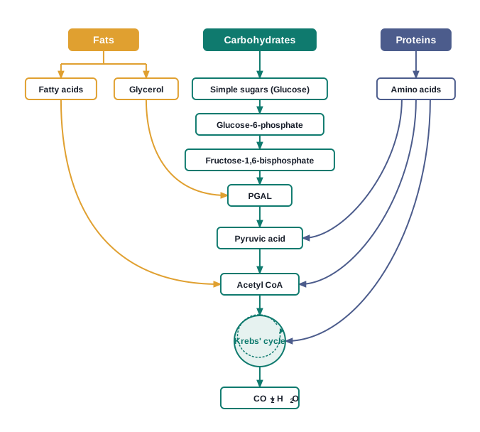
Fig. 12.8 – Interrelationship among metabolic pathways.
Where Each Substrate Enters
Fats must first be broken down into glycerol and fatty acids. If the fatty acids are to be respired, they are degraded into acetyl CoA and enter the pathway there. Glycerol takes a different door — it is converted into PGAL and joins the pathway at that stage.
Proteins are first broken down by enzymes called proteases into individual amino acids. Each amino acid, after deamination (removal of its amino group), enters the pathway at a point that depends on its particular structure — some join within the Krebs’ cycle, others enter as pyruvate or as acetyl CoA.
Flowchart
FC 12.7 – Entry points of substrates into the respiratory pathway: Carbohydrates → glucose → glycolysis → pyruvate; Fats → glycerol → PGAL, and fatty acids → acetyl CoA; Proteins → proteases → amino acids → (after deamination) → pyruvate / acetyl CoA / Krebs’-cycle intermediates → all converge on the Krebs’ cycle → CO₂ + H₂O.
Breakdown and Build-up: The Two-Way Street
Because respiration breaks substrates down, the respiratory pathway has traditionally been called a catabolic pathway. But is that the full picture?
Notice that the very compounds at which fats and proteins enter the pathway are also the compounds that must be withdrawn from it whenever the cell needs to build those same substances. Fatty acids, for instance, are broken down to acetyl CoA to be respired — but when the organism needs to make fatty acids, acetyl CoA is drawn out of the respiratory pathway to do so. The pathway is therefore involved both in the breakdown and in the synthesis of fatty acids. The same is true for proteins: respiratory intermediates form the link during both their breakdown and their build-up.
Not Just Catabolic — Amphibolic
We can now put clear names to the two directions.
Because the respiratory pathway takes part in both — catabolism and anabolism — it is better described not as merely catabolic, but as an amphibolic pathway.
12.7 Respiratory Quotient
Reading Respiration Through Two Gases
We turn finally to one more useful aspect of respiration. As you know, during aerobic respiration O₂ is consumed and CO₂ is released. If we measure the volumes of these two gases, their ratio tells us something about what the cell is burning. This ratio — the volume of CO₂ evolved to the volume of O₂ consumed — is called the respiratory quotient (RQ), or respiratory ratio.
\[RQ = \frac{\text{volume of } CO_2 \text{ evolved}}{\text{volume of } O_2 \text{ consumed}}\]
The value of the RQ depends on the type of respiratory substrate being used — and that is exactly why it is so informative.
RQ for Different Foods
Carbohydrates. When a carbohydrate is the substrate and is completely oxidised, the RQ is 1, because equal volumes of CO₂ and O₂ are exchanged:
\[C_6H_{12}O_6 + 6O_2 \rightarrow 6CO_2 + 6H_2O + \text{Energy}\]
\[RQ = \frac{6CO_2}{6O_2} = 1.0\]
Fats. When fats are respired, the RQ is less than 1. Taking the fatty substance tripalmitin as a worked example:
\[2(C_{51}H_{98}O_6) + 145O_2 \rightarrow 102CO_2 + 98H_2O + \text{energy}\]
\[RQ = \frac{102CO_2}{145O_2} = 0.7\]
Proteins. When proteins are the respiratory substrates, the ratio works out to about 0.9.
Why Fat Gives a Lower RQ
Mixed Substrates in Real Cells
12.8 Summary
Plants, unlike animals, have no special organs for breathing. Gaseous exchange happens simply by diffusion through stomata and lenticels, and almost every living cell in a plant has part of its surface exposed to air.
The breaking of the C–C bonds of complex organic molecules by oxidation, releasing a large amount of energy, is called cellular respiration. Glucose is the favoured substrate, but fats and proteins can also be broken down to yield energy.
The first stage of respiration, glycolysis, takes place in the cytoplasm. Through a series of enzyme-controlled reactions, each glucose molecule is split into two molecules of pyruvic acid.
What happens to pyruvate depends on the organism and on whether oxygen is available. Without oxygen, either lactic acid fermentation or alcoholic fermentation occurs. Fermentation takes place under anaerobic conditions in many prokaryotes, in unicellular eukaryotes, and in germinating seeds.
In eukaryotes, aerobic respiration occurs when oxygen is present. Pyruvic acid is carried into the mitochondria, where it is converted into acetyl CoA with the release of CO₂.
Acetyl CoA enters the tricarboxylic acid (Krebs’) cycle, which operates in the mitochondrial matrix and generates NADH + H⁺ and FADH₂.
The energy in these molecules, together with that in the NADH + H⁺ made during glycolysis, is used to synthesise ATP. This happens through a chain of electron carriers — the electron transport system (ETS) — on the inner mitochondrial membrane. As the electrons pass along the system, they release energy that is trapped to make ATP, a process called oxidative phosphorylation. Here O₂ is the ultimate acceptor of electrons and is reduced to water.
The respiratory pathway is an amphibolic pathway, because it takes part in both catabolism (breakdown) and anabolism (build-up).
The respiratory quotient (RQ) — the ratio of CO₂ evolved to O₂ consumed — depends on the type of respiratory substrate being used.
Flowchart
FC 12.8 – The whole respiratory sequence: Glucose → glycolysis (cytoplasm) → 2 Pyruvic acid → if no O₂ → fermentation → lactic acid OR ethanol + CO₂ (net 2 ATP); if O₂ present → into mitochondria → pyruvate → Acetyl CoA (+CO₂, NADH) → Krebs’ cycle in matrix (→ CO₂ + NADH + FADH₂ + ATP) → ETS on inner membrane (NADH & FADH₂ → electrons → O₂ → H₂O) → oxidative phosphorylation → net up to 38 ATP.
12.9 Points to Ponder
A cell could, in principle, oxidise glucose in a single burst, yet it insists on breaking it down in many small, enzyme-controlled steps. What does this stepwise strategy reveal about how living systems manage energy, and what might go wrong if all the energy were released at once?
Oxygen is used only at the very last step of aerobic respiration, and still its absence halts the entire process. What does this dependence tell you about how the steps of a metabolic pathway are linked to one another?
Fermentation yields only about 2 ATP per glucose, while aerobic respiration can yield up to 38. If fermentation is so inefficient, why has it been kept by living organisms right up to the present day?
The respiratory pathway both breaks molecules down and helps build them up. Does it really make sense to call any single pathway purely “catabolic” or purely “anabolic”? What does the amphibolic nature of respiration suggest about the economy of a living cell?
The figure of 38 ATP rests on assumptions that are never fully true in a real cell. Is a number that is never exactly reached still worth calculating? Reflect on the value of idealised models in biology.
A germinating seed’s respiratory quotient can change over several days as it uses up one kind of stored food and begins on another. What might such a shifting RQ tell you about the seed’s changing metabolism?
Photosynthesis stores energy in glucose, while respiration releases it. How do these two opposite processes, taken together, keep energy flowing through the entire living world?
12.10 NCERT Questions
Differentiate between:
- Respiration and Combustion
- Glycolysis and Krebs’ cycle
- Aerobic respiration and Fermentation
What are respiratory substrates? Name the most common respiratory substrate.
Give the schematic representation of glycolysis.
What are the main steps in aerobic respiration? Where does it take place?
Give the schematic representation of an overall view of Krebs’ cycle.
Explain ETS.
Distinguish between the following:
- Aerobic respiration and Anaerobic respiration
- Glycolysis and Fermentation
- Glycolysis and Citric acid Cycle
What are the assumptions made during the calculation of net gain of ATP?
Discuss “The respiratory pathway is an amphibolic pathway.”
Define RQ. What is its value for fats?
What is oxidative phosphorylation?
What is the significance of step-wise release of energy in respiration?
12.11 Check Your Concepts
Assertion–Reason. Assertion: Plants do not need specialised respiratory organs. Reason: Almost every living cell in a plant lies close to the surface and exchanges gases by simple diffusion. State whether each statement is true, and whether the reason correctly explains the assertion.
Why does a cell break glucose down in many small, enzyme-controlled steps rather than oxidising it in a single step?
Why must NADH + H⁺ be reoxidised to NAD⁺ during fermentation for glycolysis to keep running?
What happens to a naturally fermenting beverage as the alcohol concentration approaches about 13 per cent, and why?
Arrange these glycolytic intermediates in the correct order from start to finish: fructose-1,6-bisphosphate, glucose-6-phosphate, pyruvic acid, phosphoenolpyruvate, PGAL, glucose.
Arrange the following in the order in which electrons pass through them in the ETS: cytochrome c, ubiquinone, Complex I, O₂, Complex IV, Complex III.
Match the enzyme in Column I with its reaction in Column II.
| Column I | Column II |
|---|---|
| a. hexokinase | i. pyruvate → lactic acid |
| b. pyruvic acid decarboxylase | ii. glucose → glucose-6-phosphate |
| c. lactate dehydrogenase | iii. acetyl CoA + OAA → citric acid |
| d. citrate synthase | iv. pyruvate → acetaldehyde + CO₂ |
Draw a simple diagram of a mitochondrion and label the sites of (a) the Krebs’ cycle and (b) the electron transport system.
Assertion–Reason. Assertion: One molecule of FADH₂ yields fewer ATP than one molecule of NADH in the ETS. Reason: FADH₂ donates its electrons at Complex II, bypassing Complex I.
Oxygen is used only at the final step of aerobic respiration, yet the whole process stops when oxygen is absent. Explain why.
Odd one out. Identify the odd one and justify your choice: glycolysis, lactic acid fermentation, alcoholic fermentation, Krebs’ cycle.
What would happen to ATP production in a cell whose Complex IV is blocked by cyanide? Explain your answer.
Why is the respiratory pathway described as amphibolic rather than simply catabolic? Illustrate with an example involving fatty acids.
State the point at which each of the following enters the respiratory pathway: (a) glycerol, (b) fatty acids, (c) amino acids.
Explain why the RQ of a fat is less than 1, whereas that of a carbohydrate is exactly 1.
Assertion–Reason. Assertion: The net ATP yield per glucose is usually quoted as 38, but a living cell rarely achieves exactly this figure. Reason: In a real cell the pathways operate simultaneously and intermediates are continually withdrawn for other syntheses.
How does substrate-level phosphorylation differ from oxidative phosphorylation? Give one example of each from this chapter.
Describe what happens when pyruvate is converted to acetyl CoA. Name the enzyme involved and list the products.
12.12 Practice With Numericals
During the glycolysis of one glucose molecule, 4 ATP are produced and 2 ATP are consumed. Calculate the net gain of ATP, and state how many molecules of NADH + H⁺ are formed.
In the complete aerobic oxidation of one glucose molecule, 10 NADH + H⁺ and 2 FADH₂ are passed through the ETS. Taking 3 ATP per NADH + H⁺ and 2 ATP per FADH₂, calculate the ATP obtained from the ETS alone.
Using the respiratory balance sheet, show how the net gain of 38 ATP per glucose is made up (direct ATP + ATP from NADH + H⁺ + ATP from FADH₂).
A respiring tissue releases 30 mL of CO₂ while consuming 30 mL of O₂ in a fixed period. Calculate its respiratory quotient and name the likely respiratory substrate.
For the complete oxidation of tripalmitin, 145 volumes of O₂ are consumed and 102 volumes of CO₂ are evolved. Calculate the RQ, correct to one decimal place.
One turn of the Krebs’ cycle yields 3 NADH + H⁺, 1 FADH₂ and 1 ATP. How many of each are produced from one glucose molecule? (Remember that the cycle turns twice per glucose.)