Chapter 6: Anatomy of Flowering Plants
More than three centuries ago, two curious observers on opposite sides of Europe pointed their newly built microscopes at something most people walked past without a second thought — thin slices of plant stems, roots and leaves. In England, Nehemiah Grew, and in Italy, Marcello Malpighi, spent the 1670s and 1680s peeling, slicing and carefully sketching what they saw inside ordinary plants.
What they found astonished them. A plant that looked like a plain green rod on the outside was, on the inside, a carefully organised city — layer upon layer of differently shaped cells, long tubes running lengthwise like pipelines, a protective outer covering, and soft packing tissue in between. Grew described these internal patterns so carefully that he is often remembered as a founder of plant anatomy, the study of a plant’s internal structure. Malpighi, working on his own, arrived at strikingly similar conclusions.
Their drawings revealed something we still rely on today: the inside of a plant is not random. Every tissue sits exactly where its job requires it to be. Cut a stem straight across and you can even tell one kind of plant from another — a grass from a sunflower — just from the pattern of tissues you see.
In this chapter you will look through the same “window” they opened. You will learn how a plant is built from the inside out, and why a single cross-section can reveal a plant’s identity, its way of life, and even the environment it grew in.
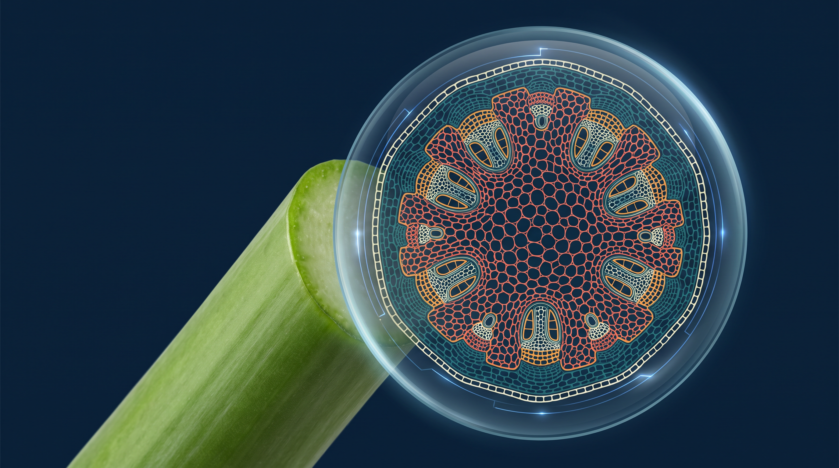
Fig. 6.0 – Looking inside a plant stem.
By the end of this chapter, you will be able to answer each of these questions for yourself.
6.1 The Tissue System
Earlier, you learnt to sort plant tissues by the type of cells they contain — for example, whether the cells are living or dead, thin-walled or thick-walled. But there is another useful way to look at tissues: by where they sit in the plant body. A tissue’s location largely decides both its structure and its job. A tissue on the outer surface will be built for protection, while a tissue deep inside will be built for support or transport.
When we group tissues by their structure and position, the whole plant body can be divided into three tissue systems — the epidermal tissue system, the ground (or fundamental) tissue system, and the vascular (or conducting) tissue system. A tissue system is simply a large functional unit made of one or more tissues that work together to carry out a particular role across the plant.
The way the three systems are classified is summarised in FC 6.1.
Flowchart
FC 6.1 – Plant tissue systems: Tissue system → branches into (1) Epidermal tissue system (epidermal cells + stomata + trichomes/hairs), (2) Ground tissue system (parenchyma + collenchyma + sclerenchyma), (3) Vascular tissue system (xylem + phloem)
6.1.1 Epidermal Tissue System
The epidermal tissue system forms the outermost covering of the entire plant body — in a sense, it is the plant’s “skin.” It is made up of epidermal cells, the stomata, and the epidermal appendages, which are the trichomes and hairs.
The epidermis is the outermost layer of the primary plant body. It is built from elongated cells that are packed closely together with no gaps, so they form a continuous, unbroken sheet. This layer is usually only one cell thick (single-layered), which is enough to cover and protect the plant without blocking light or slowing it down.
Epidermal cells are parenchymatous — living cells with thin walls, only a thin lining of cytoplasm pressed against the wall, and a large central vacuole that takes up most of the cell’s space.
The waterproof cuticle
On its outer side, the epidermis is often coated with a thick, waxy layer called the cuticle. Because it is waxy, the cuticle is water-repellent, so it greatly reduces the loss of water from the plant surface. The cuticle is absent in roots — and this makes sense, because a root’s job is to absorb water from the soil, and a waterproof coat would only get in the way.
Stomata and guard cells
Set into the epidermis, especially the epidermis of leaves, are tiny pores called stomata (singular: stoma). Stomata control two vital exchanges with the air: transpiration (the loss of water vapour) and gaseous exchange (taking in and giving out gases such as \(CO_2\) and \(O_2\)).
Each stoma is bordered by two bean-shaped cells called guard cells, which enclose a central opening, the stomatal pore. In grasses, the guard cells are shaped more like a dumb-bell than a bean.
The walls of a guard cell are unevenly thickened. The outer wall, facing away from the pore, is thin and flexible, while the inner wall, facing the pore, is highly thickened and stiff. Guard cells also contain chloroplasts, and it is the guard cells that open and close the stoma. You can compare both guard-cell shapes in Fig. 6.1.
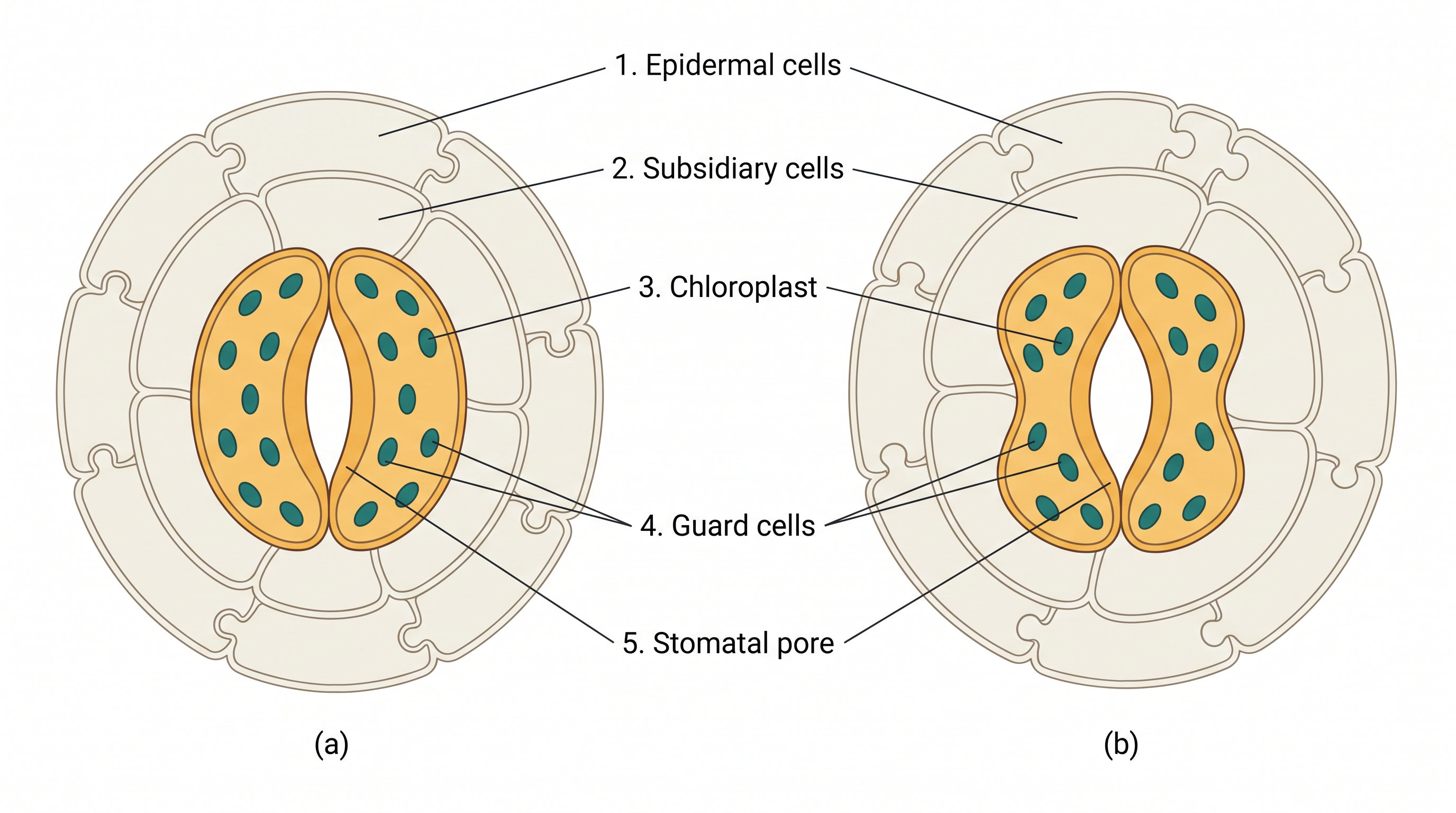
Fig. 6.1 – Stomatal apparatus with (a) bean-shaped and (b) dumb-bell-shaped guard cells.
Because the two walls differ, a guard cell bends outward when it swells with water, pulling the pore open; when it loses water, it straightens and the pore closes. This is how the plant fine-tunes both water loss and gas exchange.
Sometimes a few epidermal cells lying right next to the guard cells become special in their shape and size; these are called subsidiary cells. Together, the stomatal pore, the two guard cells and the surrounding subsidiary cells make up the stomatal apparatus.
Root hairs and trichomes
The epidermis also bears a variety of hairs. On the root, these are the root hairs — unicellular (single-celled) outgrowths of epidermal cells that greatly increase the root’s ability to absorb water and minerals from the soil.
On the stem and other shoot parts, the epidermal hairs are called trichomes. Unlike root hairs, shoot trichomes are usually multicellular. They may be branched or unbranched, soft or stiff, and some are even secretory (they release substances). By clothing the surface, trichomes help reduce water loss through transpiration.
6.1.2 The Ground Tissue System
Everything that is neither epidermis nor part of the vascular bundles belongs to the ground tissue (also called fundamental tissue). In other words, the ground tissue system is the large “filler” lying between the outer skin and the inner conducting strands, and it forms the main bulk of the plant.
The ground tissue is made of simple tissues — tissues built from just one kind of cell. There are three of these: parenchyma (living, thin-walled cells that store food and carry out many basic activities), collenchyma (living cells thickened at their corners to give flexible support), and sclerenchyma (dead, thick-walled cells that provide rigid mechanical strength).
In the primary stems and roots, parenchymatous cells make up regions such as the cortex, the pericycle, the pith, and the medullary rays. You will meet each of these regions by name when we examine root and stem sections in the next part of the chapter.
In leaves, the ground tissue takes a special form: it consists of thin-walled cells packed with chloroplasts, and is called the mesophyll. This is the tissue in which most photosynthesis takes place.
6.1.3 The Vascular Tissue System
The vascular (conducting) tissue system is the plant’s transport network. It is made of two complex tissues — tissues built from more than one kind of cell — namely the xylem and the phloem. The xylem carries water and dissolved minerals upward from the root, while the phloem carries food (mainly sugars) made in the leaves to the rest of the plant.
Where xylem and phloem occur together as a strand, they form a vascular bundle. The kinds of vascular bundle differ from plant to plant, and these differences are among the most exam-important points in this chapter. Fig. 6.2 shows the main types.
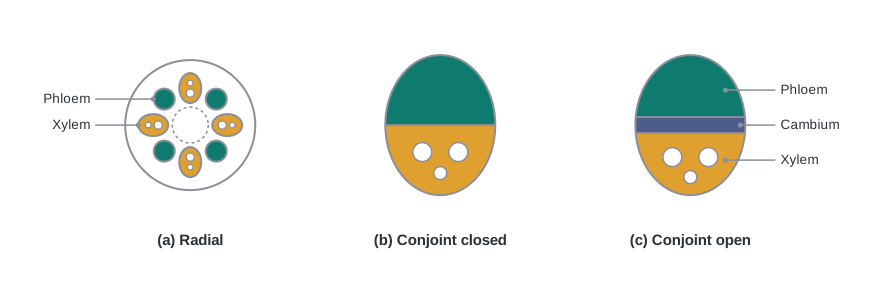
Fig. 6.2 – Types of vascular bundles.
Open and closed bundles
In the stems of dicotyledonous plants, a layer of cambium — a thin band of dividing cells — lies between the phloem and the xylem. Because of this cambium, the bundle can later form new, secondary xylem and phloem; such bundles are called open vascular bundles.
In monocotyledons, the vascular bundles have no cambium in them. Since they cannot form secondary tissue, these bundles are described as closed.
Radial and conjoint arrangement
There is a second, quite separate way to describe a bundle: by how the xylem and phloem are placed relative to each other. When xylem and phloem lie separately, on different radii, so that they alternate around the axis, the arrangement is called radial; this is what we see in roots.
When the xylem and phloem instead sit together along the same radius, the arrangement is conjoint. Conjoint bundles are common in stems and leaves, and in them the phloem usually lies only on the outer side of the xylem.
The full classification of vascular bundles is summarised in FC 6.2.
Flowchart
FC 6.2 – Vascular bundle classification. By cambium: cambium present → open (dicot stems, can form secondary tissue); cambium absent → closed (monocotyledons, no secondary tissue). By arrangement: xylem and phloem on different radii, alternating → radial (roots); xylem and phloem on the same radius → conjoint (stems and leaves, phloem outer to xylem).
6.2 Anatomy of Dicotyledonous and Monocotyledonous Plants
To understand how tissues are arranged inside roots, stems and leaves, the most convenient approach is to cut a transverse section — a thin slice taken straight across the organ — through its mature zone and study it under the microscope. This is exactly the “window” the early anatomists first opened.
As we move through each organ, watch closely for the features that separate dicots from monocots. Telling the two apart from a single section is one of the commonest ways this topic is tested — as diagram-labelling, “identify this section,” and match-the-following questions.
6.2.1 Dicotyledonous Root
Fig. 6.3 (a) shows the transverse section of a sunflower root, a typical dicot root. Reading it from the outside inward gives a clear picture of how the tissues are layered.
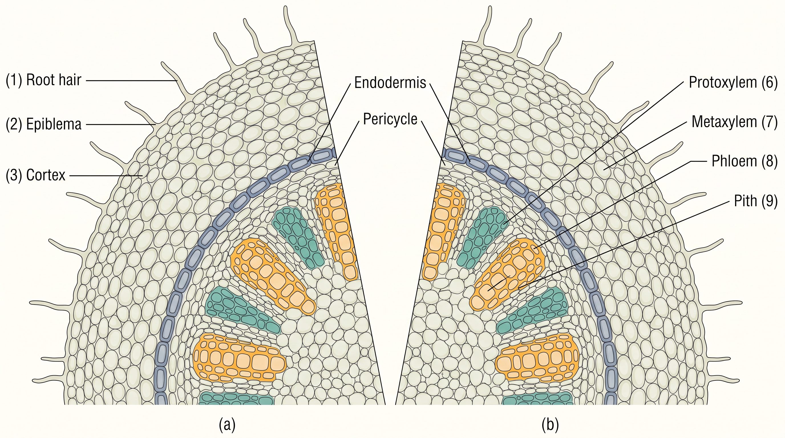
Fig. 6.3 – Transverse sections of (a) dicot root and (b) monocot root.
The outermost layer is the epiblema — which is simply the epidermis of the root. Many of its cells push out into fine, single-celled root hairs that increase water and mineral uptake.
Just inside lies the cortex, made of several layers of thin-walled parenchyma cells with intercellular spaces between them. The innermost layer of the cortex is a special ring called the endodermis, a single layer of barrel-shaped cells fitted together with no gaps.
The walls of these endodermal cells carry a band of a waxy, water-repellent material called suberin. These waterproof bands, laid down on the tangential and radial walls, are the casparian strips.
Next to the endodermis lie a few layers of thick-walled parenchyma cells called the pericycle. These cells are important: both new lateral (side) roots and the vascular cambium of later secondary growth are first formed here.
In the centre, the pith is small or barely noticeable. The parenchyma cells lying between the patches of xylem and phloem are called conjunctive tissue. A dicot root usually has only two to four patches each of xylem and phloem. Later, a cambium ring develops between the xylem and phloem, and it is this ring that goes on to produce secondary tissue.
All the tissues lying to the inner side of the endodermis — the pericycle, the vascular bundles and the pith — are together called the stele.
The order of layers from the surface to the centre is summarised in FC 6.3.
Flowchart
FC 6.3 – Dicot root, surface to centre: epiblema (with root hairs) → cortex → endodermis (with casparian strips) → pericycle → vascular bundles + conjunctive tissue → pith
6.2.2 Monocotyledonous Root
The monocot root, shown in Fig. 6.3 (b), is built on the same general plan as the dicot root. It, too, has an epidermis, cortex, endodermis, pericycle, vascular bundles and pith.
The differences lie in the details. While a dicot root has only a few xylem bundles, a monocot root usually has more than six — a condition described as polyarch. Its pith is large and well developed, rather than small. And, crucially, a monocotyledonous root does not undergo any secondary growth.
6.2.3 Dicotyledonous Stem
The transverse section of a typical young dicot stem is shown in Fig. 6.4 (a). Its outermost protective layer is the epidermis, covered by a thin cuticle, and it may carry trichomes and a few stomata.
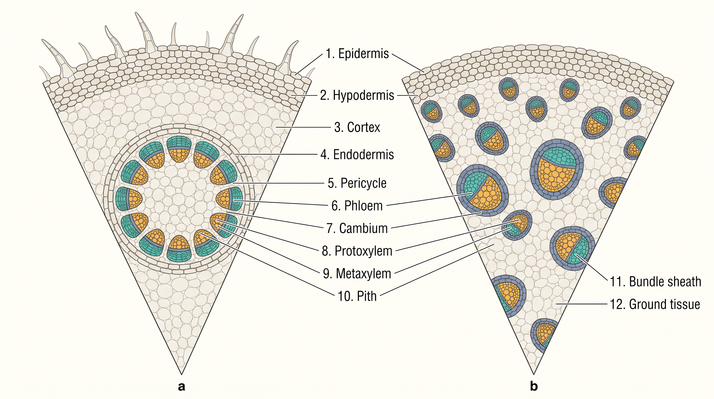
Fig. 6.4 – Transverse sections of (a) dicot stem and (b) monocot stem.
The three-zoned cortex
The tissue lying between the epidermis and the pericycle makes up the cortex, and in the dicot stem it is divided into three sub-zones.
The outermost sub-zone is the hypodermis — a few layers of collenchyma cells sitting just below the epidermis. Because collenchyma is flexible yet strong, the hypodermis gives mechanical strength to the young, still-growing stem.
Below the hypodermis are the general cortical layers, made of rounded, thin-walled parenchyma cells with conspicuous intercellular spaces. The innermost sub-zone of the cortex is the endodermis. Its cells are packed with starch grains, and for this reason the layer is also called the starch sheath.
From pericycle to pith
Just inside the endodermis lies the pericycle, which here occurs above the phloem as half-moon-shaped (semi-lunar) patches of sclerenchyma. Between neighbouring vascular bundles run a few rows of radially placed parenchyma cells called the medullary rays.
A large number of vascular bundles are arranged in a ring — and this ring arrangement is a hallmark of the dicot stem. Each bundle is conjoint (xylem and phloem on the same radius), open (with cambium, so capable of secondary growth), and has endarch protoxylem, meaning the first-formed xylem (protoxylem) lies toward the centre and the later metaxylem toward the outside. (In roots, by contrast, the protoxylem lies toward the periphery — an arrangement called exarch.)
Filling the centre of the stem is the pith: rounded parenchyma cells with large intercellular spaces.
6.2.4 Monocotyledonous Stem
The monocot stem, shown in Fig. 6.4 (b), differs from the dicot stem in several clear ways. Its hypodermis is sclerenchymatous (not collenchymatous), and it carries a large number of vascular bundles scattered through a big, conspicuous mass of parenchymatous ground tissue — there is no organised cortex-and-pith arrangement.
Each vascular bundle is wrapped in a sheath of sclerenchyma, the bundle sheath, and every bundle is conjoint and closed (no cambium, so no secondary growth). The bundles near the periphery are generally smaller than those toward the centre.
Two further points are exam-favourites: in the monocot stem the phloem parenchyma is absent, and small water-containing cavities are present within the vascular bundles.
6.2.5 Dorsiventral (Dicotyledonous) Leaf
A dorsiventral leaf is one whose upper and lower surfaces are clearly different — typical of dicots. A vertical section through the flat blade (lamina), shown in Fig. 6.5 (a), reveals three main parts: the epidermis, the mesophyll and the vascular system.
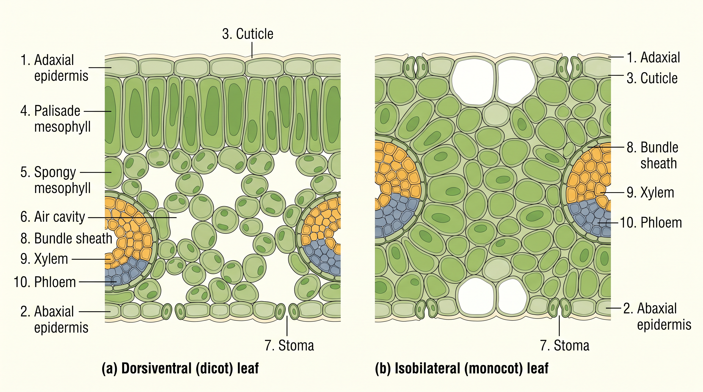
Fig. 6.5 – Transverse sections of (a) dorsiventral and (b) isobilateral leaves.
Epidermis and mesophyll
The epidermis covers both the upper surface (the adaxial epidermis) and the lower surface (the abaxial epidermis) and carries a conspicuous cuticle. The abaxial (lower) epidermis generally bears more stomata than the adaxial (upper) one, which may even have none at all.
Between the two epidermal layers lies the mesophyll — the parenchyma tissue that contains chloroplasts and carries out photosynthesis. In a dorsiventral leaf the mesophyll is made of two distinct cell types.
The palisade parenchyma lies just under the adaxial (upper) epidermis and is made of elongated cells set upright, vertical and parallel to one another, like a row of columns. Below it lies the spongy parenchyma — oval or rounded cells, loosely packed, extending down to the lower epidermis, with numerous large air spaces and cavities between them.
The vascular system
The vascular system of the leaf lies in the veins and the midrib. The size of each vascular bundle depends on the size of the vein carrying it, and in the dicot leaf the veins vary in thickness because dicots have reticulate (net-like) venation. Each vascular bundle is surrounded by a layer of thick-walled bundle sheath cells. In Fig. 6.5 (a), notice where the xylem sits within the bundle relative to the phloem.
6.2.6 Isobilateral (Monocotyledonous) Leaf
An isobilateral leaf — the grass-type leaf typical of monocots — has upper and lower surfaces that look much the same. Its anatomy resembles the dorsiventral leaf in many ways but shows a few characteristic differences, seen in Fig. 6.5 (b).
First, the stomata are present on both surfaces of the epidermis, not mainly on the lower one. Second, the mesophyll is not differentiated into palisade and spongy layers; it is fairly uniform throughout.
Bulliform cells and leaf rolling
In grasses, certain adaxial (upper) epidermal cells lying along the veins are modified into large, empty, colourless cells called bulliform cells. These act like tiny water balloons for the leaf.
When the bulliform cells are full of water and turgid, they hold the leaf blade flat and spread open. When water is scarce and they become flaccid (limp), the leaf rolls inward on itself, reducing the surface exposed to the air and so cutting down water loss.
Finally, the parallel venation of monocot leaves shows up in the section as vascular bundles of nearly the same size all across the leaf (except in the main veins), quite unlike the varied bundle sizes of a reticulately veined dicot leaf.
6.3 Summary
Anatomy is the study of a plant’s internal structure. A plant body is built from many different kinds of tissues working together.
Plant tissues fall into two broad groups: meristematic tissues (regions of dividing cells — apical, lateral and intercalary) and permanent tissues (simple and complex).
The main jobs carried out by tissues are making food and storing it, transporting water, minerals and photosynthates (the food made in photosynthesis), and providing mechanical support.
Grouped by their structure and location, tissues form three tissue systems: the epidermal, the ground (fundamental) and the vascular (conducting) systems. This organisation is condensed in FC 6.4.
Flowchart
FC 6.4 – Overall plan of plant anatomy: cell → tissue → tissue system → organ. Tissue system branches into: epidermal (epidermal cells, stomata, root hairs/trichomes); ground (cortex, pericycle, pith / mesophyll in leaves); vascular (xylem + phloem, forming vascular bundles). Each organ (root, stem, leaf) then differs between dicot and monocot.
The epidermal tissue system is the plant’s outermost covering. It is made of epidermal cells, the stomata, and the epidermal appendages — the root hairs and trichomes.
The ground tissue system forms the main bulk of the plant. It is divided into three zones: the cortex, the pericycle and the pith.
The vascular tissue system is formed by the xylem and the phloem. Depending on whether cambium is present and on how the xylem and phloem are placed, vascular bundles come in different types. These bundles make up the conducting tissue, translocating water, minerals and food material.
Monocotyledonous and dicotyledonous plants show marked differences in their internal structure — especially in the type, number and location of their vascular bundles.
Secondary growth (thickening over time) takes place in most dicotyledonous roots and stems, but does not occur in monocotyledons.
6.4 Points to Ponder
Both the epidermis and the endodermis are single “boundary” layers, yet the epidermis lets water in while the endodermis, with its casparian strips, tightly controls what passes. Why might a plant need more than one kind of internal boundary?
A dicot stem lines up its vascular bundles in a neat ring, while a monocot scatters them through the ground tissue. What advantages might a scattered arrangement give a tall, wind-blown grass that a ring might not?
Secondary growth lets a dicot tree live for centuries and grow enormously thick, yet monocots such as bamboo and palms reach great heights without it. What might a plant gain, and what might it give up, in following one strategy rather than the other?
A leaf must take in carbon dioxide but must not lose too much water — two aims that pull in opposite directions. How do the cuticle, the stomata and the arrangement of the mesophyll work together to balance them, and under what conditions might that balance fail?
Imagine you are handed an unlabelled transverse section of a plant organ. Which single feature would you look for first to decide whether it is a root or a stem, and whether it is a monocot or a dicot — and why would you trust that feature above the others?
The same few tissues — parenchyma, collenchyma, sclerenchyma, xylem and phloem — are simply rearranged differently in the root, the stem and the leaf. What might this reuse of a small “tissue toolkit” suggest about how plants are built and how they may have evolved?
6.5 NCERT Questions
Draw illustrations to bring out the anatomical difference between (a) monocot root and dicot root, and (b) monocot stem and dicot stem.
Cut a transverse section of the young stem of a plant from your school garden and observe it under the microscope. How would you ascertain whether it is a monocot stem or a dicot stem? Give reasons.
The transverse section of a plant material shows the following anatomical features — (a) the vascular bundles are conjoint, scattered and surrounded by sclerenchymatous bundle sheaths, and (b) phloem parenchyma is absent. What will you identify it as?
What is the stomatal apparatus? Explain the structure of stomata with a labelled diagram.
Name the three basic tissue systems in flowering plants. Give the tissue names under each system.
How is the study of plant anatomy useful to us?
Describe the internal structure of a dorsiventral leaf with the help of labelled diagrams.
6.6 Check Your Concepts
Assertion: The cuticle is absent on the epidermis of roots. Reason: Roots must absorb water and minerals from the soil. State whether the assertion and the reason are true, and whether the reason correctly explains the assertion.
Why are the inner and outer walls of a guard cell thickened to different degrees, and how does this uneven thickening help the stoma open and close?
Distinguish between root hairs and trichomes with respect to their cellularity (single- or many-celled), their location on the plant, and their main function.
Match each item in Column I with its correct description in Column II.
| Column I | Column II |
|---|---|
| a. Bulliform cells | i. suberin bands deposited on the endodermal walls |
| b. Casparian strips | ii. sclerenchyma surrounding a monocot-stem vascular bundle |
| c. Conjunctive tissue | iii. large, empty upper-epidermal cells that bring about leaf rolling |
| d. Bundle sheath | iv. parenchyma lying between the xylem and phloem in a dicot root |
Arrange the following tissues of a dicot root in the correct order from the surface to the centre: pericycle, epiblema, pith, endodermis, cortex, vascular bundles.
A transverse section shows vascular bundles arranged in a ring, each conjoint and open, with a collenchymatous hypodermis and an endodermis rich in starch grains. Identify the organ and state whether it belongs to a monocot or a dicot, giving your reasons.
What would happen to the ability of a young dicot stem to grow thicker if the cambium within its vascular bundles were absent? Explain your answer.
Explain why a monocot root has a large, well-developed pith while the pith of a dicot root is small or inconspicuous.
Compare a dorsiventral leaf and an isobilateral leaf with respect to differentiation of the mesophyll, distribution of stomata, and type of venation.
Draw a labelled diagram of the transverse section of a dicotyledonous stem, showing the epidermis, hypodermis, cortex, endodermis, pericycle, a vascular bundle and the pith.
Why is the palisade parenchyma located towards the upper (adaxial) surface of a dorsiventral leaf rather than towards the lower surface? Relate your answer to its function.
Define the term ‘stele’. Name the tissues that it includes in a dicot root.
Assertion: Monocotyledonous roots do not undergo secondary growth. Reason: Their vascular bundles are closed and lack cambium. State whether both statements are true, and whether the reason correctly explains the assertion.
During a period of drought, how do bulliform cells help a grass leaf conserve water? Describe the change that takes place in these cells and its effect on the leaf.
Differentiate between open and closed vascular bundles, and between radial and conjoint bundles, giving one example location for each of the four terms.
A gardener notices that seedlings wilt for a few days after they are transplanted. Using your knowledge of root anatomy, explain why this happens and suggest how the wilting can be reduced.