Chapter 17: Locomotion and Movement
In the summer of 1954, two separate research teams in England were staring at the same puzzle: what exactly happens inside a muscle when it shortens? For decades, most scientists assumed the muscle proteins themselves must coil up or fold, the way a stretched spring recoils. But the answer turned out to be far stranger.
Working with the newly powerful tools of electron microscopy and X-ray diffraction, Hugh Huxley and Jean Hanson at one laboratory, and Andrew Huxley and Rolf Niedergerke at another, reached the same surprising conclusion at almost the same moment. The two sets of protein filaments inside a muscle do not shrink at all. Instead, one set simply slides past the other, like two overlapping combs pushed together. When their papers appeared side by side in the journal Nature, the “sliding filament theory” was born.
It is a beautiful example of how careful observation can overturn common sense. The muscle you use to lift this book is not squeezing its molecules smaller — it is sliding microscopic filaments over one another, thousands of times, in perfect coordination with your bones, joints and nerves.
This chapter is about that teamwork: how muscles are built, how they contract, how the skeleton gives them something to pull against, and how joints turn that pull into movement.
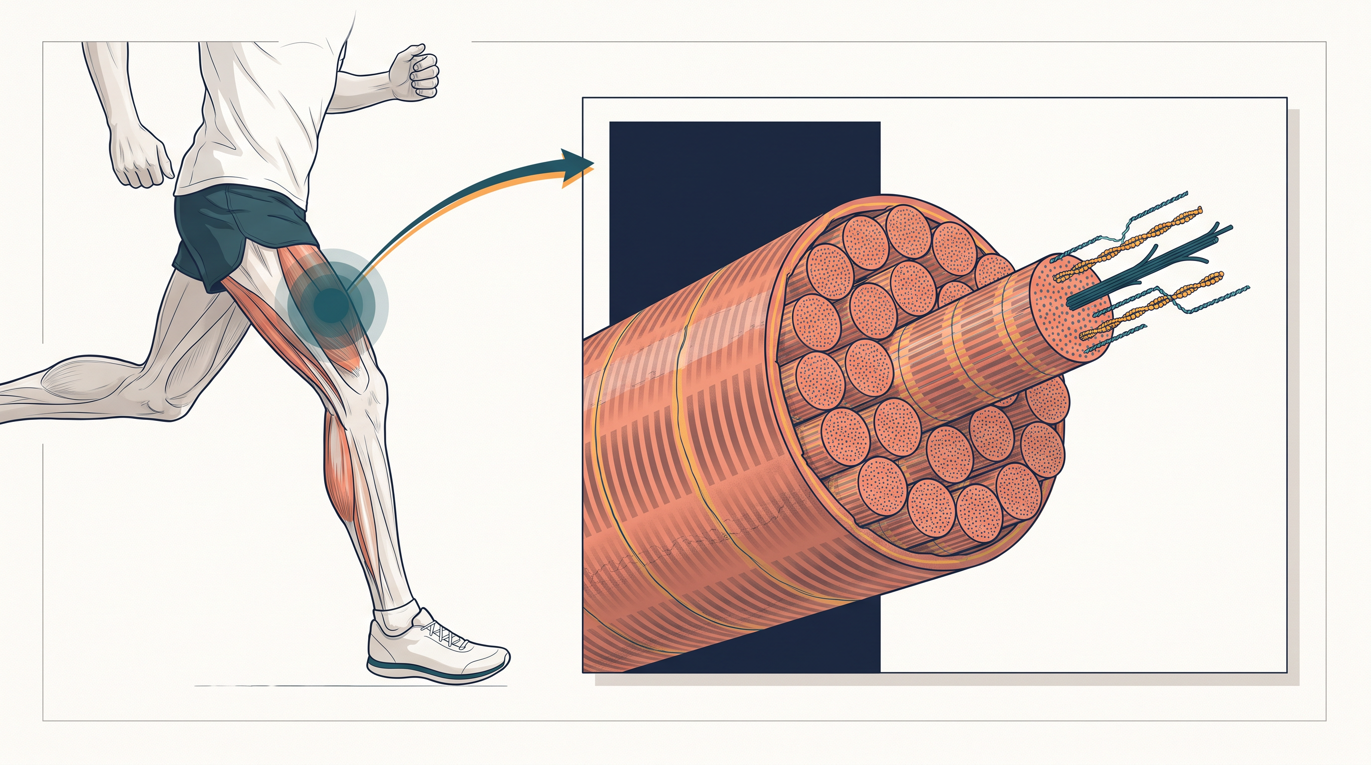
Fig. 17.0 – Runner mid-stride and inset of a striated muscle fibre.
Keep these questions in mind as you read — by the end of the chapter, you will be able to answer every one of them.
17.1 Types of Movement
Movement Is a Sign of Life
One of the clearest signs that something is alive is that it moves. Both animals and plants show a huge variety of movements. In a single-celled organism like Amoeba, even the flowing of the jelly-like living material inside the cell — called protoplasmic streaming — is a simple form of movement. Many organisms move their cilia (tiny hair-like structures) and flagella (longer whip-like structures), and some, like the small freshwater animal Hydra, wave their tentacles.
Human beings move too — our limbs, jaws, eyelids and tongue are all moving parts. But there is an important difference between kinds of movement. Some movements simply shift a part of the body, while others move the whole organism from one place to another.
When a movement carries the entire body from one place to another, and is under the animal’s own control, we call it locomotion. Walking, running, climbing, flying and swimming are all examples of locomotory movement.
Movement and Locomotion Are Linked
An interesting point is that the same structures an animal uses for locomotion are often also used for other kinds of movement — the two are not carried out by separate machinery. In Paramoecium, for example, the cilia sweep food into the cytopharynx (the cell’s “food channel”) and also row the organism forward. Hydra uses its tentacles both to capture prey and to move about. We ourselves use our limbs for locomotion and for changing our body posture.
Because of this overlap, movement and locomotion cannot really be studied as separate topics. The neatest way to state the relationship is this: all locomotion is movement, but not all movement is locomotion.
Why Animals Move from Place to Place
The way an animal moves depends on where it lives and on what the situation demands — a fish, a bird and a snake all solve the problem of locomotion very differently. Yet the reasons for moving are surprisingly similar across the animal world. Animals generally move in search of food, shelter, a mate, and suitable breeding grounds, to reach favourable climatic conditions, or to escape from enemies and predators.
Three Types of Movement in Human Cells
Even inside our own body, movement is not carried out in only one way. The cells of the human body show three main types of movement: amoeboid, ciliary and muscular.
Amoeboid movement is shown by certain specialised cells such as macrophages and leucocytes (white blood cells) in the blood. Just as Amoeba crawls, these cells throw out temporary “false feet” called pseudopodia, formed by the streaming of protoplasm. Cytoskeletal elements — in particular thread-like proteins called microfilaments — provide the machinery for this crawling.
Ciliary movement takes place in most of our internal tubular organs, which are lined by ciliated epithelium (a covering tissue whose cells bear cilia). The cilia beat in a coordinated, wave-like rhythm. In the trachea (windpipe), this beating helps sweep out dust and other foreign particles that we breathe in with the air. Ciliary movement also gently moves the ovum (egg) along the female reproductive tract.
Muscular movement is needed whenever we move our limbs, jaws, tongue and so on. It depends on the ability of muscle tissue to shorten, or contract. This contractile property is the basis of locomotion and most other movements in human beings and in the majority of multicellular animals.
An important idea to carry forward is that locomotion is never the job of muscles alone. It requires the perfectly coordinated activity of three systems working together — the muscular system (which pulls), the skeletal system (which provides the framework to pull against), and the neural system (which sends the commands). This teamwork is the thread that runs through the whole chapter.
In the sections that follow, you will study muscle in detail — the types of muscle, their structure, the mechanism by which they contract — and then the skeletal system that makes their pulling useful.
17.2 Muscle
From Flagella to Muscle
You already know that cilia and flagella are outgrowths of the cell membrane. Flagellar movement, driven by the whip-like beating of a flagellum, is what lets spermatozoa swim, maintains the water current in the canal system of sponges, and powers the locomotion of protists such as Euglena. These are elegant ways of moving, but for large, active animals a far more powerful tissue is needed — muscle.
Muscle is a specialised tissue of mesodermal origin. (Recall that the mesoderm is the middle of the three germ layers formed in the early animal embryo; many of the body’s connective and contractile tissues arise from it.) Muscle is remarkably abundant: in a human adult, about 40–50 per cent of the total body weight is made up of muscle.
The Four Properties of Muscle
Muscle tissue owes its usefulness to four special properties. It has excitability — the ability to respond to a stimulus; contractility — the ability to shorten and generate force; extensibility — the ability to be stretched; and elasticity — the ability to return to its original length after being stretched or contracted. Together, these let a muscle receive a command, pull, and reset itself, over and over.
Three Types of Muscle
Muscles have been classified using several criteria — their location, their appearance, and the nature of regulation of their activity (whether or not we can control them at will). Based on location, three types of muscle are recognised: skeletal, visceral and cardiac.
Skeletal muscles are closely attached to the bones (the skeletal components) of the body. Under a microscope they show a striped, or striated, appearance, so they are also called striated muscles. Because we can move them at will — they are under the voluntary control of the nervous system — they are also called voluntary muscles. They are chiefly responsible for locomotion and for changing body posture.
Visceral muscles are found in the inner walls of the hollow visceral organs, such as the alimentary canal and the reproductive tract. They show no striations and look smooth, so they are called smooth muscles (or non-striated muscles). Their activity is not under our conscious control, which makes them involuntary muscles. They quietly do jobs such as pushing food along the digestive tract and moving gametes through the genital tract.
Cardiac muscles, as the name tells us, are the muscles of the heart. Many cardiac muscle cells join together in a branching pattern to build the cardiac muscle. By appearance they are striated, yet they are involuntary, because the nervous system does not control their beating directly.
Inside a Skeletal Muscle: From Bundle to Fibre
To understand how contraction actually works, let us take a skeletal muscle apart, layer by layer. Each organised skeletal muscle is built from a number of muscle bundles, also called fascicles. These bundles are held together by a common sheet of collagen-rich connective tissue called the fascia. Each muscle bundle, in turn, contains many muscle fibres, as shown in Fig. 17.1.
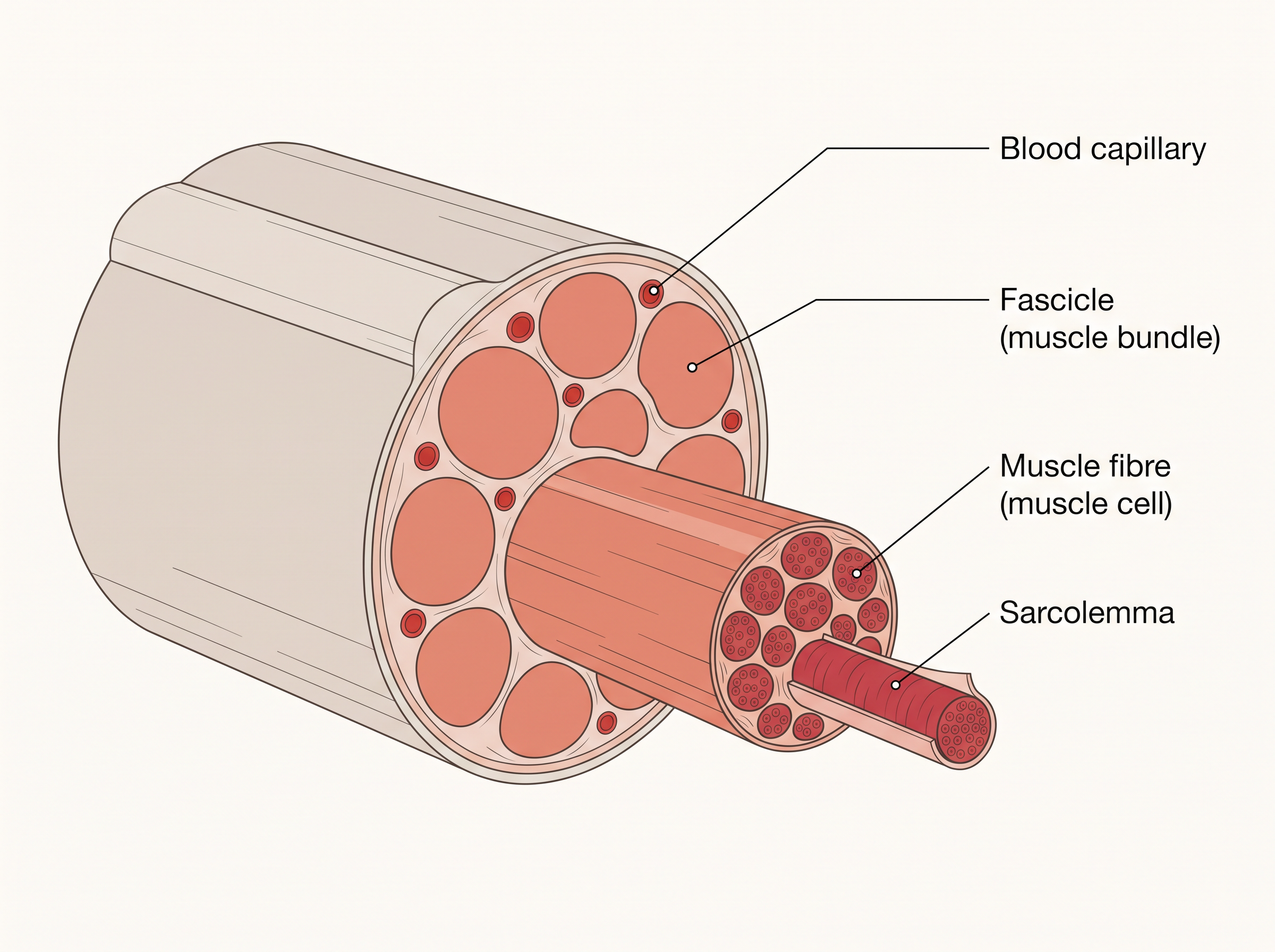
Fig. 17.1 – Cross-sectional view of a skeletal muscle.
Each muscle fibre is a single muscle cell. It is wrapped in its own plasma membrane, here given the special name sarcolemma, which encloses the cell’s cytoplasm, called the sarcoplasm. A muscle fibre is unusual in that it is a syncytium — its sarcoplasm contains many nuclei rather than just one.
Within the sarcoplasm lies the muscle’s endoplasmic reticulum, called the sarcoplasmic reticulum. This structure is the storehouse of calcium ions (\(Ca^{2+}\)) — a detail that will become crucial when we study how contraction is switched on.
The whole arrangement forms a neat biological hierarchy, summarised in FC 17.1.
Flowchart
FC 17.1 – Structural hierarchy of skeletal muscle: Muscle → Fascicles (muscle bundles, wrapped by fascia) → Muscle fibres (muscle cells, each bounded by sarcolemma) → Myofibrils → Myofilaments (thin actin + thick myosin)
Bands, Filaments and the Sarcomere
The most striking feature of a muscle fibre is the presence of a very large number of long, parallel filaments in the sarcoplasm, called myofilaments or myofibrils. Each myofibril carries alternating dark and light bands along its length, and it is this pattern that gives skeletal muscle its striped look.
Careful study has shown that the striations come from the ordered arrangement of two important proteins — actin and myosin. The light bands contain actin and are called I-bands (Isotropic bands). The dark bands, called A-bands (Anisotropic bands), contain myosin.
Both proteins are arranged as rod-like structures, running parallel to one another and to the long axis of the myofibril. Actin filaments are thinner than myosin filaments, so they are commonly called thin filaments and thick filaments respectively.
In the centre of each I-band lies an elastic fibre called the Z-line, which bisects it; the thin filaments are firmly anchored to this Z-line. The thick filaments in the A-band are held together in the middle by a thin fibrous membrane called the M-line. The A- and I-bands alternate all the way along the myofibril.
The portion of a myofibril lying between two successive Z-lines is the functional unit of contraction, called the sarcomere, shown in Fig. 17.2.
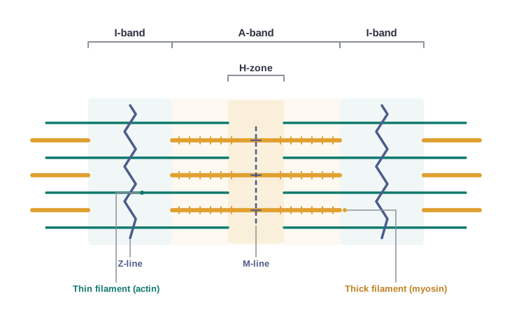
Fig. 17.2 – Anatomy of a muscle fibre showing one sarcomere.
In a resting muscle, the edges of the thin filaments on either side of the thick filaments only partially overlap the free ends of the thick filaments, leaving the central portion of the thick filaments uncovered. This central part of the thick filament, not overlapped by any thin filament, is called the H-zone.
With the sarcomere and its bands now clearly defined, we are ready to look more closely at the two contractile proteins themselves.
17.2.1 Structure of Contractile Proteins
We have seen that thin filaments are made of actin and thick filaments of myosin. To follow how a muscle contracts, we first need to look closely at how these two proteins are built.
The Thin Filament
Each actin (thin) filament is made of two strands of ‘F’ (filamentous) actin wound helically around each other, like two beaded threads twisted together. Each F-actin strand is itself a polymer — a long chain built from many identical ‘G’ (globular) actin subunits joined end to end.
Running alongside the F-actin strands, throughout their length, are two filaments of another protein called tropomyosin. Sitting at regular intervals on the tropomyosin is a complex protein called troponin.
The position of these two proteins matters greatly. In the resting state, one subunit of troponin masks the active binding sites on actin — the very sites to which myosin would otherwise attach, as shown in Fig. 17.3a. In other words, a resting muscle is “switched off” because these sites are physically covered.
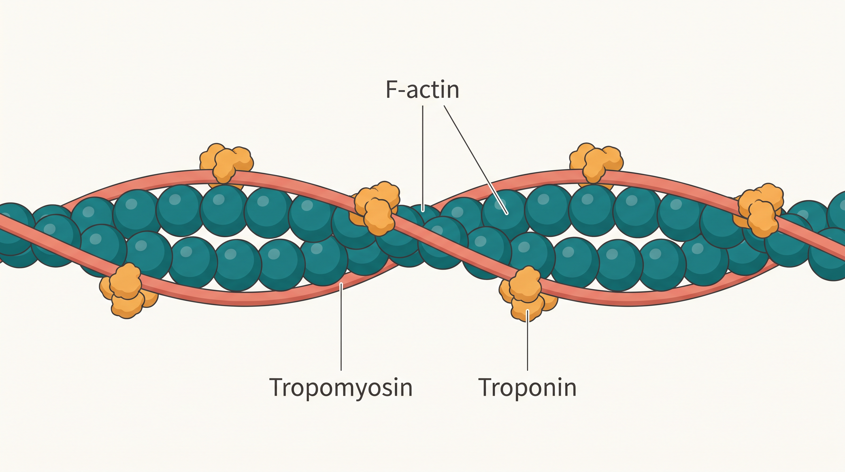
Fig. 17.3a – Structure of an actin (thin) filament.
The Thick Filament
Each myosin (thick) filament is also a polymerised protein, built from many monomer proteins called meromyosins (Fig. 17.3b).
Each meromyosin has two important parts: a globular head with a short arm, and a tail. The head-and-short-arm portion is called the heavy meromyosin (HMM), while the tail is called the light meromyosin (LMM).
The HMM component — the head together with its short arm — projects outwards from the surface of the polymerised myosin filament, at regular distances and angles from each other. This projecting part is known as the cross arm, because it will later reach across to the thin filament.
The globular head is not just a passive knob. It is an active ATPase enzyme (an enzyme that splits ATP to release energy), and it carries binding sites for ATP as well as active sites for actin.
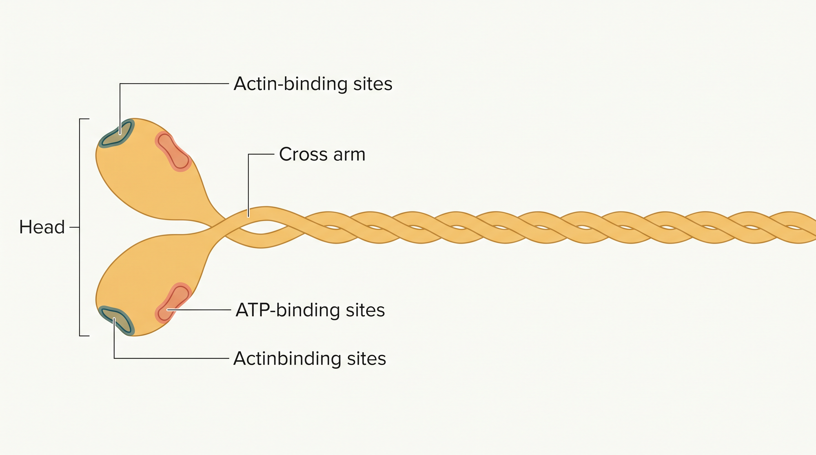
Fig. 17.3b – Structure of a myosin monomer (meromyosin).
With both filaments now understood — the thin filament that can be “unmasked” and the thick filament whose heads can grip and pull — we are ready to see how they work together to shorten a muscle.
17.2.2 Mechanism of Muscle Contraction
The Sliding Filament Theory
How does a sarcomere actually shorten? The best explanation is the sliding filament theory. It states that a muscle fibre contracts when the thin filaments slide over the thick filaments — not by the filaments themselves becoming shorter.
From Nerve to Muscle: The Signal Arrives
A muscle does not contract on its own — it waits for a command. Muscle contraction is initiated by a signal sent from the central nervous system (CNS) along a motor neuron (a nerve cell that carries instructions to a muscle).
A motor neuron together with all the muscle fibres connected to it is called a motor unit.
The point where a motor neuron meets the sarcolemma of a muscle fibre is called the neuromuscular junction (also called the motor-end plate).
When the neural signal reaches this junction, it releases a chemical messenger — a neurotransmitter called acetylcholine. Acetylcholine triggers an action potential (a wave of electrical change) in the sarcolemma.
Switching On: Calcium Unmasks the Actin
The action potential spreads through the muscle fibre and causes calcium ions (\(Ca^{2+}\)) to be released from the sarcoplasmic reticulum into the sarcoplasm.
This rise in \(Ca^{2+}\) is the crucial switch. The calcium binds to a subunit of troponin on the actin filaments, which shifts the troponin–tropomyosin cover aside and removes the masking of the myosin-binding sites. The active sites on actin are now exposed.
Using the energy released by ATP hydrolysis, \(ATP \rightarrow ADP + P_i\), the myosin head now reaches across and binds to the exposed active sites on actin, forming a cross bridge (Fig. 17.4).
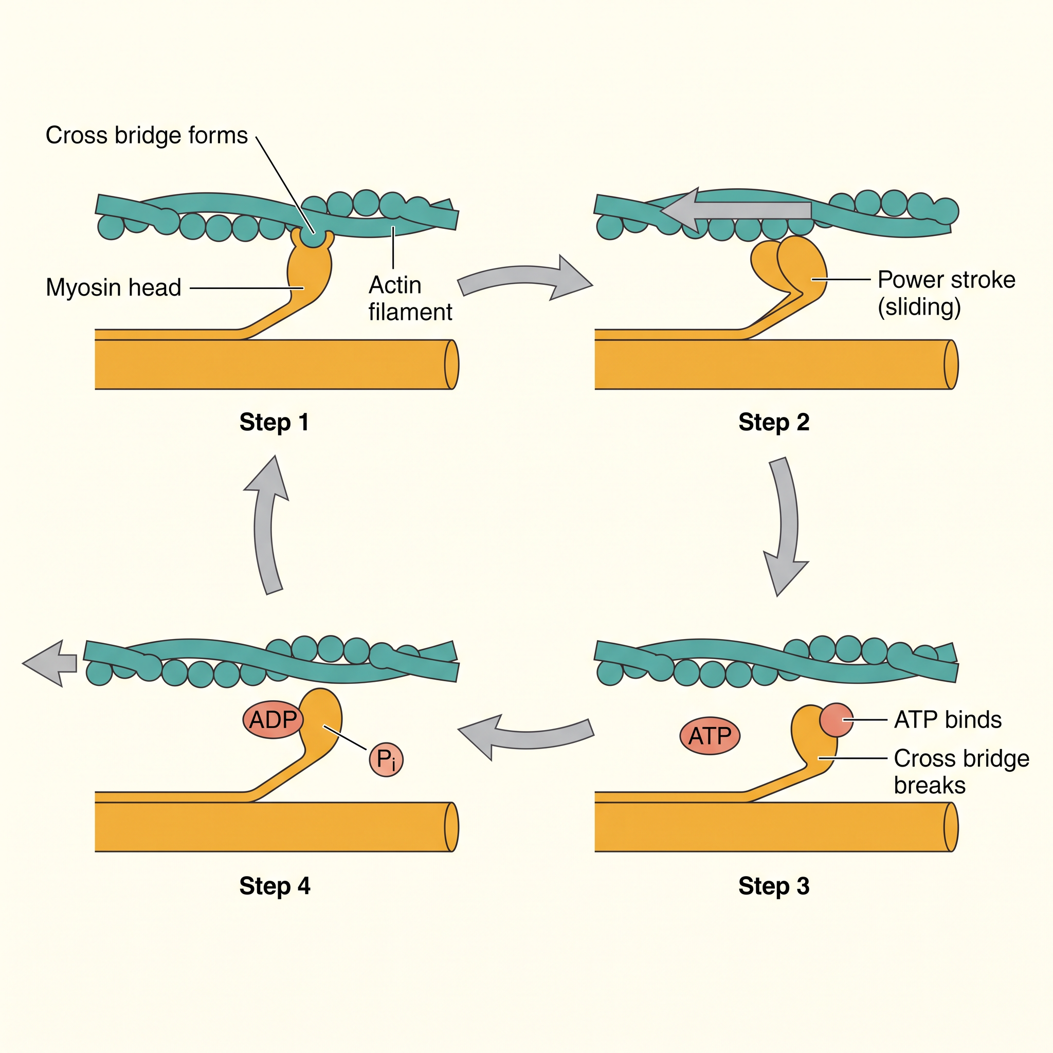
Fig. 17.4 – Stages of the cross-bridge cycle.
The Power Stroke: Filaments Slide In
Once the cross bridge forms, the myosin head rotates and pulls the attached actin filaments towards the centre of the A-band. Because the thin filaments are anchored to the Z-lines, the Z-lines are dragged inward too. This brings the two Z-lines of a sarcomere closer, shortening it — and that is exactly what we mean by contraction.
A helpful clue about which parts move can be seen in the banding pattern (Fig. 17.5). During contraction, the I-bands get reduced, while the A-bands keep their length. This makes sense: the A-band is the full length of the thick filaments, which do not shorten, whereas the I-band is the actin-only region that is progressively pulled into the overlap.
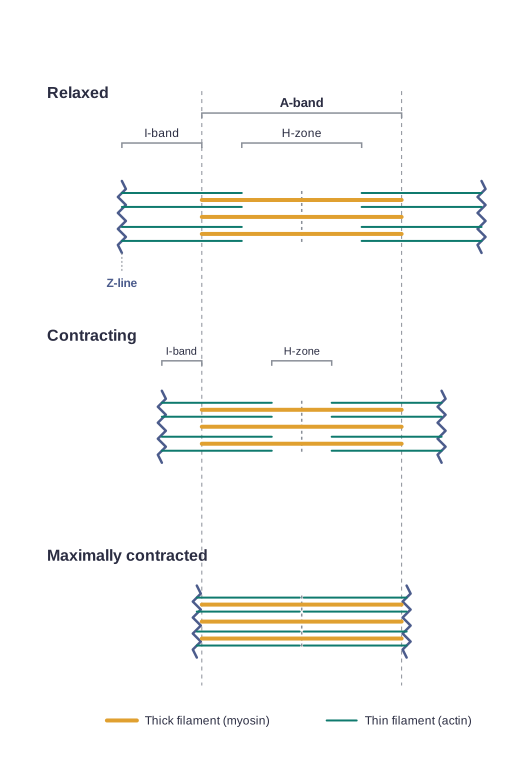
Fig. 17.5 – Sliding-filament theory of muscle contraction.
The whole sequence, from nerve signal to sliding, is summarised in FC 17.2.
Flowchart
FC 17.2 – Steps of muscle contraction: CNS signal → motor neuron → neuromuscular junction → acetylcholine released → action potential in sarcolemma → spreads through fibre → \(Ca^{2+}\) released from sarcoplasmic reticulum → \(Ca^{2+}\) binds troponin → myosin-binding sites unmasked → myosin head (ATP energy) binds actin → cross bridge → head rotates, actin slides in → Z-lines pulled together → sarcomere shortens (contraction)
Breaking the Cross Bridge and Relaxing
After the power stroke, the myosin releases ADP and \(P_i\) and returns to its relaxed state. A new ATP molecule then binds to the head, and the cross bridge is broken (Fig. 17.4). This ATP is again hydrolysed by the myosin head, and the cycle of cross-bridge formation and breakage repeats, producing further sliding.
This continues only as long as calcium is available. Relaxation begins when the \(Ca^{2+}\) ions are actively pumped back into the sac-like parts (cisternae) of the sarcoplasmic reticulum. With calcium gone, the troponin–tropomyosin cover slides back and once again masks the active sites on actin. The cross bridges can no longer form, the thin filaments slide back out, and the Z-lines return to their original positions — the muscle relaxes.
The reaction time of the fibres — how quickly they respond — can differ from one muscle to another.
Why Muscles Tire
Muscles cannot keep contracting forever. Repeated, prolonged activation can lead to the build-up of lactic acid in the muscle. This lactic acid comes from the anaerobic breakdown of glycogen (the stored carbohydrate) when oxygen supply cannot keep pace with demand. The accumulation of lactic acid is what causes fatigue — the tired, aching feeling in an overworked muscle.
Red Fibres and White Fibres
Not all skeletal muscle fibres are alike. Muscle contains a red, oxygen-storing pigment called myoglobin. In some muscles the myoglobin content is high, giving them a reddish colour; these are called Red fibres. Red fibres also contain plenty of mitochondria, which can use the large store of oxygen to produce ATP. Because they rely on oxygen, they are also called aerobic muscles.
Other muscles contain very little myoglobin and therefore look pale or whitish; these are the White fibres. They have few mitochondria, but a high amount of sarcoplasmic reticulum, and they depend on anaerobic processes for their energy.
17.3 Skeletal System
The skeletal system is the framework of bones and a few cartilages that gives the body its shape and support. Its role in movement is easy to overlook until you imagine trying to chew food without jaw bones, or walking around without the bones of your limbs — the muscles would have nothing firm to pull against.
Bone and Cartilage
Both bone and cartilage are specialised connective tissues. The difference lies in their matrix — the non-living material in which their cells are embedded. Bone has a very hard matrix, made rigid by deposits of calcium salts. Cartilage has a slightly pliable matrix, made flexible by chondroitin salts. This is why bones are stiff while cartilage (as in your ear or nose tip) can bend a little.
In human beings, the skeletal system is made up of 206 bones and a few cartilages. It is grouped into two principal divisions — the axial skeleton and the appendicular skeleton — as summarised in FC 17.3.
Flowchart
FC 17.3 – Divisions of the human skeleton: Skeleton → Axial skeleton (80 bones: skull, vertebral column, sternum, ribs) and Appendicular skeleton (limb bones + pectoral and pelvic girdles)
The Axial Skeleton and the Skull
The axial skeleton comprises 80 bones distributed along the main axis of the body. The skull, vertebral column, sternum and ribs together constitute the axial skeleton.
The skull (Fig. 17.6) is composed of two sets of bones — cranial and facial — which total 22 bones. The cranial bones are 8 in number; they form the hard protective outer covering, the cranium, that shields the brain. The facial region is made of 14 skeletal elements that form the front of the skull.
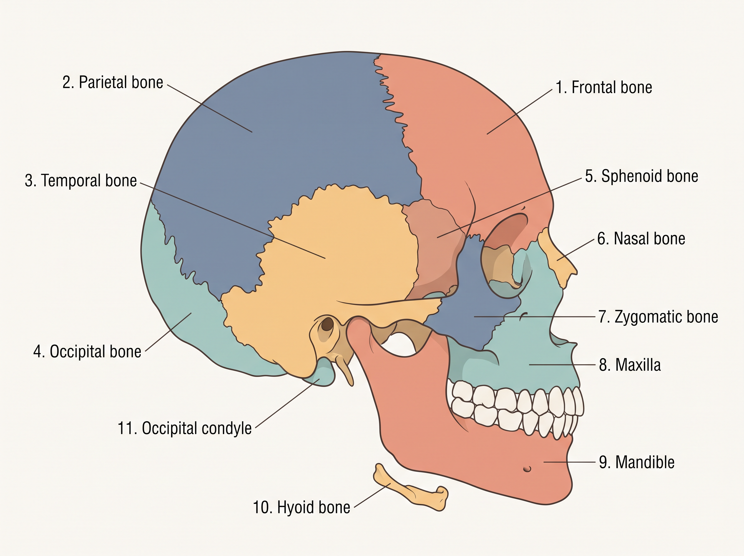
Fig. 17.6 – Lateral view of the human skull.
Two more sets of small bones are associated with this region. A single U-shaped bone called the hyoid lies at the base of the buccal cavity (mouth). And each middle ear contains three tiny bones — the malleus, incus and stapes — collectively called the ear ossicles.
The skull rests on and connects with the top of the vertebral column through two occipital condyles — knob-like surfaces at the base of the skull. Because there are two such condyles, the human skull is described as a dicondylic skull. (To articulate simply means to form a joint or movable connection with another bone.)
The Vertebral Column
Our vertebral column (Fig. 17.7) is formed by 26 serially arranged units called vertebrae, and it lies dorsally (along the back). It extends from the base of the skull and forms the main framework of the trunk.
Each vertebra has a central hollow portion, the neural canal, through which the spinal cord passes. The very first vertebra, the atlas, is the one that articulates with the occipital condyles of the skull.
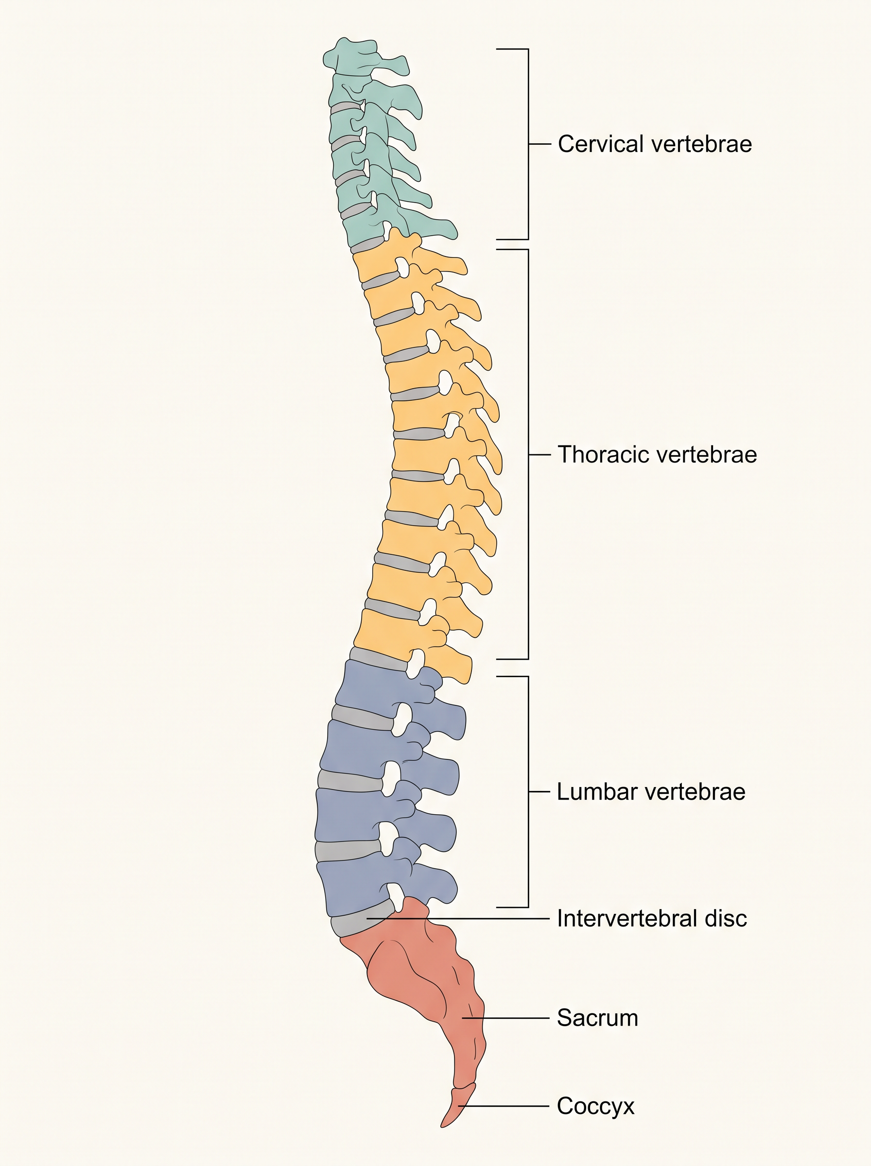
Fig. 17.7 – Right lateral view of the vertebral column.
Starting from the skull, the vertebral column is differentiated into five regions: cervical (7), thoracic (12), lumbar (5), sacral (1, fused) and coccygeal (1, fused). Interestingly, the number of cervical vertebrae is seven in almost all mammals, including human beings — whether it is a mouse or a giraffe.
The vertebral column does three important jobs: it protects the spinal cord, supports the head, and serves as the point of attachment for the ribs and the muscles of the back.
The Rib Cage: Sternum and Ribs
The sternum is a flat bone lying on the ventral midline of the thorax (the front centre of the chest).
There are 12 pairs of ribs. Each rib is a thin, flat bone joined dorsally to the vertebral column and ventrally to the sternum. Because each rib has two articulation surfaces on its dorsal end, it is described as bicephalic (two-headed).
The ribs fall into three groups, as shown in FC 17.4. The first seven pairs are the true ribs: dorsally they attach to the thoracic vertebrae, and ventrally they connect to the sternum through hyaline cartilage. The 8th, 9th and 10th pairs do not reach the sternum directly; instead each joins the seventh rib through hyaline cartilage, so they are called vertebrochondral (false) ribs. The last two pairs (11th and 12th) are not connected ventrally at all and are therefore called floating ribs.
Flowchart
FC 17.4 – Classification of ribs: 12 pairs → True ribs (pairs 1–7, joined to sternum directly) → False/vertebrochondral ribs (pairs 8–10, joined to the 7th rib) → Floating ribs (pairs 11–12, not joined ventrally)
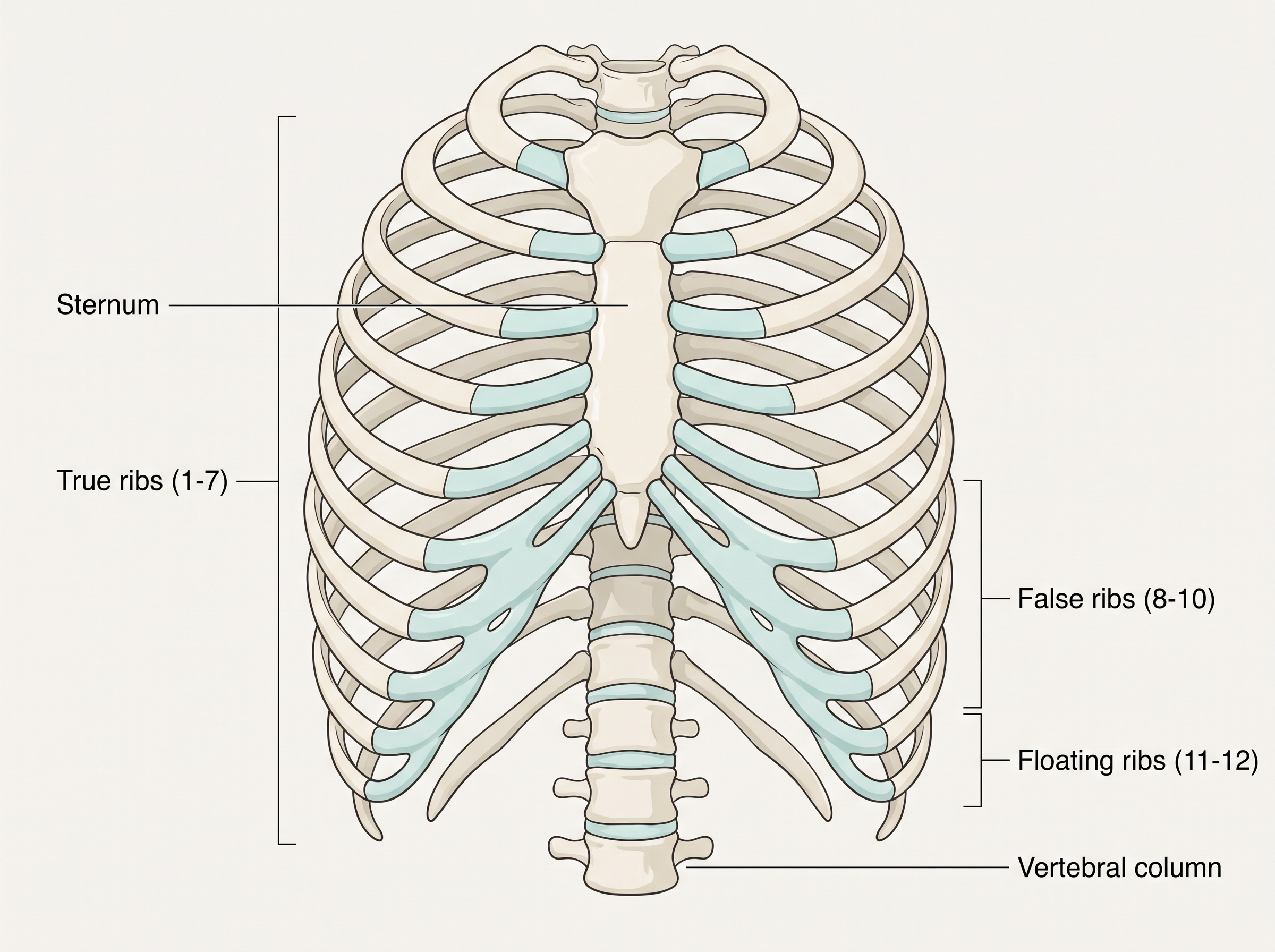
Fig. 17.8 – The human rib cage.
Together, the thoracic vertebrae, the ribs and the sternum form the rib cage, which shields the heart and lungs.
The Appendicular Skeleton: Bones of the Limbs
The bones of the limbs, along with their girdles, make up the appendicular skeleton. Each limb is made of 30 bones.
In the hand (fore limb), the bones are the humerus, the radius and ulna, the carpals (wrist bones — 8 in number), the metacarpals (palm bones — 5 in number) and the phalanges (digits — 14 in number), as shown in Fig. 17.9.
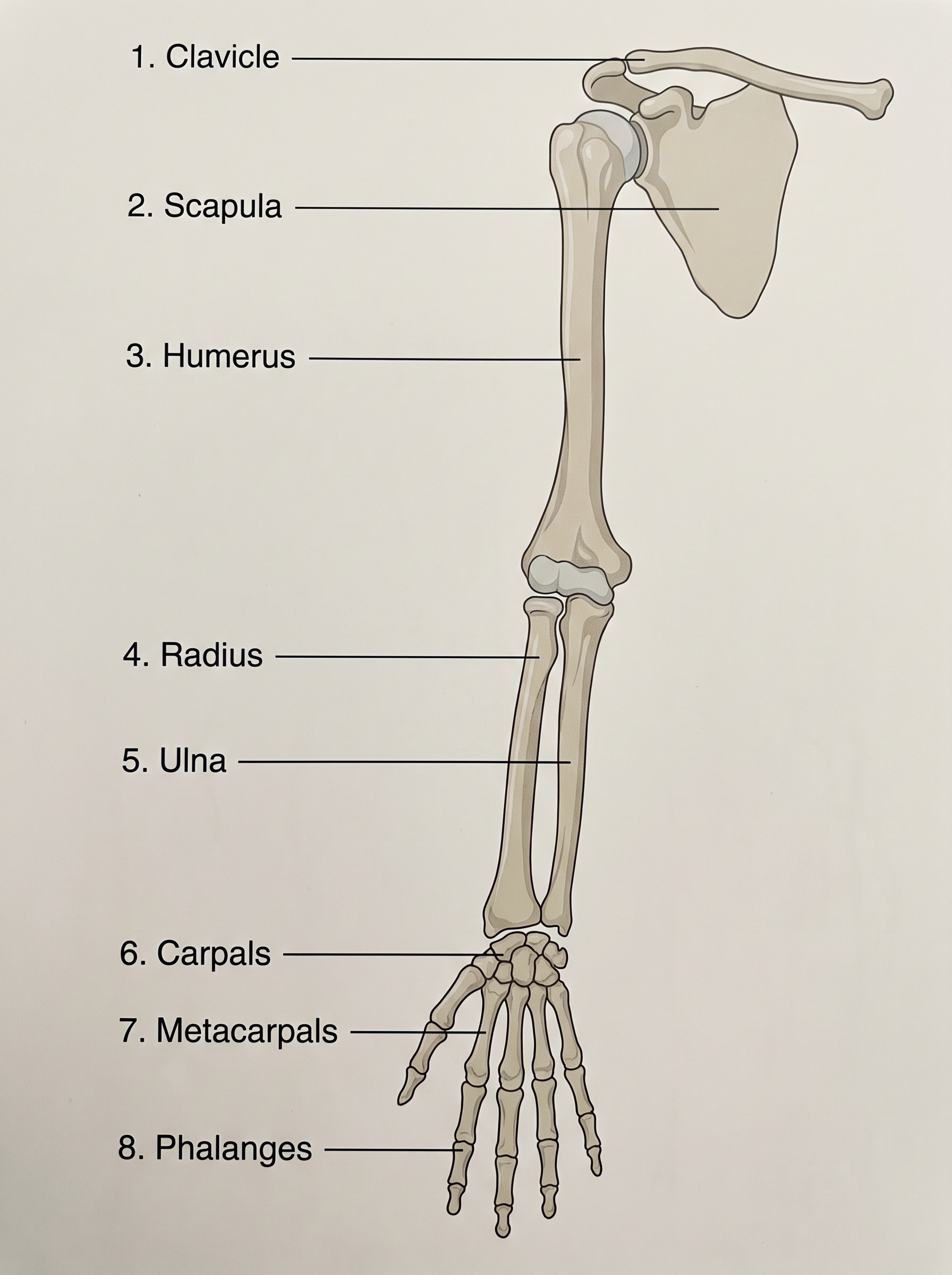
Fig. 17.9 – Right pectoral girdle and upper arm.
In the leg (hind limb), the bones are the femur (the thigh bone, which is the longest bone in the body), the tibia and fibula, the tarsals (ankle bones — 7 in number), the metatarsals (5 in number) and the phalanges (digits — 14 in number), as shown in Fig. 17.10. In addition, a cup-shaped bone called the patella covers the knee ventrally — this is the knee cap.
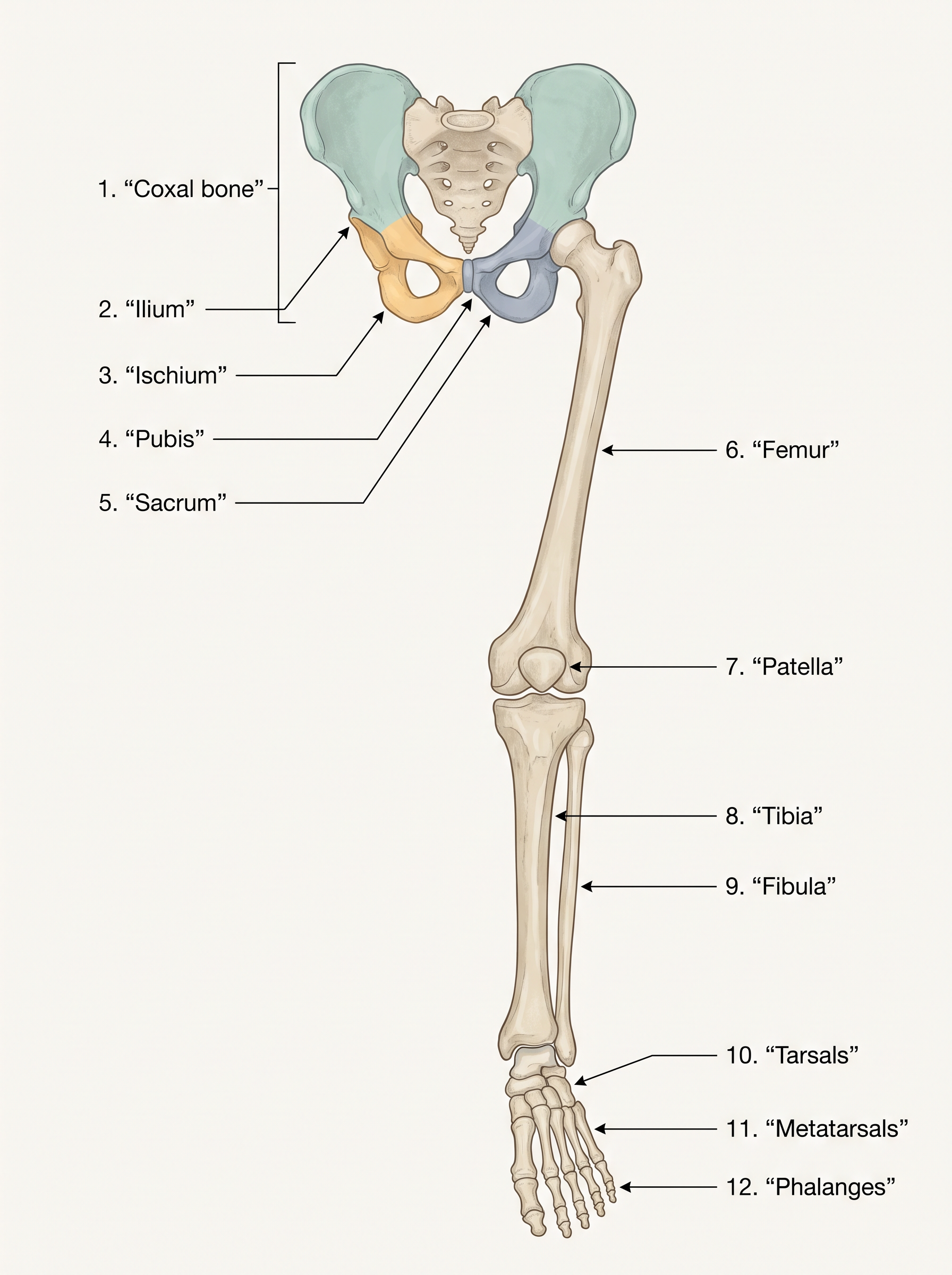
Fig. 17.10 – Right pelvic girdle and lower limb.
The Girdles That Anchor the Limbs
The pectoral and pelvic girdles connect the upper and lower limbs, respectively, to the axial skeleton. Each girdle is formed of two halves.
Each half of the pectoral girdle consists of a clavicle and a scapula (Fig. 17.9). The scapula is a large, triangular, flat bone in the dorsal part of the thorax, lying between the second and the seventh ribs. Its flat triangular body carries a slightly raised ridge called the spine, which projects as a flat, expanded process called the acromion; the clavicle articulates with this. Just below the acromion is a depression, the glenoid cavity, which receives the head of the humerus to form the shoulder joint. Each clavicle is a long, slender, doubly curved bone — commonly known as the collar bone.
The pelvic girdle consists of two coxal bones (Fig. 17.10). Each coxal bone is formed by the fusion of three bones — the ilium, ischium and pubis. At the point where these three fuse lies a cavity called the acetabulum, into which the head of the thigh bone (femur) fits. The two halves of the pelvic girdle meet ventrally to form the pubic symphysis, which contains fibrous cartilage.
17.4 Joints
Muscles and bones together can generate force, but that force only becomes movement at the places where bones meet. These meeting points are joints, and they are essential for every kind of movement involving the bony parts of the body — locomotion included.
What a Joint Is and Does
A joint is a point of contact between two bones, or between a bone and a cartilage. When a muscle contracts, the force it generates is transmitted through a joint, and the joint acts as a fulcrum — the fixed pivot around which a lever turns. The bones act as the levers, the muscle supplies the effort, and the joint is the point about which movement swings.
The amount of movement allowed at a joint — its movability — varies depending on several factors, chiefly the way the bones are held together. On this basis, joints are classified into three major structural forms: fibrous, cartilaginous and synovial (FC 17.5).
Flowchart
FC 17.5 – Classification of joints: Joints → Fibrous (immovable) / Cartilaginous (limited movement) / Synovial (free movement); Synovial → ball-and-socket, hinge, pivot, gliding, saddle
Fibrous Joints
Fibrous joints do not allow any movement at all. They are seen in the flat bones of the skull, which fuse end-to-end with the help of dense fibrous connective tissue in the form of sutures, forming the rigid cranium that protects the brain.
Cartilaginous Joints
In cartilaginous joints, the bones are joined together with the help of cartilages. The joint between adjacent vertebrae in the vertebral column is of this type, and it permits only limited movement — enough for the spine to bend a little, but not freely.
Synovial Joints
Synovial joints are the freely movable joints. They are characterised by a fluid-filled synovial cavity between the articulating surfaces of the two bones. This fluid-filled gap lets the bone surfaces glide smoothly, so these joints allow considerable movement and are the ones most involved in locomotion and other body movements.
There are several kinds of synovial joint, each suited to a particular type of motion: the ball-and-socket joint (between the humerus and the pectoral girdle), the hinge joint (the knee joint), the pivot joint (between the atlas and the axis), the gliding joint (between the carpals) and the saddle joint (between the carpal and metacarpal of the thumb).
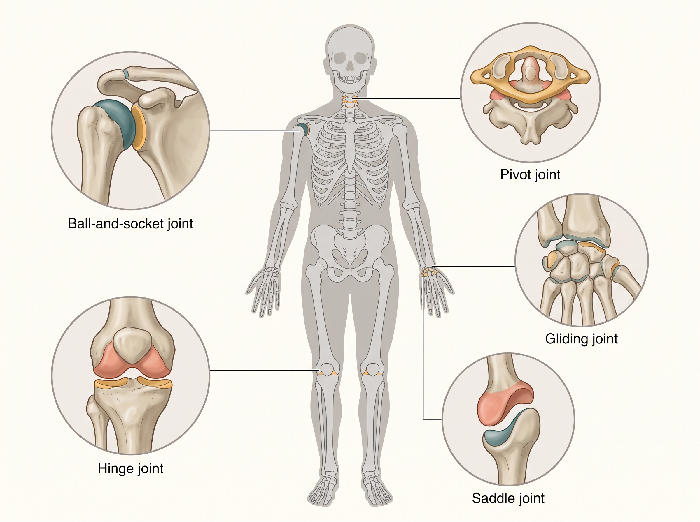
Fig. 17.11 – Main types of synovial joints in the human body.
17.5 Disorders of Muscular and Skeletal System
Having seen how muscles, bones and joints normally work, it helps to know what happens when they go wrong. Several disorders can affect the muscular and skeletal systems; each can be understood by linking it back to a structure or process you have already studied.
Disorders Affecting Muscles
Myasthenia gravis is an auto-immune disorder — one in which the body’s own immune system attacks its own tissues. Here the attack is directed at the neuromuscular junction, the very site where a nerve normally signals a muscle to contract. As a result, signals fail to pass properly, leading to fatigue, weakening and, in severe cases, paralysis of the skeletal muscle.
Muscular dystrophy is a progressive degeneration of skeletal muscle — the muscle tissue gradually breaks down and weakens over time. It is mostly a genetic disorder, inherited through faulty genes.
Tetany refers to rapid spasms, or wild, uncontrolled contractions, of muscle. It is caused by a fall in the level of calcium ions (\(Ca^{2+}\)) in the body fluid. Recall that calcium is the switch that controls muscle contraction; when its balance in the body fluid is disturbed, the muscles can fire in an uncontrolled way.
Disorders Affecting Bones and Joints
Osteoporosis is an age-related disorder marked by decreased bone mass and an increased chance of fractures. A common cause is a fall in the level of the hormone estrogen, which is why it is frequently seen in older women. With less bone mass, the bones become fragile and break easily.
Arthritis is the inflammation of joints, which makes them painful and stiff.
Gout is a specific kind of joint inflammation caused by the accumulation of uric acid crystals in the joints.
17.6 Summary
Movement is an essential feature of all living beings. Protoplasmic streaming, ciliary movement, and the movement of fins, limbs and wings are some of the forms shown by animals.
A voluntary movement that shifts the whole animal to a new place is called locomotion. Animals generally move in search of food, shelter, a mate or breeding ground, better climate, or to protect themselves.
The cells of the human body show three kinds of movement — amoeboid, ciliary and muscular. Locomotion and most other movements need coordinated muscular activity.
There are three types of muscle. Skeletal muscles are attached to skeletal elements; they are striated and voluntary. Visceral (smooth) muscles line the inner walls of visceral organs; they are non-striated and involuntary. Cardiac muscles form the heart; they are striated, branched and involuntary.
All muscle shares four properties: excitability, contractility, extensibility and elasticity.
The muscle fibre is the anatomical unit of muscle. Each fibre contains many parallel myofibrils, and each myofibril is made of serially arranged sarcomeres, the functional units of contraction.
Each sarcomere has a central A-band of thick myosin filaments, flanked by two half I-bands of thin actin filaments, bounded by Z-lines. Actin and myosin are polymerised, contractile proteins.
In a resting muscle, the myosin-binding sites on actin are masked by the protein troponin. The myosin head carries ATPase activity, with binding sites for ATP and active sites for actin.
Contraction begins when a motor neuron signals the muscle fibre, generating an action potential. This releases \(Ca^{2+}\) from the sarcoplasmic reticulum. Calcium unmasks the actin sites; the myosin head binds actin to form a cross bridge, and the cross bridges pull the thin filaments to slide over the thick filaments — this is contraction. When \(Ca^{2+}\) returns to the sarcoplasmic reticulum, the sites are masked again, the cross bridges break, and the muscle relaxes.
Flowchart
FC 17.6 – Condensed contraction cycle: motor neuron signal → action potential → \(Ca^{2+}\) released → actin unmasked → cross bridge forms → thin filaments slide (contraction) → \(Ca^{2+}\) pumped back → actin masked → cross bridges break (relaxation)
Repeated stimulation leads to fatigue. Based mainly on the amount of the red pigment myoglobin, muscles are classified as Red fibres (aerobic) and White fibres (anaerobic).
Bones and cartilages make up the skeletal system, which is divided into the axial and appendicular skeletons. The skull, vertebral column, ribs and sternum form the axial skeleton; the limb bones and girdles form the appendicular skeleton.
Three types of joints connect bones, or bone and cartilage — fibrous, cartilaginous and synovial. Synovial joints allow considerable movement and so play a significant role in locomotion.
17.7 Points to Ponder
All locomotion is movement, but not all movement is locomotion. Look at what your own body is doing right now — which of its movements are not locomotion, and what would happen if these quiet, non-locomotory movements stopped?
During contraction, the actin and myosin filaments do not themselves shorten — they slide past one another. Why might sliding be a more efficient and reliable way for a muscle to shorten than actually shrinking its protein molecules?
Calcium ions act as the switch that both starts contraction and, when pumped away, ends it. What might happen to a muscle if calcium could no longer be pumped back into the sarcoplasmic reticulum after each contraction?
Red fibres are rich in myoglobin and mitochondria and work aerobically, while white fibres rely on anaerobic energy. If you were “designing” the flight muscles of a long-distance migratory bird versus a bird that flies only in short, sudden bursts, which fibre type should dominate in each, and why?
The number of cervical vertebrae is seven in almost all mammals — the same in a mouse, a human and a giraffe. What might this shared number tell us about how these very different animals are related?
The skull bones meet at immovable fibrous joints, while the limb bones meet at freely movable synovial joints. Why is it an advantage for the skull to be rigid but the limbs to be flexible? What problems would arise if this arrangement were reversed?
Bone is a living tissue that can gain and lose mass throughout life. Given that osteoporosis involves a loss of bone mass with age, how might a person’s habits during their youth influence the strength of their bones decades later?
17.8 NCERT Questions
Draw the diagram of a sarcomere of skeletal muscle showing different regions.
Define the sliding filament theory of muscle contraction.
Describe the important steps in muscle contraction.
Write true or false. If false, change the statement so that it is true.
- Actin is present in the thin filament.
- The H-zone of a striated muscle fibre represents both thick and thin filaments.
- The human skeleton has 206 bones.
- There are 11 pairs of ribs in man.
- The sternum is present on the ventral side of the body.
Write the difference between:
- Actin and Myosin
- Red and White muscles
- Pectoral and Pelvic girdle
Match Column I with Column II:
| Column I | Column II |
|---|---|
| a. Smooth muscle | i. Myoglobin |
| b. Tropomyosin | ii. Thin filament |
| c. Red muscle | iii. Sutures |
| d. Skull | iv. Involuntary |
What are the different types of movements exhibited by the cells of the human body?
How do you distinguish between a skeletal muscle and a cardiac muscle?
Name the type of joint between the following:
- atlas/axis
- carpal/metacarpal of thumb
- between phalanges
- femur/acetabulum
- between cranial bones
- between pubic bones in the pelvic girdle
Fill in the blank spaces:
- All mammals (except a few) have __________ cervical vertebra.
- The number of phalanges in each limb of human is __________.
- The thin filament of a myofibril contains 2 ‘F’ actins and two other proteins, namely __________ and __________.
- In a muscle fibre, Ca²⁺ is stored in the __________.
- __________ and __________ pairs of ribs are called floating ribs.
- The human cranium is made of __________ bones.
17.9 Check Your Concepts
Assertion–Reason. A: During muscle contraction, the width of the A-band stays the same. R: The thick myosin filaments do not shorten during contraction; only the thin filaments slide inward. State whether both A and R are true and whether R correctly explains A.
Assertion–Reason. A: A resting muscle fibre does not contract even when ATP is present in it. R: In the resting state, a subunit of troponin masks the active sites for myosin on the actin filament. State whether both A and R are true and whether R correctly explains A.
Assertion–Reason. A: Cardiac muscle is involuntary even though it appears striated. R: The nervous system does not directly control the activity of cardiac muscle. State whether both A and R are true and whether R correctly explains A.
Arrange the following events of muscle contraction in the correct sequence: (i) a cross bridge forms between the myosin head and actin; (ii) acetylcholine is released at the neuromuscular junction; (iii) Ca²⁺ is released from the sarcoplasmic reticulum; (iv) an action potential is generated in the sarcolemma; (v) the thin filaments slide inward and the sarcomere shortens; (vi) troponin shifts and unmasks the active sites on actin.
Arrange these structural levels of skeletal muscle from largest to smallest: myofibril, muscle, myofilament, fascicle, muscle fibre.
Match the type of synovial joint in Column I with its example in Column II.
| Column I | Column II |
|---|---|
| a. Ball-and-socket joint | i. Between the carpals |
| b. Hinge joint | ii. Between atlas and axis |
| c. Pivot joint | iii. Between humerus and pectoral girdle |
| d. Gliding joint | iv. Between carpal and metacarpal of thumb |
| e. Saddle joint | v. Knee joint |
- Match each structure of the muscle fibre in Column I with its correct description in Column II.
| Column I | Column II |
|---|---|
| a. Sarcolemma | i. Stores calcium ions |
| b. Z-line | ii. Plasma membrane of the muscle fibre |
| c. Sarcoplasmic reticulum | iii. Holds thick filaments together in the middle of the A-band |
| d. M-line | iv. Bisects the I-band and anchors the thin filaments |
Why does prolonged, intense exercise lead to a fatigued, aching feeling in the muscles? Name the substance responsible and the process that produces it.
What would happen to muscle relaxation if the calcium pumps of the sarcoplasmic reticulum stopped working? Explain your reasoning.
Using the structure and function of the myosin head, explain why ATP is required both for muscle contraction and for the breaking of the cross bridge during relaxation.
Draw a labelled diagram of a myosin monomer (meromyosin) and mark the head, the cross arm, the ATP-binding site and the actin-binding site.
With the help of a labelled sketch of the rib cage, identify the true ribs, the false (vertebrochondral) ribs and the floating ribs, and state how each group is connected (or not connected) to the sternum.
Differentiate between heavy meromyosin (HMM) and light meromyosin (LMM).
Distinguish between the axial and the appendicular skeleton, listing the main bones that make up each.
A person’s blood calcium level falls sharply. Predict the effect on their muscles and name the resulting condition.
Explain why a patient with myasthenia gravis experiences muscle weakness and fatigue, even though the muscle proteins themselves may be structurally normal. Which part of the muscle system is affected?
The number of cervical vertebrae is the same (seven) in a mouse, a human and a giraffe. What does this constancy suggest, and why is it a useful example of a shared feature among mammals?