Chapter 7: Structural Organisation in Animals
If you have ever stood near a village pond or a flooded paddy field after the first heavy monsoon rains, you will remember the sudden chorus of frogs calling into the night — where only days earlier there was dry, cracked mud. Look into the shallow water a little later and you find something stranger still: floating clusters of jelly-like eggs, and soon after, hundreds of tiny black tadpoles wriggling like little fish.
Weeks pass. The tails shrink, four legs push out, and small frogs begin climbing onto the bank to gulp air (Fig. 7.0). A single animal has quietly lived two completely different lives — one in water with a fish-like, gill-breathing body, and one on land with lungs and legs.
This double life is why the frog belongs to the class Amphibia, from Greek words meaning “both kinds of life.” It is also why biologists have long chosen the frog as the animal to study first when learning how a backboned (vertebrate) body is built. Inside that small, familiar body lie neatly arranged organ systems — for digestion, breathing, circulation, waste removal, control and reproduction — each doing its own share of the work. In this chapter you will open up the frog, at least in your imagination, and trace how countless cells are organised into tissues, organs and organ systems that together keep the animal alive.
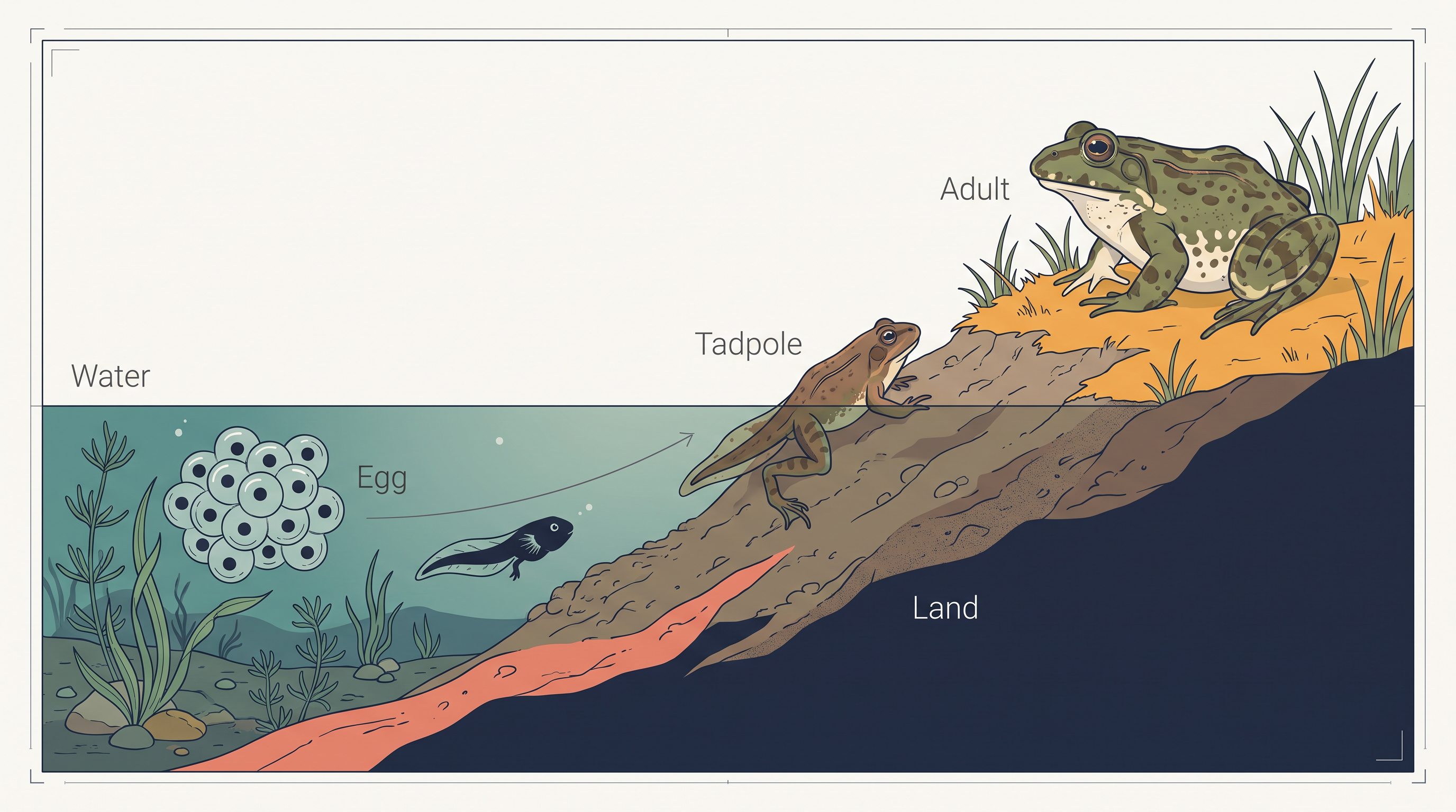
Fig. 7.0 – The life cycle of a frog.
You cannot answer these on this page — but by the end of the chapter, each one will make clear sense.
7.1 Organ and Organ System
In the preceding chapters you came across a large variety of animals — some built from just a single cell, others from countless cells. In a unicellular organism, that one cell must do everything by itself: it digests food, exchanges gases for respiration, and reproduces, all within the same tiny package. A large, complex animal cannot run on this plan. Its body contains so many cells that the different tasks of staying alive are shared out among different groups of cells, each group specialised for a particular job.
Even a simple animal like Hydra already shows this sharing of work. Its body is made of several distinct kinds of cells, and each kind may number in the thousands. The human body carries this far further, with billions of cells belonging to many different types. This raises an obvious question: how do so many cells manage to act together, instead of each going its own way? The answer lies entirely in how they are organised.
From Cells to Organ Systems
When a group of similar cells, together with the material lying between them, works as a single unit to carry out a particular function, that unit is called a tissue. That in-between material is the intercellular substance — a non-living matrix that surrounds the cells and binds them into one working whole. By pooling their effort, cells of the same kind achieve as a tissue what no single cell could achieve alone.
You may be surprised to learn that the bodies of all complex animals are built from only four basic types of tissue. These four tissues are combined in fixed proportions and patterns to build an organ — a distinct body part such as the stomach, lung, heart or kidney. It is this exact proportion and arrangement that gives each organ its particular shape and its particular job. An organ, then, is made of one or more tissue types working together.
The heart makes this clear. We picture it as a single pump, yet it actually contains all four tissue types at once, each supplying something the organ needs.
Organs seldom act on their own. When two or more organs work towards one common function through physical and/or chemical interaction, they together form an organ system — the digestive system and the respiratory system are familiar examples.
The body is therefore organised in rising levels: cells build tissues, tissues build organs, organs build organ systems, and the organ systems together make up the whole organism. This ladder of organisation is summarised in FC 7.1.
Flowchart
FC 7.1 – Levels of structural organisation in a complex animal: cell → tissue → organ → organ system → organism
Notice what this arrangement achieves. Instead of every cell struggling to perform every task, the work is divided up — a plan we call division of labour. Each level specialises in its own share, and specialists working as a team keep the millions of cells of the body active in an efficient and well-coordinated way. This is precisely why such organisation is essential in any large animal, and why it contributes to the survival of the body as a whole.
When we compare the organs and organ systems of different animals, we find that their complexity does not differ at random; it changes along a recognisable pattern.
Two Ways of Studying an Animal
To understand an animal fully, biologists examine it in two complementary ways. The first is its morphology — its form and the features that can be seen from the outside.
For plants and microbes, the word morphology means only this outward form. In animals, it refers to the external appearance of the organs or parts of the body. The second way looks inward, and is called anatomy.
Students often confuse these two terms in examinations, so it is worth fixing the difference firmly in mind.
In this chapter you will study both the morphology and the anatomy of the frog — an animal chosen because it represents the vertebrates, the great group of backboned animals to which we ourselves belong.
7.2 Frogs
Frogs are equally at home on land and in freshwater, and this ability to live in two habitats places them in the class Amphibia of the phylum Chordata — the phylum whose members possess a backbone. The frog most commonly found in India is the Indian bullfrog, Rana tigrina, and it is this species we take as our example throughout the chapter.
A frog does not keep a fixed, constant body temperature. Instead, its body temperature rises and falls along with the temperature of its surroundings. Animals of this kind are described as cold-blooded, or poikilotherms. One advantage of this arrangement is that the frog spends almost no energy generating body heat, so it needs far less food than a warm-blooded animal of similar size. The drawback is that it is left at the mercy of the weather — active in warm conditions, and sluggish in cold ones.
You may have noticed that a frog’s colour is not always the same on green grass as on dry ground. Frogs can change their body colour to blend with their surroundings and so hide from their enemies — a protective ability called camouflage. A predator that cannot spot its prey cannot catch it, so this blending-in is an effective defence. This protective coloration is referred to as mimicry.
Surviving the Extremes
Frogs are also hard to find during the peak of summer and the depth of winter. Because they cannot control their own body temperature, fierce heat could dry out their moist bodies while severe cold would leave them helpless. To escape both, they take shelter deep inside burrows, where their life processes slow right down until the harsh season passes. The summer rest is called aestivation (summer sleep) and the winter rest is called hibernation (winter sleep).
7.2.1 Morphology
A frog’s outward form is closely matched to its life in and around water, and the details described here can be followed on the external view in Fig. 7.1.
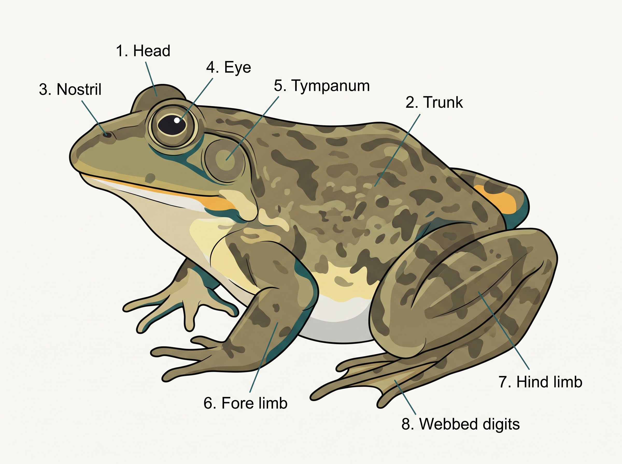
Fig. 7.1 – External features of a frog.
The Skin
Have you ever touched the skin of a frog? It feels smooth and slippery because it is covered with mucus, and it is always kept moist. The upper (dorsal) surface of the body is usually olive green marked with dark, irregular spots, while the lower (ventral) surface is a uniform pale yellow. This colour pattern is itself a way of hiding: seen from above, the mottled green back blends with pond weeds and muddy water, and the pale belly is hard to make out against bright light coming from below.
Interestingly, a frog never drinks water in the way we do. Instead, it takes up water directly through its skin, which is one more reason the skin must stay damp at all times.
Body, Head and Sense Organs
The body of a frog is divided into just two parts — a head and a trunk (Fig. 7.1); there is no distinct neck and no tail. Just above the mouth lies a pair of nostrils, through which air enters.
The eyes are large and bulging, and each is covered by a special transparent fold called the nictitating membrane.
On either side of the eyes lies a rounded membrane called the tympanum, which serves as the frog’s ear and receives sound signals from its surroundings.
Limbs and Sexual Dimorphism
The forelimbs and hind limbs together allow the frog to swim, walk, leap and burrow. The hind limbs are distinctly larger and more muscular than the forelimbs, and they end in five digits, whereas the forelimbs end in four. The extra muscle in the hind limbs is exactly what powers the frog’s strong leaps and swimming strokes.
Between the digits of the feet is a stretch of skin called a web.
Male and female frogs are not alike in appearance — they show sexual dimorphism.
In frogs, the male can be told apart by two features that the female lacks: a pair of sound-producing vocal sacs, and a copulatory pad on the first digit of each forelimb.
7.2.2 Anatomy
Packed inside the frog’s body cavity are all the organ systems that keep it alive — the digestive, circulatory, respiratory, nervous, excretory and reproductive systems — each well developed and each carrying out its own set of functions. Many of these organs can be seen in their natural positions in Fig. 7.2.
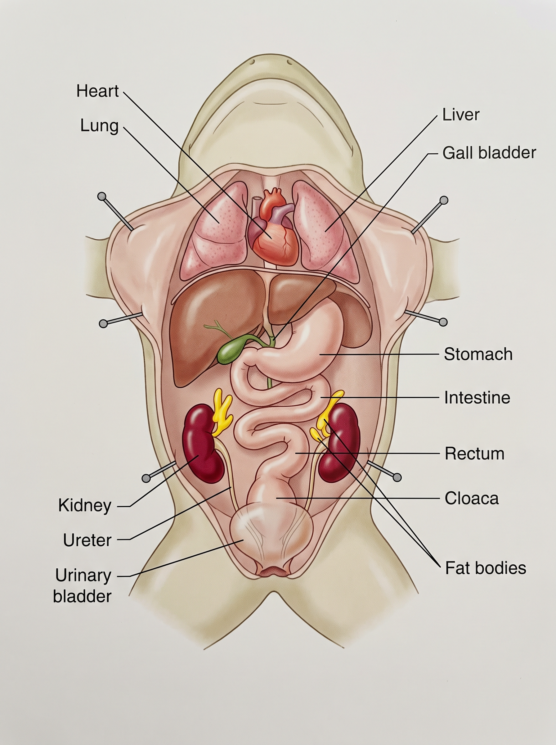
Fig. 7.2 – Internal organs of a frog.
The Digestive System
The digestive system has two parts: the alimentary canal (the long tube through which food travels) and the digestive glands that pour their juices into it. In the frog, the alimentary canal is short. This is because frogs are carnivores — they eat animal food such as insects, which is easier to break down than plant food — so a long intestine is not needed, and its length is reduced.
Food travels along the canal in a fixed order. The mouth opens into the buccal cavity, which leads through the pharynx into the oesophagus. The oesophagus is a short tube that opens into the stomach; the stomach continues as the intestine, then the rectum, and finally opens to the outside through the cloaca. This whole passage is summarised in FC 7.2.
Flowchart
FC 7.2 – Passage of food through the alimentary canal: mouth → buccal cavity → pharynx → oesophagus → stomach → duodenum (first part of small intestine) → intestine → rectum → cloaca → exterior
Two large digestive glands assist the canal. The liver secretes bile, which is stored in a small sac called the gall bladder. The pancreas is a digestive gland that produces pancreatic juice, and this juice contains digestive enzymes.
Now follow what happens to a meal. Food is first captured by the bilobed tongue — a tongue that is divided into two lobes at its tip. In the stomach, the food is acted upon by hydrochloric acid (HCl) and gastric juices secreted by the walls of the stomach. The partly digested food, now a semi-liquid mass called chyme, is passed from the stomach into the first part of the small intestine, the duodenum.
The duodenum receives two secretions through a common bile duct: bile from the gall bladder and pancreatic juice from the pancreas. Bile itself carries no enzymes; instead it emulsifies fat — breaking large fat droplets into many tiny ones so that enzymes can act on them more easily. The pancreatic juice then digests carbohydrates and proteins. The final steps of digestion are completed in the intestine.
The digested food must now be taken into the body. The inner wall of the intestine is thrown into countless finger-like folds called villi, and the surface of these is folded still further into microvilli.
Whatever remains undigested — the solid waste — is moved into the rectum and finally passed out through the cloaca.
The Respiratory System
A frog can breathe in two quite different ways, and it is this that allows it to live both in water and on land.
When the frog is in water, its skin serves as the respiratory organ. Oxygen dissolved in the water passes straight through the moist skin into the blood by diffusion. Breathing through the skin in this way is called cutaneous respiration.
On land, three surfaces can be used for breathing — the buccal cavity, the skin, and the lungs. Breathing with the help of the lungs is called pulmonary respiration. The lungs are a pair of elongated, pink, sac-like structures lying in the upper part of the trunk region (the thorax). Air reaches them by entering through the nostrils into the buccal cavity, and then passing on into the lungs.
There is also one special situation. During aestivation and hibernation — when the frog is shut away in its burrow — gaseous exchange takes place only through the skin.
The Circulatory System
The circulatory (vascular) system of the frog is well developed and of the closed type — meaning the blood always flows within vessels and is never let loose into open body spaces. Alongside this blood system, the frog also possesses a lymphatic system.
The blood vascular system is made up of the heart, the blood vessels, and the blood itself. The lymphatic system, in turn, consists of lymph, lymph channels and lymph nodes.
The heart is a muscular organ lying in the upper part of the body cavity, wrapped in a protective membrane called the pericardium. Unlike our own four-chambered heart, the frog’s heart has three chambers — two atria (the upper chambers) and a single ventricle (the lower chamber). Its structure is shown in Fig. 7.3.
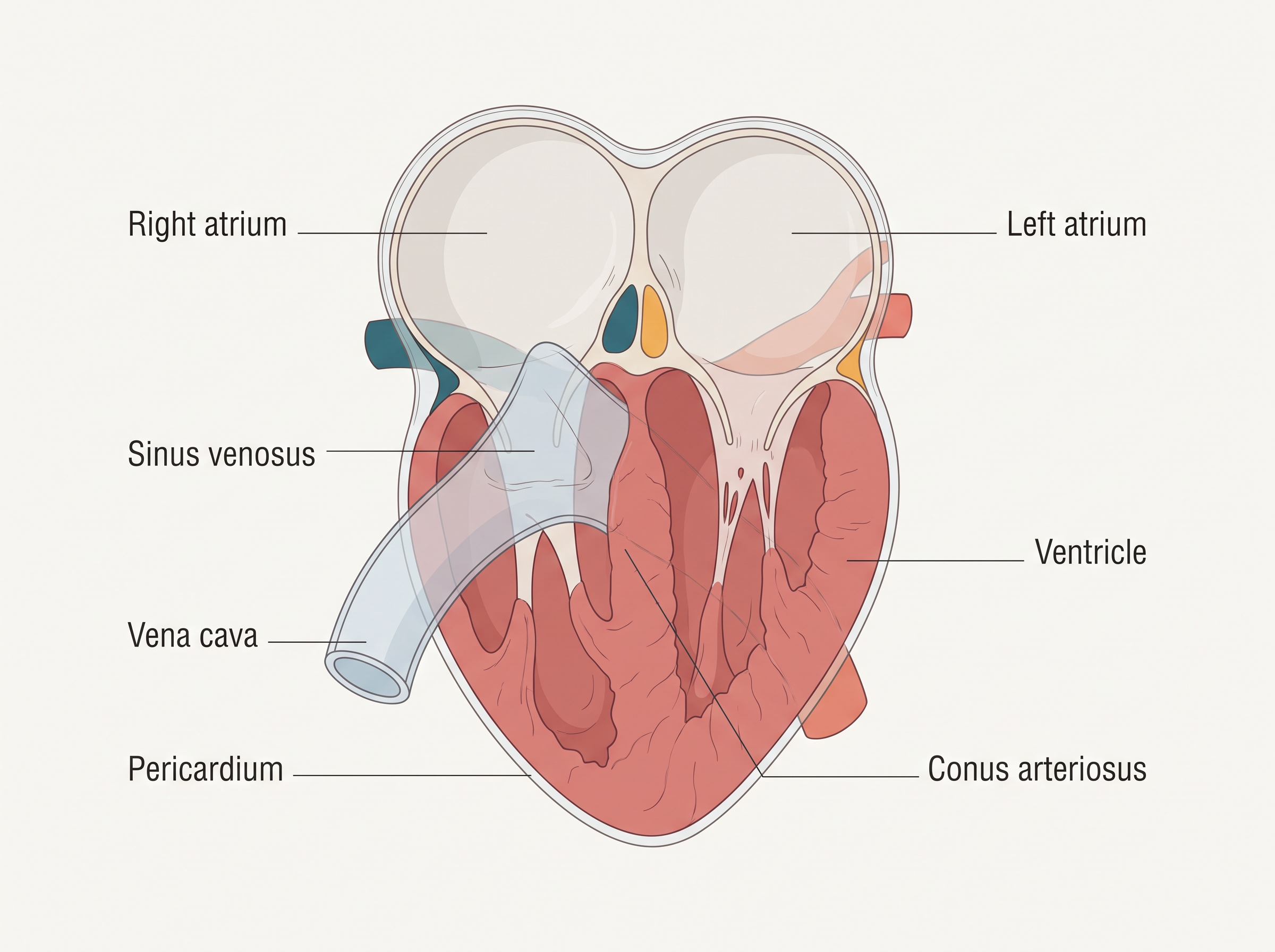
Fig. 7.3 – The three-chambered heart of a frog.
A triangular structure called the sinus venosus joins the right atrium; it collects blood arriving through the large veins known as the vena cava. On the ventral side of the heart, the ventricle opens into a sac-like tube, the conus arteriosus, which carries blood out of the heart.
From the heart, blood is carried to every part of the body by the arteries; together these make up the arterial system. The veins do the opposite work, collecting blood from the various parts of the body and returning it to the heart to form the venous system.
The frog also has two special venous connections worth remembering. One links the liver with the intestine, and the other links the kidney with the lower parts of the body.
The blood is made of a fluid part, the plasma, and cells suspended in it. The blood cells are of three kinds: red blood cells (RBCs) or erythrocytes, white blood cells (WBCs) or leucocytes, and platelets. The RBCs contain a red-coloured pigment called haemoglobin, which carries the gases. One point often asked in exams: the frog’s RBCs are nucleated — they keep their nucleus — unlike our own mature red blood cells, which lose theirs.
Lymph is not the same as blood. It is a similar fluid, but it lacks a few of the proteins and has no RBCs. Through all this plumbing, the blood carries nutrients, gases and water to wherever they are needed, and the whole circulation is driven by the pumping action of the muscular heart.
The Excretory System
The task of removing nitrogenous (nitrogen-containing) wastes from the body falls to a well-developed excretory system. It is made up of a pair of kidneys, their ureters, a urinary bladder, and the cloaca.
The kidneys are compact, dark-red, bean-shaped organs lying a little towards the back of the body cavity, one on each side of the vertebral column (backbone). Each kidney is built from very large numbers of tiny units called uriniferous tubules, or nephrons — these are the structural and functional units that actually clean the blood.
From the kidneys arise the ureters. In the male frog, two ureters emerge and act as a urinogenital duct — that is, a single duct that carries both urine and sperms — before opening into the cloaca. In the female, the arrangement is different: the ureters and the oviduct open separately into the cloaca. A thin-walled urinary bladder lies just below (ventral to) the rectum and also opens into the cloaca.
The frog gets rid of its nitrogen waste in the form of urea, and an animal that excretes urea is described as ureotelic.
The waste materials are carried by the blood into the kidneys, where they are separated out and finally excreted.
Control and Coordination
The systems that keep all the organs working together in harmony are highly developed in the frog. Control and coordination are shared between two partners: the neural (nervous) system, which sends fast electrical messages, and the endocrine glands, which send slower chemical messages.
The endocrine glands achieve chemical coordination by releasing hormones — chemical messengers that travel in the blood and control the activity of various organs. The main endocrine glands found in the frog are the pituitary, thyroid, parathyroid, thymus, pineal body, pancreatic islets, adrenals and gonads.
The nervous system is organised into three parts: a central nervous system made of the brain and the spinal cord; a peripheral nervous system made of the cranial and spinal nerves; and an autonomic nervous system with its sympathetic and parasympathetic divisions. Ten pairs of cranial nerves arise from the brain. This overall plan is shown in FC 7.3.
Flowchart
FC 7.3 – Organisation of the frog’s nervous system: Nervous system branches into → Central nervous system (brain + spinal cord); Peripheral nervous system (cranial nerves + spinal nerves); Autonomic nervous system (sympathetic + parasympathetic)
The brain itself is protected inside a bony case called the brain box, or cranium, and it is divided into three regions — the fore-brain, the mid-brain and the hind-brain. The fore-brain includes the olfactory lobes, a pair of cerebral hemispheres, and an unpaired diencephalon. The mid-brain is marked by a pair of optic lobes. The hind-brain consists of the cerebellum and the medulla oblongata. These divisions are laid out in FC 7.4.
Flowchart
FC 7.4 – Divisions of the frog’s brain: Brain → Fore-brain (olfactory lobes + paired cerebral hemispheres + unpaired diencephalon); Mid-brain (pair of optic lobes); Hind-brain (cerebellum + medulla oblongata)
At the base of the brain, the medulla oblongata passes out through an opening called the foramen magnum and continues as the spinal cord, which runs protected inside the vertebral column.
Sense Organs
The frog gathers information about its surroundings through several kinds of sense organs: organs of touch (the sensory papillae), of taste (the taste buds), of smell (the nasal epithelium), of sight (the eyes), and of hearing (the tympanum, together with the internal ears).
Of all these, only the eyes and the internal ears are well-organised sense organs; the rest are simply groups of cells gathered around nerve endings.
The eyes are a pair of spherical structures set in sockets, called orbits, in the skull. They are simple eyes — each is a single unit, unlike the many-unit compound eyes of insects. As noted earlier, a frog has no external ear; only the tympanum is visible on the outside. Importantly, the ear is not only for hearing — it also serves as the organ of balance, helping the frog to keep its equilibrium.
The Reproductive System
Frogs have well-organised reproductive systems, which are separate in the male and the female.
In the male, the reproductive organs are a pair of yellowish, oval (ovoid) testes. These are held against the upper part of the kidneys by a double fold of a membrane (peritoneum) called the mesorchium (Fig. 7.4). From the testes arise 10–12 fine tubes called the vasa efferentia. These enter the kidney on their own side and open into a channel known as Bidder’s canal. From there the connection finally leads into the urinogenital duct, which emerges from the kidney and opens into the cloaca. The cloaca itself is a small, median chamber that serves as a common exit, passing out faecal matter, urine and sperms. The route taken by the sperms is traced in FC 7.5.
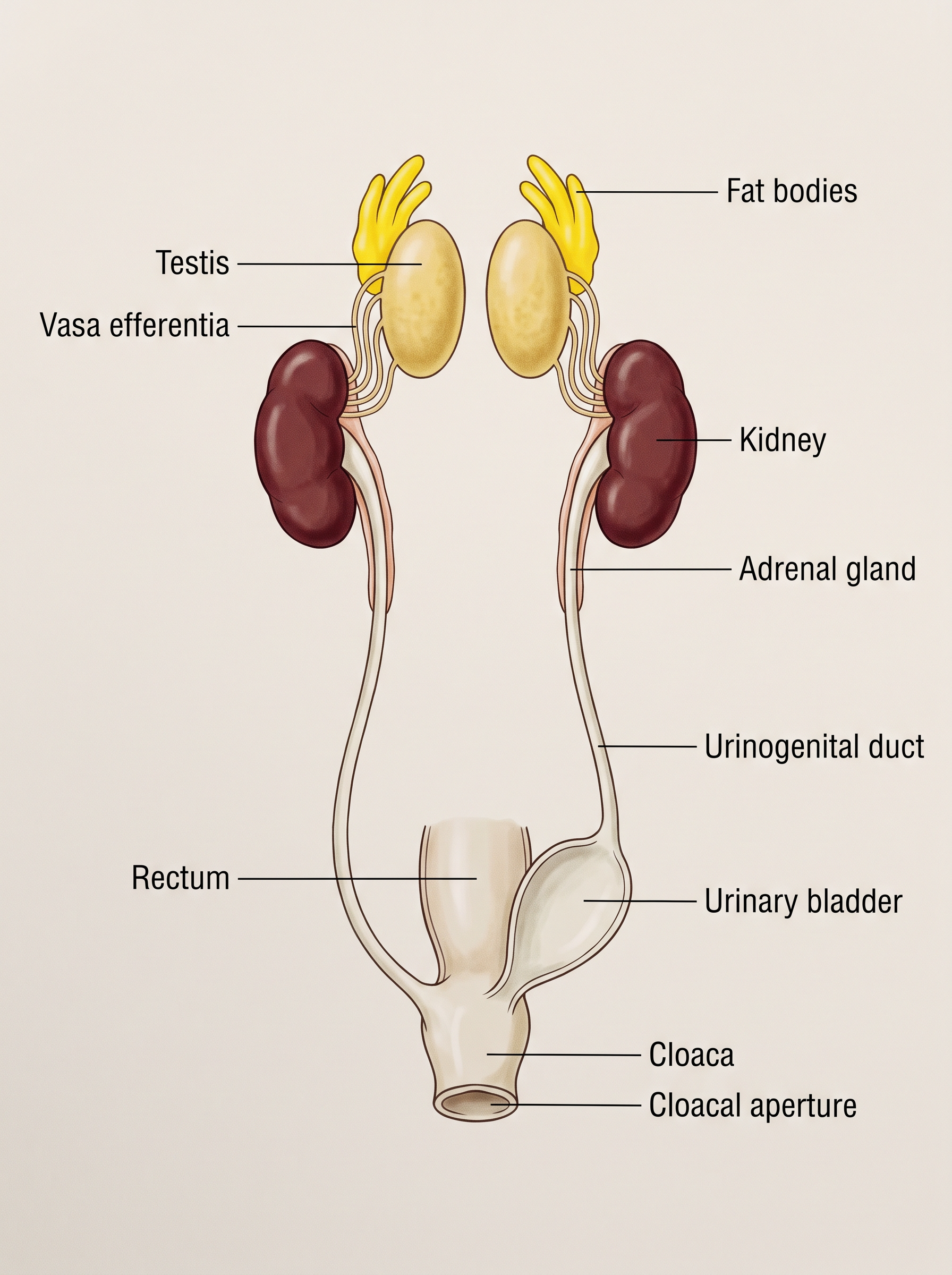
Fig. 7.4 – Male reproductive system of a frog.
Flowchart
FC 7.5 – Path of sperms in the male frog: testes → vasa efferentia (10–12) → kidney → Bidder’s canal → urinogenital duct → cloaca → exterior
In the female, the reproductive organs are a pair of ovaries lying near the kidneys — but, unlike in the male, there is no functional connection between the ovaries and the kidneys (Fig. 7.5). A pair of oviducts arising from the ovaries opens separately into the cloaca. A mature female can lay a remarkable 2500 to 3000 ova (eggs) at a single time.
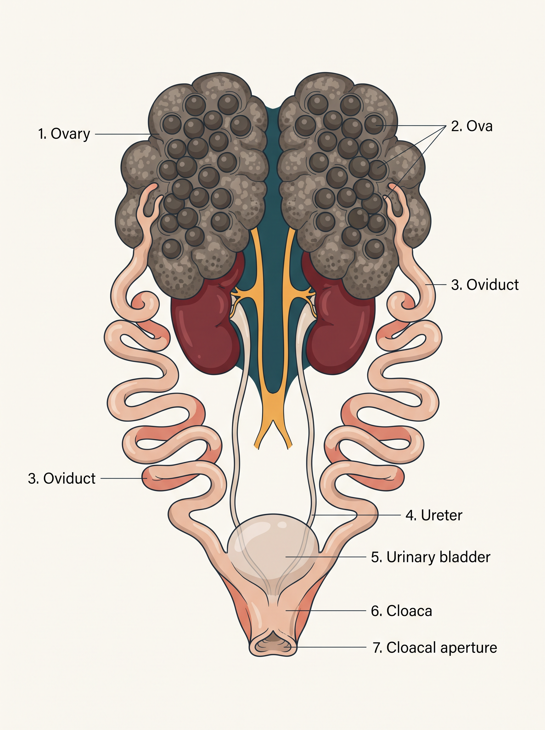
Fig. 7.5 – Female reproductive system of a frog.
Fertilisation in frogs is external — the male and female release their gametes into the water, where the sperms meet the eggs outside the body. Development then passes through a larval stage called the tadpole. The tadpole later undergoes metamorphosis — a dramatic change of body form — to become the adult frog.
Why Frogs Matter
Frogs are useful to human beings in more ways than one. They feed on insects, and in doing so they protect crops from pests. They also help to maintain the balance of nature, forming an important link in the food chains and food webs of their ecosystem — both as predators of insects and as prey for larger animals. In some countries, the muscular hind legs of frogs are even eaten as food.
7.3 Summary
- Cells, tissues, organs and organ systems divide the body’s work among themselves — a plan called division of labour — and this shared effort keeps the body as a whole alive.
- A tissue is a group of cells, together with the intercellular substances between them, that carries out one or more functions in the body.
- Epithelia are sheet-like tissues that line the body’s outer surface and its internal cavities, ducts and tubes. Each epithelium has one free surface facing either a body fluid or the outside environment, and its cells are joined to one another — both in structure and in function — at junctions.
- The Indian bullfrog, Rana tigrina, is the common frog of India. Its body is covered by skin containing mucous glands; the skin is richly supplied with blood vessels and helps in respiration both in water and on land.
- The body is divided into a head and a trunk. A muscular tongue, bilobed at its tip, is used to capture prey.
- The alimentary canal runs from the oesophagus to the stomach, intestine and rectum, all finally opening into the cloaca. The main digestive glands are the liver and the pancreas.
- The frog respires through its skin while in water and through its lungs while on land.
- The circulatory system is of the closed type and works with single circulation. The red blood cells (RBCs) are nucleated.
- The nervous system is organised into central, peripheral and autonomic parts.
- The organs of the urinogenital system are the kidneys and the urinogenital ducts, which open into the cloaca.
- The male reproductive organ is a pair of testes, and the female reproductive organ is a pair of ovaries. A female lays 2500 to 3000 ova at a time.
- Both fertilisation and development are external: the eggs hatch into tadpoles, which then metamorphose into adult frogs. The whole cycle is shown in FC 7.6.
Flowchart
FC 7.6 – Life cycle of the frog (a cycle): adult frog → female lays 2500–3000 eggs in water → external fertilisation → tadpole (larva) → metamorphosis → adult frog (returns to start)
7.4 Points to Ponder
In a single-celled animal, one cell carries out every function of life; in a large animal, these same functions are shared among many specialised groups of cells. What does a body gain by dividing its work in this way, and can you think of anything it might risk by depending so heavily on specialisation?
A frog can take in oxygen through its moist skin while in water and through its lungs while on land. Why is it useful for an animal living in two habitats to have two different ways of breathing — and what might happen to such an animal if its skin could no longer stay moist?
A frog’s body temperature rises and falls with its surroundings, so it must retreat into a burrow during the hottest and the coldest parts of the year. A warm-blooded animal, by contrast, stays active all year round but must constantly spend energy to keep its body warm. What advantages and disadvantages can you see in each of these two ways of living?
A female frog releases 2500 to 3000 eggs at a time, and both fertilisation and development take place outside the body, in open water. How might laying such a large number of eggs be connected to the fact that the eggs and young are neither protected inside the parent nor cared for by it?
You have studied the frog as a representative of the vertebrates — the animals with a backbone. In what ways do you think examining the organ systems of a comparatively simple vertebrate like the frog can help us understand the workings of more complex vertebrate bodies, including our own?
7.5 NCERT Questions
Draw a neat diagram of the digestive system of a frog.
Mention the function of the ureters in a frog.
7.6 Check Your Concepts
Arrange the following levels of biological organisation in the correct order, from the simplest to the most complex: organ, cell, organism, tissue, organ system.
Define a tissue. Name the four basic types of tissue that together make up an organ such as the heart.
Why is the division of labour among cells, tissues and organs considered essential for the survival of a large, complex animal?
Distinguish between morphology and anatomy, giving one example of what each would study in a frog.
To which class and phylum does the frog belong? Write the scientific name of the common frog of India.
Why is the frog described as a cold-blooded animal, or poikilotherm?
Differentiate between aestivation and hibernation in the frog.
A frog is kept for a long time in a dry container with no water. Using what you know about the frog’s skin, explain why this would seriously endanger the animal.
Explain how each of the following features helps the frog: (a) webbed digits on the feet, and (b) the nictitating membrane covering the eye.
What is sexual dimorphism? State two features by which a male frog can be distinguished from a female frog.
Draw a well-labelled diagram of the external features of a frog, labelling the head, trunk, eye, nostril, tympanum, fore limb and hind limb.
Why is the alimentary canal, and in particular the intestine, comparatively short in the frog?
Arrange the following parts of the frog’s alimentary canal in the order in which food passes through them: rectum, buccal cavity, oesophagus, stomach, cloaca, mouth, intestine.
State the role of (a) bile and (b) the villi in the digestion and absorption of food in the frog.
Assertion–Reason. Assertion (A): A frog can remain under water for long periods without breathing air. Reason (R): The moist skin of the frog can exchange gases with the surrounding water. — State whether A and R are true, and whether R is the correct explanation of A.
Name and briefly describe the two methods by which a frog respires. Through which organ does gaseous exchange take place during hibernation?
How many chambers are present in the heart of a frog? Name them.
Distinguish between the hepatic portal system and the renal portal system of the frog.
Assertion–Reason. Assertion (A): The red blood cells of a frog are nucleated. Reason (R): The red blood cells of a frog lack haemoglobin. — State whether A and R are true or false, and whether R correctly explains A.
Give two differences between blood and lymph in the frog.
Draw and label the three-chambered heart of a frog, showing the two atria, the ventricle, the sinus venosus and the conus arteriosus.
- What is meant by a ureotelic animal? (b) Name the structural and functional unit of the frog’s kidney. (c) Why is the duct arising from the kidney of a male frog called a urinogenital duct, and how is the arrangement different in the female?
Into which three parts is the nervous system of the frog organised? How many pairs of cranial nerves arise from its brain?
Match the brain region in Column I with the structures it contains in Column II.
| Column I | Column II |
|---|---|
| a. Fore-brain | i. A pair of optic lobes |
| b. Mid-brain | ii. Cerebellum and medulla oblongata |
| c. Hind-brain | iii. Olfactory lobes, cerebral hemispheres and diencephalon |
Which two sense organs of the frog are described as well-organised? State the two functions performed by the ear.
Match each sense in Column I with its receptor organ in Column II.
| Column I | Column II |
|---|---|
| a. Touch | i. Taste buds |
| b. Taste | ii. Nasal epithelium |
| c. Smell | iii. Sensory papillae |
Name the double fold of peritoneum that attaches the testes to the kidneys in the male frog. How many vasa efferentia are present?
Arrange the following in the correct sequence through which sperms travel in a male frog: Bidder’s canal, cloaca, testes, urinogenital duct, vasa efferentia.
A female frog lays 2500 to 3000 eggs at a time, and fertilisation is external. Why is the release of such a large number of eggs important for the survival of the species?
What is metamorphosis? Name the larval stage that appears in the life cycle of a frog.
Give two reasons why frogs are considered beneficial to human beings.