Chapter 16: Excretory Products and their Elimination
```markdown
In a small hospital in the Netherlands during the Second World War, a physician named Willem Kolff was troubled by a hard truth: patients whose kidneys had stopped working were dying, and there was nothing anyone could do. Their blood slowly filled with waste that their bodies could no longer remove. Kolff wondered — if the kidney’s job is really just to clean the blood, could a machine be built to do the same thing?
Working with scarce materials in wartime, he built a strange-looking device from a rotating wooden drum, lengths of thin cellophane tubing (the kind used for sausage casings), and a tank of fluid. Blood from a patient flowed through the tubing while the drum turned; wastes seeped out through the cellophane into the surrounding fluid, and the cleaned blood returned to the body. In the mid-1940s, one of his patients — a woman who had slipped into a coma from kidney failure — woke up after treatment and recovered.
Kolff had not cured the kidney. He had copied one of its most important tricks: letting small waste molecules leave the blood while keeping the useful things behind. To understand why his machine worked, and why losing your kidneys is so dangerous in the first place, we must first understand what the kidney quietly does inside you every single minute of your life.
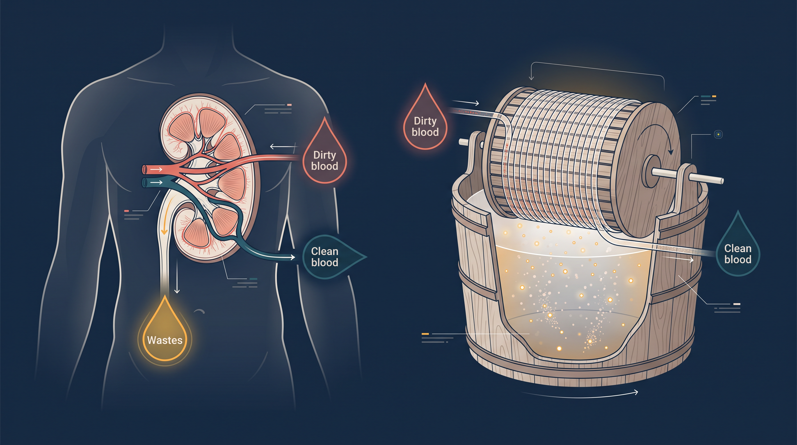
Fig. 16.0 – The human kidney and the early dialysis machine.
By the end of this chapter, you will be able to answer each of these questions for yourself.
16.1 Human Excretory System
Every living cell is constantly at work, and all that work leaves waste behind. As the body breaks down food, builds new molecules, and repairs itself, it steadily produces substances it can no longer use. On top of this, we often take in more of certain things than the body needs, simply through our food and water.
The body therefore accumulates a mixture of unwanted substances: ammonia, urea and uric acid, along with carbon dioxide, water, and ions such as \(Na^+\), \(K^+\), \(Cl^-\), phosphate and sulphate. Some of these are produced by metabolic activities inside our cells; others enter through excess ingestion. Whatever their source, they must be removed — either completely or in part — so that the internal environment of the body stays steady and safe.
This removal of metabolic wastes from the body is called excretion. In this chapter, our main focus is on the nitrogen-containing wastes, which are among the most important to manage carefully.
The Nitrogenous Wastes
When the body breaks down proteins and nucleic acids, the nitrogen locked inside them is released and ends up in nitrogen-containing waste molecules. The three major nitrogenous wastes excreted by animals are ammonia, urea and uric acid.
These three wastes are not equally dangerous, and — just as importantly — they do not cost the same amount of water to throw out. Ammonia is the most toxic of the three, and eliminating it safely needs a large amount of water. Uric acid is the least toxic, and it can be removed with only a minimum loss of water. Urea sits in between the two.
This single trade-off — toxicity balanced against water cost — is the key to the whole story. It explains why animals living in different habitats have evolved different ways of getting rid of their nitrogen waste.
Three Patterns of Nitrogen Excretion
The excretory pattern an animal uses depends mainly on how much water is available in its surroundings.
Ammonotelism is the excretion of nitrogen waste as ammonia. Many bony fishes, aquatic amphibians and aquatic insects are ammonotelic. Because ammonia is readily soluble in water, it is generally excreted straight out by simple diffusion across the body surface, or across the gill surfaces in fish, in the form of ammonium ions. Since it leaves directly across these surfaces, the kidneys play no significant role in removing ammonia.
Moving onto land changed everything. On land, water is precious and cannot be wasted, so a terrestrial lifestyle made it necessary to produce less toxic nitrogen wastes — urea and uric acid — that need far less water to remove.
Ureotelism is the excretion of nitrogen waste mainly as urea. Mammals, many terrestrial amphibians and marine fishes are ureotelic. In these animals, the ammonia produced by metabolism is converted into the far less toxic urea in the liver. This urea is released into the blood, which is then filtered by the kidneys, and the urea is excreted out. In some of these animals, a certain amount of urea is deliberately held back in the kidney matrix to help maintain a desired osmolarity (the concentration of dissolved particles in a fluid).
Uricotelism is the excretion of nitrogen waste as uric acid. Reptiles, birds, land snails and insects are uricotelic. They release uric acid in the form of a pellet or paste, losing almost no water in the process — a great advantage for animals that must survive where water is scarce.
| Feature | Ammonotelic | Ureotelic | Uricotelic |
|---|---|---|---|
| Main nitrogen waste | Ammonia | Urea | Uric acid |
| Toxicity | Highest | Moderate | Lowest |
| Water needed to excrete it | Very high | Moderate | Very low (minimal) |
| Typical animals | Many bony fishes, aquatic amphibians, aquatic insects | Mammals, many terrestrial amphibians, marine fishes | Reptiles, birds, land snails, insects |
| How/where it leaves | Diffusion across body/gill surfaces as ammonium ions | Made in liver, filtered out by kidneys | Excreted as a pellet or paste |
Excretory Structures Across the Animal Kingdom
A survey of the animal kingdom shows a wide variety of excretory structures. In most invertebrates these are simple tubular forms, whereas vertebrates have complex tubular organs called kidneys.
In flatworms (Platyhelminthes, for example Planaria), in rotifers, in some annelids, and in the cephalochordate Amphioxus, the excretory structures are protonephridia, also called flame cells. Protonephridia are mainly concerned with osmoregulation — the regulation of the body’s water content and ion balance (that is, its ionic and fluid volume regulation).
Nephridia are the tubular excretory structures of earthworms and other annelids. They help remove nitrogenous wastes and also maintain the body’s fluid and ionic balance.
Malpighian tubules are the excretory structures of most insects, including cockroaches. They help both in removing nitrogenous wastes and in osmoregulation. In crustaceans such as prawns, this excretory job is done by antennal glands (also called green glands).
| Animal group | Example | Excretory structure | Main role |
|---|---|---|---|
| Flatworms, rotifers, some annelids, cephalochordate | Planaria, Amphioxus | Protonephridia (flame cells) | Osmoregulation (ionic & fluid volume regulation) |
| Annelids | Earthworm | Nephridia | Remove nitrogenous wastes; fluid & ionic balance |
| Insects | Cockroach | Malpighian tubules | Remove nitrogenous wastes; osmoregulation |
| Crustaceans | Prawn | Antennal (green) glands | Excretion |
| Vertebrates | Human | Kidneys | Excretion, osmoregulation, ionic & acid–base balance |
The Human Urinary System
In humans, the excretory system is made up of a pair of kidneys, one pair of ureters, a urinary bladder and a urethra, as shown in Fig. 16.1. The kidneys do the filtering; the ureters carry urine away; the bladder stores it; and the urethra releases it.
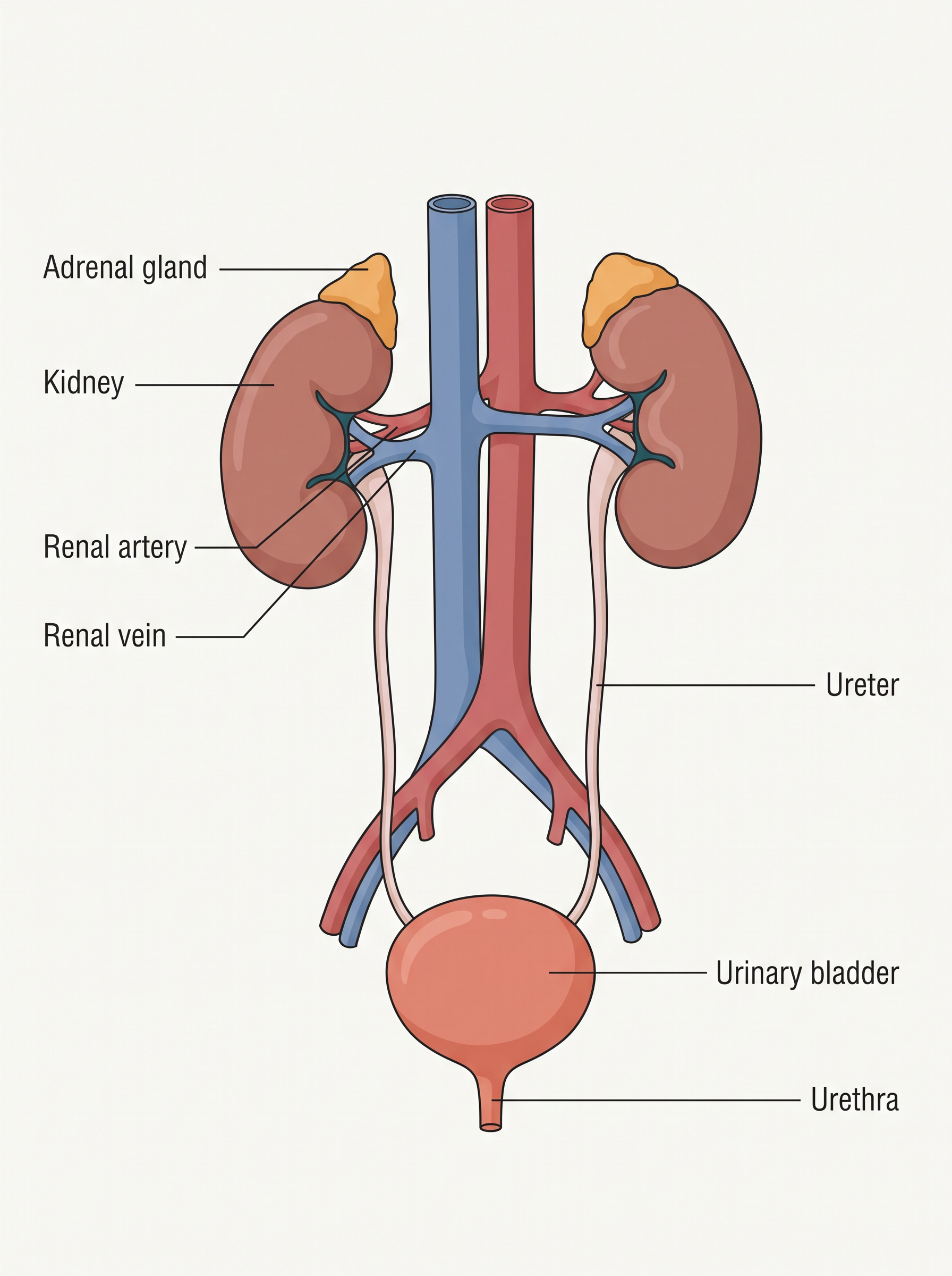
Fig. 16.1 – The human urinary system.
The kidneys are reddish-brown, bean-shaped organs. They lie between the levels of the last thoracic and the third lumbar vertebra, close to the dorsal (back) inner wall of the abdominal cavity. Each kidney of an adult human measures about 10–12 cm in length, 5–7 cm in width and 2–3 cm in thickness, with an average weight of 120–170 g.
Inside the Kidney
At the centre of the inner concave surface of each kidney is a notch called the hilum. This is the gateway through which the ureter, blood vessels and nerves enter or leave the kidney. Just inside the hilum lies a broad, funnel-shaped space called the renal pelvis, which sends out finger-like projections called calyces (singular: calyx).
A tough protective capsule covers the outer surface of the kidney. Inside, the kidney is organised into two zones — an outer cortex and an inner medulla, as seen in Fig. 16.2. The medulla is divided into a few conical masses called medullary pyramids, which project into the calyces. Between these pyramids, the cortex dips inward as extensions called renal columns, known as the Columns of Bertini.
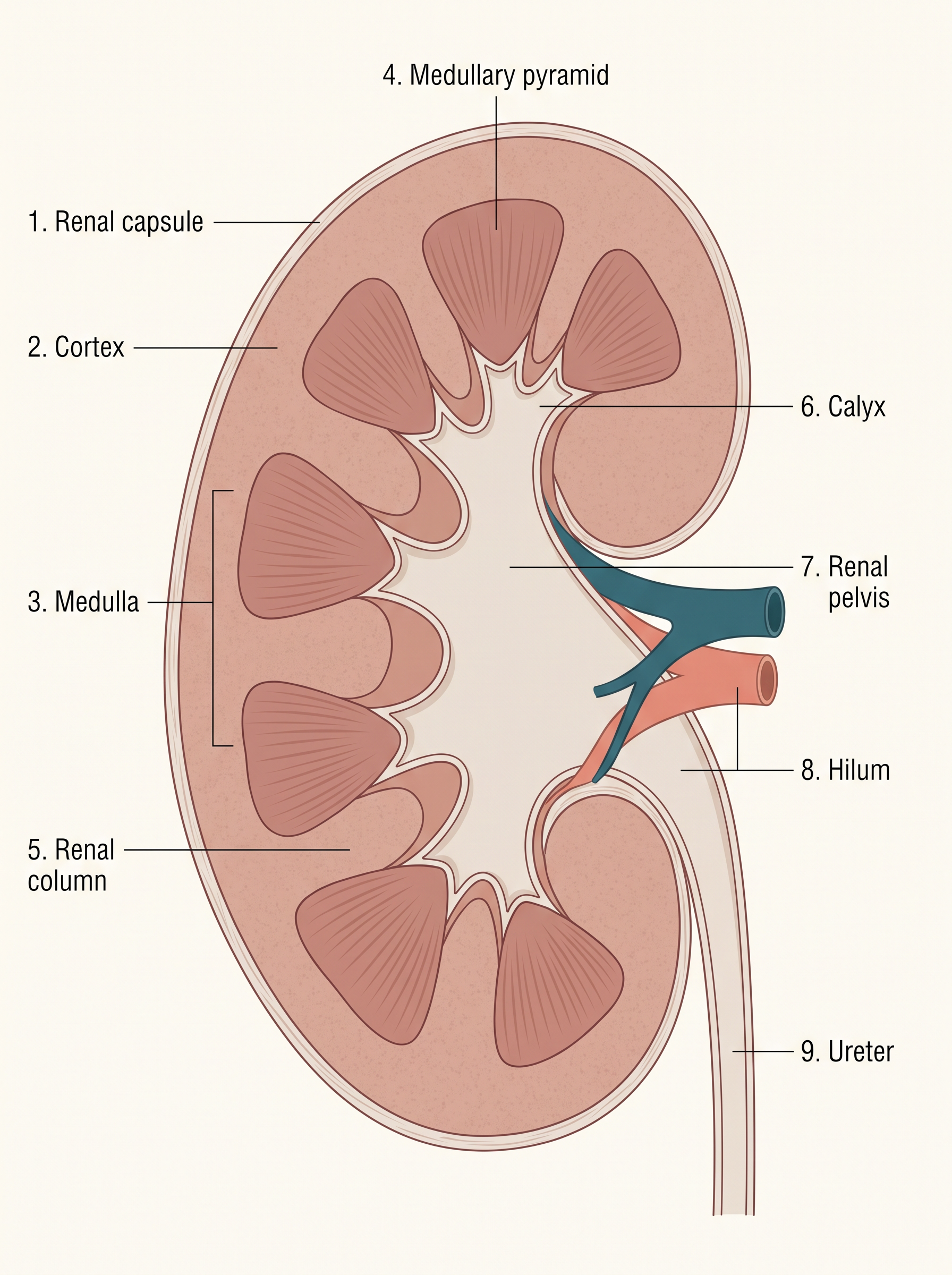
Fig. 16.2 – Longitudinal section of the human kidney.
The Nephron — The Functional Unit
Each kidney contains nearly one million tiny, complex tubular structures called nephrons, shown in Fig. 16.3. The nephron is the functional unit of the kidney — the smallest structure that can actually carry out the kidney’s job of making urine.
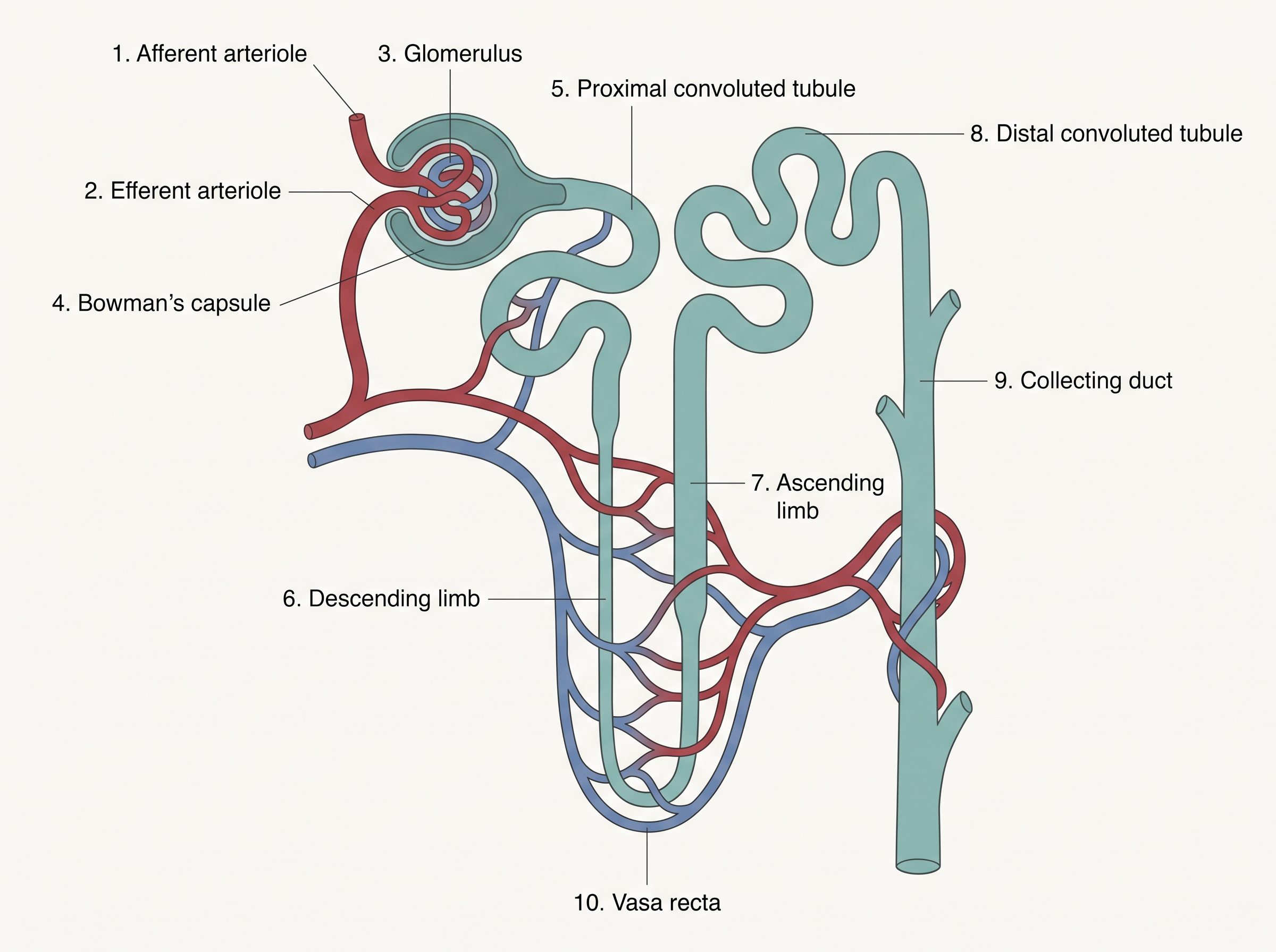
Fig. 16.3 – Structure of a nephron.
Each nephron has two parts — the glomerulus and the renal tubule. The glomerulus is a tuft (a compact bundle) of capillaries formed by the afferent arteriole, a fine branch of the renal artery. After passing through this tuft, blood is carried away by the efferent arteriole.
The renal tubule begins with a double-walled, cup-like structure called Bowman’s capsule, which surrounds and encloses the glomerulus. The glomerulus together with Bowman’s capsule is called the Malpighian body, or renal corpuscle, seen in Fig. 16.4.
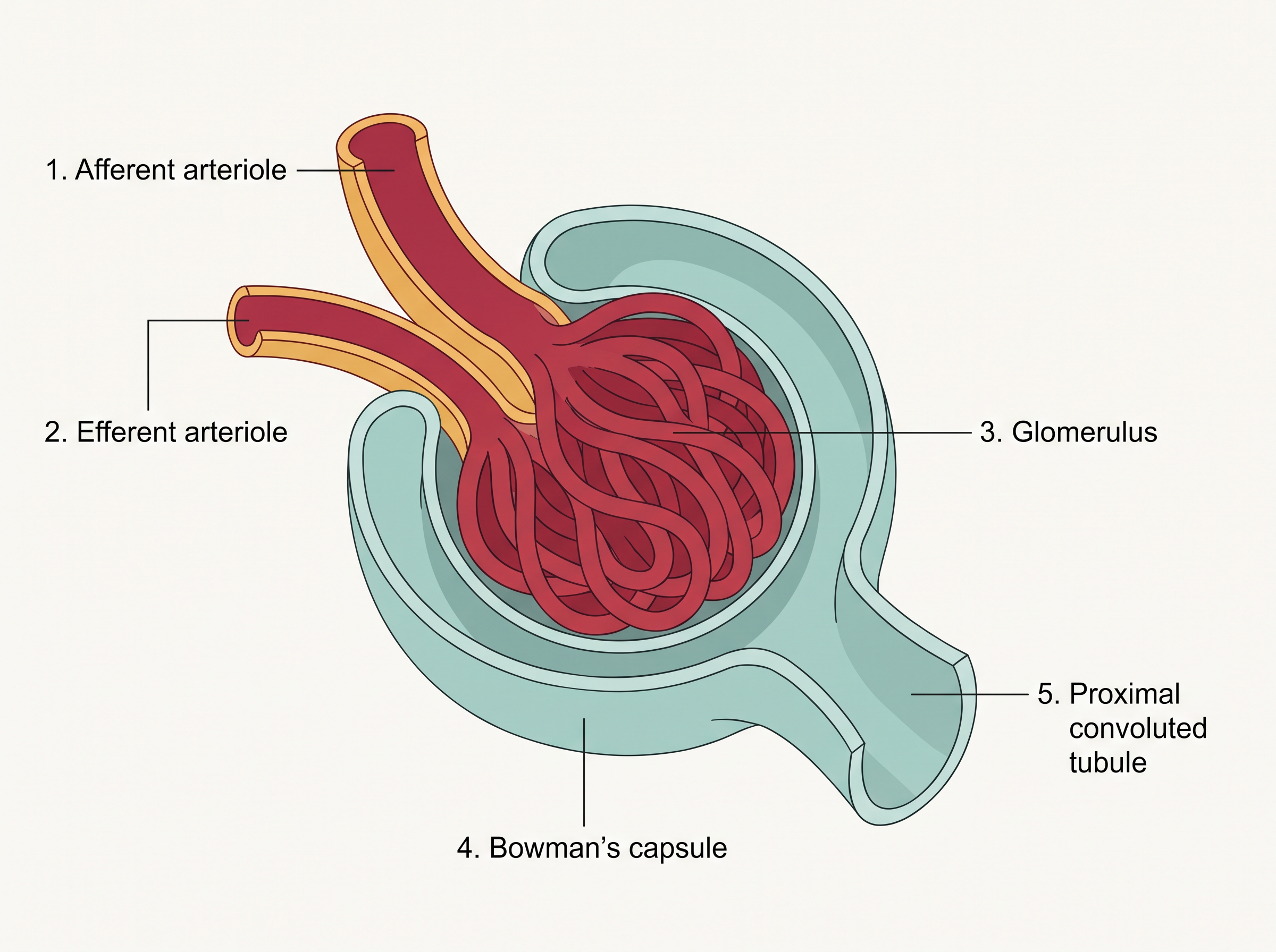
Fig. 16.4 – The Malpighian body (renal corpuscle).
From Bowman’s capsule, the tubule continues as a highly coiled region called the proximal convoluted tubule (PCT). Next comes a hairpin-shaped Henle’s loop, which has a descending limb and an ascending limb. The ascending limb then continues as another coiled region, the distal convoluted tubule (DCT). The DCTs of many nephrons open into a straight tube called the collecting duct. Many collecting ducts converge and open into the renal pelvis through the medullary pyramids in the calyces. This ordered path is summarised in FC 16.1.
Flowchart
FC 16.1 – Path of filtrate through the nephron tubule: Bowman’s capsule → proximal convoluted tubule (PCT) → descending limb of Henle’s loop → ascending limb of Henle’s loop → distal convoluted tubule (DCT) → collecting duct → renal pelvis (via medullary pyramids and calyces)
Cortical and Juxtamedullary Nephrons
The Malpighian corpuscle, the PCT and the DCT of a nephron all sit in the cortical region of the kidney, while the loop of Henle dips down into the medulla. However, not all nephrons dip equally deep.
In the majority of nephrons, the loop of Henle is quite short and extends only a little way into the medulla. These are called cortical nephrons. In some nephrons, the loop of Henle is very long and runs deep into the medulla. These are called juxtamedullary nephrons.
Blood Supply Around the Tubule
The efferent arteriole leaving the glomerulus does not simply drain away. It forms a fine capillary network wrapped around the renal tubule, called the peritubular capillaries. This close wrapping is important, because it allows substances to move back and forth between the tubule and the blood.
One special part of this network is a slender, ‘U’-shaped vessel that runs alongside the loop of Henle, called the vasa recta. In cortical nephrons, the vasa recta is absent or highly reduced; it is well developed in juxtamedullary nephrons, where — as we will see in a later section — it plays a key role in concentrating urine.
16.2 Urine Formation
Making urine is not a single event but a sequence of steps carried out in different parts of the nephron. Urine formation involves three main processes: glomerular filtration, reabsorption and secretion. Together, these three turn ordinary blood into the final urine that leaves the body.
Flowchart
FC 16.2 – The three steps of urine formation: blood enters glomerulus → (1) glomerular filtration (in the glomerulus/Bowman’s capsule) → filtrate enters the tubule → (2) reabsorption (useful substances returned to blood) → (3) secretion (extra wastes added to filtrate) → final urine
Step 1 — Glomerular Filtration
The very first step in urine formation is the filtration of blood, carried out by the glomerulus. This is called glomerular filtration.
On an average, about 1100–1200 ml of blood is filtered by the kidneys every minute. This is a huge quantity — it works out to roughly \(\frac{1}{5}\) of the blood pumped out by each ventricle of the heart in one minute. In other words, a large share of every heartbeat’s output is sent to the kidneys for cleaning.
What actually drives this filtration is pressure. The glomerular capillary blood pressure pushes the fluid part of the blood out through three layers stacked together: the endothelium of the glomerular blood vessels, the epithelium of Bowman’s capsule, and a basement membrane sitting between these two layers. As shown in Fig. 16.5, these three layers together act as a fine filter.
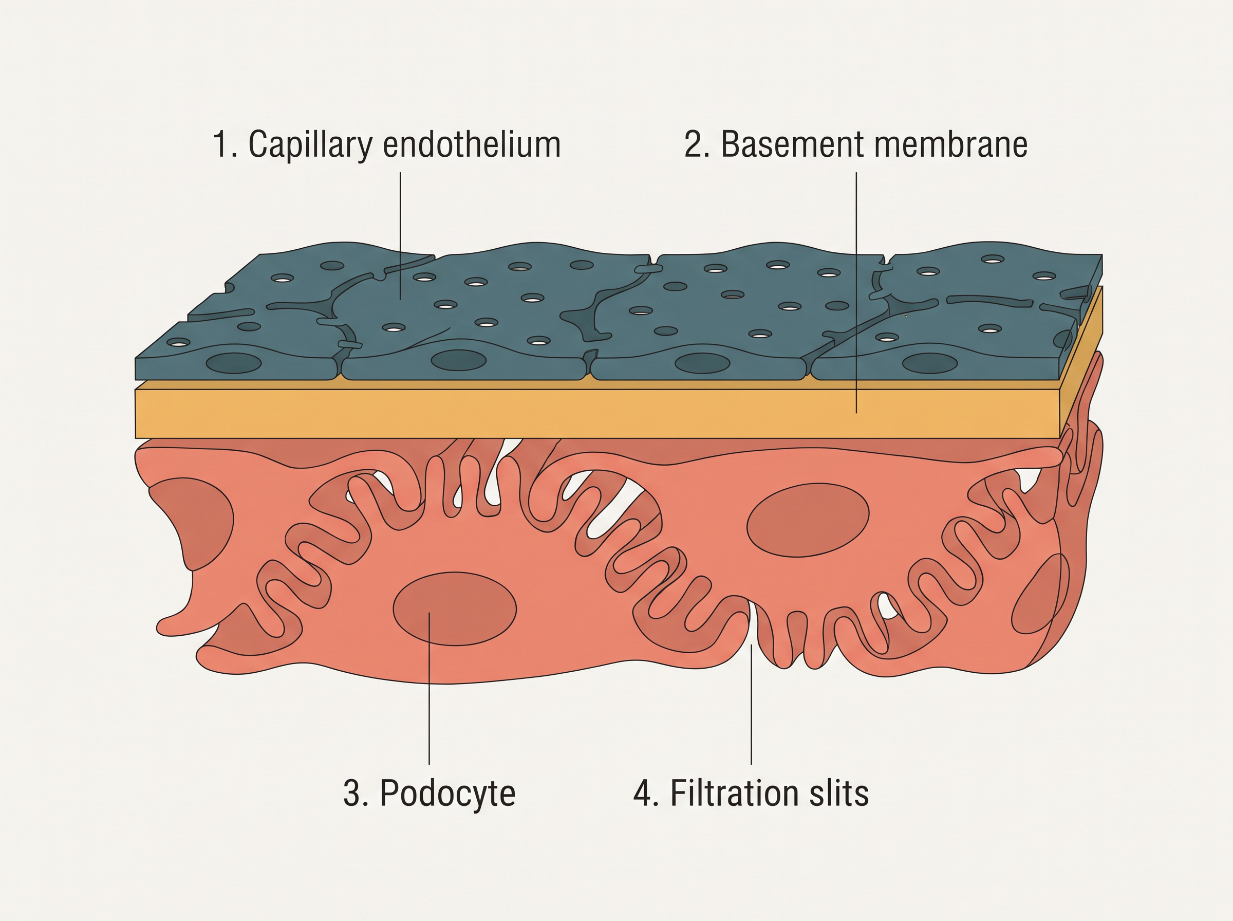
Fig. 16.5 – The three-layered filtration barrier.
Why It Is Called Ultrafiltration
The epithelial cells of Bowman’s capsule are special cells called podocytes. They are arranged in an intricate, interlocking way that leaves tiny gaps between them, called filtration slits or slit pores. Blood is filtered so finely through these membranes and slits that almost all the constituents of the plasma pass into the lumen of Bowman’s capsule — except the proteins, which are too large to get through.
Because the filter is so extremely fine, allowing everything small through but holding back large molecules like proteins, this process is called ultrafiltration.
Glomerular Filtration Rate (GFR)
The amount of filtrate formed by the kidneys per minute is called the glomerular filtration rate (GFR). In a healthy individual, GFR is approximately 125 ml per minute — which adds up to about 180 litres per day.
Keeping GFR Steady — The Juxtaglomerular Apparatus
The kidneys have built-in mechanisms to keep GFR from swinging too high or too low. One efficient mechanism is carried out by the juxtaglomerular apparatus (JGA).
The JGA is a special, sensitive region formed by cellular modifications in the distal convoluted tubule and the afferent arteriole, right at the point where these two come into contact. It acts like a sensor that can respond to changes in filtration.
If GFR falls, the JG cells are activated to release an enzyme called renin. Renin stimulates the glomerular blood flow, which raises the GFR back towards normal. This self-correcting loop is summarised in FC 16.3.
Flowchart
FC 16.3 – Autoregulation of GFR: GFR falls → JG cells of the JGA are activated → release renin → glomerular blood flow increases → GFR rises back to normal → JG cells switch off (returns to normal)
Step 2 — Reabsorption
Now compare two numbers: the filtrate formed per day is about 180 litres, but the urine actually released is only about 1.5 litres. This means nearly 99 per cent of the filtrate has to be taken back into the blood. This taking-back is called reabsorption.
The tubular epithelial cells in different segments of the nephron carry out reabsorption either by active mechanisms (which use energy) or by passive mechanisms (which do not). For example, substances such as glucose, amino acids and \(Na^+\) in the filtrate are reabsorbed actively, whereas nitrogenous wastes are absorbed by passive transport. Reabsorption of water also happens passively in the initial segments of the nephron, as shown in Fig. 16.6.
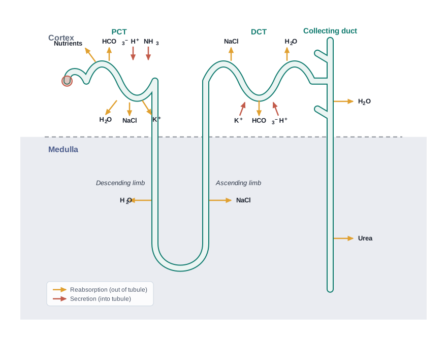
Fig. 16.6 – Reabsorption and secretion in the nephron.
Step 3 — Tubular Secretion
Reabsorption takes substances out of the filtrate, but the tubule also does the reverse. During urine formation, the tubular cells secrete substances such as \(H^+\), \(K^+\) and ammonia into the filtrate.
This tubular secretion is an important step, because it helps maintain the ionic balance and the acid–base (pH) balance of the body fluids. By actively adding these ions and ammonia to the filtrate, the tubules fine-tune the composition of the blood.
16.3 Function of the Tubules
The filtrate that enters the tubule is not yet urine. As it travels along the nephron, each region of the tubule works on it in its own way — reabsorbing some substances, secreting others, and adjusting how much water stays behind. Let us follow the tubule region by region and see what each part does.
Proximal Convoluted Tubule (PCT)
The PCT is lined by simple cuboidal epithelium with a brush border — a dense fringe of tiny finger-like projections on the cell surface. This brush border greatly increases the surface area available for reabsorption, so the PCT is built for taking substances back efficiently.
This is where the bulk of reabsorption happens. Nearly all of the essential nutrients, and about 70–80 per cent of the electrolytes and water, are reabsorbed by this single segment. The PCT also helps maintain the pH and the ionic balance of the body fluids: it does this by selectively secreting hydrogen ions (\(H^+\)) and ammonia into the filtrate, and by absorbing bicarbonate (\(HCO_3^-\)) from it.
Henle’s Loop
Reabsorption is at a minimum in the ascending limb of Henle’s loop. Even so, this region plays a very significant role — it helps build and maintain the high osmolarity (high concentration of dissolved particles) of the medullary interstitial fluid, which is the fluid filling the spaces between the tubules deep in the medulla.
The two limbs behave in opposite ways because they differ in what they let through. The descending limb is permeable to water but almost impermeable to electrolytes. As the filtrate moves down this limb, water leaves it while salts stay behind, so the filtrate becomes more and more concentrated on the way down.
The ascending limb is the reverse: it is impermeable to water but allows electrolytes to be transported out, either actively or passively. So as the now-concentrated filtrate passes upward, it loses electrolytes to the surrounding medullary fluid and becomes diluted again.
Distal Convoluted Tubule (DCT)
In the DCT, conditional reabsorption of \(Na^+\) and water takes place — “conditional” meaning it is not automatic but happens according to the body’s needs, under hormonal control. When the body needs to save water and sodium, more is reabsorbed here; when it does not, less is.
The DCT is also capable of reabsorbing bicarbonate (\(HCO_3^-\)) and of selectively secreting hydrogen and potassium ions and ammonia (\(NH_3\)). Through this give-and-take, the DCT helps maintain the pH and the sodium–potassium balance of the blood.
Collecting Duct
The collecting duct is a long tube that extends all the way from the cortex of the kidney to the inner parts of the medulla. Because it runs through the increasingly concentrated medulla, large amounts of water can be reabsorbed from this region, which is how the kidney finally produces a concentrated urine.
The collecting duct also allows small amounts of urea to pass out into the medullary interstitium, which helps keep up the high osmolarity there. In addition, it helps maintain the pH and ionic balance of the blood by the selective secretion of \(H^+\) and \(K^+\) ions, as shown in Fig. 16.6.
Segment-by-Segment Summary
Putting it all together, each segment of the tubule has its own specialised job, summarised below.
| Segment | Water handling | Main reabsorption | Main secretion / special role |
|---|---|---|---|
| PCT | Reabsorbs ~70–80% of water (passive) | Nearly all nutrients; 70–80% of electrolytes; \(HCO_3^-\) | Secretes \(H^+\) and ammonia; controls pH & ionic balance |
| Descending limb (Henle’s loop) | Permeable to water (water leaves) | — | Concentrates the filtrate |
| Ascending limb (Henle’s loop) | Impermeable to water | Electrolytes leave (to medulla) | Builds high medullary osmolarity; dilutes the filtrate |
| DCT | Conditional reabsorption of water | Conditional \(Na^+\); \(HCO_3^-\) | Secretes \(H^+\), \(K^+\), \(NH_3\); maintains pH & Na–K balance |
| Collecting duct | Large water reabsorption → concentrated urine | Water; passes urea to medulla | Secretes \(H^+\), \(K^+\); maintains pH & ionic balance |
16.4 Mechanism of Concentration of the Filtrate
One of the most remarkable abilities of mammals is that they can produce a concentrated urine — urine that holds far more dissolved waste per drop than blood does. This lets them get rid of wastes while losing as little water as possible. Two structures make this possible: the loop of Henle and the vasa recta.
Counter Current: Flow in Opposite Directions
The secret lies in the geometry of these two structures. In the loop of Henle, the filtrate flows down the descending limb and up the ascending limb — that is, in opposite directions in the two limbs that lie right next to each other. Flow in opposite directions in adjacent tubes is called a counter current.
The vasa recta, the ‘U’-shaped blood vessel running alongside the loop, does the same thing: blood flows through its two limbs in a counter current pattern as well. Because the loop and the vasa recta lie so close together, and because both carry counter currents, they can work as a team to build and preserve a special gradient in the medulla, as shown in Fig. 16.7.
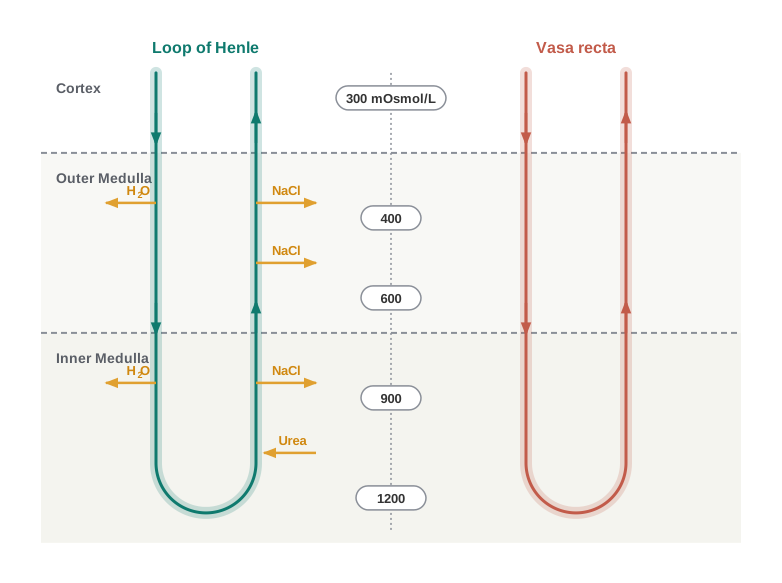
Fig. 16.7 – The counter current mechanism.
Building the Osmolarity Gradient
The result of this arrangement is a steadily rising osmolarity as you move from the cortex deep into the inner medulla — from about 300 mOsmol/L in the cortex to about 1200 mOsmol/L in the inner medulla. This gradient is caused mainly by two substances: NaCl (common salt) and urea.
Here is how each is kept in the medulla. NaCl is transported out by the ascending limb of Henle’s loop, and it is exchanged with the descending limb of the vasa recta; the ascending portion of the vasa recta then returns this NaCl to the interstitium instead of carrying it away. In a similar recycling loop, small amounts of urea enter the thin segment of the ascending limb of Henle’s loop, and this urea is later transported back into the interstitium by the collecting tubule.
This transport of substances, made possible by the special side-by-side arrangement of Henle’s loop and the vasa recta, is called the counter current mechanism. Its whole purpose is to maintain the concentration gradient in the medullary interstitium.
Flowchart
FC 16.4 – How the gradient concentrates urine: ascending limb pumps out NaCl → medullary interstitium becomes highly concentrated (300 → 1200 mOsmol/L) → urea from the collecting duct adds to this concentration → as filtrate passes down the collecting duct through this concentrated medulla → water is drawn out of the collecting duct into the interstitium → urine left behind becomes concentrated
Using the Gradient to Concentrate Urine
The gradient is not built for its own sake — it is a tool. As the filtrate passes down the collecting duct, it travels through the increasingly concentrated medulla. Because the interstitium outside is so concentrated, water is easily drawn out of the collecting duct into the interstitium. Losing this water leaves behind a concentrated filtrate — that is, concentrated urine.
How Concentrated Can Urine Get
Thanks to this mechanism, human kidneys can produce urine that is nearly four times as concentrated as the initial filtrate formed in Bowman’s capsule. This is an excellent water-conservation strategy — we throw out our wastes in a small, concentrated volume rather than flooding them out with litres of water.
16.5 Regulation of Kidney Function
The kidneys do not work blindly. Their functioning is constantly monitored and adjusted so that the body’s water, salt and blood pressure stay within safe limits. This fine control is achieved through hormonal feedback mechanisms — loops in which a change triggers a hormone, the hormone corrects the change, and the correction then switches the hormone off. Three players share this job: the hypothalamus (a control centre in the brain), the JGA, and, to a certain extent, the heart.
The ADH (Vasopressin) Mechanism
Scattered in the body are sensors called osmoreceptors, which are activated by changes in blood volume, body fluid volume and the concentration of ions. They act as an early-warning system for the body’s water balance.
Suppose the body loses too much fluid — say, through heavy sweating or diarrhoea. This excessive loss activates the osmoreceptors, which stimulate the hypothalamus to release antidiuretic hormone (ADH), also called vasopressin, from the neurohypophysis (the posterior part of the pituitary gland). ADH then facilitates the reabsorption of water from the latter parts of the tubule, thereby preventing diuresis — the excessive loss of water as urine.
Once enough water has been saved and the body fluid volume rises again, the osmoreceptors are switched off and ADH release is suppressed. This completes the feedback loop, shown in FC 16.5.
Flowchart
FC 16.5 – ADH feedback loop: excessive fluid loss → osmoreceptors activated → hypothalamus stimulated → ADH released from neurohypophysis → water reabsorption increases in the tubule → body fluid volume restored → osmoreceptors switched off → ADH suppressed (returns to normal)
ADH has a second effect too. It can act on blood vessels, causing them to constrict. This constriction raises the blood pressure, and a higher blood pressure increases the glomerular blood flow and thereby the GFR.
The Renin–Angiotensin Mechanism
The JGA plays a more complex regulatory role, built around a chain of chemical changes. When there is a fall in glomerular blood flow, glomerular blood pressure or GFR, the JG cells are activated to release renin.
Renin sets off a cascade. It converts a blood protein called angiotensinogen into angiotensin I, and this is then converted further into angiotensin II. Angiotensin II is a powerful vasoconstrictor: by narrowing blood vessels, it increases the glomerular blood pressure and thereby raises the GFR.
Angiotensin II does one more thing — it activates the adrenal cortex (the outer part of the adrenal gland, which sits atop the kidney) to release the hormone aldosterone. Aldosterone causes the reabsorption of \(Na^+\) and water from the distal parts of the tubule. This too raises the blood pressure and the GFR. This whole chain is known as the Renin–Angiotensin mechanism, traced in FC 16.6.
Flowchart
FC 16.6 – Renin–Angiotensin mechanism: fall in glomerular blood flow / blood pressure / GFR → JG cells release renin → angiotensinogen converted to angiotensin I → angiotensin I converted to angiotensin II → (branch 1) angiotensin II acts as vasoconstrictor → glomerular blood pressure and GFR rise; (branch 2) angiotensin II activates adrenal cortex → aldosterone released → Na⁺ and water reabsorbed from distal tubule → blood pressure and GFR rise
The ANF Check
If every mechanism only ever raised blood pressure, the system would run out of control. The heart provides a counterbalance. When blood flow to the atria of the heart increases, the heart releases Atrial Natriuretic Factor (ANF).
ANF causes vasodilation — the widening of blood vessels — which lowers the blood pressure. In this way, the ANF mechanism acts as a check on the renin–angiotensin mechanism, keeping it from pushing blood pressure too high.
The Three Regulators at a Glance
| Feature | ADH (vasopressin) | Renin–Angiotensin | ANF |
|---|---|---|---|
| Triggered by | Fluid loss / rise in ionic concentration (via osmoreceptors) | Fall in glomerular blood flow / pressure / GFR | Increased blood flow to the atria |
| Source | Hypothalamus → neurohypophysis | JG cells (JGA); aldosterone from adrenal cortex | Atria of the heart |
| Main action | Increases water reabsorption; constricts vessels | Vasoconstriction; aldosterone drives Na⁺ & water reabsorption | Vasodilation |
| Effect on blood pressure | Raises it | Raises it | Lowers it |
| Overall role | Conserves water, prevents diuresis | Restores GFR and blood pressure | Checks the renin–angiotensin system |
16.6 Micturition
Once urine has been formed by the nephrons, it does not leave the body drop by drop. It is collected and stored, and released only at a suitable moment. This controlled release is the subject of this section.
Storing and Releasing Urine
The urine formed by the nephrons is ultimately carried to the urinary bladder, where it is stored until a voluntary signal is given by the central nervous system (CNS) — the brain and spinal cord. This is why urination can usually be held back until a convenient time; the final “go-ahead” is under voluntary control.
The signal to empty the bladder is set off by the stretching of the bladder wall as it fills with urine. As the bladder swells, stretch receptors on its walls detect the stretch and send signals to the CNS. In response, the CNS sends motor messages that do two things at once: they make the smooth muscles of the bladder contract, and at the same time they make the urethral sphincter relax. With the bladder squeezing and the sphincter open, urine is released.
Flowchart
FC 16.7 – The micturition reflex: bladder fills with urine → bladder wall stretches → stretch receptors send signals to the CNS → CNS sends motor messages → smooth muscles of the bladder contract AND urethral sphincter relax → urine is released (micturition)
What Normal Urine Is Like
On an average, an adult human excretes about 1 to 1.5 litres of urine per day. The urine formed is a light yellow coloured, watery fluid. It is slightly acidic, with a pH of about 6.0, and has a characteristic odour.
A large part of the nitrogen waste leaves in this fluid: on an average, about 25–30 g of urea is excreted per day. These features — volume, colour, pH and composition — describe healthy urine, and any marked departure from them can be a clue that something is wrong.
Urine as a Diagnostic Window
Various conditions can change the characteristics of urine, and this makes urine a valuable window into health. Analysis of urine helps in the clinical diagnosis of many metabolic disorders as well as of malfunctioning of the kidney.
A well-known example concerns diabetes mellitus. Normally the tubules reabsorb virtually all the glucose from the filtrate, so healthy urine contains almost none. But when blood glucose is abnormally high, more glucose is filtered than the tubules can reabsorb, and the excess spills into the urine. The presence of glucose in urine is called glycosuria, and the presence of ketone bodies is called ketonuria; both are indicative of diabetes mellitus.
16.7 Role of Other Organs in Excretion
The kidneys are the main excretory organs, but they are not the only ones. Other than the kidneys, the lungs, the liver and the skin also help in the elimination of excretory wastes. Each removes a particular set of substances, and together they lighten the load on the kidneys.
The Lungs
Our lungs remove large amounts of carbon dioxide — approximately 200 mL per minute — produced by the body’s cells during respiration. Along with this gas, the lungs also give off significant quantities of water every day in the form of water vapour in the exhaled air.
The Liver
The liver, the largest gland in our body, is a major site of waste processing. It secretes bile, a fluid that carries several waste substances out of the body. Bile contains bilirubin and biliverdin (pigments left over from the breakdown of old red blood cells), along with cholesterol, degraded steroid hormones, vitamins and drugs. Most of these substances ultimately pass out of the body along with the digestive wastes in the faeces.
The Skin — Sweat and Sebaceous Glands
The skin contains two kinds of glands — sweat glands and sebaceous glands — that can eliminate certain substances through their secretions.
The sweat produced by the sweat glands is a watery fluid containing NaCl, small amounts of urea, lactic acid and so on. It is important to be clear about its main job: the primary function of sweat is to cool the body surface (thermoregulation), and only secondarily does it help remove some of these wastes.
The sebaceous glands eliminate substances such as sterols, hydrocarbons and waxes through an oily secretion called sebum. Here too, the main role is protective — the sebum provides a protective oily covering for the skin — with excretion being a side benefit.
Summary of Extra Excretory Routes
| Organ | Secretion / route | Substances eliminated | Primary function |
|---|---|---|---|
| Lungs | Exhaled air | \(CO_2\) (~200 mL/min) and water vapour | Gas exchange (breathing) |
| Liver | Bile | Bilirubin, biliverdin, cholesterol, degraded steroid hormones, vitamins, drugs | Digestion & metabolism |
| Skin (sweat glands) | Sweat | NaCl, small amounts of urea, lactic acid | Cooling the body (thermoregulation) |
| Skin (sebaceous glands) | Sebum | Sterols, hydrocarbons, waxes | Protective oily covering for skin |
16.8 Disorders of the Excretory System
Because the kidneys handle such a vital job, any failure in their working has serious consequences for the whole body. This section looks at what goes wrong when the kidneys malfunction, and at the methods medicine uses to cope with it.
Uremia and Kidney Failure
When the kidneys malfunction, they can no longer clear urea efficiently, so urea begins to accumulate in the blood. This condition is called uremia. It is highly harmful, and if it continues it may lead to kidney failure.
Haemodialysis — The Artificial Kidney
In patients with uremia, the excess urea can be removed artificially by a process called hemodialysis, carried out by a machine that copies the kidney’s basic trick of separating wastes from blood.
Here is how the process works. Blood is first drained from a convenient artery and, before anything else, an anticoagulant such as heparin is added to it so that it does not clot inside the machine. This blood is then pumped into a dialysing unit — the artificial kidney, shown in Fig. 16.8.
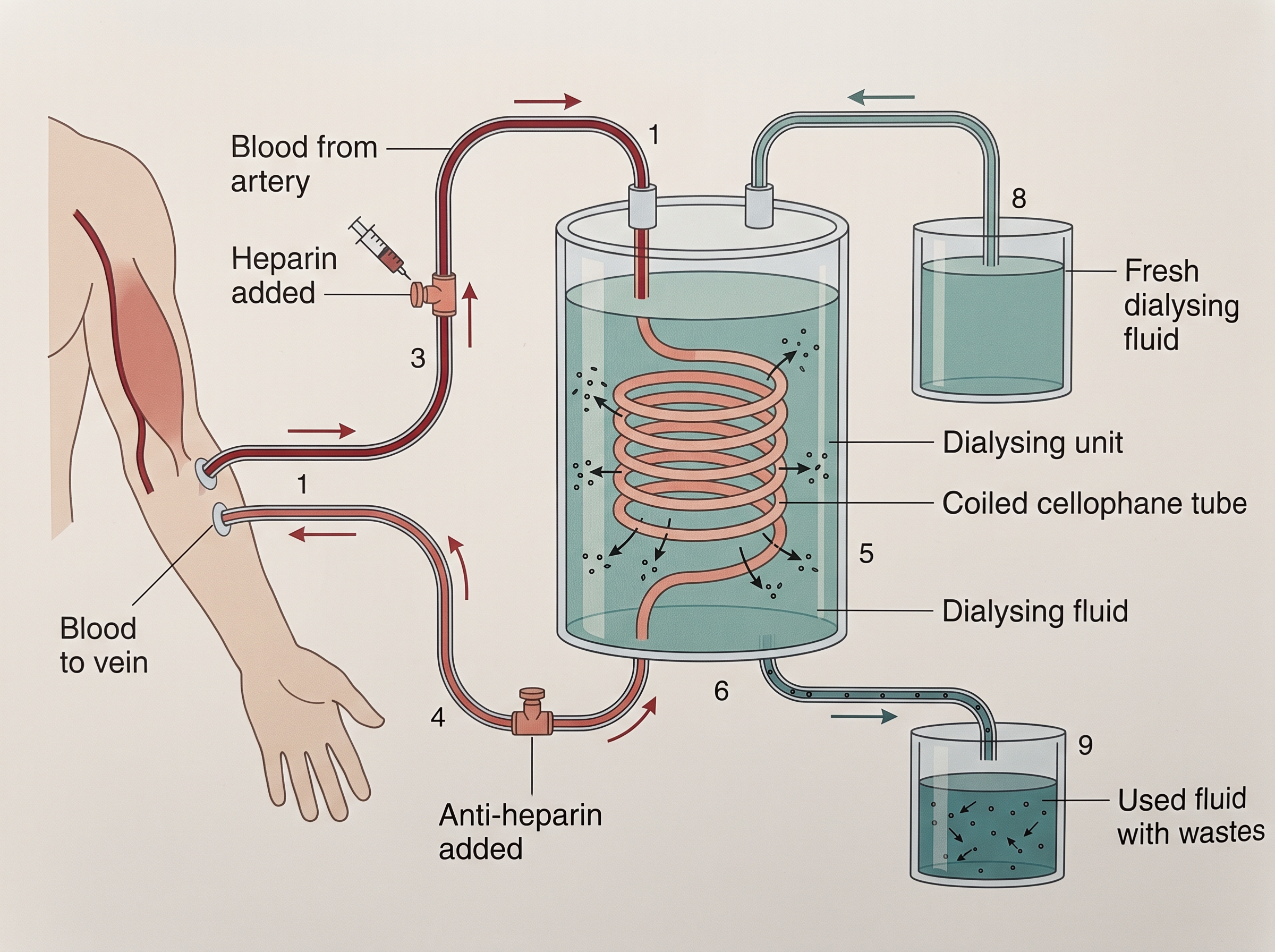
Fig. 16.8 – Haemodialysis (the artificial kidney).
The dialysing unit contains a coiled cellophane tube surrounded by a fluid called the dialysing fluid, which has the same composition as blood plasma — except that it contains no nitrogenous wastes. The cellophane membrane of the tube is porous, so it allows molecules to pass through based on their concentration gradient (from where they are more concentrated to where they are less concentrated).
Since nitrogenous wastes are present in the blood but absent from the dialysing fluid, these wastes freely move out of the blood into the fluid, thereby clearing the blood. The cleared blood is then pumped back into the body through a vein, after adding anti-heparin to it to restore normal clotting. This method has been a boon for thousands of uremic patients all over the world.
Flowchart
FC 16.8 – Haemodialysis: blood drained from an artery → heparin added (prevents clotting) → blood enters coiled cellophane tube in the dialysing unit → nitrogenous wastes diffuse out across the porous membrane into the dialysing fluid (down the concentration gradient) → cleared blood collected → anti-heparin added → blood returned to the body through a vein
Kidney Transplantation
Haemodialysis manages the problem but does not solve it permanently. The ultimate method for correcting acute renal failure (kidney failure) is kidney transplantation.
In a transplant, a functioning kidney taken from a donor — preferably a close relative — is placed in the patient. A close relative is preferred because the tissues are more similar, which minimises the chances of the new kidney being rejected by the immune system of the host (the recipient). Modern clinical procedures have greatly increased the success rate of this complicated technique.
Other Disorders
Two more common kidney disorders are worth knowing.
Renal calculi are stones — insoluble masses of crystallised salts, such as oxalates — that form within the kidney.
Glomerulonephritis is the inflammation of the glomeruli of the kidney.
16.9 Summary
- Many nitrogen-containing substances, ions, carbon dioxide, water and similar wastes build up in the body and have to be eliminated. The kind of nitrogenous waste an animal forms, and the way it excretes it, vary from animal to animal — mainly depending on the habitat and how much water is available. The three major nitrogenous wastes are ammonia, urea and uric acid.
- Different animals use different excretory organs: protonephridia, nephridia, malpighian tubules, green (antennal) glands and, in vertebrates, the kidneys. These organs not only remove nitrogenous wastes but also help maintain the ionic and acid–base balance of the body fluids.
- In humans, the excretory system consists of one pair of kidneys, a pair of ureters, a urinary bladder and a urethra. Each kidney contains over a million nephrons, and the nephron is the functional unit of the kidney.
- Each nephron has two portions — a glomerulus and a renal tubule. The glomerulus is a tuft of capillaries formed from the afferent arterioles, which are fine branches of the renal artery. The renal tubule begins with a double-walled Bowman’s capsule and is then differentiated into the proximal convoluted tubule (PCT), Henle’s loop (HL) and distal convoluted tubule (DCT). The DCTs of many nephrons join a common collecting duct, many of which open into the renal pelvis through the medullary pyramids. Bowman’s capsule enclosing the glomerulus forms the Malpighian (renal) corpuscle.
- Urine formation involves three main processes: filtration, reabsorption and secretion. Filtration is a non-selective process carried out by the glomerulus using the glomerular capillary blood pressure. About 1200 ml of blood is filtered by the glomerulus per minute, forming about 125 ml of filtrate in Bowman’s capsule per minute — the glomerular filtration rate (GFR).
- The JGA, a specialised portion of the nephrons, plays a significant role in regulating the GFR. Nearly 99 per cent of the filtrate is reabsorbed as it passes through the different parts of the nephron. The PCT is the major site of reabsorption and selective secretion. Henle’s loop mainly helps maintain an osmolar gradient (about 300–1200 mOsmol/L) within the kidney interstitium. The DCT and the collecting duct allow extensive reabsorption of water and certain electrolytes, helping in osmoregulation. Ions such as \(H^+\), \(K^+\) and \(NH_3\) can be secreted into the filtrate by the tubules to maintain the ionic balance and the pH of the body fluids.
- A counter current mechanism operates between the two limbs of the loop of Henle and those of the vasa recta (the capillary running parallel to Henle’s loop). The filtrate becomes concentrated as it moves down the descending limb and is diluted by the ascending limb. Electrolytes and urea are retained in the interstitium by this arrangement. Together, the DCT and collecting duct concentrate the filtrate about four times (from about 300 to 1200 mOsmol/L) — an excellent mechanism for the conservation of water.
- Urine is stored in the urinary bladder until a voluntary signal from the CNS releases it through the urethra; this release is called micturition. In addition to the kidneys, the skin, lungs and liver also assist in excretion.
Flowchart
FC 16.9 – Overview of urine formation and release: blood enters glomerulus → filtration (~125 ml filtrate/min) → filtrate passes through PCT (bulk reabsorption) → Henle’s loop (sets up 300–1200 mOsmol/L gradient) → DCT (conditional reabsorption + secretion) → collecting duct (water reabsorbed, urine concentrated ~4×) → renal pelvis → ureter → urinary bladder (storage) → urethra → micturition
16.10 Points to Ponder
- Ammonia is cheap for the body to produce but very expensive in water to remove, while urea and uric acid cost energy to make yet save water. Why might natural selection push animals in different habitats toward different points on this toxicity-versus-water trade-off?
- The kidney first filters almost everything out of the blood — including glucose, amino acids and useful ions — and then works hard to reabsorb about 99 per cent of it. Why might this “filter everything, then take back what you need” design be better than filtering out only the wastes in the first place?
- Maintaining the 300–1200 mOsmol/L gradient in the medulla costs the body energy every moment of the day. What would happen to an animal’s ability to survive in a dry habitat if this gradient could no longer be kept up?
- The renin–angiotensin–aldosterone system protects the body by restoring blood pressure and GFR when they fall. How might a mechanism that is life-saving in the short term become harmful if it stays switched on for too long?
- A dialysis machine can clear urea from the blood, yet a kidney transplant is described as the “ultimate” correction for kidney failure. What jobs of a real kidney — beyond simply removing wastes — might an artificial kidney be unable to carry out?
- Skin, lungs, liver and even saliva all remove some wastes, and yet a person cannot survive complete kidney failure. What does this tell you about how specialised and irreplaceable the kidney’s particular role really is?
16.11 NCERT Questions
Define Glomerular Filtration Rate (GFR).
Explain the autoregulatory mechanism of GFR.
Indicate whether the following statements are true or false:
- Micturition is carried out by a reflex.
- ADH helps in water elimination, making the urine hypotonic.
- Protein-free fluid is filtered from blood plasma into the Bowman’s capsule.
- Henle’s loop plays an important role in concentrating the urine.
- Glucose is actively reabsorbed in the proximal convoluted tubule.
Give a brief account of the counter current mechanism.
Describe the role of liver, lungs and skin in excretion.
Explain micturition.
Match the items of column I with those of column II:
| Column I | Column II |
|---|---|
| a. Ammonotelism | i. Birds |
| b. Bowman’s capsule | ii. Water reabsorption |
| c. Micturition | iii. Bony fish |
| d. Uricotelism | iv. Urinary bladder |
| e. ADH | v. Renal tubule |
What is meant by the term osmoregulation?
Terrestrial animals are generally either ureotelic or uricotelic, not ammonotelic, why?
What is the significance of juxta glomerular apparatus (JGA) in kidney function?
Name the following:
- A chordate animal having flame cells as excretory structures.
- Cortical portions projecting between the medullary pyramids in the human kidney.
- A loop of capillary running parallel to the Henle’s loop.
Fill in the gaps:
- Ascending limb of Henle’s loop is _______ to water whereas the descending limb is _______ to it.
- Reabsorption of water from distal parts of the tubules is facilitated by hormone _______.
- Dialysis fluid contain all the constituents as in plasma except _______.
- A healthy adult human excretes (on an average) _______ gm of urea/day.
16.12 Check Your Concepts
Name the three major nitrogenous wastes and arrange them in increasing order of toxicity. State how the water needed for their removal changes across this order.
Assertion (A): Most terrestrial animals are ureotelic or uricotelic rather than ammonotelic. Reason (R): Excreting ammonia safely requires a large amount of water. State whether A and R are true, and whether R correctly explains A.
Why is glomerular filtration described as “ultrafiltration” rather than ordinary filtration? In your answer, mention the role of the podocytes and filtration slits.
Assertion (A): The descending limb of Henle’s loop concentrates the filtrate as it moves down. Reason (R): The descending limb is permeable to water but almost impermeable to electrolytes. State whether A and R are true, and whether R correctly explains A.
Arrange the following parts of the renal tubule in the correct order in which the filtrate flows through them: distal convoluted tubule, Bowman’s capsule, collecting duct, proximal convoluted tubule, Henle’s loop.
Distinguish between cortical nephrons and juxtamedullary nephrons, and explain why the vasa recta is well developed in one but reduced or absent in the other.
Distinguish between reabsorption and tubular secretion, giving one example of a substance handled by each.
What would happen to the kidney’s ability to produce concentrated urine if the loop of Henle were much shorter than normal? Justify your answer using the counter current mechanism.
Explain why the dialysing fluid used in haemodialysis has the same composition as blood plasma except for the nitrogenous wastes. What would happen if glucose were left out of the dialysing fluid?
Arrange the steps of the renin–angiotensin mechanism in the correct sequence: angiotensin II, renin, angiotensinogen, angiotensin I, aldosterone.
The renin–angiotensin mechanism raises blood pressure while the ANF mechanism lowers it. Explain how these two mechanisms together keep blood pressure balanced, and predict what might happen if the ANF mechanism failed.
Assertion (A): Glucose does not normally appear in the urine of a healthy person. Reason (R): The proximal convoluted tubule reabsorbs almost all of the filtered glucose. State whether A and R are true, and whether R correctly explains A. Also explain why glucose appears in the urine in uncontrolled diabetes mellitus.
Differentiate between diabetes mellitus and diabetes insipidus in terms of their cause and their effect on urine.
Identify the odd one out in each group and give a reason:
- Ammonia, urea, uric acid, aldosterone.
- PCT, DCT, Henle’s loop, ureter.
- Bilirubin, biliverdin, cholesterol, renin.
Draw a labelled diagram of a nephron and label the following: glomerulus, Bowman’s capsule, PCT, descending and ascending limbs of Henle’s loop, DCT, collecting duct and vasa recta.
In a longitudinal section of the human kidney, label the cortex, medulla, a medullary pyramid, a renal column (Column of Bertini), a calyx and the renal pelvis.
Match each structure in Column I with its correct role in Column II:
| Column I | Column II |
|---|---|
| a. Podocytes | i. Increases surface area for reabsorption in the PCT |
| b. Brush border | ii. Releases renin when GFR falls |
| c. JG cells | iii. Form filtration slits for ultrafiltration |
| d. Vasa recta | iv. Runs parallel to Henle’s loop and helps maintain the medullary gradient |
- Why can a healthy person survive and lead a normal life with only one functioning kidney? Relate your answer to the number of nephrons and the concept of functional reserve.