Chapter 15: Body Fluids and Circulation
For nearly fourteen centuries, physicians believed something about the human body that turned out to be completely wrong. Following the ancient Greek physician Galen, they taught that the liver constantly manufactured fresh blood from the food we eat, and that the body’s organs simply soaked it up and used it — like water poured onto dry soil. Blood, in this view, was made and consumed, made and consumed, never travelling in a loop.
In 1628, an English physician named William Harvey challenged this idea with a startlingly simple piece of arithmetic. He estimated how much blood the heart pushes out with each beat, then multiplied it by the number of beats in an hour. The answer was staggering — the heart moved far more blood in a single hour than the entire body could ever manufacture from food. There was only one explanation: it had to be the same blood, going around and around.
Harvey then tested his idea. Tying a cord around a volunteer’s arm, he showed that the valves inside veins let blood move in only one direction — back toward the heart. Piece by piece, he demonstrated that blood flows in a closed circuit, driven by the heart’s muscular pump. His small book, De Motu Cordis, quietly overturned a belief older than most nations, and founded the modern science of circulation.
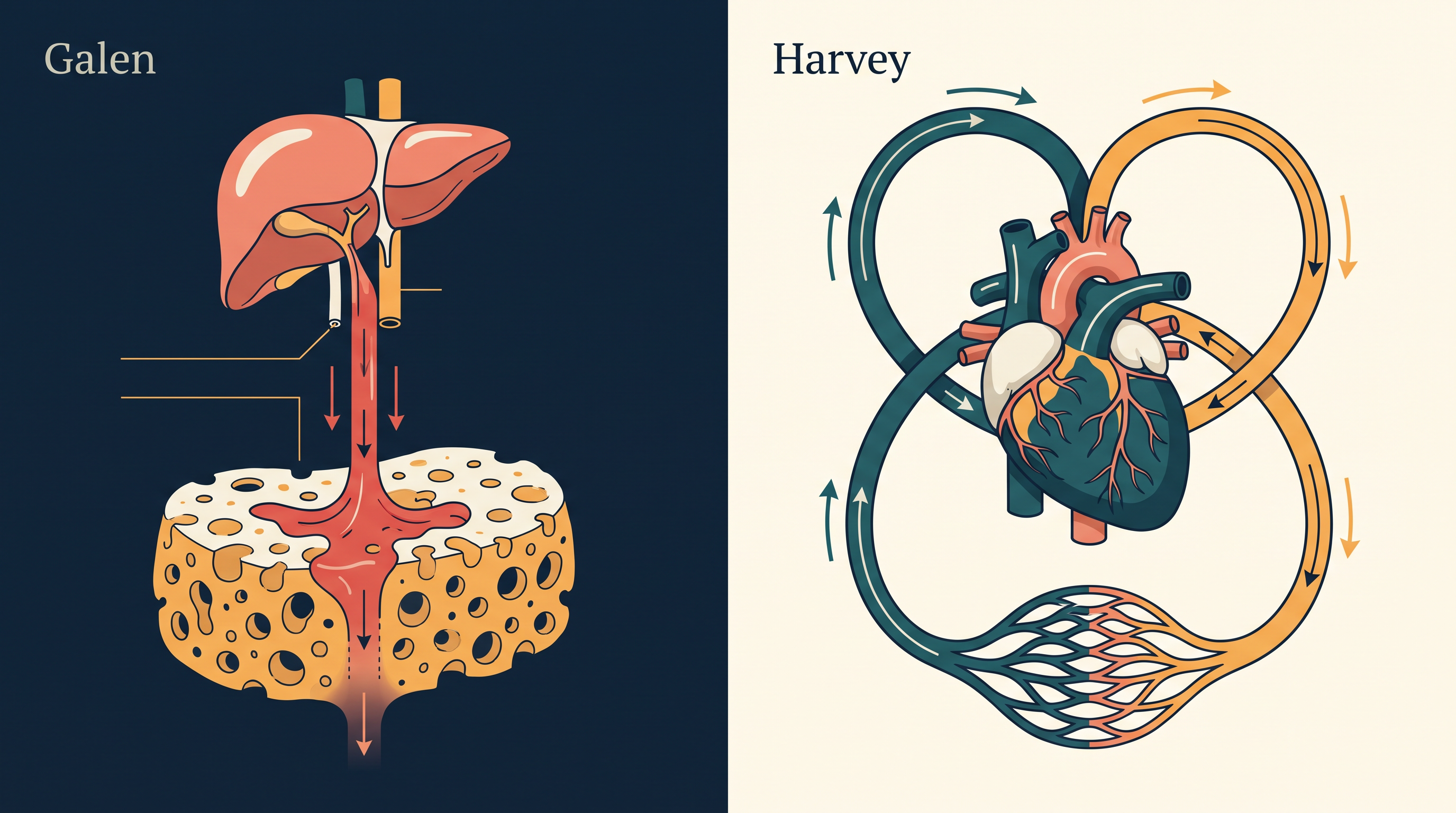
Fig. 15.0 – The Galenic view of blood versus Harvey’s closed circuit.
Keep these questions in mind as you read — by the end of this chapter, you will be able to answer every one of them.
15.1 Blood
Every living cell in the body must be supplied with nutrients, oxygen (\(O_2\)) and other essential substances. At the same time, the wastes and harmful substances that cells produce have to be removed continuously, or the tissues cannot function properly. The body therefore needs an efficient system for moving substances to its cells and away from them.
Different groups of animals solve this transport problem in different ways. Very simple animals such as sponges and coelenterates do not use a special transport fluid at all. Instead, they circulate water from their surroundings through their body cavities, and the cells lining those cavities exchange substances directly with the water.
More complex animals cannot depend on outside water reaching every cell. They use special fluids inside the body to carry materials from one place to another. In most higher animals, including humans, the main such fluid is blood. A second body fluid, lymph, helps transport certain substances as well.
This chapter looks at the composition and properties of both fluids — blood and lymph (also called tissue fluid) — and then explains how blood is circulated through the body.
Blood as a connective tissue
Although blood is a flowing liquid, it is classified as a tissue — and, more exactly, as a special kind of connective tissue. (Recall that a tissue is a group of similar cells, along with the material around them, that carry out a shared function.)
In a connective tissue, the cells are not tightly packed. They lie scattered within a large amount of surrounding non-living material called the matrix. In familiar connective tissues such as bone and cartilage, this matrix is hard or firm.
Blood is called “special” because its matrix is a liquid instead of a solid. Its cells float freely in this fluid — and that is exactly what allows blood to be pumped through vessels and delivered to every part of the body, which a solid tissue could never do.
Blood thus has two distinct parts. The first is its fluid matrix, a pale liquid called plasma. The second is the cells and cell fragments suspended in the plasma, known together as the formed elements. The next two subsections take up each part in turn — plasma first, then the formed elements.
15.1.1 Plasma
Plasma is the liquid part of blood — the fluid matrix in which the blood cells float. It is a straw-coloured (pale yellow), slightly thick or viscous fluid, and it makes up nearly 55 per cent of the blood.
By far the largest part of plasma is water: about 90–92 per cent. This is what lets plasma dissolve and carry so many substances at once. Proteins make up another 6–8 per cent of plasma, and these plasma proteins matter a great deal despite their small share.
Three proteins are present in the largest amounts — fibrinogen, globulins and albumins. Fibrinogen is needed for the clotting (coagulation) of blood. Globulins are mainly involved in the body’s defence mechanisms — that is, in fighting infection. Albumins, the most abundant of the three, help maintain the osmotic balance of blood: they hold water within the vessels by osmotic pull, so that the right amount of fluid stays in the blood rather than leaking away into the surrounding tissues.
Plasma also carries small amounts of minerals in the form of ions — such as \(Na^+\), \(Ca^{2+}\), \(Mg^{2+}\), \(HCO_3^-\) and \(Cl^-\). Alongside these are substances that are always in transit through the body, such as glucose, amino acids and lipids, being ferried between the gut, the liver and the body’s cells.
Plasma also contains the factors needed for blood clotting, but keeps them in an inactive form until an injury calls for them. This is a safety feature — the clotting machinery is present and ready, yet it does not switch on unless there is a genuine need.
One term is worth fixing in mind now: plasma from which the clotting factors have been removed is called serum. In short, serum is plasma minus its clotting factors.
15.1.2 Formed Elements
The cells and cell fragments that float in the plasma are together called the formed elements. There are three kinds — erythrocytes (red blood cells), leucocytes (white blood cells) and platelets — and together they make up nearly 45 per cent of the blood. The different formed elements are shown in Fig. 15.1.
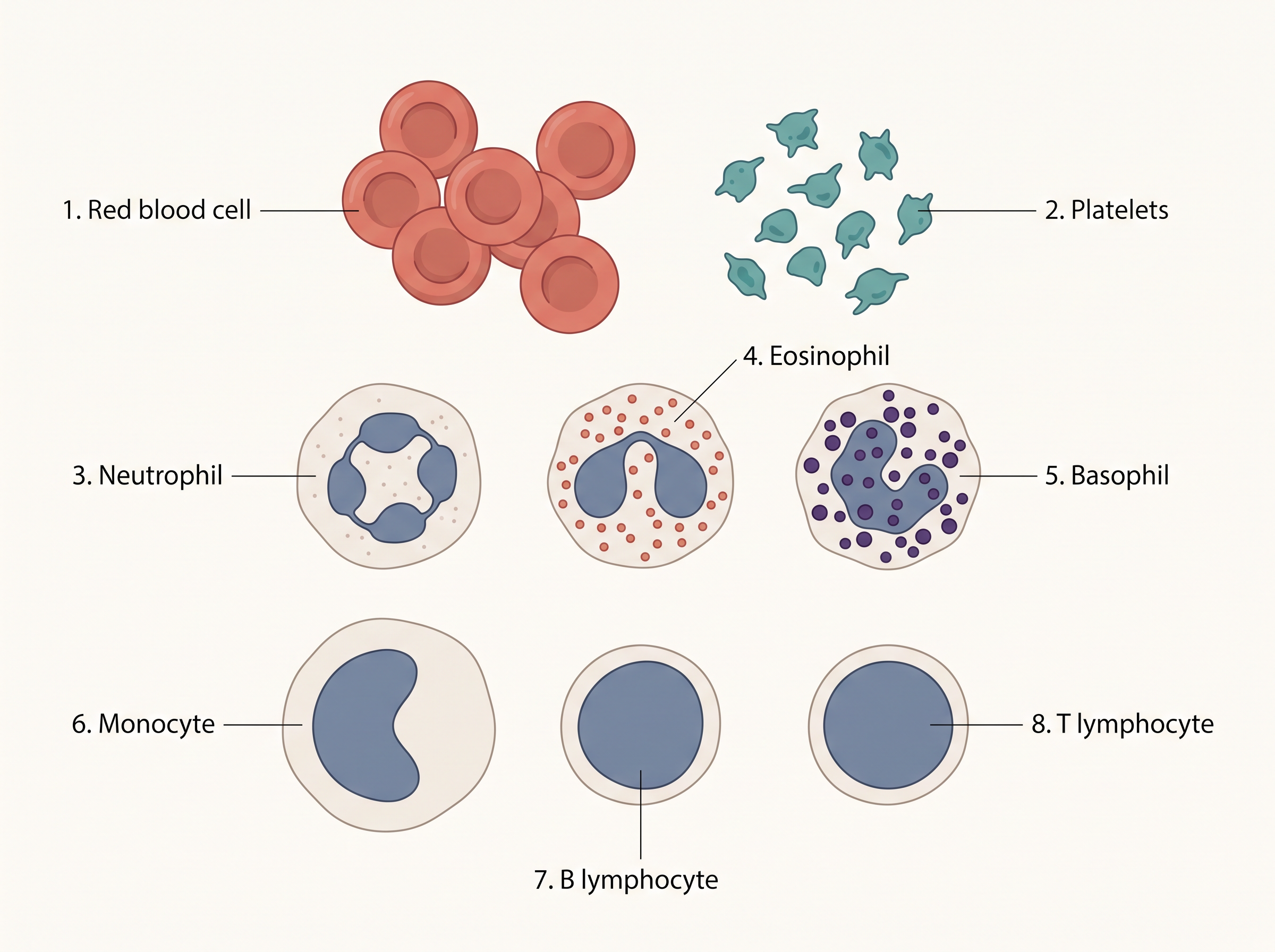
Fig. 15.1 – The formed elements of blood.
Red blood cells (erythrocytes)
Erythrocytes, or red blood cells (RBCs), are by far the most numerous cells in the blood. In a healthy adult man there are, on average, about 5 million to 5.5 million RBCs in every cubic millimetre (mm³) of blood. In adults, they are formed in the red bone marrow.
In most mammals, including humans, a mature RBC has no nucleus, and its shape is biconcave — like a disc pressed inward on both faces. Each RBC is packed with haemoglobin, a red, iron-containing protein. It is this pigment that gives blood its red colour and gives these cells their name. A healthy person has about 12–16 g of haemoglobin in every 100 mL of blood, and this haemoglobin is what carries the respiratory gases — chiefly oxygen — around the body.
The shape and contents of the RBC are closely tied to the job it has to do.
An RBC lives for about 120 days on average. Worn-out RBCs are then broken down in the spleen, which is why the spleen is often called the “graveyard of RBCs.”
White blood cells (leucocytes)
Leucocytes, or white blood cells (WBCs), are colourless — they lack haemoglobin, so they carry no red pigment. Unlike mature RBCs, they are nucleated (they keep their nucleus). They are far fewer than RBCs, averaging about 6000–8000 per mm³ of blood, and are generally short-lived.
White blood cells fall into two main groups, depending on whether their cytoplasm contains visible granules: granulocytes (with granules) and agranulocytes (without granules). This classification is summarised in FC 15.1.
Flowchart
FC 15.1 – Classification of leucocytes (WBCs): Leucocytes → two groups. (i) Granulocytes → Neutrophils, Eosinophils, Basophils; (ii) Agranulocytes → Lymphocytes (B and T types), Monocytes.
The three granulocytes are neutrophils, eosinophils and basophils; the two agranulocytes are lymphocytes and monocytes. Each type is present in a different proportion and does a different job.
Neutrophils are the most abundant of all WBCs (60–65 per cent), while basophils are the fewest (0.5–1 per cent). Neutrophils, along with monocytes (6–8 per cent), are phagocytic cells — they engulf and destroy foreign organisms that enter the body. Basophils secrete substances such as histamine, serotonin and heparin, and take part in inflammatory reactions. Eosinophils (2–3 per cent) help resist infections and are also linked with allergic reactions. Lymphocytes (20–25 per cent) come in two major forms — B and T lymphocytes — and both are responsible for the body’s immune responses.
Platelets
Platelets, also called thrombocytes, are not whole cells but small cell fragments. They are pinched off from large cells in the bone marrow called megakaryocytes. Normal blood contains about 1,50,000 to 3,50,000 platelets per mm³.
Platelets can release a variety of substances, most of which help in the coagulation (clotting) of blood. Because of this role, a fall in platelet numbers can cause clotting disorders, in which the blood fails to clot properly and too much blood may be lost from the body.
15.1.3 Blood Groups
One person’s blood looks much like another’s — all red and similar. Yet human blood is not identical from person to person; it differs in certain features invisible to the naked eye. Because of these hidden differences, blood can be sorted into distinct types called blood groups.
Several grouping systems have been worked out, but two are used all over the world: the ABO grouping and the Rh grouping. Knowing a person’s blood group matters most when blood must be transferred from one person to another, as the next two subsections — on ABO and Rh grouping — will make clear.
15.1.3.1 ABO grouping
The ABO system sorts blood according to two special marker molecules, called antigens, that may sit on the surface of the red blood cells. These two antigens are named A and B. (An antigen is any chemical that the body can recognise as foreign and that can therefore trigger an immune response.)
A person’s red cells may carry antigen A, antigen B, both A and B, or neither. This gives the four familiar ABO blood groups: A, B, AB and O.
Matching each antigen there is an antibody — a defensive protein that the body makes to act against a particular antigen. Remarkably, the plasma of each person naturally contains antibodies against exactly those A/B antigens that their own red cells do not carry. So a group A person (with A antigen) carries anti-B antibodies in the plasma; a group B person carries anti-A; a group AB person, having both antigens, carries neither antibody; and a group O person, having no A or B antigen, carries both anti-A and anti-B. The full pattern is set out in Table 15.1.
This pattern matters enormously during a blood transfusion. As you may already know, one cannot simply use any blood at all — before blood is given, the donor’s blood must be carefully matched with the recipient’s. The danger being avoided is clumping (also called agglutination): if the recipient’s plasma antibodies meet a matching antigen on the donor’s red cells, those cells stick together in clumps and are destroyed. This can be fatal.
The rightmost column of Table 15.1 shows which groups can safely donate to whom, and two special cases stand out. Group O blood carries no A or B antigen on its red cells, so there is nothing on those cells for any recipient’s antibodies to attack — which is why O blood can be given to a person of any ABO group. For this reason, group O individuals are called universal donors. At the other extreme, group AB plasma carries no anti-A or anti-B antibodies, so it will not attack the red cells of any donor — which is why an AB person can receive blood from any ABO group. Such persons are called universal recipients.
15.1.3.2 Rh grouping
Besides the A and B antigens, red blood cells may carry yet another antigen, the Rh antigen. It gets its name because it is similar to an antigen first found in Rhesus monkeys (“Rh” is short for Rhesus). This antigen is present on the red cells of the majority of humans — nearly 80 per cent. People who have it are called Rh positive (Rh+ve), and those who lack it are called Rh negative (Rh-ve).
The Rh antigen behaves much like A and B in one respect: if an Rh-ve person is exposed to Rh+ve blood, their body treats the Rh antigen as foreign and makes specific anti-Rh antibodies against it. For this reason, the Rh group — not just the ABO group — must also be matched before any blood transfusion.
Rh grouping becomes especially important in a special situation during pregnancy, known as Rh incompatibility. It can arise when an Rh-ve mother is carrying an Rh+ve foetus (the baby having inherited the Rh antigen from its father).
During the first such pregnancy, the baby is normally safe. The mother’s blood and the foetus’s blood are kept well apart by the placenta, so the foetal Rh antigens are not exposed to the mother’s Rh-ve blood.
The turning point comes at the delivery of the first child. As the baby is born, small amounts of its Rh+ve blood can leak across into the mother’s circulation. Meeting the Rh antigen for the first time, the mother’s immune system responds by beginning to make anti-Rh antibodies, which then remain in her blood.
The problem shows up in her later pregnancies. If a subsequent foetus is again Rh+ve, the mother’s anti-Rh antibodies can now cross the placenta into the foetus and attack its red blood cells, destroying them. This may cause severe anaemia and jaundice in the baby, and can even be fatal to the foetus. This condition is called erythroblastosis foetalis.
The whole sequence, from the first pregnancy to the harm caused in a later one, is summarised in FC 15.2.
Flowchart
FC 15.2 – How erythroblastosis foetalis develops: Rh-ve mother carrying an Rh+ve foetus → (first pregnancy) placenta keeps the two bloods apart, foetal Rh antigen not exposed to the mother → (delivery of first child) small amount of Rh+ve foetal blood enters the mother’s circulation → mother’s immune system makes anti-Rh antibodies → (a later Rh+ve pregnancy) maternal anti-Rh antibodies cross the placenta into the foetus → they destroy the foetal RBCs → erythroblastosis foetalis (severe anaemia and jaundice, possibly fatal to the foetus).
Fortunately, this danger can be prevented. The mother is given anti-Rh antibodies immediately after the delivery of her first child, which stops her from becoming sensitised in the first place.
15.1.4 Coagulation of Blood
When you cut a finger or hurt yourself, the wound does not keep bleeding for long — after a little while, the blood stops flowing. This is because blood coagulates, or clots, in response to an injury or trauma. Clotting is a protective mechanism that seals the wound and prevents excessive loss of blood from the body.
If you look at a cut after some time, you will notice a dark reddish-brown scum over it. This is the clot (also called a coagulum). A clot is essentially a mesh — a network of fine threads called fibrin, in which the dead and damaged formed elements of blood become trapped, plugging the wound.
How a clot forms: the cascade
But where do these fibrin threads come from? They are made from an inactive protein already present in the plasma, called fibrinogen. Fibrinogen is converted into fibrin by an enzyme called thrombin. (Recall from the plasma section that blood stripped of these clotting factors is what we called serum.)
Thrombin, however, is not floating around ready-made — that would be dangerous, since blood would then clot inside intact vessels. Instead, thrombin is produced only when needed, from another inactive plasma substance called prothrombin. This conversion of prothrombin into active thrombin requires an enzyme complex called thrombokinase.
Thrombokinase, in turn, does not exist ready-made either. It is built up through a series of linked enzyme reactions — a “cascade” — involving a number of clotting factors that lie in the plasma in an inactive state, waiting to be switched on. Building the clot through so many small steps may seem roundabout, but it gives the body fine control: the reaction can be triggered quickly and strongly exactly when and where it is needed, yet stays switched off the rest of the time.
What sets this cascade in motion? An injury stimulates the platelets to release certain factors that start the coagulation process. In addition, certain factors released by the damaged tissues at the site of injury can also trigger clotting. Throughout the whole process, calcium ions (\(Ca^{2+}\)) play a very important role.
The complete cascade is summarised in FC 15.3.
Flowchart
FC 15.3 – The blood clotting cascade: injury/trauma → platelets release clotting factors (and damaged tissues release factors too) → a cascade of linked reactions among inactive plasma factors → forms the enzyme complex thrombokinase → thrombokinase (with \(Ca^{2+}\)) converts prothrombin (inactive) → thrombin (active enzyme) → thrombin converts fibrinogen (inactive) → fibrin (threads) → fibrin network traps blood cells → clot (coagulum) that seals the wound.
15.2 Lymph (Tissue Fluid)
As blood flows through the tiny capillaries within the tissues, some of its water — along with many small, water-soluble substances dissolved in it — seeps out through the thin capillary walls into the spaces between the cells. The larger plasma proteins and most of the formed elements are too big to pass through and stay behind inside the vessels. The fluid that filters out in this way is called the interstitial fluid, or tissue fluid.
Because it comes straight from plasma, tissue fluid has the same mineral distribution as plasma. It bathes the cells directly, and it is through this fluid that the exchange of nutrients, gases and wastes between the blood and the body’s cells always takes place. In other words, blood never touches the cells directly — tissue fluid is the go-between.
Not all of this fluid returns straight to the blood. An elaborate network of vessels, the lymphatic system, collects the tissue fluid and drains it back into the major veins. Once it is inside this network, the fluid is given a new name: lymph.
This journey — from blood, to tissue fluid, to lymph, and back to blood — is summarised in FC 15.4.
Flowchart
FC 15.4 – Formation and return of tissue fluid and lymph: blood flowing through tissue capillaries → water and small solutes filter out through the capillary walls (large proteins and formed elements stay in the vessel) → tissue fluid (interstitial fluid) in the spaces between cells → exchange of nutrients, gases and wastes with the cells → tissue fluid collected by lymphatic vessels → now called lymph → lymphatic system drains it back into the major veins → rejoins the blood.
Lymph is a colourless fluid — colourless because it lacks the red, haemoglobin-filled RBCs. It does, however, contain specialised lymphocytes, the white blood cells responsible for the body’s immune responses. Besides its role in defence, lymph is an important carrier of nutrients and hormones. It also has a special job in digestion: the fats absorbed from digested food enter the body through lymph, by way of tiny lymphatic vessels called lacteals that lie inside the villi of the intestine.
15.3 Circulatory Pathways
Blood can be moved around an animal’s body in one of two broad patterns — an open circulatory system or a closed one.
In an open circulatory system, the blood pumped by the heart does not stay inside vessels the whole time. It travels through a few large vessels and is then released into open spaces or body cavities called sinuses, where it directly bathes the tissues before making its way back to the heart. This arrangement is found in arthropods (such as insects) and molluscs.
In a closed circulatory system, by contrast, the blood pumped by the heart always stays within a closed network of blood vessels and never flows freely into open cavities. This pattern is found in annelids (such as earthworms) and in all chordates, including us. A closed system is considered more advantageous because, with the blood kept inside vessels, its flow can be regulated far more precisely — the body can send more or less blood to particular organs as needed.
Hearts and circulation across the vertebrates
Every vertebrate has a muscular, chambered heart, but the number of chambers — and how well the heart keeps “clean” (oxygenated) blood apart from “used” (deoxygenated) blood — differs from group to group, and this shows a clear evolutionary trend.
Fishes have a 2-chambered heart, with a single atrium and a single ventricle. The heart pumps out only deoxygenated blood, which is sent to the gills to be oxygenated and then supplied directly to the body; deoxygenated blood then returns to the heart. Because the blood passes through the heart only once in each complete circuit, this is called single circulation.
Amphibians and most reptiles (all except crocodiles) have a 3-chambered heart — two atria but only a single ventricle. Here the left atrium receives oxygenated blood (from the gills, lungs or skin) and the right atrium receives deoxygenated blood from the rest of the body. But because there is only one ventricle, the two kinds of blood partly mix before being pumped out. Since the blood passes through the heart twice, yet the two streams are not kept fully separate, this is called incomplete double circulation.
Crocodiles, birds and mammals have a fully 4-chambered heart — two atria and two ventricles. Oxygenated blood entering the left atrium and deoxygenated blood entering the right atrium pass down into the ventricles of the same side, and the ventricles pump them out with no mixing at all. Two entirely separate circulatory pathways therefore exist, which is called (complete) double circulation.
With this background, we can now look closely at the human circulatory system.
15.3.1 Human Circulatory System
The human circulatory system — also called the blood vascular system — has three parts working together: a muscular, chambered heart that pumps; a network of closed, branching blood vessels that carry the blood; and the blood itself, the fluid that is circulated. In effect, the heart is a double pump, its two sides driving blood along two different routes at the same time.
The heart develops from the mesoderm (it is a mesodermally derived organ). It lies in the thoracic cavity, between the two lungs and tilted slightly to the left, and is roughly the size of a clenched fist. It is protected inside a double-walled membranous bag called the pericardium, which encloses a lubricating pericardial fluid that lets the heart beat with very little friction. A cut-away view of the heart is shown in Fig. 15.2.
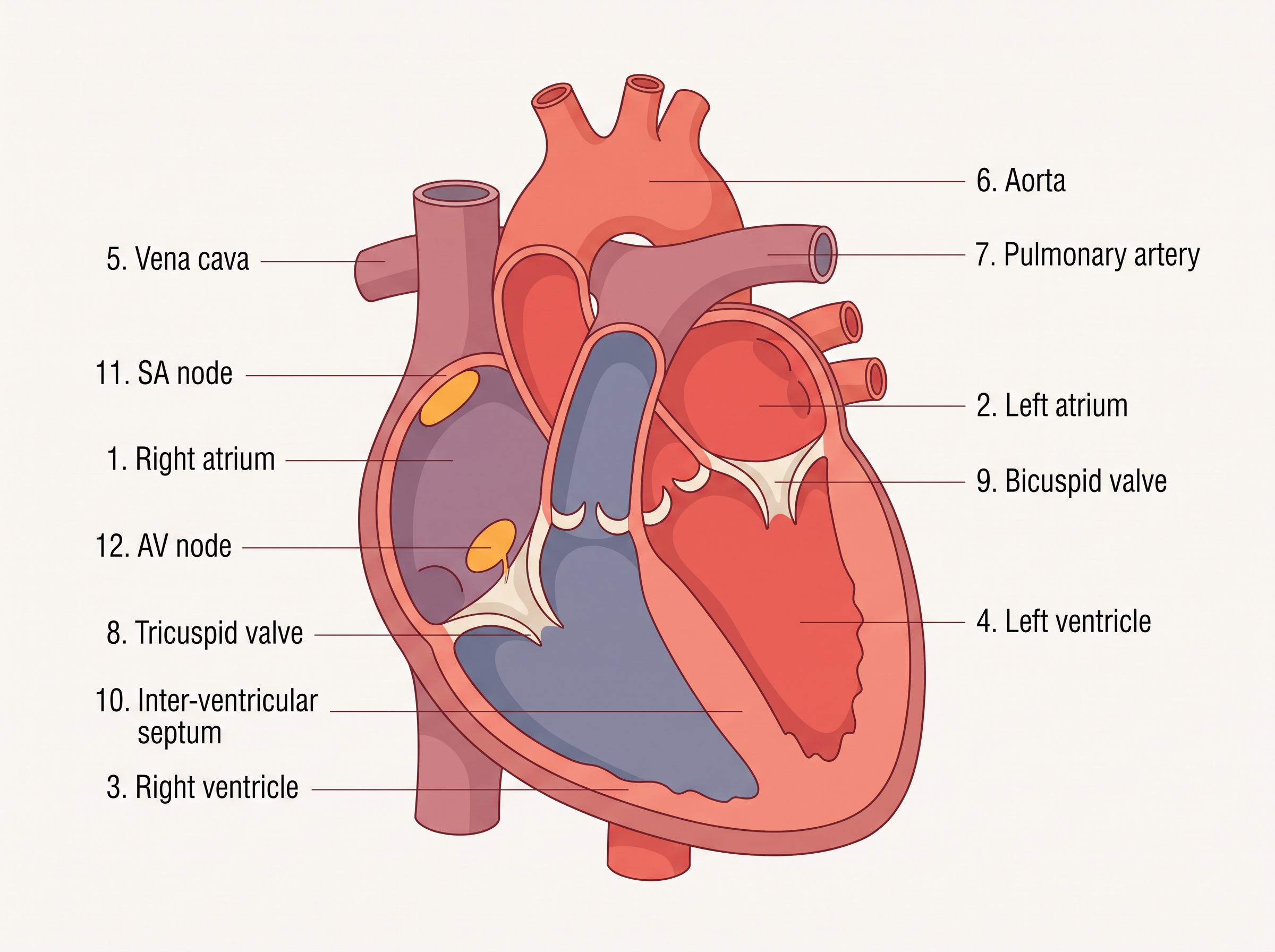
Fig. 15.2 – Section of the human heart.
Chambers and septa
The human heart has four chambers. The two upper chambers are smaller and are called atria (singular: atrium); the two lower chambers are larger and are called ventricles.
The chambers are separated by muscular walls called septa. A thin, muscular inter-atrial septum separates the right atrium from the left atrium, while a thicker inter-ventricular septum separates the right ventricle from the left ventricle. In addition, the atrium and the ventricle of the same side are separated by a thick fibrous atrio-ventricular septum.
These septa are not completely sealed, however — each carries an opening through which the two chambers on the same side stay connected, so that blood can pass from an atrium down into the ventricle below it.
The valves of the heart
Where blood passes from one chamber to the next, a valve guards the opening and makes sure blood flows only forward, never backward.
The opening between the right atrium and the right ventricle is guarded by the tricuspid valve, made of three muscular flaps (cusps). The opening between the left atrium and the left ventricle is guarded by the bicuspid valve (also called the mitral valve), made of two cusps. Where the ventricles open into the great arteries — the right ventricle into the pulmonary artery and the left ventricle into the aorta — the openings are guarded by semilunar valves.
Together, these valves allow blood to move in only one direction: from the atria into the ventricles, and from the ventricles into the pulmonary artery or aorta, snapping shut to prevent any backward flow. This one-way design is essential — it keeps blood moving forward through the circuit with each beat, so no pumping effort is wasted pushing blood the wrong way.
The whole heart is built of a special muscle called cardiac muscle. Notice in Fig. 15.2 that the walls of the ventricles are much thicker than the walls of the atria.
The conducting system and the pacemaker
Besides ordinary cardiac muscle, the heart contains a special nodal tissue — cardiac muscle modified to generate and conduct electrical signals (see Fig. 15.2).
A patch of this tissue sits in the upper right corner of the right atrium and is called the sino-atrial node (SAN). A second mass lies in the lower left corner of the right atrium, near the atrio-ventricular septum, and is called the atrio-ventricular node (AVN). From the AVN a bundle of nodal fibres, the atrio-ventricular bundle (AV bundle) — also known as the bundle of His — passes through the atrio-ventricular septa and reaches the top of the inter-ventricular septum, where it immediately splits into a right and a left bundle. These branch further into fine fibres that spread throughout the muscle of both ventricles, called the Purkinje fibres. The order in which a signal travels along this system is shown in FC 15.5.
Flowchart
FC 15.5 – The heart’s impulse-conducting pathway: SA node (SAN) → AV node (AVN) → AV bundle (bundle of His) → right and left bundle branches → Purkinje fibres spreading through the ventricular muscle.
There is a neat reason for this particular layout. The fibrous atrio-ventricular septum acts as an electrical insulator between the atria and the ventricles, so the only route by which a signal can cross from the atria to the ventricles is through the AVN and the AV bundle. This single, controlled pathway ensures that the ventricles are triggered in a coordinated way, a fraction of a moment after the atria.
A remarkable feature of nodal tissue is that it can generate electrical signals (action potentials) entirely on its own, without any nerve telling it to — a property called being autoexcitable. Because of this, the heart does not depend on the brain to keep beating. Different parts of the nodal system can fire at different rates, but the SAN fires fastest, producing about 70–75 action potentials per minute. Firing fastest, the SAN sets the pace for the whole heart: it starts each heartbeat and keeps the rhythm steady. For this reason the SAN is called the pacemaker. Driven by it, the heart normally beats about 70–75 times per minute (on average, 72 beats per minute).
15.3.2 Cardiac Cycle
How does the heart actually pump? Its work is carried out in a fixed, repeating sequence of steps. We can follow that sequence one phase at a time.
Joint diastole. When the heart is resting between beats, all four chambers are relaxed together — a state called joint diastole (diastole means relaxation; systole, which we meet shortly, means contraction). The AV valves (tricuspid and bicuspid) are open, while the semilunar valves are closed. Blood returning from the body through the vena cava and from the lungs through the pulmonary veins flows into the atria and passes on, through the open AV valves, into the ventricles. Much of the ventricles’ filling happens quietly in this way, even before the atria contract.
Atrial systole. Now the SAN fires an action potential. It sweeps through both atria and makes them contract together — this is atrial systole. The contracting atria squeeze the last of their blood down into the ventricles, topping up the ventricular filling by about 30 per cent.
Ventricular systole. The same electrical signal is now carried to the ventricles by the AVN and the AV bundle, from where the bundle of His spreads it through the entire ventricular muscle. There is a brief, deliberate delay as the signal passes through the AVN; this pause gives the atria just enough time to finish emptying before the ventricles contract, so the ventricles fill as fully as possible. The ventricles then contract powerfully — this is ventricular systole — while the atria relax (atrial diastole) at the same moment.
As the ventricles squeeze, the pressure inside them rises sharply. This rising pressure first pushes the tricuspid and bicuspid valves shut, as blood tries to flow back into the atria — producing the first heart sound. As the pressure climbs still higher, it forces open the semilunar valves at the mouths of the pulmonary artery (right side) and the aorta (left side), and blood is driven out of the ventricles into these great vessels and away into the circulation.
Ventricular diastole. The ventricles now relax — ventricular diastole — and the pressure inside them falls. As it drops, the semilunar valves snap shut, stopping blood from flowing back into the ventricles (this makes the second heart sound). As ventricular pressure falls lower still, the pressure of blood collecting in the atria pushes the tricuspid and bicuspid valves open again, and blood once more flows freely into the ventricles. All four chambers are now relaxed — joint diastole — exactly as we began. Soon the SAN fires a fresh action potential, and the whole sequence repeats, beat after beat, for as long as the heart lives.
Note that the heart’s valves have no muscles of their own to open and shut them; they move passively, purely because of the differences in blood pressure on their two sides. This simple pressure-driven teamwork — atria filling the ventricles, ventricles ejecting the blood — is what makes each beat efficient. The phases of one complete cardiac cycle are summarised in FC 15.6.
Flowchart
FC 15.6 – The cardiac cycle (repeats continuously): joint diastole (all chambers relaxed; AV valves open, semilunar valves closed; ventricles fill) → SAN fires → atrial systole (atria contract, push extra blood into ventricles) → ventricular systole (ventricles contract; AV valves close = “lub”; semilunar valves open; blood ejected into pulmonary artery and aorta) → ventricular diastole (ventricles relax; semilunar valves close = “dub”; AV valves open again) → back to joint diastole.
Duration, stroke volume and cardiac output
This complete, repeating set of events is called the cardiac cycle. Each cycle includes the systole (contraction) and diastole (relaxation) of both the atria and the ventricles.
Since the heart beats about 72 times a minute, 72 cardiac cycles are completed each minute. From this we can work out how long a single cycle lasts: dividing 60 seconds by 72 beats gives a duration of about 0.8 seconds per cardiac cycle.
During each cardiac cycle, each ventricle pumps out roughly 70 mL of blood. This volume, pumped per beat, is called the stroke volume.
If we multiply the stroke volume by the heart rate (the number of beats per minute), we obtain the cardiac output — the volume of blood pumped out by each ventricle in one minute:
\[\text{Cardiac output} = \text{Stroke volume} \times \text{Heart rate}\]
Here the stroke volume is measured in mL (per beat) and the heart rate in beats per minute, so the cardiac output comes out in mL per minute. For a healthy person at rest this is about \(70 \text{ mL} \times 72 \approx 5000 \text{ mL}\), that is, roughly 5 litres per minute.
The body can adjust both the stroke volume and the heart rate, and so can raise or lower its cardiac output to suit its needs. During heavy exercise, for example, the heart pumps far more blood each minute — which is why the cardiac output of a trained athlete can be much higher than that of an ordinary person.
Heart sounds
During every cardiac cycle, two clear sounds are produced that can be heard through a stethoscope, usually written as “lub” and “dub.” The first sound, lub, is made by the closing of the tricuspid and bicuspid (AV) valves at the start of ventricular systole. The second sound, dub, is made by the closing of the semilunar valves at the start of ventricular diastole. Doctors listen to these sounds closely, because changes in them can point to problems with the heart’s valves — so they carry real clinical diagnostic value.
15.3.3 Electrocardiogram (ECG)
You may have seen this scene in a hospital drama: a patient lies connected to a beeping monitor, a jagged line marching across the screen — “pip… pip… pip” — until, at the moment of cardiac arrest, the line goes flat and the beeps stretch into one long “peeeee.” That monitor is an electro-cardiograph, and the trace it draws is an electrocardiogram, or ECG.
An ECG is a graphical record of the electrical activity of the heart during a cardiac cycle. Recall that each heartbeat is set off by electrical signals from the heart’s nodal tissue; the ECG captures these signals as a pattern of waves.
To record a standard ECG, the patient is connected to the machine by three electrical leads — one attached to each wrist and one to the left ankle — which continuously pick up the heart’s electrical activity. For a more detailed examination, many more leads are attached across the chest region; but here we will consider only the standard ECG shown in Fig. 15.3.
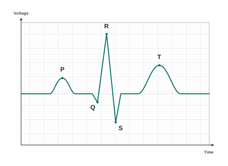
Fig. 15.3 – A standard electrocardiogram (ECG) trace.
The P, QRS and T waves
Each peak or wave in the ECG is named with a letter from P to T, and each corresponds to a particular electrical event in the heart.
The P-wave represents the electrical excitation — the depolarisation — of the two atria. This excitation is what makes both atria contract. (Depolarisation is the electrical excitation of heart muscle that triggers it to contract; repolarisation, which we meet at the T-wave, is its recovery back to the resting electrical state.)
The QRS complex represents the depolarisation of the ventricles, which sets off the ventricular contraction. The contraction begins just after the Q point, marking the start of ventricular systole.
The T-wave represents the ventricles returning from their excited state back to normal — their repolarisation. The end of the T-wave marks the end of systole.
Notice that each electrical wave appears just before the muscle movement it triggers — the P-wave just before the atria contract, the QRS just before the ventricles contract. In this way the ECG mirrors, in electrical form, the very same cardiac cycle we traced earlier.
What an ECG reveals
The ECG is not just a pattern on a screen — it is a powerful diagnostic tool. Because a QRS complex appears with every heartbeat, simply counting the number of QRS complexes in a known span of time tells us the person’s heart rate.
Moreover, a healthy ECG has a fairly standard, recognisable shape (for a given arrangement of leads), and it looks much the same from one healthy person to another. So any deviation from this normal shape — an odd wave, a missing complex, or irregular spacing — can signal a heart abnormality or disease. This is what gives the ECG its great clinical significance. It is also why, in the hospital scene we began with, a flat line spells danger: it means the heart’s electrical activity has stopped altogether.
15.4 Double Circulation
Blood does not slosh about randomly; it flows along a fixed route through the blood vessels — the arteries and veins. If you were to cut across an artery or a vein and look at its wall, you would find it built of three layers (Fig. 15.4).
The innermost layer is a smooth lining of flattened (squamous) cells, the endothelium, forming the tunica intima. Around it lies a middle layer of smooth muscle and elastic fibres, the tunica media. The outermost layer is tough fibrous connective tissue rich in collagen fibres, the tunica externa.
All three layers are present in both arteries and veins, but the tunica media is comparatively thin in veins. This makes sense: arteries carry blood pumped directly from the heart at high pressure, so their thick, muscular, elastic middle layer helps them withstand and smooth out that pressure, whereas veins carry blood back to the heart at low pressure and need far less muscle.
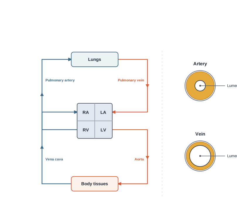
Fig. 15.4 – Schematic plan of double circulation.
Pulmonary and systemic circulation
In humans, the blood travels not one but two separate loops in each complete round of the body — this is what we mean by double circulation. Let us follow each loop in turn.
The right ventricle pumps deoxygenated blood into the pulmonary artery. This blood is carried to the lungs, where it gives up carbon dioxide and takes up oxygen. The now-oxygenated blood is carried back by the pulmonary veins to the left atrium. This loop — heart → lungs → heart — is the pulmonary circulation.
The left ventricle then pumps this oxygenated blood into the aorta. From the aorta, a branching network of arteries, arterioles and capillaries delivers the blood to all the tissues of the body. Having given up its oxygen, the now-deoxygenated blood is collected by a system of venules, veins and the vena cava and emptied into the right atrium. This loop — heart → body → heart — is the systemic circulation.
The systemic circulation is the body’s main supply line: it delivers nutrients, oxygen (\(O_2\)) and other essential substances to the tissues, and carries carbon dioxide (\(CO_2\)) and other harmful wastes away to be eliminated.
The full double-circulation route is traced in FC 15.7.
Flowchart
FC 15.7 – Double circulation in humans. Pulmonary circuit: right ventricle → pulmonary artery → lungs (blood becomes oxygenated) → pulmonary veins → left atrium. Systemic circuit: left ventricle → aorta → arteries → arterioles → capillaries in the tissues (blood becomes deoxygenated) → venules → veins → vena cava → right atrium.
Two special vascular routes
Two special arrangements of blood vessels deserve mention.
The first is the hepatic portal system, a unique vascular connection between the digestive tract and the liver. The hepatic portal vein carries blood from the intestine to the liver first, before that blood is passed on into the general systemic circulation. This detour lets the liver process and adjust the freshly absorbed nutrients before they reach the rest of the body.
The second is the coronary system — a special set of blood vessels that exists solely to supply blood to and from the heart muscle (the cardiac musculature) itself. Even though the heart is always full of blood, its own muscular walls need a dedicated blood supply of their own, and that is exactly what the coronary vessels provide.
15.5 Regulation of Cardiac Activity
The heart, as we have seen, is autoexcitable — its own nodal tissue generates the electrical signals that set the beat, with no outside command needed. Because the heartbeat originates within its own muscle in this way, the heart is described as myogenic.
This does not mean the heart is beyond all outside influence. While the beat starts on its own, its rate and force can be adjusted to match the body’s changing needs. A control centre in the medulla oblongata of the brain does this fine-tuning, acting through the autonomic nervous system (ANS), the part of the nervous system that governs involuntary activities.
The ANS influences the heart in two opposite ways. Signals travelling along the sympathetic nerves speed things up: they increase the heart rate, increase the strength of ventricular contraction, and so raise the cardiac output. Signals along the parasympathetic nerves do the reverse: they slow the heart rate, slow the conduction of the action potential, and so lower the cardiac output. On top of this neural control, hormones from the adrenal medulla can also raise the cardiac output.
15.6 Disorders of Circulatory System
Even a system as well-built as the circulatory system can develop problems. Some of its more common disorders are described below.
High Blood Pressure (Hypertension)
Blood pressure is the pressure that blood exerts against the walls of the arteries, and it is written as two numbers — for example, 120/80 mm Hg (mm Hg stands for millimetres of mercury, the unit of pressure). The upper figure, 120 mm Hg, is the systolic pressure — the pressure while the heart is pumping (during ventricular systole). The lower figure, 80 mm Hg, is the diastolic pressure — the pressure while the heart is resting (during diastole). A reading of 120/80 is taken as normal.
Hypertension, or high blood pressure, is the term for blood pressure that is higher than normal. If repeated checks of a person’s blood pressure come out at 140/90 (140 over 90) mm Hg or higher, it indicates hypertension. High blood pressure is dangerous because it strains the heart, leading to heart diseases, and it also damages other vital organs such as the brain and the kidneys.
Coronary Artery Disease (CAD)
Coronary Artery Disease (CAD), often called atherosclerosis, affects the coronary vessels — the ones that supply blood to the heart muscle itself. It develops when deposits of calcium, fat, cholesterol and fibrous tissue build up inside the walls of these arteries. These deposits make the lumen (the internal space) of the arteries narrower, so that less blood can flow through to the heart muscle.
Angina
Angina — also called angina pectoris — is a symptom rather than a disease in itself: it is a feeling of acute chest pain that appears when the heart muscle is not receiving enough oxygen. Angina can affect both men and women and can occur at any age, though it is more common in the middle-aged and the elderly. It results from conditions that reduce the blood flow to the heart.
Heart Failure
Heart failure is the state in which the heart is not pumping blood effectively enough to meet the needs of the body. It is sometimes called congestive heart failure, because a build-up (congestion) of fluid in the lungs is one of its main symptoms.
Heart failure should not be confused with two other conditions. It is not the same as cardiac arrest, which is when the heart stops beating altogether, nor the same as a heart attack, which is when the heart muscle is suddenly damaged by an inadequate blood supply.
15.7 Summary
- Vertebrates circulate blood — a fluid connective tissue — to carry essential substances to their cells and to remove wastes from them. Lymph (tissue fluid) also helps transport certain substances.
- Blood is made of a fluid matrix, plasma, together with the formed elements: red blood cells (RBCs/erythrocytes), white blood cells (WBCs/leucocytes) and platelets (thrombocytes).
- Human blood is grouped into the A, B, AB and O types, based on the presence or absence of two surface antigens (A and B) on the RBCs.
- Blood is also grouped by the presence or absence of another RBC antigen, the Rhesus factor (Rh).
- The fluid derived from blood that fills the spaces between tissue cells is tissue fluid; once collected into the lymphatic vessels it is called lymph. Lymph is almost the same as blood, except that it has less protein and mostly lacks the formed elements.
- All vertebrates (and a few invertebrates) have a closed circulatory system. Ours consists of a muscular pumping heart, a network of vessels, and the fluid blood.
- The heart has two atria and two ventricles, and its muscle is auto-excitable.
- The sino-atrial node (SAN) produces the greatest number of action potentials per minute (70–75/min), so it sets the heart’s pace and is called the pacemaker.
- Each action potential makes the atria and then the ventricles contract (systole) and then relax (diastole). Systole drives blood from the atria into the ventricles and on into the pulmonary artery and the aorta.
- These events, repeated over and over, make up the cardiac cycle; a healthy person completes about 72 cardiac cycles per minute.
- Each ventricle pumps out about 70 mL of blood per cycle — the stroke (beat) volume. The cardiac output — the volume pumped by each ventricle per minute — equals stroke volume × heart rate, about 5 litres.
- The heart’s electrical activity can be recorded from the body surface as an electrocardiogram (ECG), which is of clinical importance.
- We have a complete double circulation, made of two pathways. In the pulmonary circulation, the right ventricle pumps deoxygenated blood to the lungs, where it is oxygenated and returned to the left atrium.
- In the systemic circulation, the left ventricle pumps oxygenated blood through the aorta to the body tissues; the deoxygenated blood is then collected by the veins and returned to the right atrium.
- Although the heart is auto-excitable, its activity can be moderated by neural and hormonal mechanisms.
The complete circulatory route is summarised in FC 15.8.
Flowchart
FC 15.8 – The complete circulation at a glance: right atrium → right ventricle → (pulmonary circulation) pulmonary artery → lungs (blood oxygenated) → pulmonary veins → left atrium → left ventricle → (systemic circulation) aorta → body tissues (blood deoxygenated) → vena cava → back to right atrium.
15.8 Points to Ponder
Group O is called the “universal donor” and group AB the “universal recipient.” Yet doctors still prefer to transfuse blood of a patient’s own exact group whenever they can. Why might exactly matching the blood group be safer than relying on a “universal” one?
As a mammalian red blood cell matures, it loses its nucleus — making room for more haemoglobin, but also leaving the cell unable to repair itself or divide. Do you think this is a worthwhile trade-off for the body? What might the hidden costs of a cell without a nucleus be?
The factors that make blood clot are kept in an inactive form until an injury switches them on. Why would it be dangerous if these factors were active all the time? What might go wrong if a clot were to form inside a healthy, uninjured blood vessel?
Amphibians and most reptiles manage well with a three-chambered heart, in which oxygenated and deoxygenated blood partly mix — yet birds and mammals have a fully separated four-chambered heart. Why might a partly mixing heart be “good enough” for some animals but not for others?
The heart can beat entirely on its own, without any signal from the brain. So why might it still be useful for the body to be able to speed the heart up or slow it down through nerves and hormones? In what situations would such control matter most?
A blood pressure reading is given as two numbers — the systolic over the diastolic. Why do you think doctors look carefully at both numbers, rather than just the higher one? What might each number reveal about how the heart and the arteries are working?
15.9 NCERT Questions
Name the components of the formed elements in the blood and mention one major function of each of them.
What is the importance of plasma proteins?
Match Column I with Column II:
| Column I | Column II |
|---|---|
| a. Eosinophils | i. Coagulation |
| b. RBC | ii. Universal Recipient |
| c. AB Group | iii. Resist Infections |
| d. Platelets | iv. Contraction of Heart |
| e. Systole | v. Gas transport |
Why do we consider blood as a connective tissue?
What is the difference between lymph and blood?
What is meant by double circulation? What is its significance?
Write the differences between:
- Blood and Lymph
- Open and Closed system of circulation
- Systole and Diastole
- P-wave and T-wave
Describe the evolutionary change in the pattern of heart among the vertebrates.
Why do we call our heart myogenic?
Sino-atrial node is called the pacemaker of our heart. Why?
What is the significance of atrio-ventricular node and atrio-ventricular bundle in the functioning of the heart?
Define a cardiac cycle and the cardiac output.
Explain heart sounds.
Draw a standard ECG and explain the different segments in it.
15.10 Check Your Concepts
Assertion (A): Mature mammalian red blood cells are biconcave and lack a nucleus. Reason (R): These features increase the amount of haemoglobin each cell can carry and let it bend through the narrowest capillaries. State whether A and R are true, and whether R is the correct explanation of A.
Odd one out — pick the leucocyte that does not belong with the others, and justify your choice: Neutrophils, Eosinophils, Basophils, Lymphocytes.
Arrange the following white blood cells in order of decreasing abundance in blood: Basophils, Lymphocytes, Neutrophils, Eosinophils, Monocytes.
Serum and plasma are almost the same fluid. Why, then, can plasma take part in blood clotting while serum cannot?
Match each blood group in Column I with the antibodies present in its plasma in Column II:
| Column I (Blood group) | Column II (Antibodies in plasma) |
|---|---|
| a. A | i. anti-A and anti-B |
| b. B | ii. no antibodies |
| c. AB | iii. anti-B |
| d. O | iv. anti-A |
Group O is called the universal donor. Explain why, by referring to the antigens present on its red blood cells.
Assertion (A): An Rh-negative mother’s first Rh-positive child is usually unaffected, but a later Rh-positive child may develop erythroblastosis foetalis. Reason (R): The mother becomes sensitised to the Rh antigen only around the time of the first delivery. State whether A and R are true, and whether R is the correct explanation of A.
Arrange the following in the correct order in which they appear during blood clotting: thrombin, fibrinogen, thrombokinase, fibrin, prothrombin.
What might happen to a person if the number of platelets in their blood fell far below normal? Give a reason.
If the lymphatic vessels draining a leg became blocked, what would you expect to happen to the leg, and why?
Assertion (A): The wall of the left ventricle is the thickest of all four heart chambers. Reason (R): The left ventricle must pump blood all the way around the entire body. State whether A and R are true, and whether R is the correct explanation of A.
On a diagram of the human heart (as in Fig. 15.2), identify and label: (a) the chamber that pumps blood into the aorta, (b) the valve guarding the opening between the right atrium and the right ventricle, and (c) the node that acts as the pacemaker.
Arrange the parts of the heart’s conducting system in the order in which an electrical impulse passes through them: AV bundle, Purkinje fibres, SA node, bundle branches, AV node.
During ventricular systole, why must the tricuspid and bicuspid valves close before the semilunar valves open?
Assertion (A): The QRS complex marks the beginning of ventricular systole. Reason (R): The QRS complex represents the depolarisation of the ventricles, which sets off their contraction. State whether A and R are true, and whether R is the correct explanation of A.
Why is the double circulation of mammals considered more efficient than the single circulation of fishes?
Why does blood from the intestine flow through the liver (via the hepatic portal system) before joining the general circulation?
How would strong stimulation of the parasympathetic nerves change the cardiac output, and why?
In atherosclerosis, the coronary arteries become narrowed. Why does this endanger the heart muscle, even though the chambers of the heart are always full of blood?
15.11 Practice With Numericals
A person has a stroke volume of 80 mL and a heart rate of 75 beats per minute. Calculate the cardiac output, in mL per minute and in litres per minute.
In a healthy adult, the cardiac output is 5 litres per minute and the heart rate is 72 beats per minute. Calculate the stroke volume (in mL).
A person’s heart beats 75 times per minute. Calculate the duration of one cardiac cycle, in seconds.
During intense exercise, an athlete’s stroke volume rises to 120 mL and heart rate to 150 beats per minute. Calculate the cardiac output, and find how many times greater it is than a resting cardiac output of 5 litres per minute.