Chapter 10: Cell Cycle and Cell Division
In a laboratory in Germany in the 1880s, a biologist named Walther Flemming was doing something that seems ordinary today but was quietly revolutionary then: he was staining cells with coloured dyes and watching them through a microscope. The dyes soaked into a thread-like material inside the nucleus that soaked up colour so strongly that Flemming named it chromatin, from the Greek word for colour.
When he looked at dividing cells from salamander larvae, he saw those threads do something remarkable. They thickened, lined up neatly across the middle of the cell, split into two matching sets, and were pulled to opposite ends — after which the cell pinched into two. No one had ever traced this orderly dance step by step before. Flemming drew the stages carefully and gave the whole process a name built from the Greek word for thread, mitos: mitosis.
What Flemming was witnessing is the reason a single fertilised egg can become a human being of many trillions of cells, each carrying the same genetic instructions. Every one of those cells came from an earlier cell that grew, copied its DNA exactly, and divided. This chapter follows that same journey — how a cell prepares itself, how it shares out its chromosomes without error, and how a special kind of division makes the sperm and eggs that let life continue across generations.
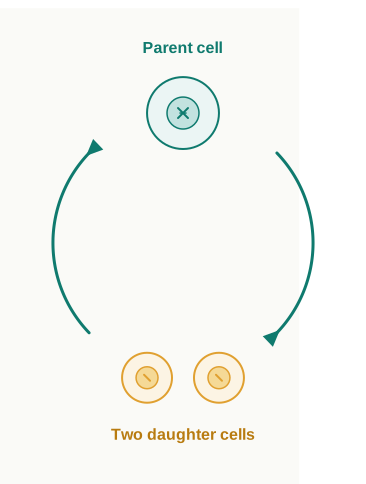
Fig. 10.0 – The cell cycle.
By the end of this chapter, you will be able to answer each of these questions for yourself.
10.1 Cell Cycle
Every organism you have ever seen — a towering banyan tree, a blue whale, or you yourself — began life as a single cell. It is natural to wonder how one microscopic cell can build a body made of millions or even trillions of cells. The answer lies in two properties shared by all living things: growth and reproduction.
Cells achieve both by dividing. Every cell reproduces by splitting into two, so that one parent cell gives rise to two daughter cells each time it divides. These daughter cells are not the end of the story — each one can grow and divide again, and so on. Repeated over and over, such cycles of growth and division let a single starting cell produce a whole population of cells, and eventually an entire multicellular structure.
Why cell division must be tightly coordinated
Cell division is one of the most important processes in living organisms, but it cannot happen carelessly. When a cell divides, two other things must also be arranged: the cell’s DNA has to be copied, and the cell itself has to grow large enough to be shared between two offspring cells.
These three events — cell growth, DNA replication, and cell division — must be timed and coordinated with one another. If they are not, a daughter cell might end up with too little DNA, a broken set of instructions, or too small a share of the cell’s contents. Coordination is what guarantees that each new cell receives a complete and intact copy of the genome (the full set of an organism’s DNA).
The whole ordered sequence — a cell copying its genome, building the other parts it needs, and finally splitting into two daughter cells — is what biologists call the cell cycle.
An important detail sits inside this definition. Growth of the cell — meaning the steady increase in its cytoplasm — happens more or less continuously. DNA synthesis, however, does not. The copying of DNA is squeezed into one specific stage of the cell cycle, not spread across the whole of it. Once the chromosomes (the DNA) have been copied, they are shared out to the two daughter nuclei through a complex, carefully ordered series of steps. And crucially, none of this is random: these events are themselves controlled by genes.
10.1.1 Phases of Cell Cycle
A convenient way to study a typical eukaryotic cell cycle is to look at human cells growing in culture (in a laboratory dish). Such cells divide roughly once every 24 hours, and the overall pattern of the cycle can be pictured as a wheel, as shown in Fig. 10.1.
This 24-hour figure is only a representative value. The length of the cell cycle varies widely from one organism to another, and even from one cell type to another within the same body. Yeast, for example, can race through an entire cell cycle in about 90 minutes.
The two basic phases
The cell cycle is divided into two basic phases:
- Interphase
- M Phase (Mitosis phase)
The M Phase is the phase in which the actual cell division, or mitosis, takes place. Interphase is simply the phase that lies between two successive M phases — the long stretch of time separating one division from the next.
Here is something worth pausing on. In the 24-hour average cell cycle of a human cell, the division itself — the M Phase — occupies only about one hour. The remaining time, more than 95% of the whole cycle, is interphase.
The M Phase begins with nuclear division, the separation of the duplicated chromosomes into two sets, which is called karyokinesis. It usually ends with the division of the cytoplasm, which is called cytokinesis. Interphase, despite often being nicknamed the “resting phase,” is anything but restful — it is the time during which the cell actively prepares for division by carrying out both cell growth and DNA replication in an orderly way.
The three parts of interphase
Interphase itself is divided into three further phases, which follow one another in a fixed order:
- G₁ phase (Gap 1)
- S phase (Synthesis)
- G₂ phase (Gap 2)
G₁ phase is the interval between the end of mitosis and the start of DNA replication. During \(G\_1\), the cell is metabolically active and grows steadily, but it does not yet copy its DNA.
S phase, or the synthesis phase, is the period during which DNA synthesis (replication) actually happens. During this phase the amount of DNA in the cell doubles. If the starting amount of DNA is written as \(2C\), then by the end of S phase it has increased to \(4C\).
This is a point students often stumble on, so read it carefully: although the amount of DNA doubles, the number of chromosomes does not change. A cell that had a diploid or \(2n\) number of chromosomes at \(G\_1\) still has exactly \(2n\) chromosomes after S phase. What has happened is that each chromosome now consists of two identical copies (sister chromatids) joined together — more DNA per chromosome, but not more chromosomes.
G₂ phase is the second gap. In animal cells, DNA replication during S phase begins in the nucleus, while the centriole is duplicated separately in the cytoplasm. Then, during \(G\_2\), the cell synthesises the proteins it will need for mitosis, and cell growth continues right up to the start of division.
The overall order of the cell cycle can be summarised as a repeating loop, as shown in FC 10.1.
Flowchart
FC 10.1 – The cell cycle sequence: G₁ → S (DNA replication, 2C → 4C) → G₂ → M Phase (karyokinesis → cytokinesis) → back to G₁; with a branch from G₁ → G₀ (quiescent stage) for cells that stop dividing.
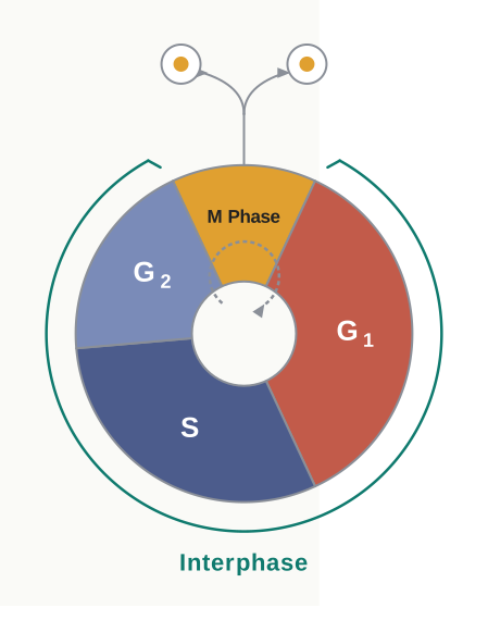
Fig. 10.1 – Phases of the cell cycle.
When a cell stops dividing: the G₀ stage
Not every cell keeps cycling. Some cells in adult animals appear never to divide again — heart muscle cells are a familiar example. Many other cells divide only occasionally, stepping in only when they are needed to replace cells lost through injury or natural cell death.
Cells like these leave the cycle at the \(G\_1\) phase and enter an inactive stage called the quiescent stage, written as \(G\_0\) (G-zero). A cell in \(G\_0\) is not dead or shut down — it stays metabolically active — but it no longer prepares to divide unless the organism specifically calls on it to do so.
Which cells divide by mitosis?
There is also a pattern in which kinds of cells divide this way. In animals, mitotic cell division is normally seen only in the diploid somatic cells (the ordinary body cells). There are a few exceptions in which haploid cells divide by mitosis — male honey bees, for instance, develop from unfertilised eggs and are haploid, yet their cells still divide by mitosis.
Plants are more flexible: they can carry out mitotic divisions in both haploid and diploid cells. Recalling the alternation of generations you studied in the plant kingdom will help you identify the plant species and life-cycle stages at which mitosis occurs in haploid cells.
10.2 M Phase
The M Phase is the most dramatic period of the whole cell cycle. In this short window, virtually every component of the cell is reorganised so that the duplicated contents can be shared cleanly between two new cells. Because the number of chromosomes in the parent cell and in each progeny cell stays exactly the same, mitosis is also called equational division — the chromosome number is “kept equal” from one generation of cells to the next.
For convenience, the nuclear division part of the M Phase (karyokinesis) is described in four stages. It is important to remember, though, that cell division is a smooth, progressive process — the stages flow into one another, and no sharp line can really be drawn between them. The four stages of karyokinesis, in order, are:
- Prophase
- Metaphase
- Anaphase
- Telophase
The full sequence of the M Phase, from prophase through to the splitting of the cytoplasm, is summarised in FC 10.2.
Flowchart
FC 10.2 – Stages of mitosis (M Phase): Prophase → Metaphase → Anaphase → Telophase → Cytokinesis → two daughter cells.
10.2.1 Prophase
Prophase is the first stage of karyokinesis, and it follows the S and G₂ phases of interphase. During those interphase stages, the newly copied DNA molecules are not yet visible as separate, distinct threads — they lie intertwined and tangled together.
Prophase begins as this chromosomal material starts to condense. As the loosely spread chromatin coils up tightly (a process called chromatin condensation), the tangled threads become untangled and gradually appear as distinct, compact chromosomes, as shown in Fig. 10.2 a.
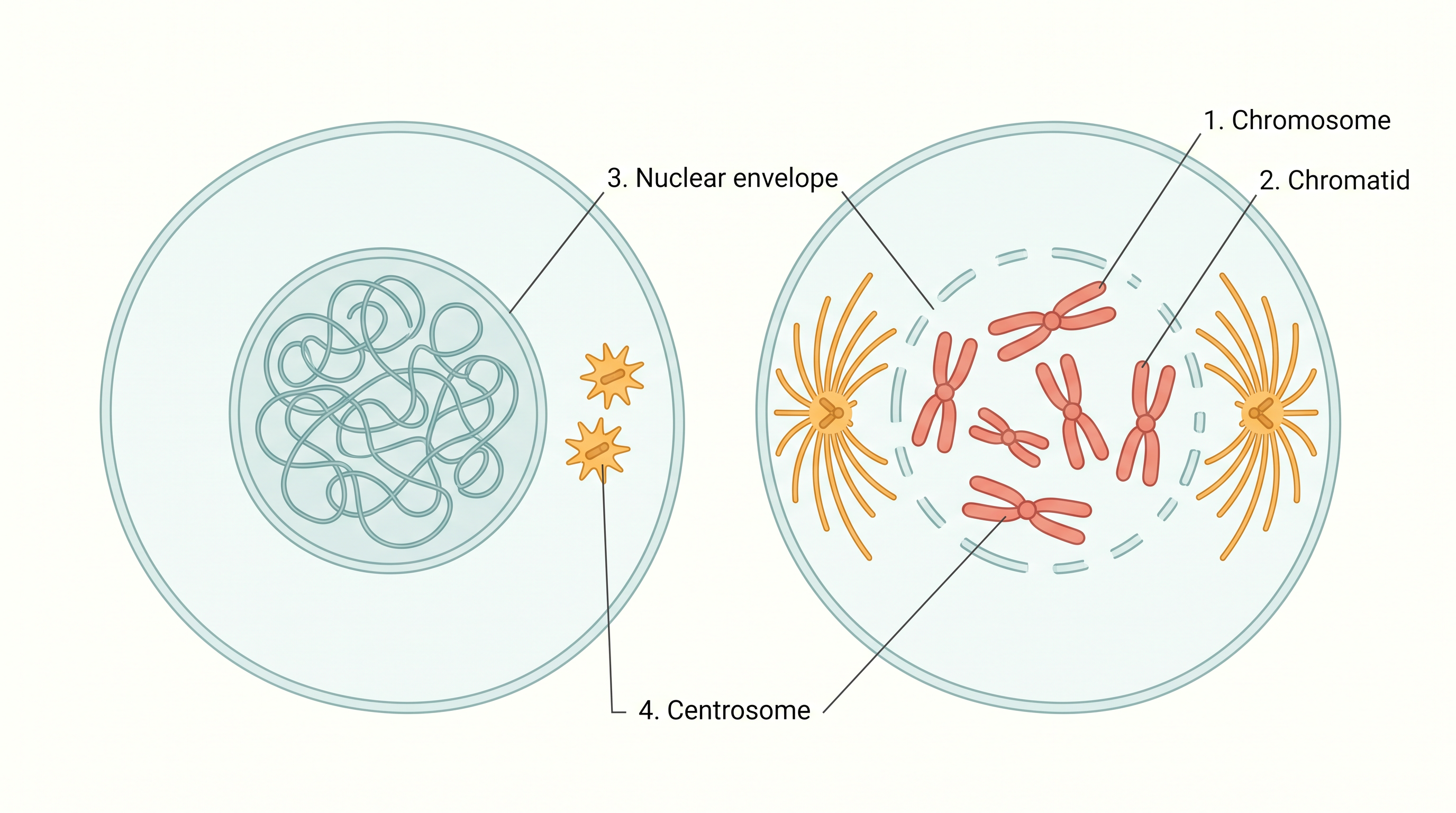
Fig. 10.2 a – Early and late prophase.
At the same time, the centrosome — a structure that had already been duplicated during the S phase — begins to move towards opposite poles (ends) of the cell. The completion of prophase can therefore be recognised by two characteristic events.
First, the chromosomal material condenses into compact mitotic chromosomes. Each chromosome is now clearly seen to be made of two threads, called sister chromatids, joined together at a point called the centromere.
Second, the two centrosomes move to opposite poles. From each centrosome, microtubules radiate outward in a star-like array called an aster. Together, the two asters and the spindle fibres between them make up the mitotic apparatus, the machinery that will later pull the chromosomes apart.
By the end of prophase, if the cell is viewed under a microscope, several familiar structures can no longer be seen: the golgi complex, the endoplasmic reticulum, the nucleolus, and the nuclear envelope have all disappeared or broken down.
10.2.2 Metaphase
The start of metaphase is marked by the complete disintegration of the nuclear envelope. With the nucleus’s boundary gone, the chromosomes are now free to spread out through the cytoplasm of the cell.
By this stage, condensation of the chromosomes is fully complete, so they can be seen very clearly under the microscope. For this reason, metaphase is the best stage for studying the morphology (shape and structure) of chromosomes. A metaphase chromosome is made up of two sister chromatids held together by the centromere, as shown in Fig. 10.2 b.
Sitting on the surface of each centromere are small, disc-shaped structures called kinetochores. These act as the attachment points where spindle fibres grip the chromosomes and move them into position at the centre of the cell.
Metaphase is therefore characterised by all the chromosomes coming to lie at the equator (middle) of the cell. Each chromosome is anchored so that one of its chromatids is connected, through its kinetochore, to spindle fibres from one pole, while its sister chromatid is connected, through its kinetochore, to spindle fibres from the opposite pole (Fig. 10.2 b). The imaginary plane along which all the chromosomes line up is called the metaphase plate.
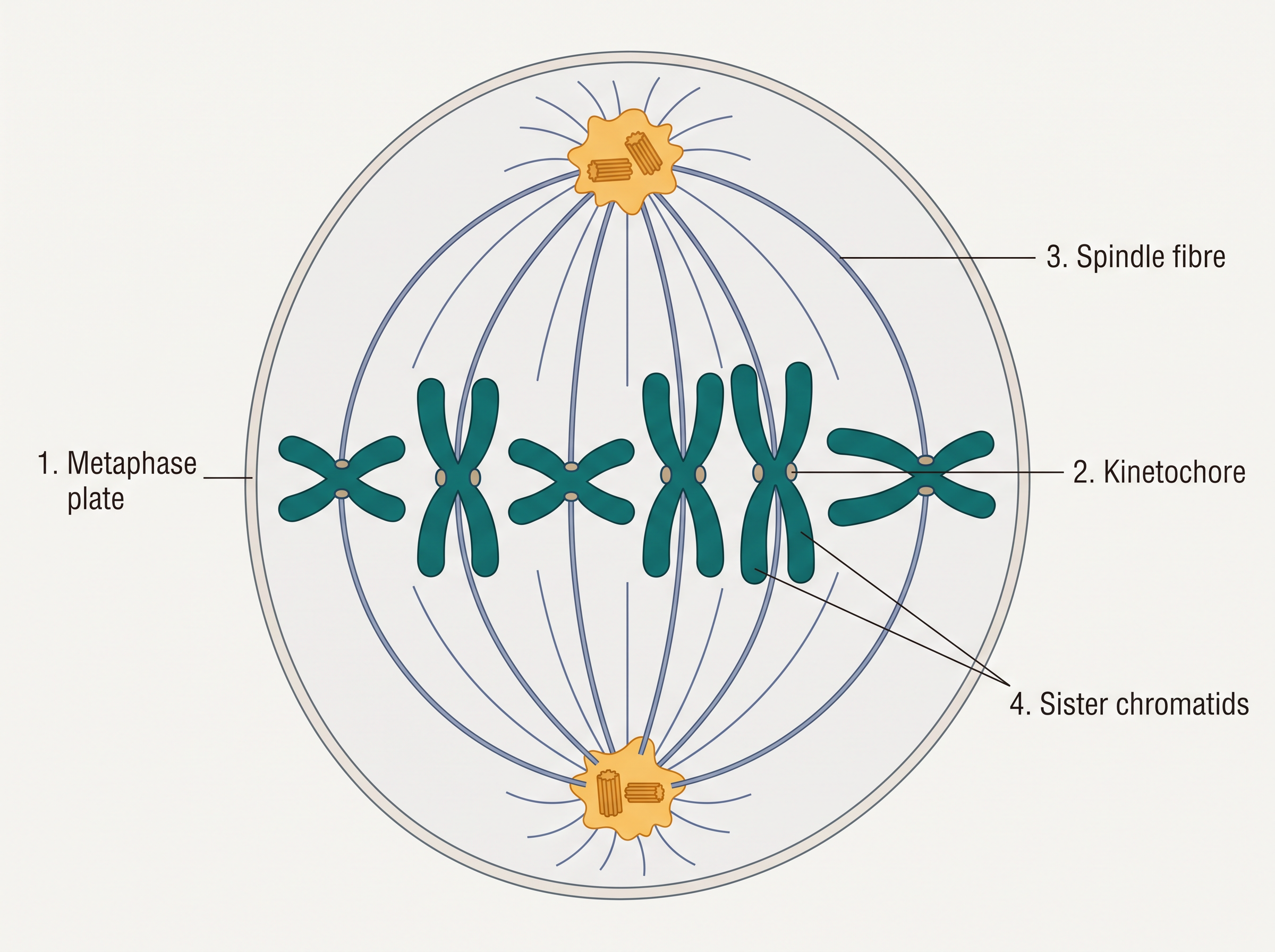
Fig. 10.2 b – Metaphase.
The key features of metaphase are:
- Spindle fibres attach to the kinetochores of the chromosomes.
- Chromosomes are moved to the spindle equator and become aligned along the metaphase plate, held by spindle fibres running to both poles.
10.2.3 Anaphase
At the onset of anaphase, each chromosome lined up on the metaphase plate splits simultaneously. The centromere holding the two sister chromatids together divides, and the two chromatids — now called daughter chromosomes of the future daughter nuclei — begin to move towards the two opposite poles.
As each chromosome is pulled away from the equatorial plate, its centromere stays directed towards the pole, so the centromere leads the way while the arms of the chromosome trail behind it, as shown in Fig. 10.2 c.
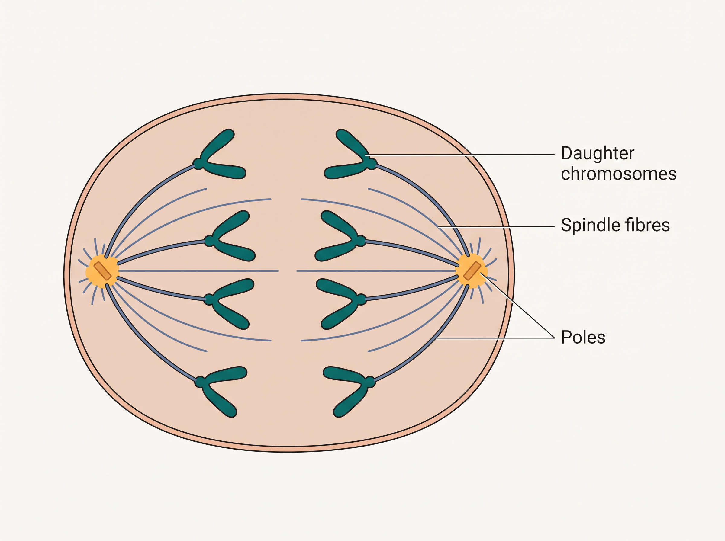
Fig. 10.2 c – Anaphase.
The key events of anaphase are:
- Centromeres split and the sister chromatids separate.
- The separated chromatids move to opposite poles.
10.2.4 Telophase
Telophase is the final stage of karyokinesis. The chromosomes that have now reached their respective poles begin to decondense — they uncoil and lose their distinct, individual identity. The separate chromosomes can no longer be made out, and each set of chromatin material gathers together at its pole, as shown in Fig. 10.2 d.
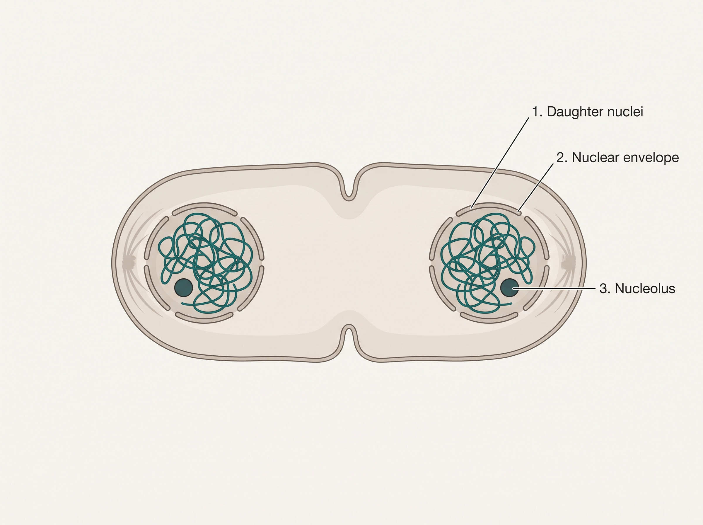
Fig. 10.2 d – Telophase.
The key events of telophase are:
- The chromosomes cluster at the two opposite spindle poles, and their identity as separate elements is lost.
- A nuclear envelope forms around each chromosome cluster, producing two daughter nuclei.
- The nucleolus, golgi complex, and endoplasmic reticulum — all of which had disappeared during prophase — now re-form.
Notice the symmetry: the structures that broke down in prophase are exactly the ones rebuilt in telophase. Telophase essentially reverses prophase, leaving two complete nuclei where there was one.
10.2.5 Cytokinesis
Karyokinesis shares the duplicated chromosomes out into two daughter nuclei — but the cell still has a single body of cytoplasm. Division is only truly finished when the cell itself is split into two, by the division of the cytoplasm. This cytoplasmic division is called cytokinesis, and it marks the completion of cell division (Fig. 10.2 e).
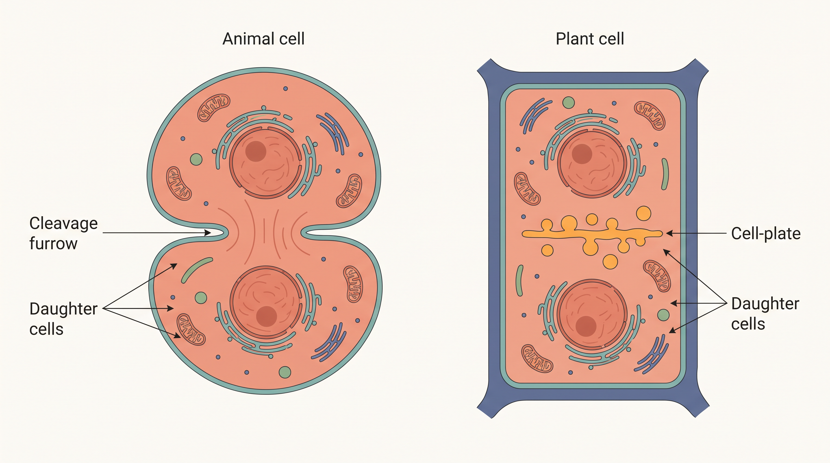
Fig. 10.2 e – Cytokinesis in animal and plant cells.
Animal cells and plant cells achieve cytokinesis by two different mechanisms, and the reason lies in their structure.
In an animal cell, cytokinesis is achieved by the appearance of a furrow (a groove) in the plasma membrane. This furrow gradually deepens from the outside inward and finally joins at the centre, pinching the cytoplasm into two.
A plant cell cannot pinch inward like this, because it is enclosed by a relatively inextensible (rigid) cell wall. So plant cells divide the cytoplasm by a different route. Wall formation begins in the centre of the cell and grows outward to meet the existing lateral walls. This new wall starts as a simple precursor called the cell-plate, which represents the middle lamella — the shared boundary layer — between the walls of the two new adjacent cells.
During cytoplasmic division, the cell’s organelles — such as mitochondria and plastids — are shared out between the two daughter cells, so that each new cell receives the parts it needs to function.
Finally, there is an interesting exception. In some organisms, karyokinesis (nuclear division) takes place but is not followed by cytokinesis. Repeated nuclear divisions without cytoplasmic division produce a cell with many nuclei, a multinucleate condition, and the resulting structure is called a syncytium.
10.3 Significance of Mitosis
Having followed mitosis step by step, it is worth asking a bigger question: why does it matter? What does this equational division actually do for a living organism?
As a rule, mitosis is restricted to the diploid cells of the body. There are exceptions, though: in some lower plants and in certain social insects, even haploid cells can divide by mitosis (recall the haploid male honey bees mentioned earlier). Whatever the ploidy of the parent cell, the outcome of mitosis is the same in principle — daughter cells that carry an identical genetic complement to the parent.
Growth of the organism
The most obvious role of mitosis is growth. The steady increase in cell number that turns a fertilised egg into a full-sized multicellular organism is due to repeated mitotic divisions.
Growth also creates a problem that mitosis then solves. As a cell grows, its cytoplasm expands, and this disturbs the balance between the amount of nucleus and the amount of cytoplasm — the nucleo-cytoplasmic ratio. A single nucleus can effectively manage only so much cytoplasm; if the cytoplasm keeps growing, the nucleus can no longer control it efficiently. Dividing splits the enlarged cell into two smaller ones, restoring a healthy nucleo-cytoplasmic ratio in each.
Cell repair and replacement
A second, equally significant role of mitosis is cell repair — replacing cells that are worn out, damaged, or lost. Several tissues in the body are renewed constantly by mitosis: the cells of the upper layer of the epidermis (the outer skin), the cells lining the gut, and the blood cells are all being replaced continuously.
Continuous growth in plants
In plants, mitosis is also the engine of lifelong growth. Certain regions retain cells that keep dividing by mitosis throughout the plant’s life. Mitotic divisions in the meristematic tissues — the apical meristems (at root and shoot tips) and the lateral cambium — allow plants to keep growing continuously, in length and in girth, for as long as they live.
10.4 Meiosis
Sexual reproduction produces offspring by the fusion of two gametes (sex cells) — for example, a sperm and an egg. Each gamete carries a complete haploid set of chromosomes, meaning a single set, written as \(n\). When two gametes fuse at fertilisation, their two haploid sets combine to restore the full diploid set, \(2n\).
This raises a problem. Gametes are themselves made from specialised diploid cells. If those diploid cells divided by ordinary mitosis, each gamete would end up diploid, and every fertilisation would then double the chromosome number — \(2n\) becoming \(4n\), then \(8n\), and so on, generation after generation. Clearly, something must halve the chromosome number before the gametes form.
That “something” is a specialised kind of cell division that reduces the chromosome number by half, producing haploid daughter cells. This division is called meiosis.
Meiosis therefore ensures the production of the haploid phase in the life cycle of a sexually reproducing organism, while fertilisation restores the diploid phase. We encounter meiosis during gametogenesis — the formation of gametes — in both plants and animals, where it gives rise to haploid gametes.
Key features of meiosis
Before looking at the stages, keep these defining features of meiosis in mind:
- Meiosis involves two sequential cycles of nuclear and cell division — called meiosis I and meiosis II — but only a single cycle of DNA replication.
- Meiosis I begins only after the parental chromosomes have already replicated during the S phase, producing identical sister chromatids.
- Meiosis involves the pairing of homologous chromosomes and recombination (exchange of parts) between the non-sister chromatids of those homologous chromosomes.
- Four haploid cells are formed at the end of meiosis II.
The two divisions are made up of matching phases, which can be grouped as follows:
| Meiosis I | Meiosis II |
|---|---|
| Prophase I | Prophase II |
| Metaphase I | Metaphase II |
| Anaphase I | Anaphase II |
| Telophase I | Telophase II |
The complete flow of meiosis, from one diploid cell to four haploid cells, is summarised in FC 10.3.
Flowchart
FC 10.3 – Meiosis overview: one diploid cell (2n, DNA already replicated) → Meiosis I (Prophase I → Metaphase I → Anaphase I → Telophase I) → interkinesis (no DNA replication) → Meiosis II (Prophase II → Metaphase II → Anaphase II → Telophase II) → four haploid cells (n).
10.4.1 Meiosis I
Prophase I
Prophase of the first meiotic division is typically much longer and more complex than the prophase of mitosis. Based on the way the chromosomes behave, it is further subdivided into five stages, in this order: leptotene, zygotene, pachytene, diplotene, and diakinesis.
During the leptotene stage, the chromosomes gradually become visible under the light microscope, and their compaction (condensation) continues throughout this stage.
This is followed by zygotene, in which the chromosomes begin to pair up. This pairing of chromosomes is called synapsis, and the paired chromosomes — one from each parent, matching in length and gene content — are called homologous chromosomes. Electron micrographs show that synapsis is accompanied by the formation of a complex structure called the synaptonemal complex, which zips the pair together.
The structure formed by a pair of synapsed homologous chromosomes is called a bivalent, or a tetrad (because it contains four chromatids in all). Bivalents are more clearly visible in the next stage.
The first two substages are relatively short-lived compared with pachytene, which comes next. During pachytene, the four chromatids of each bivalent become distinct and the bivalents clearly appear as tetrads.
Pachytene is marked by the appearance of recombination nodules — the specific sites on the bivalent where crossing over takes place, between the non-sister chromatids of the homologous chromosomes. Crossing over is the exchange of segments of genetic material between two homologous chromosomes.
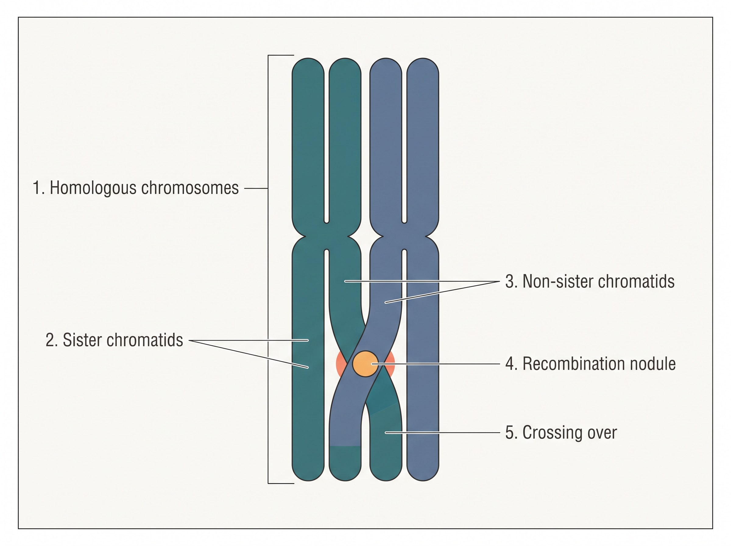
Fig. 10.3 – Crossing over during pachytene.
Crossing over is an enzyme-mediated process, and the enzyme responsible is recombinase. It leads to the recombination (reshreshuffling) of genetic material between the two chromosomes. Recombination between the homologous chromosomes is completed by the end of pachytene, but the chromosomes remain physically linked at the points where crossing over occurred.
The next substage, diplotene, begins with the dissolution of the synaptonemal complex. The recombined homologous chromosomes of each bivalent now tend to pull apart from each other — except at the sites of crossing over, where they stay attached. These X-shaped points of attachment are called chiasmata (singular: chiasma).
Diplotene can be remarkably long-lasting. In the oocytes (developing egg cells) of some vertebrates, this stage can persist for months or even years.
The final substage of prophase I is diakinesis. It is marked by the terminalisation of chiasmata — the chiasmata gradually shift towards the ends of the chromosomes. During diakinesis the chromosomes become fully condensed, and the meiotic spindle assembles to prepare the homologous chromosomes for separation. By the end of diakinesis, the nucleolus disappears and the nuclear envelope breaks down. Diakinesis thus represents transition to metaphase I.
Metaphase I
At metaphase I, the bivalent chromosomes line up on the equatorial plate, as shown in Fig. 10.4. The microtubules coming from the opposite poles of the spindle attach to the kinetochores of the homologous chromosomes.
Note an important contrast with mitosis: here it is whole homologous chromosomes (each still made of two sister chromatids) that face opposite poles, not individual chromatids.
Anaphase I
At anaphase I, the homologous chromosomes separate and move towards opposite poles, while the sister chromatids of each chromosome remain associated at their centromeres (Fig. 10.4). This is the step that actually reduces the chromosome number: each pole receives only one chromosome from each homologous pair.
Telophase I
In telophase I, the nuclear membrane and nucleolus reappear, and cytokinesis follows. The two-celled structure formed at this point is called a dyad of cells (Fig. 10.4). Although the chromosomes may disperse somewhat, they do not return to the fully extended state seen in an interphase nucleus.
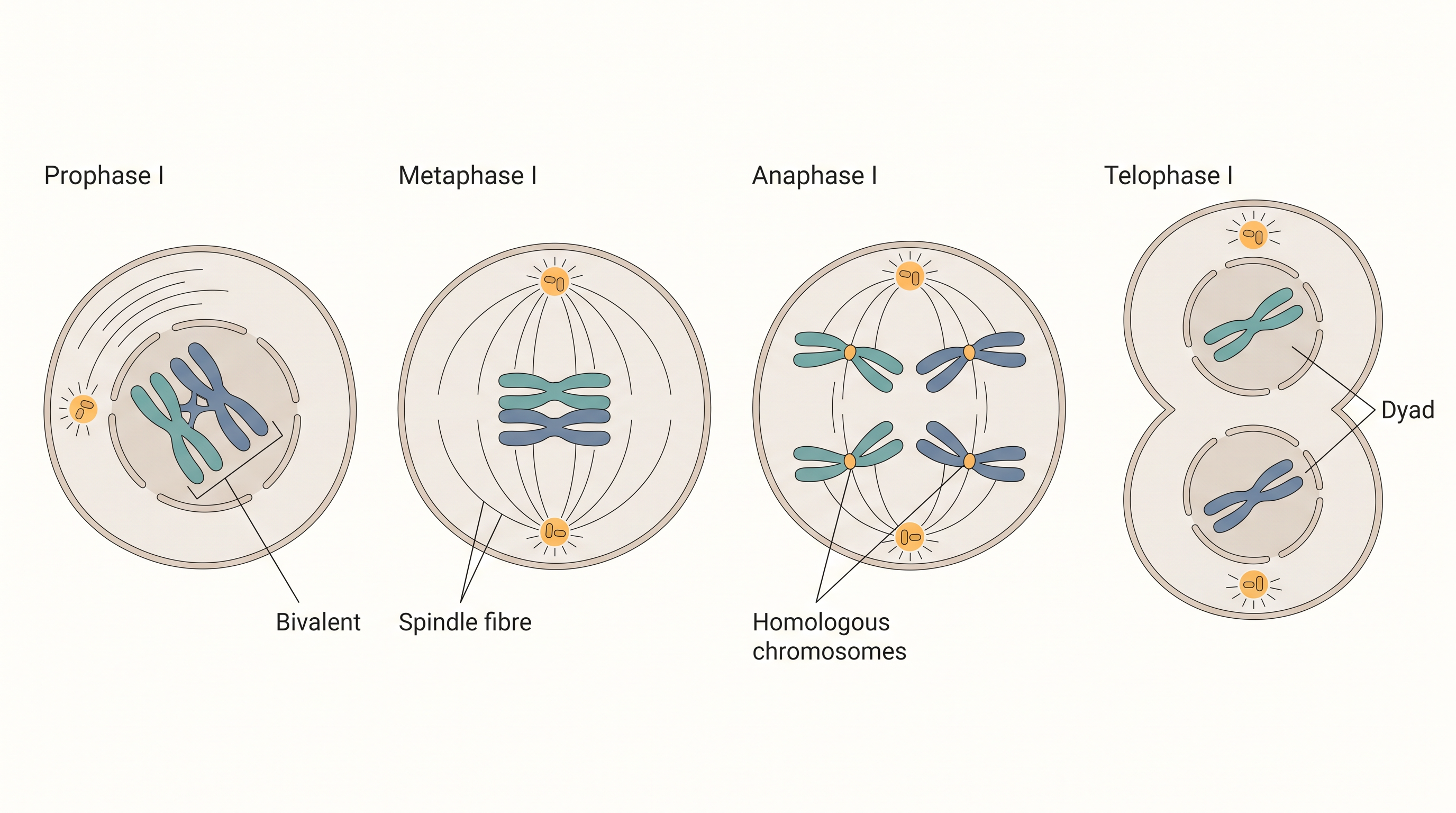
Fig. 10.4 – Stages of Meiosis I.
The stage between the two meiotic divisions is called interkinesis, and it is generally short-lived. Crucially, there is no replication of DNA during interkinesis — this is what keeps meiosis to a single round of DNA copying. Interkinesis is followed by prophase II, a much simpler prophase than prophase I.
10.4.2 Meiosis II
Prophase II
Meiosis II begins immediately after the cytokinesis of meiosis I, usually before the chromosomes have fully elongated. In contrast to meiosis I, meiosis II closely resembles a normal mitosis. The nuclear membrane disappears by the end of prophase II (Fig. 10.5), and the chromosomes become compact once again.
Metaphase II
At metaphase II, the chromosomes line up at the equator, and the microtubules from the opposite poles of the spindle attach to the kinetochores of the sister chromatids (Fig. 10.5).
Anaphase II
Anaphase II begins with the simultaneous splitting of the centromere of each chromosome — the centromere that had been holding the two sister chromatids together. Freed from each other, the sister chromatids move towards opposite poles of the cell (Fig. 10.5), pulled by the shortening of the microtubules attached to their kinetochores.
Telophase II
Meiosis ends with telophase II. The two groups of chromosomes at each cell are once again enclosed by a nuclear envelope, and cytokinesis follows. The final result is a tetrad of cells — that is, four haploid daughter cells (Fig. 10.5).
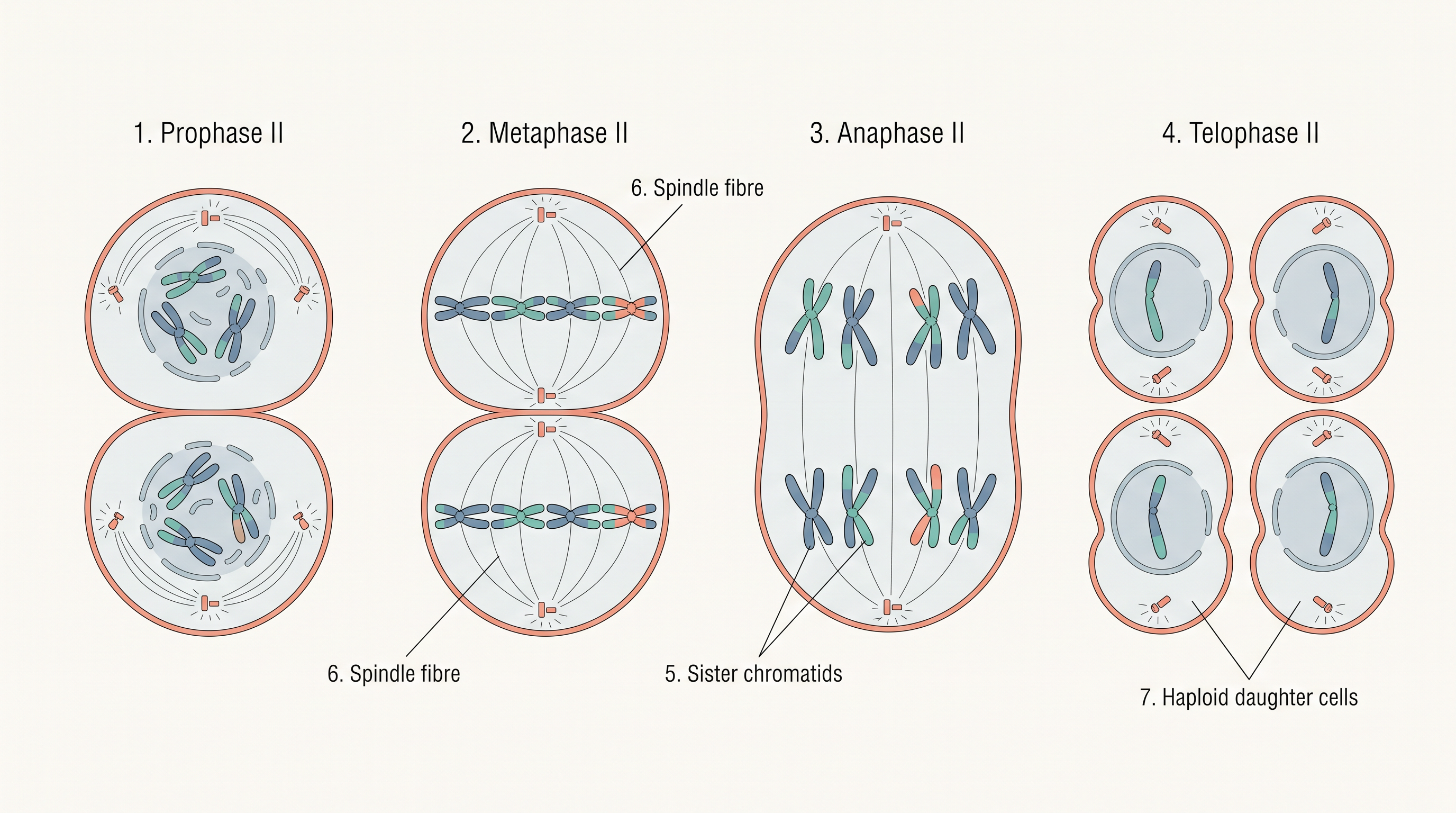
Fig. 10.5 – Stages of Meiosis II.
10.5 Significance of Meiosis
Meiosis performs two great services for sexually reproducing organisms, and the first of them sounds like a contradiction.
Conserving the species’ chromosome number
Meiosis is the mechanism that conserves the specific chromosome number of each species across generations — and yet the process itself reduces the chromosome number by half. How can halving the number be the thing that keeps it constant?
The answer lies in pairing meiosis with fertilisation. Meiosis halves the chromosome number when making gametes, so each gamete is haploid (\(n\)). At fertilisation, two haploid gametes fuse and their sets add back together, restoring the diploid number (\(2n\)) of the species. Without the halving step, fertilisation would double the number every generation. So the reduction is precisely what allows the number to stay stable from one generation to the next.
Increasing genetic variability
The second service of meiosis is that it increases the genetic variability of a population from one generation to the next. Crossing over during prophase I reshuffles segments between homologous chromosomes, and the random way each homologous pair lines up and separates at meiosis I mixes maternal and paternal chromosomes into fresh combinations in the gametes. Together, these produce offspring that are genetically varied rather than identical.
10.6 Summary
- According to the cell theory, all cells arise from pre-existing cells, and the process by which this happens is cell division. Every sexually reproducing organism begins its life as a single-celled zygote, and cell division does not stop once the mature organism has formed — it continues throughout the organism’s life.
- The stages a cell passes through from one division to the next make up the cell cycle. The cycle has two main phases: interphase, the period of preparation for division, and the mitosis (M) phase, the actual period of division.
- Interphase is subdivided into three phases. In G₁, the cell grows and carries out normal metabolism, and most organelle duplication takes place. In the S phase, DNA replication and chromosome duplication occur (the DNA doubles, \(2C → 4C\), but the chromosome number stays \(2n\)). In G₂, cytoplasmic growth continues and the cell prepares for division.
- Cells that stop dividing leave the cycle at G₁ and enter the quiescent stage, G₀, where they stay metabolically active but no longer proliferate.
- Mitosis is divided into four stages: prophase, metaphase, anaphase, and telophase. In prophase, the chromosomes condense while the centrosomes (bearing the centrioles) move to opposite poles; the nuclear envelope and nucleolus disappear and spindle fibres begin to appear.
- Metaphase is marked by the alignment of the chromosomes at the equatorial plate (the metaphase plate). During anaphase, the centromeres divide and the chromatids move to the two opposite poles.
- Once the chromatids reach the poles, they decondense and elongate, and the nucleolus and nuclear membrane reappear — this is telophase. Nuclear division (karyokinesis) is then followed by cytoplasmic division, or cytokinesis.
- Mitosis is thus an equational division: the chromosome number of the parent is conserved unchanged in the daughter cells.
- In contrast, meiosis occurs in diploid cells that are destined to form gametes. It is called a reduction division because it halves the chromosome number while making the gametes. When two gametes fuse during sexual reproduction, the chromosome number is restored to the value found in the parent.
- Meiosis is divided into two phases, meiosis I and meiosis II. In meiosis I, homologous chromosomes pair to form bivalents and undergo crossing over. Meiosis I has a long prophase, subdivided into five stages: leptotene, zygotene, pachytene, diplotene, and diakinesis.
- During metaphase I, the bivalents arrange on the equatorial plate. This is followed by anaphase I, in which the homologous chromosomes move to opposite poles, each still carrying both its chromatids — so each pole receives half the chromosome number of the parent cell. In telophase I, the nuclear membrane and nucleolus reappear.
- Meiosis II is similar to mitosis; during anaphase II, the sister chromatids separate. Thus, at the end of meiosis, four haploid cells are formed.
- The whole chapter’s sequence, from a single cell to its dividing outcomes, is condensed in FC 10.4.
Flowchart
FC 10.4 – Chapter overview: Cell cycle [Interphase (G₁ → S → G₂) → M phase]; branch A → Mitosis (Prophase → Metaphase → Anaphase → Telophase → Cytokinesis) → two diploid daughter cells (equational); branch B → Meiosis (Meiosis I: Prophase I → Metaphase I → Anaphase I → Telophase I → interkinesis → Meiosis II: Prophase II → Metaphase II → Anaphase II → Telophase II) → four haploid cells (reductional).
10.7 Points to Ponder
- Interphase is often called the “resting phase,” yet it fills more than 95% of the cell cycle and is when the cell does most of its work — growing and copying its DNA. Why might such a busy stage have been given a misleading name, and what does this suggest about the risk of naming a biological stage by how quiet it looks under a microscope?
- Meiosis reduces a cell’s chromosome number by half, and yet this very reduction is what keeps a species’ chromosome number constant across generations. How can a process that appears to “lose” chromosomes be exactly the thing that “conserves” the chromosome number? Where else in biology might a similar paradox appear?
- The events of the cell cycle are described as being “under genetic control.” What do you imagine could happen to a tissue, or to a whole organism, if this control were to fail and cells began dividing without restraint?
- Crossing over reshuffles genes into new combinations, so that offspring differ from their parents. From an organism’s point of view, is generating this variation a risk, an advantage, or both? Would your answer change for a species living in a stable environment compared with one living in a rapidly changing environment?
- Some cells, such as heart muscle cells, enter the G₀ stage and rarely divide again, while others, such as the cells lining the gut, divide almost constantly. What might a cell gain, and what might it give up, by stopping division and entering G₀? Why might it actually be safer for certain tissues never to divide?
- A syncytium forms when karyokinesis (nuclear division) is repeated without cytokinesis, producing a single cell with many nuclei. Why might it be advantageous for some structures — such as the liquid endosperm of a coconut or a skeletal muscle fibre — to keep many nuclei within one shared cytoplasm rather than dividing into separate cells?
10.8 NCERT Questions
- What is the average cell cycle span for a mammalian cell?
- Distinguish cytokinesis from karyokinesis.
- Describe the events taking place during interphase.
- What is G₀ (quiescent phase) of the cell cycle?
- Why is mitosis called equational division?
- Name the stage of the cell cycle at which each of the following events occurs:
- Chromosomes are moved to the spindle equator.
- Centromere splits and chromatids separate.
- Pairing between homologous chromosomes takes place.
- Crossing over between homologous chromosomes takes place.
- Describe the following: (a) synapsis (b) bivalent (c) chiasmata. Draw a diagram to illustrate your answer.
- How does cytokinesis in plant cells differ from that in animal cells?
- Find examples where the four daughter cells from meiosis are equal in size, and where they are found unequal in size.
- Distinguish anaphase of mitosis from anaphase I of meiosis.
- List the main differences between mitosis and meiosis.
- What is the significance of meiosis?
- Can there be mitosis without DNA replication in the ‘S’ phase?
- Can there be DNA replication without cell division?
- Analyse the events during every stage of the cell cycle and notice how the following two parameters change:
- number of chromosomes (N) per cell
- amount of DNA content (C) per cell
10.9 Check Your Concepts
- Assertion–Reason. Assertion (A): During the S phase, the amount of DNA in a cell doubles, but the chromosome number does not change. Reason (R): In the S phase, each chromosome is replicated into two sister chromatids that remain joined at the centromere. Choose the correct option: (a) Both A and R are true and R is the correct explanation of A; (b) Both A and R are true but R is not the correct explanation of A; (c) A is true but R is false; (d) A is false but R is true.
- Assertion–Reason. Assertion (A): Interphase is called the resting phase of the cell cycle. Reason (R): The cell carries out no metabolic activity during interphase. Choose the correct option: (a) Both A and R are true and R is the correct explanation of A; (b) Both A and R are true but R is not the correct explanation of A; (c) A is true but R is false; (d) A is false but R is true.
- Explain why meiosis, and not mitosis, is essential for maintaining a constant chromosome number in a sexually reproducing species from one generation to the next.
- What would happen to the chromosome number of a species over successive generations if its gametes were formed by mitosis instead of meiosis?
- Predict the consequence if karyokinesis occurred normally in a cell but cytokinesis failed to follow. Name the resulting condition and give one example.
- Arrange the following substages of prophase I in their correct sequence: diakinesis, leptotene, diplotene, pachytene, zygotene.
- Arrange the following events of mitosis in the correct order: alignment of chromosomes at the metaphase plate; condensation of chromatin; decondensation of chromosomes and reformation of the nuclear envelope; separation of sister chromatids to opposite poles.
- Match the following. Match each structure in Column I with its correct description in Column II.
| Column I | Column II |
|---|---|
| a. Kinetochore | i. Precursor of the new wall during plant cytokinesis |
| b. Chiasma | ii. Disc-shaped structure on the centromere where spindle fibres attach |
| c. Cell-plate | iii. X-shaped point marking the site of crossing over |
| d. Synaptonemal complex | iv. Structure that zips together a pair of synapsed homologous chromosomes |
- Identify the odd one out and justify your choice: Leptotene, Zygotene, Metaphase, Pachytene, Diakinesis.
- Identify the odd one out and give your reason: Prophase, Metaphase, Interkinesis, Anaphase, Telophase.
- Draw a labelled diagram of a bivalent (tetrad) undergoing crossing over. Label the homologous chromosomes, sister chromatids, non-sister chromatids, and the recombination nodule.
- Draw and label a metaphase chromosome, marking the sister chromatids, centromere, and kinetochore.
- Distinguish between the anaphase of mitosis and anaphase II of meiosis, and explain why the two are essentially similar even though they occur in different types of division.
- A cell has 24 chromosomes at G₁, with a DNA content taken as \(2C\). State the number of chromosomes and the DNA content (in terms of \(C\)) at each of the following points: at the end of the S phase, at the end of mitosis, and at the end of meiosis II. Explain your reasoning.
- Explain why metaphase is considered the best stage for studying the morphology of chromosomes.
- Once damaged, heart muscle heals poorly compared with skin or the lining of the gut. Relate this difference to the behaviour of these cells in the cell cycle.
- Crossing over is completed by the end of pachytene, yet the homologous chromosomes remain attached at chiasmata through diplotene and diakinesis. Explain the significance of these chiasmata for the dividing cell.