Chapter 2: Host–Vector System
In the early 1970s, two American scientists met at a conference in Hawaii and got talking over dinner about a problem that seemed impossible at the time. Stanley Cohen, working on small circular pieces of bacterial DNA called plasmids, knew how to get those circles into bacterial cells. Herbert Boyer, working on enzymes that cut DNA at precise points, knew how to open a DNA molecule cleanly. Neither idea alone was enough. Together, they saw something remarkable: if you cut open a plasmid, stitched a foreign piece of DNA into the gap, and then put that plasmid back into a living bacterium, the bacterium would copy the foreign DNA every time it divided.
They tried it. The plasmid they used was pSC101, and the bacterium was the ordinary gut microbe Escherichia coli. Within hours, a flask of bacteria was quietly manufacturing millions of copies of a gene that had never belonged to it. The experiment, reported in 1973, is usually taken as the birth of recombinant DNA technology.
Notice what the experiment actually needed. It needed a carrier — a DNA molecule able to replicate on its own. And it needed a cell willing to accept that carrier and treat it as its own. Every genetic engineering experiment since then, from insulin production to gene therapy, still rests on that same pairing.
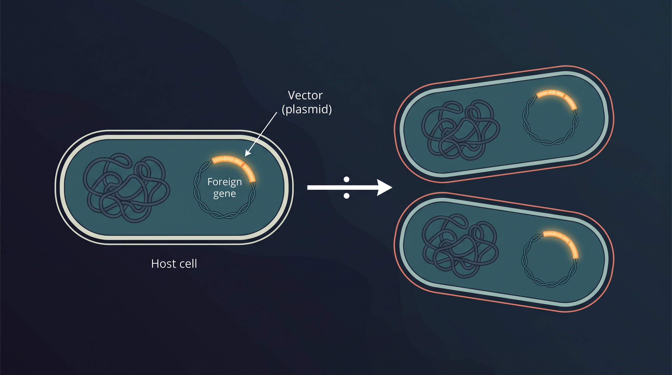
Fig. 2.0 – Recombinant DNA propagation in a bacterial host.
This chapter will answer each of these questions as you work through it — keep them in mind as you read.
2.1 Two Key Components of Recombinant DNA Technology
As you learnt in Chapter 1, recombinant DNA (rDNA) technology means joining two different DNA molecules together. The purpose behind this joining is practical: once two DNA molecules are combined, a gene can be isolated from the crowd of thousands of other genes, propagated in large numbers, characterised in detail, and manipulated for a chosen application.
The Two Major Steps
The whole technique rests on two major steps, shown in Fig. 2.1. In the first step, the desired DNA molecule is isolated from its source organism. This desired piece is generally called the insert, or the target gene — it is the gene you actually want.
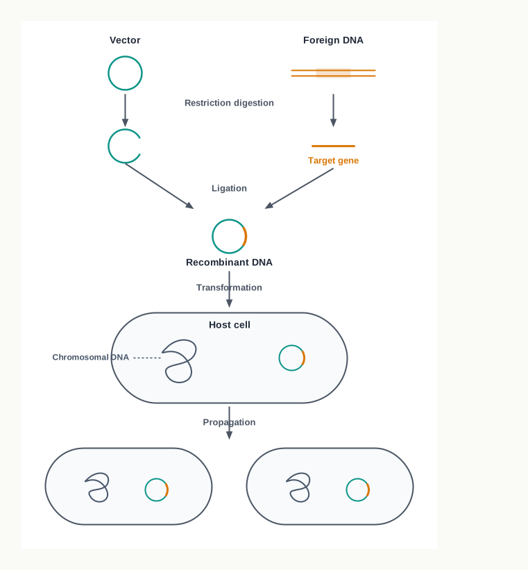
Fig. 2.1 – The major steps of gene cloning.
In the second step, this target gene is inserted into a convenient carrier DNA molecule called a vector. A vector is simply a DNA molecule that can carry the insert into a cell and keep it there in working condition. The vector that now contains the insert is called recombinant DNA (rDNA) — recombinant because it is a new combination of DNA that did not exist before in nature.
Entry Into a Living Cell
The recombinant DNA is next introduced into a living organism, and that organism is referred to as the host. Notice why a host is unavoidable. A DNA molecule sitting in a test tube cannot copy itself; the enzymes and proteins that carry out replication belong to a living cell. Once the rDNA is inside a suitable host, the host’s own genetic machinery takes over and the rDNA undergoes propagation — it is copied again and again as the cell multiplies — and, in many applications, expression as well, meaning the inserted gene is transcribed and translated into protein.
This entire process, from isolating the insert to multiplying it inside a host, is covered under the single term gene cloning.
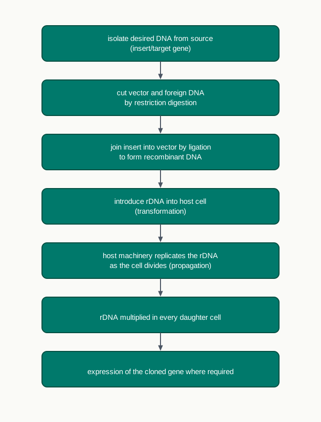
Fig. 2.2 – The gene cloning workflow.
A Two-Component System
Gene cloning may therefore be considered a two component system: a compatible host and a vector, working as a matched pair. The division of labour between them is worth remembering, because almost every practical decision in a cloning experiment follows from it.
The vector supplies the essential sequences required for its own replication — the address, so to speak, that tells the cell “copy me”. The host supplies the various replication functions, that is, the actual enzymes and proteins that do the copying. Neither partner works alone. A vector carrying every necessary sequence is inert without a cell around it, and a host cell, however cooperative, cannot replicate a piece of DNA that lacks the sequences it recognises.
The word compatible in “compatible host” is doing real work here as well. A vector built to replicate in a bacterium will not be recognised by a yeast cell, and vice versa. Matching the vector to the host is therefore the first design decision in any cloning experiment — and it is the reason the rest of this chapter is organised around the different hosts and vectors available, both prokaryotic and eukaryotic.
2.2 Host
A large number of host organisms, both prokaryotic and eukaryotic, are used for gene cloning. Fig. 2.3 sets out the common choices side by side, so that you can see at a glance which hosts and which vectors belong to each group.
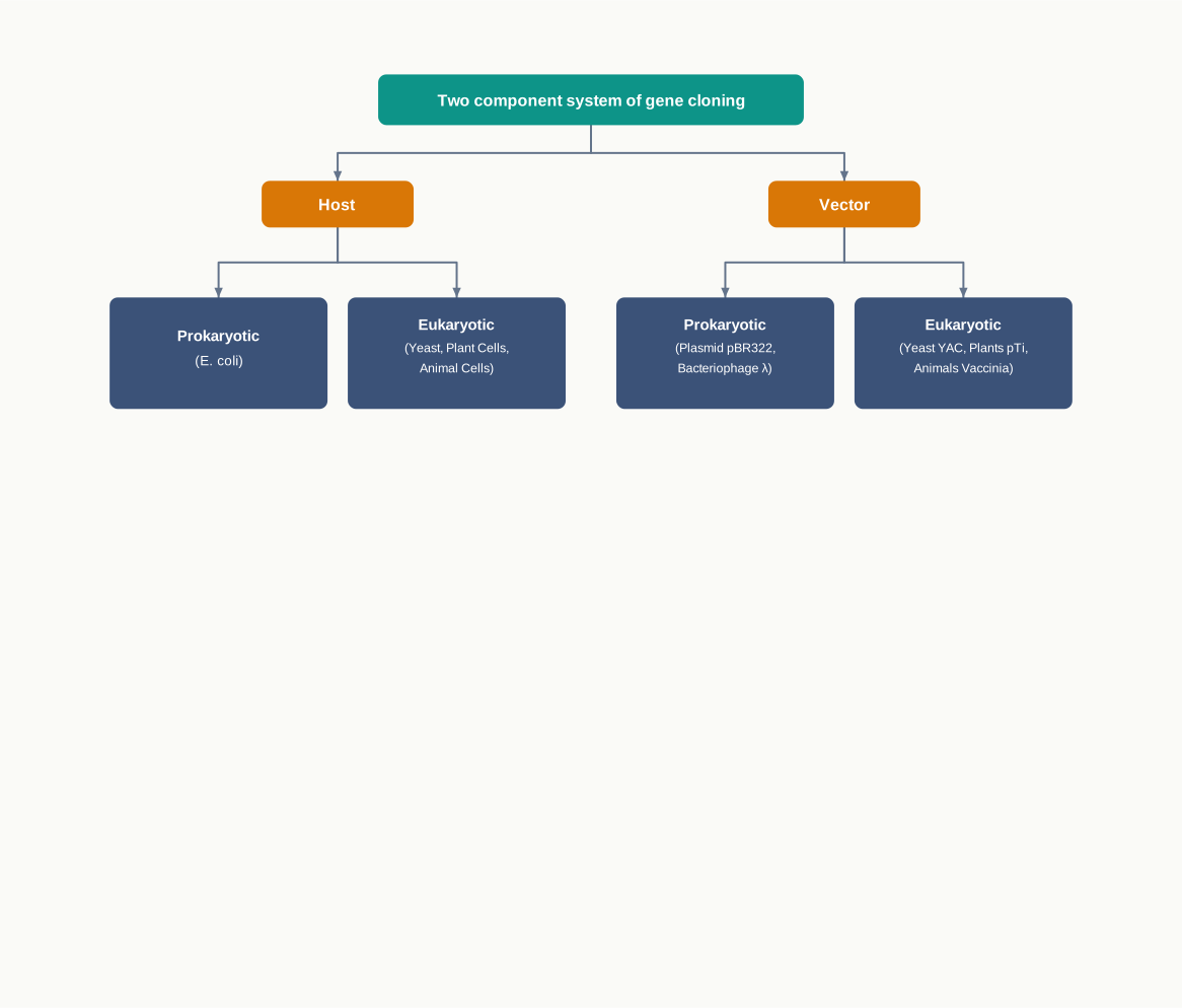
Fig. 2.3 – Classification of hosts and vectors.
What Makes a Good Host
Not every cell will do. A host must satisfy three requirements, and each of them exists for a definite reason.
First, the host should allow easy entry of the rDNA into the cell. If the recombinant molecule cannot get past the cell wall and membrane, nothing that follows can happen at all.
Second, the host should not treat the recombinant DNA as foreign DNA and degrade it. Bacteria naturally defend themselves against invading DNA by cutting it up with their own restriction enzymes — the same class of enzymes that genetic engineers use as molecular scissors. A host with an aggressive defence system would destroy the very molecule the experiment depends on, so strains with these defences weakened are deliberately chosen.
Third, the host must supply all the required enzymes and proteins for smooth replication of the vector DNA along with the insert. As established in Section 2.1, this is the host’s half of the bargain in the two-component system.
Because these demands are so specific, hosts are not picked casually from nature. A wide variety of genetically defined strains — strains whose genetic make-up is fully known and recorded — are available for the purpose, and this predictability is itself a reason for using them.
Prokaryotic Hosts
Among prokaryotic hosts, Escherichia coli is the most extensively used. A typical E. coli is a rod-shaped, Gram-negative bacterium commonly found in the lower intestine of warm-blooded organisms.
Its great advantage is speed. E. coli is able to reproduce and grow rapidly, doubling its population about every 20 minutes. A gene inserted into a few cells in the morning can therefore be present in billions of copies by the afternoon, which is exactly what “propagation” means in practical terms.
The K12 strain of E. coli is one of the most commonly used hosts in gene cloning. It has been maintained in laboratories for decades and is thoroughly characterised, which is precisely why researchers trust it.
Other prokaryotic hosts have also been developed for particular jobs. For example, Bacillus subtilis constitutes an important alternative host where the aim is secretion of a protein encoded by a cloned gene. B. subtilis naturally exports proteins out of the cell into the surrounding medium, so the product can be harvested from the culture fluid instead of having to break open the cells to get at it. The choice of host, in other words, follows from what you want out of the experiment.
Eukaryotic Hosts
Prokaryotes cannot do everything. When the cloned gene comes from a higher organism, its protein product may need folding and chemical finishing that a bacterium simply cannot carry out. For such work a eukaryotic host is required, and among eukaryotic hosts the most widely used is yeast. Plant cells and animal cells are also used, as Fig. 2.3 shows. The special vectors that these eukaryotic hosts demand are taken up later, in Section 2.4.
2.3 Vector
In principle, any molecule of DNA which can replicate itself inside a host cell can work as a vector for gene cloning. That single sentence is the whole idea of a vector — self-replication inside a living cell. A DNA fragment that cannot copy itself is a dead end, however valuable the gene it carries.
In practice, however, a plasmid must fulfil four definite features before it can act as a vector.
The Four Essential Features of a Vector
1. Small size. For easy incorporation into the host cell, a vector should itself be small in size and be able to integrate a large size of the insert. There is a practical reason behind both halves of this requirement. A small circular molecule slips into a cell far more readily than a bulky one, and the smaller the vector’s own DNA, the more room is left for the insert without the recombinant molecule becoming unmanageable.
2. An origin of replication. The vector should have an origin of replication, or ori, so that the vector is capable of autonomous replication inside the host organism. The ori is the specific sequence at which the host’s replication machinery binds and begins copying. Without it, the recombinant molecule may enter the cell perfectly well and still be lost, because it is never duplicated and gets diluted away as the cells divide.
3. Unique restriction sites. The vector needs to have unique restriction sites. A restriction site is the short sequence recognised and cut by a particular restriction enzyme. “Unique” means the site occurs only once in the whole vector, so that the enzyme opens the circle at exactly one point. If the vector possessed too many restriction sites, then it would be fragmented into several pieces on digestion — and a shattered vector can neither be re-sealed around the insert nor replicate afterwards.
4. At least one selectable marker. The vector needs to have at least one selectable marker. Selectable markers are required to screen out transformants — that is, to pick out the few cells that actually took up the vector from the enormous majority that did not. Resistance to antibiotics such as tetracycline (\(tet^R\)) and ampicillin (\(amp^R\)) is the standard example. Cells are simply plated on a medium containing the antibiotic; those without the vector die, and every colony that grows must be carrying it.
A variety of both prokaryotic as well as eukaryotic vectors is set out in Fig. 2.3, which you saw in the previous section. The rest of this section deals with the prokaryotic ones — plasmids, bacteriophages, and the hybrids built from both.
2.3.1 Plasmid as a vector
Plasmids are circular, double-stranded (ds) and extra-chromosomal DNAs capable of autonomous replication. “Extra-chromosomal” is the key word: a plasmid sits in the cytoplasm as a separate small circle, quite apart from the main bacterial chromosome, and carries its own ori.
They occur naturally in many bacteria, archaea, and even in eukaryotes such as yeast. The size of plasmids ranges from a few thousand base pairs to more than 100 kilobase pairs (kbp).
Like the host-cell chromosomal DNA, plasmid DNA is replicated before every cell division. During cell division, at least one copy of the plasmid DNA is segregated to each daughter cell, assuring continued propagation of the plasmid through successive generations of the host cell. This is precisely the behaviour a cloning experiment needs — the insert is carried forward automatically, generation after generation, without any further help from the experimenter.
Some plasmids can integrate into chromosomes. Such plasmids are called episomes. Once integrated, an episome is replicated as part of the chromosome itself.
Naturally Occurring Plasmids and Their Genes
Several naturally occurring plasmids contain genes that provide some benefit to the host cell. Plasmids are not passengers; they usually earn their keep, and the traits they carry are the reason particular plasmids were noticed and studied in the first place.
For example, some bacterial plasmids encode enzymes that deactivate antibiotics, such as ampicillin, tetracycline and chloramphenicol. Such drug-resistance providing plasmids are called R-plasmids, and they constitute a major category of plasmids used for gene cloning. Their usefulness is obvious once you recall the fourth vector requirement — the resistance gene they carry is ready-made as a selectable marker.
Another category of plasmids produces a variety of toxins called ‘colicins’, which kill other bacteria. These are called Col plasmids.
Some plasmids contain ‘transfer genes’ encoding proteins that can form a macromolecular tube, or pilus, through which a copy of the plasmid can be transferred to other host cells of the same or related bacterial species. These are called F-plasmids. Remember the pilus — it reappears later in this section, because one bacteriophage vector can infect only those cells that possess one.
Classification by Copy Number
Similarly, based on the copy number, plasmids can also be classified as follows.
(a) High or multi copy plasmids: Replication and segregation of these plasmids are free from the control of replication of the bacterial chromosomal DNA. That freedom allows for many copies of these plasmids per cell (10–30 copies), as shown in Fig. 2.4(a). Replication of these plasmids is referred to as relaxed replication.
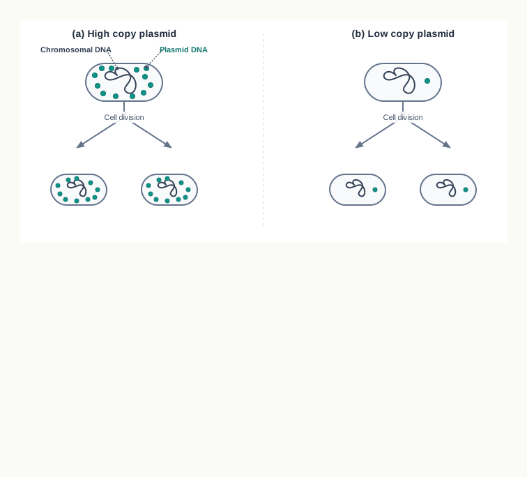
Fig. 2.4 – Replication and segregation of (a) high copy and (b) low copy plasmids.
Use of a high copy number plasmid gives rise to higher yields and increased strength of the signals during screening of recombinants. Both benefits follow from the same arithmetic: more copies of the plasmid per cell means more copies of the cloned gene, hence more DNA to recover and a stronger detectable signal when the recombinants are being identified.
(b) Low or single-copy plasmids: Replication and segregation of these plasmids are under the same control as replication of the bacterial chromosomal DNA. As a result of this, the copy number of these plasmids is restricted to only one or a few copies per cell, as shown in Fig. 2.4(b). Replication of these plasmids is referred to as stringent replication.
Development of plasmid based vectors
Natural plasmids are not vectors. They evolved for the convenience of bacteria, not of experimenters, so in order to use plasmids as a vector and to enhance their utility — either for general purposes or to suit particular experimental designs — plasmids originally found in nature are modified, shortened, reconstructed and recombined under both in vivo as well as in vitro conditions, so as to incorporate the properties indicated above into them.
The history of one such vector shows the process clearly. The plasmid based vector pBR313 replicates in relaxed fashion, giving rise to a high copy number. It contains two selectable markers, \(tet^R\) and \(amp^R\) — these selectable markers were derived from two other naturally occurring plasmids — and it carried a number of unique restriction sites, that is, cloning sites.
However, the size of this plasmid was too large: 9 kb. It was observed that more than half of its DNA was not essential for its role as a vector. Therefore, the first phase of this plasmid vector development led to the construction of vector pBR322, whose size was reduced to 4,361 bp by deleting most of the non-essential sequences of pBR313. Cutting away the dead weight left more room for the insert and made the vector easier to take up — exactly the first of the four requirements. The vector pBR322 became the most widely used vector for gene cloning, and its map is given in Fig. 2.5.
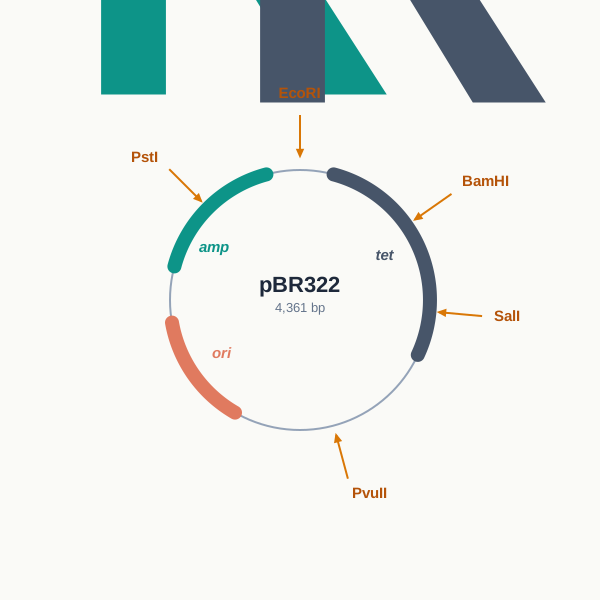
Fig. 2.5 – Vector map of pBR322.
Read Fig. 2.5 as a working document rather than a picture. Each labelled arrow is a place where one particular enzyme will cut, once and only once; the two coloured arcs are the genes that will let you select transformants; and the small segment marked ori is what makes the whole circle self-replicating. Notice also that PstI cuts within \(amp^R\) and BamHI and SalI cut within \(tet^R\) — inserting DNA at one of those sites destroys that marker gene, which is itself a way of recognising recombinants.
Multiple Cloning Sites and Easier Selection
Subsequent development included the introduction of synthetic cloning sites, termed the multiple cloning site (MCS), or poly cloning site, or polylinker. The MCS is a short synthetic DNA fragment containing a large number of unique restriction sites. This enables the insertion of a foreign DNA cleaved at any of these restriction sites into that region, as shown in Fig. 2.6.
The advantage is flexibility. With a single cloning site the experimenter must cut the insert with that one enzyme, whatever the insert’s own sequence might prefer. With a polylinker, whichever of a dozen enzymes suits the insert, the vector has a matching site waiting.
Further development involved the introduction of easy and convenient selection markers. Thus, for example, selection of the recombinants on the basis of blue/white selection was introduced, which facilitated the selection of recombinants utilising β-galactosidase (lacZ) of E. coli. The details of how blue and white colonies are produced are dealt with in Section 3.5 of Chapter 3; for the present, note only that it allows a recombinant colony to be told apart from a non-recombinant one by eye, without any further test.
A common example of such an improved cloning vector is the pUC19 vector, shown in Fig. 2.6. Many other plasmids are used as vectors for gene cloning in E. coli.
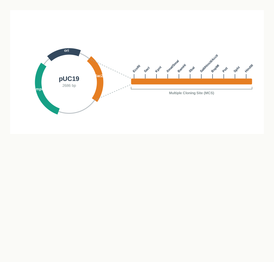
Fig. 2.6 – Vector map of pUC19.
Compare Fig. 2.6 with Fig. 2.5 and the direction of vector design becomes obvious: pUC19 is smaller than pBR322, its many cloning sites are gathered into one compact stretch instead of being scattered around the circle, and that stretch sits inside lacZ so that any successful insertion announces itself.
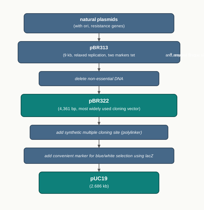
Fig. 2.7 – Development of plasmid based vectors.
2.3.2 Bacteriophage as vectors
Another important category of vectors for gene cloning is derived out of the genome of bacteriophages (phages), that is, viruses which infect bacterial cells. The logic of using them is straightforward: a virus has spent its whole evolutionary history perfecting the job of getting DNA into a bacterium, so a vector built from a phage genome enters the host far more efficiently than a bare plasmid.
In general, bacteriophage vectors are more efficient than plasmids in two respects. First, they are better for cloning large inserts. Second, they allow screening large numbers of bacteriophage plaques — plaques being the clear patches of lysed bacterial cells produced by phage infection on a bacterial lawn — which is far easier than screening bacterial colonies one by one for a specific desired insert.
The bacteriophage lambda (λ) and M13 are the two most common phages whose genomes have been frequently used to make cloning vectors for the E. coli host. They are described below.
Lambda (λ) Phage Vector
The bacteriophage lambda, a virus that infects E. coli, has been widely used as a cloning vector. The virus is easy to propagate, and has therefore long been a model system in molecular biology.
It consists of a head (capsid) and a tail, as shown in Fig. 2.8. The head contains the phage genome. The phage particles bind to the surface of E. coli and insert their genome via the tail into the cytoplasm of the bacterial cell. Note that only the DNA enters; the protein coat stays outside.
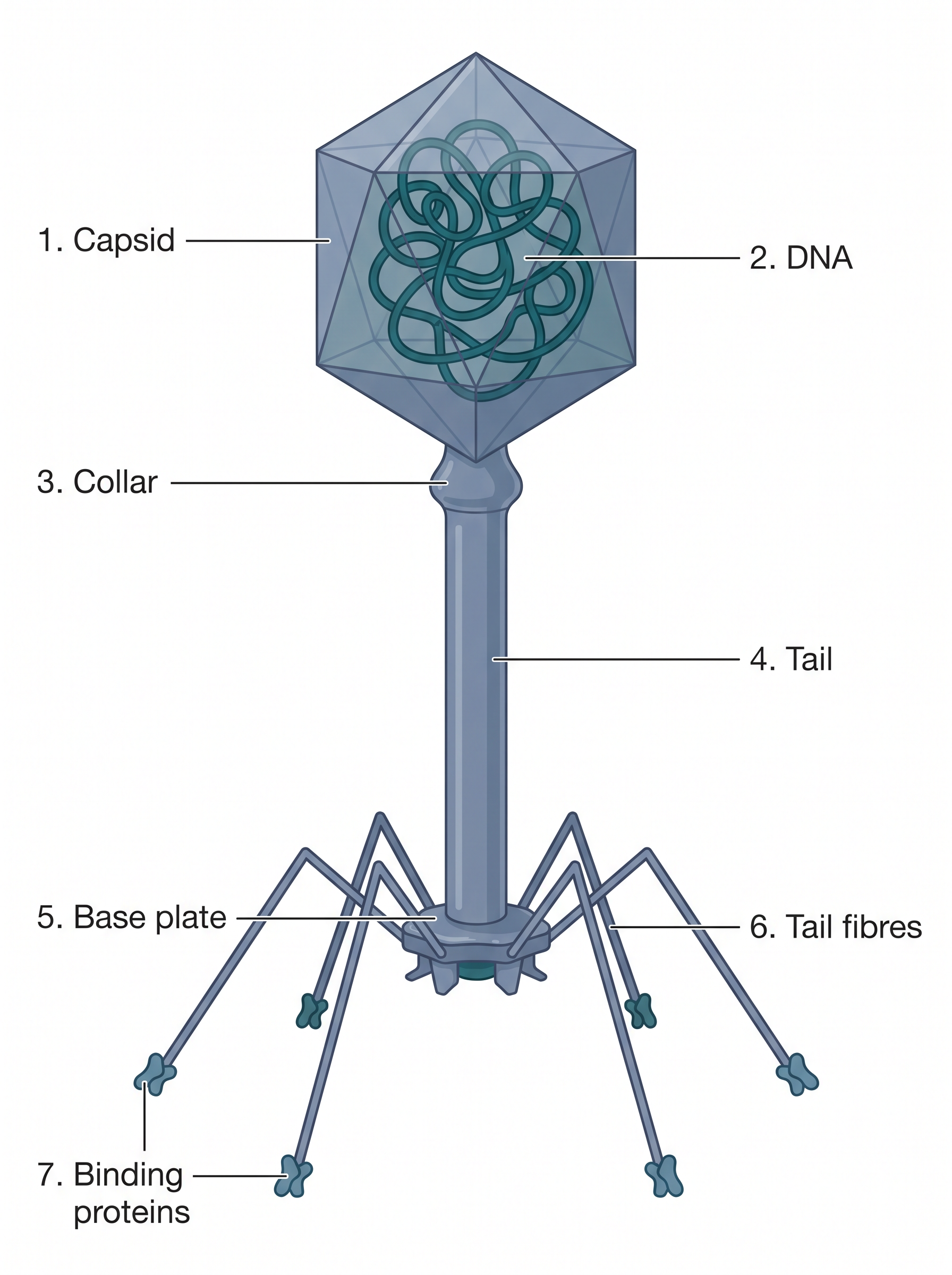
Fig. 2.8 – Structure of the lambda bacteriophage.
The Two Life Cycles of λ
λ phage is a virus with both lytic and lysogenic alternatives to its life cycle, shown in Fig. 2.9. The choice between them matters for vector design, because a cloning vector built from λ is meant to follow only one of the two.
Usually a ‘lytic cycle’ is followed, in which the λ DNA is replicated and new phage particles are produced within the cell. This causes cell lysis, releasing the newly formed phage particles.
However, under certain conditions the phage DNA may integrate itself into the host cell chromosome in the lysogenic pathway. In this state the λ DNA is called a prophage, and it stays resident within the host’s genome without causing apparent harm to the host. When conditions change, the prophage is excised from the bacterial DNA and enters the lytic cycle.
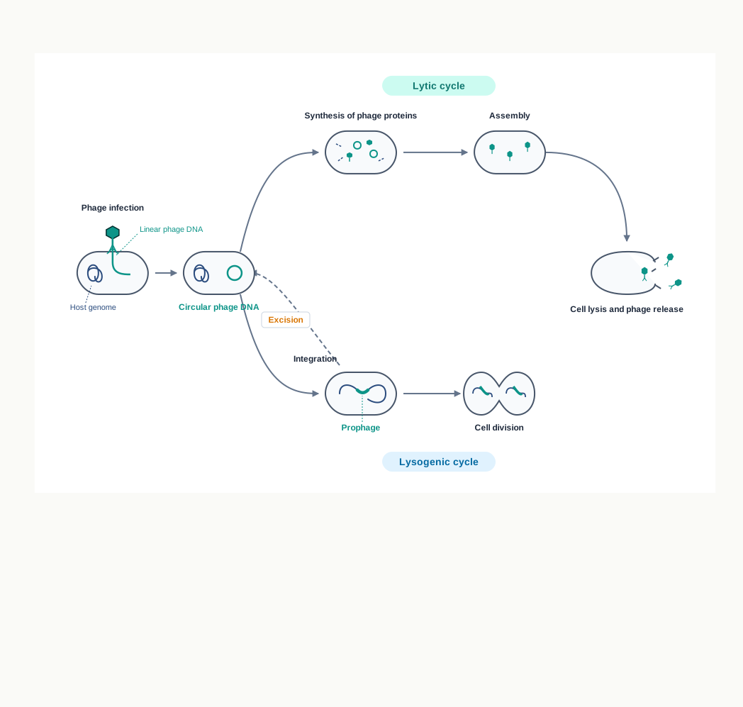
Fig. 2.9 – Lytic and lysogenic life cycles of lambda phage.
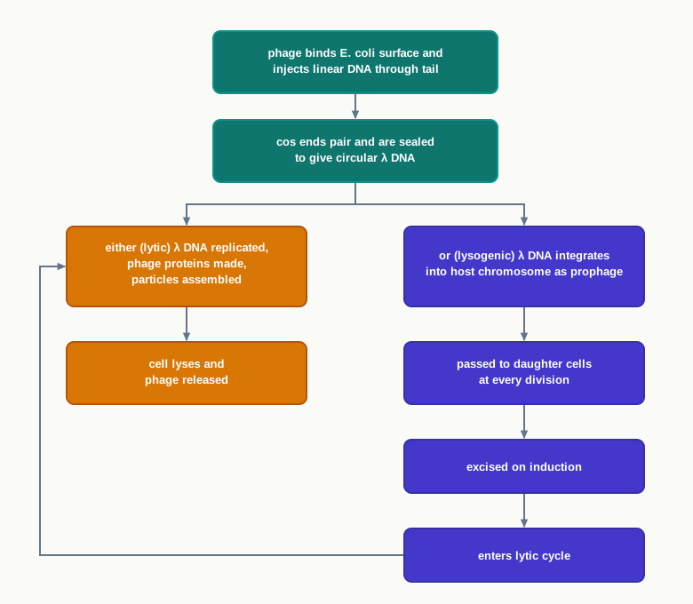
Fig. 2.10 – The two alternatives open to λ after infection.
Structure of the λ Genome
The λ phage genome is linear double stranded DNA containing 48,490 base pairs. Each end has a 12 bases single stranded segment known as the cos sequences, or cohesive ends. The two cohesive ends are complementary to each other, which is what allows them to stick together.
The λ genome has a replication origin site, genes for viral head and tail proteins, and enzymes of replication involved in the lytic and lysogenic cycles.
The λ genome remains linear within the phage head, as shown in Fig. 2.11(a). Once inside the E. coli host cell, the cos sequences pair up and the cohesive ends are ligated together by a host enzyme, forming the circular version of the lambda genome, shown in Fig. 2.11(b). The sealed cohesive end is called the cos site.
The 12 nucleotides long single stranded DNA at both ends pair with each other to form double stranded DNA. Therefore, in its circular form the phage genome is 48,502 base pairs in length. The increase of 12 base pairs over the linear figure is not an error — it is exactly the 12 unpaired nucleotides at each end becoming a paired double-stranded stretch once the circle closes.
The phage λ genome can be inserted into the host chromosome and is then called a prophage (Fig. 2.9).
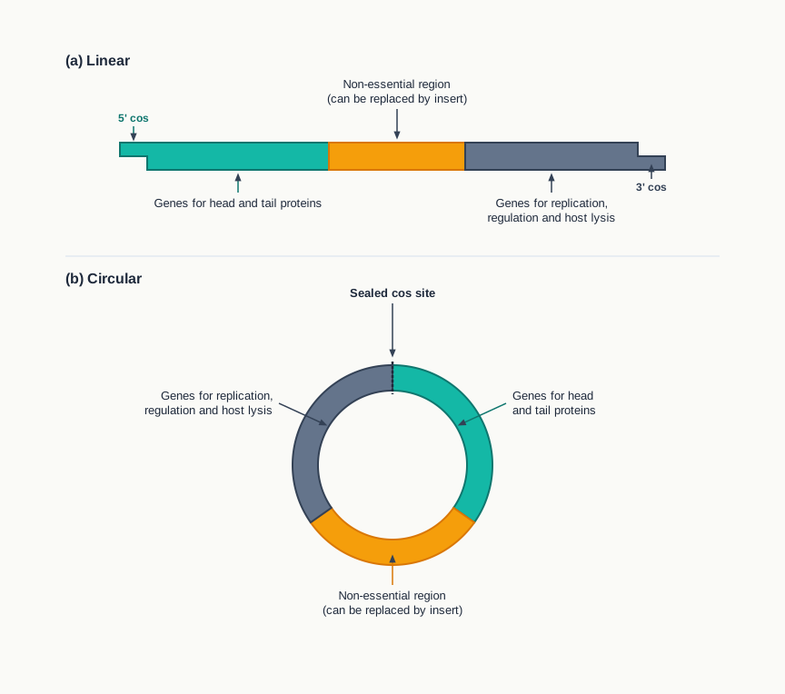
Fig. 2.11 – Lambda genome in (a) linear and (b) circular forms.
Replication and Packaging
During multiplication, the phage genome undergoes rolling circle replication, in which a template strand rotates, churning out a chain of many copies of λ genomes to form a long concatemer joined end to end and separated by cos sites.
The concatemer is then cleaved at the cos sites to yield one unit of phage DNA. This is followed by the packaging of a single copy of the λ genome into each empty head, followed by the attachment of the tail and the release of newly formed phage particles by lysis of the bacterial cells (Fig. 2.9).
The cos sites are therefore doing two distinct jobs — they circularise the genome after infection, and they later act as the measuring marks that tell the packaging machinery where to cut one genome’s worth of DNA out of a long chain.
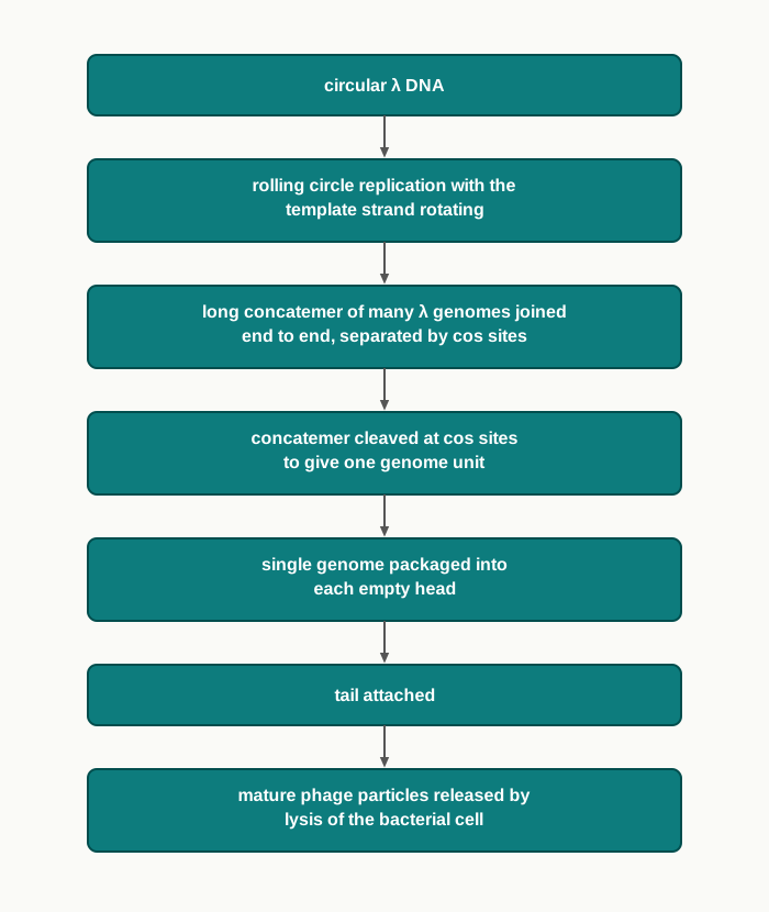
Fig. 2.12 – Multiplication and packaging of λ.
Building a Vector Out of λ
Approximately one-third of the lambda genome — the middle region of the λ genome, which contains the genes required for lysogeny — is dispensable, that is non-essential, for successful lytic infection (Fig. 2.11). This is the opening that vector design exploits: genes needed only for the lysogenic option can be thrown away without disturbing the lytic cycle.
Therefore, in order to construct a vector for gene cloning out of the lambda phage DNA, all or part of the middle dispensable region is replaced with an insert segment of appropriate length, such that the size of the recombinant DNA (rDNA) lies between 38 to 52 kbp for its efficient packing into the λ head.
That size window is a hard physical constraint, not a guideline. The λ head is a container of fixed dimensions: a recombinant molecule shorter than about 38 kbp is packaged badly or not at all, and one longer than about 52 kbp will not fit. So the insert size is decided as much by the capsid as by the experimenter.
Typical vectors coming out of the lambda genome fall into two broad classes, namely insertion vectors and replacement vectors. Insertion vectors have a single restriction enzyme site (cloning site) for insertion of the insert DNA, whereas replacement vectors have a pair of cloning sites flanking a segment of non-essential bacteriophage λ DNA — the central stuffer region — that can be replaced by the insert DNA.
A large array of λ vectors has been constructed for different purposes. Some commonly used λ bacteriophage based vectors for E. coli are given in Table 2.1.
Table 2.1: Commonly used λ bacteriophage based vectors for E. coli as host.
| S.No. | Vector name | Type of vector | Selection of recombinants | Maximum insert size (kbp) |
|---|---|---|---|---|
| 1. | λgt10 | Insertion vector | Lytic (plaque formation) | 6 |
| 2. | λgt11 | Insertion vector | Blue white | 7.2 |
| 3. | λ EMBL3 | Replacement vector | Lytic (plaque formation) | 20 |
Read the last two columns of Table 2.1 together. The replacement vector accepts a much larger insert than either insertion vector, because it gives up its stuffer region to make room — a direct illustration of the packaging limit discussed above.
Bacteriophage M13
M13 is a filamentous bacteriophage of E. coli. “Filamentous” means it is a long thread rather than the head-and-tail shape of λ. Its genome consists of a single stranded circular DNA of about 6.4 kb nucleotides, which is packaged in a tube-like capsid, as shown in Fig. 2.13.
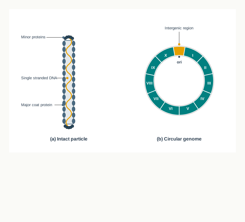
Fig. 2.13 – M13 bacteriophage: (a) intact particle and (b) circular genome.
Almost the full genome encodes genes (I–X) required for coat protein, viral replication and viral assembly, except for a small non-essential region flanking ori called the intergenic region. That small non-essential stretch is the only spare space M13 offers, and it is exactly where the cloning site is placed.
Bacteriophage M13 infects only E. coli cells harbouring the F plasmid. These cells have a tube-like structure called a ‘pilus’ extending from their membranes, and this pilus is required for attachment and adsorption of the phage to the cell (Fig. 2.14). Here is where the F-plasmid mentioned earlier becomes practical: a host strain for M13 work must carry an F plasmid, or the phage cannot attach at all.
The M13 Life Cycle and Its Advantage
During the life cycle of the M13 bacteriophage, the single stranded M13 phage DNA undergoes replication inside the host to generate a double stranded replicative form (RF), as shown in Fig. 2.14. Subsequently, the mode of replication changes to generate a single stranded genomic DNA (+) from the double stranded replicative form.
The circular single stranded genomic DNA (+) is assembled with capsid proteins during their passage through the host cell without causing cell lysis. This is the crucial difference from λ. The host survives, keeps growing, and keeps exporting phage particles into the medium.
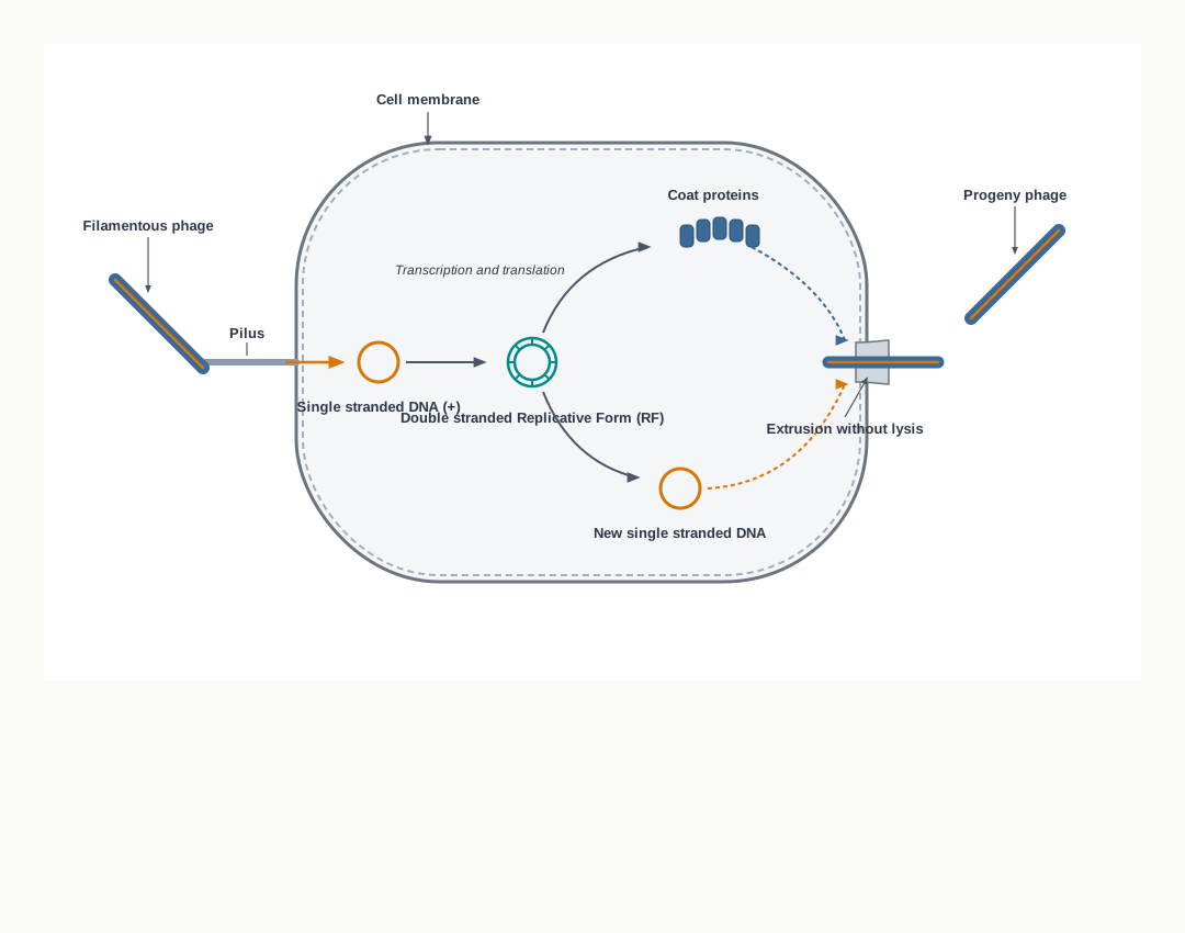
Fig. 2.14 – Life cycle of bacteriophage M13.
This life cycle gives the experimenter two convenient products at once. The double stranded RF DNA is readily isolated from infected cells for use as a vector, and the single stranded genome is abundantly available in the form of phage in the growth medium.
Thus, for M13 vector construction, the double stranded RF of the M13 genome is used — a double stranded circle can be cut with restriction enzymes and ligated exactly like a plasmid, which a single strand cannot. The intergenic region is exploited for putting the insert, so that it does not interfere with M13 replication.
A large sized DNA (more than 42 kb) can be cloned by M13 bacteriophage. An example of an M13 based vector for E. coli is M13mp18, which facilitates blue/white selection of the recombinants. Blue/white selection is taken up in Chapter 3 of this book.
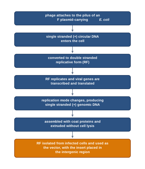
Fig. 2.15 – M13 life cycle and its use as a vector.
2.3.3 Cosmid vector
A cosmid is a type of hybrid (combination) vector that replicates like a plasmid but can also be packaged in vitro into lambda phage coats. The name itself records the combination — cos sites from λ, joined to a plasmid.
A typical cosmid has replication functions, unique restriction endonuclease sites, and selective markers contributed by plasmid DNA, combined with a λ DNA segment that includes the joined cohesive ends (cos sites), as shown in Fig. 2.16.
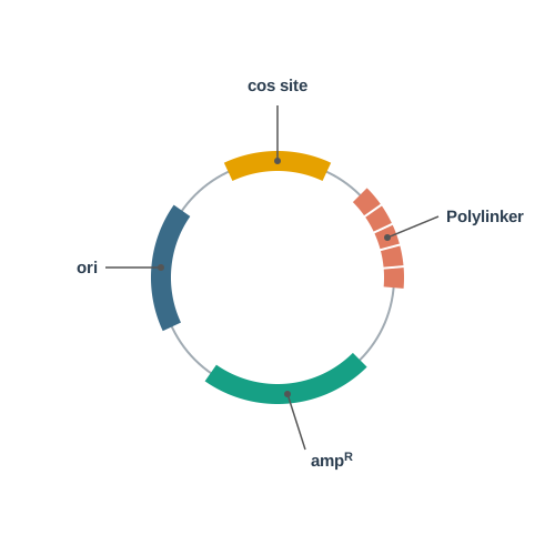
Fig. 2.16 – A typical cosmid vector.
As little as 250 bp of λ DNA is sufficient to provide the cos junction, including the sequences required for binding to and cleavage by an enzyme called terminase. This is the economy that makes the cosmid work: 250 bp is a trivial amount of DNA to carry, yet it is enough to persuade the λ packaging machinery to treat the whole molecule as a phage genome and pack it into a head.
The advantage of most cosmid cloning vectors is that they accommodate DNA inserts as large as 45 kbp — far beyond what an ordinary plasmid can hold. Once packaged and injected into E. coli, the cosmid does not go on to make phage particles, because it carries no phage genes; it simply circularises and replicates as a plasmid, and the transformants are selected using its antibiotic resistance marker.
2.3.4 Phasmids (Phagemids)
Phasmids are true combination (hybrid) vectors between phage and plasmid. Where a cosmid borrows only the cos sites from λ, a phasmid keeps whole functional blocks of both parents.
They are linear duplex DNAs whose ends are lambda phage DNA that contain all the genes required for a lytic infection, and whose middle region is linearised plasmid. Both the lambda phage and the plasmid replication functions are intact, as shown in Fig. 2.17.
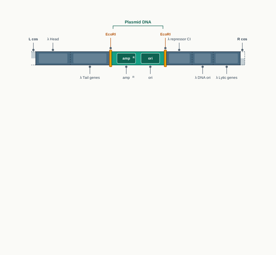
Fig. 2.17 – A typical phasmid vector.
Phasmid recombinants are packaged in vitro before infection, exactly as cosmids are. Once inside an E. coli cell, the phasmid can replicate like a phage and form plaques in the normal way.
However, if the vector contains the gene that encodes the lambda repressor, then the phasmid replicates as a plasmid rather than as a phage. The repressor shuts down the lytic genes, so the λ half of the vector falls silent and the plasmid ori takes charge. This switchable behaviour is the point of a phasmid: the same molecule can be handled as a phage when high-efficiency infection and plaque screening are wanted, and as a plasmid when the DNA simply has to be maintained and multiplied quietly.
Both cosmids and phasmids exist for the same underlying reason. A plasmid enters the host inefficiently but replicates simply; a phage enters superbly but imposes its own life cycle. The hybrids take the entry mechanism from one parent and the quiet, controllable replication from the other — which is a good example of how vectors are designed to order rather than found in nature.
2.4 Eukaryotic Host Vector System
Everything discussed so far — plasmids, phages, cosmids, phasmids — was built around a bacterial host. The moment the DNA to be cloned comes from a higher organism, that system begins to run out of room.
Why Prokaryotic Vectors Are Not Enough
Genome analysis of the higher organisms requires the cloning of much larger fragments. The reason lies in the structure of eukaryotic genes themselves: they contain introns, the non-coding stretches that interrupt the coding sequence, and these may be hundreds of kilobases in length. A eukaryotic gene, taken whole from the chromosome, is therefore an enormous piece of DNA.
Nor does removing the introns solve the problem completely. Even if a eukaryotic gene without introns is to be inserted, its size exceeds the maximum length that can be carried as an insert by a plasmid.
Therefore, special vectors are required for such large DNA fragments. Consequently, ‘artificial chromosomes’ have been developed to carry huge lengths of eukaryotic DNA. The name is exact rather than decorative: if a vector is to behave like a chromosome, it must be given the parts a real chromosome uses — a replication origin, a centromere and telomeres.
Yeast as the Eukaryotic Host
Among eukaryotic host vector systems, the most common is the baker’s yeast, Saccharomyces cerevisiae. It occupies a useful middle position: it is a true eukaryote, yet it can be handled with the convenience of a microorganism.
Although yeast can reproduce sexually, the cells usually multiply asexually by budding. They grow as single cells in suspension and produce colonies in solid medium, much as in E. coli. In other words, the familiar routines of streaking plates, picking colonies and growing liquid cultures all carry over unchanged.
A large collection of metabolic, biosynthetic, and cell cycle defective mutants are known and genetically mapped for this organism. Such a catalogue of well-characterised mutants is invaluable, because a mutant that cannot make a particular nutrient becomes a ready-made partner for a selectable marker: supply the missing gene on the vector, and only the transformed cells will grow on a medium lacking that nutrient.
Because of a long history of safe use of S. cerevisiae in the food industry, it belongs to the category which is ‘Generally Recognised As Safe’ (GRAS) organisms.
The 2 µm Plasmid and Yeast Artificial Chromosomes
S. cerevisiae also harbours a double stranded circular plasmid called the 2 µm plasmid, which has been used to develop a number of vectors. Yeast, in other words, offered a natural plasmid of its own to build upon, just as bacteria did.
One important category of vectors that has been designed for cloning large size DNA insert (200–500 kb) is known as Yeast Artificial Chromosomes (YACs).
The YAC vector consists of:
- two copies of a yeast telomeric sequence — telomeres being the sequences at the ends of chromosomes;
- a yeast centromeric sequence;
- a yeast ARS, an autonomously replicating sequence, where DNA replication begins; and
- appropriate selectable markers.
Each part earns its place. The ARS is the yeast equivalent of an ori, so the molecule can be replicated. The centromere is the attachment point for the spindle fibres, so that the artificial chromosome is pulled correctly into the daughter cells at division instead of being lost. The telomeres cap and protect the two ends of a linear molecule, which would otherwise be attacked and shortened. Together they turn a piece of DNA into something the yeast cell handles as one of its own chromosomes.
The Two Forms of a YAC
A YAC has two forms: a circular form for growing in bacteria, and a linear form for growing in yeast, as shown in Fig. 2.18.
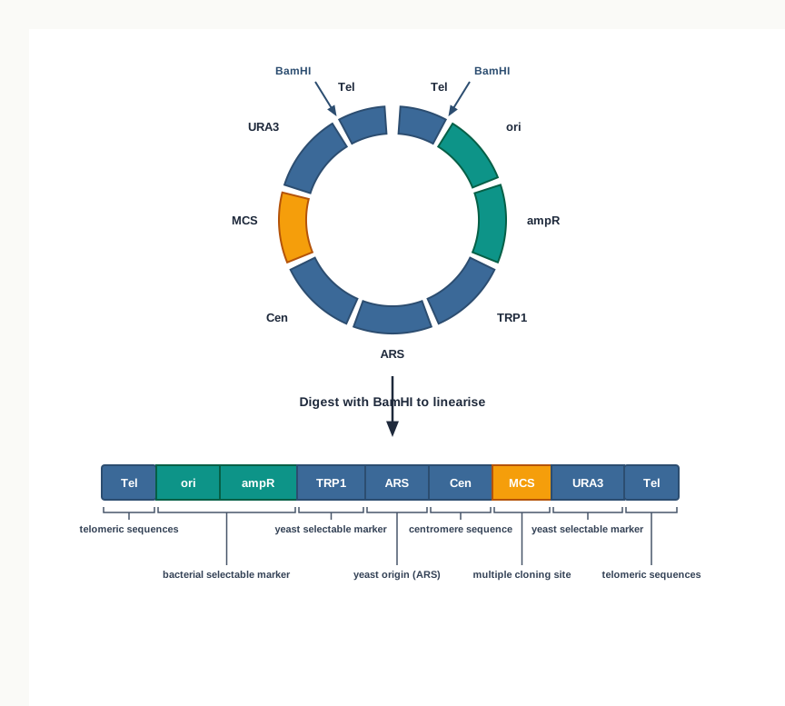
Fig. 2.18 – Yeast Artificial Chromosome (YAC) in circular and linear forms.
The circular form can be manipulated and grown like any other plasmid in bacteria, since it has a bacterial origin of replication and an antibiotic resistance gene. This is a matter of convenience: all the cutting, ligating and multiplying of vector DNA is far quicker in E. coli.
In order to use this in yeast, the circular form is isolated and linearised, such that the yeast telomere sequences are on each end. Follow that step carefully in Fig. 2.18 — digestion with BamHI opens the circle at exactly the two points that leave a telomere at either end, which is what a chromosome requires.
High Capacity Cloning Vectors
Vectors like YACs are sometimes called high capacity cloning vectors. Some other high capacity cloning vectors, coming from bacterial plasmid and phage, are called Bacterial Artificial Chromosomes (BACs) and Phage Artificial Chromosomes (PACs).
A list of commonly used vectors for gene cloning, having different capacities to take up different sized inserts, is shown in Table 2.2.
Table 2.2: DNA insert size with different cloning vectors
| S. No. | Vectors | Insert size (kb) |
|---|---|---|
| 1. | Plasmid | ≤ 10 kb |
| 2. | Bacteriophage | 8–25 kb |
| 3. | Cosmids | 23–40 kb |
| 4. | PAC | 100–300 kb |
| 5. | BAC | ≤ 300 kb |
| 6. | YAC | 200–500 kb |
Table 2.2 is worth learning as a ladder rather than as six separate facts. Each rung was built because the one below it ran out of capacity: plasmids gave way to phage vectors, phage vectors to cosmids, and cosmids to the artificial chromosomes. In an examination, a question that gives you an insert size and asks which vector to choose is really asking you to read this ladder — and note that the ranges overlap, so more than one vector may be workable for a mid-sized fragment.
2.5 Expression Vectors
Till now we have discussed the different types of cloning vectors, which are used to propagate DNA. Their job ends with multiplication: they make many copies of the insert, and nothing more is asked of them.
Very often, however, the copies are not the point. What is actually wanted is the protein the gene codes for — insulin, a vaccine antigen, an industrial enzyme. For that, a different class of vector is needed.
What an Expression Vector Does
Another type of vector are expression vectors, which act as vehicles for the DNA insert and also allow the DNA insert (gene) to be expressed efficiently, as shown in Fig. 2.19. These may be plasmid or virus based vectors.
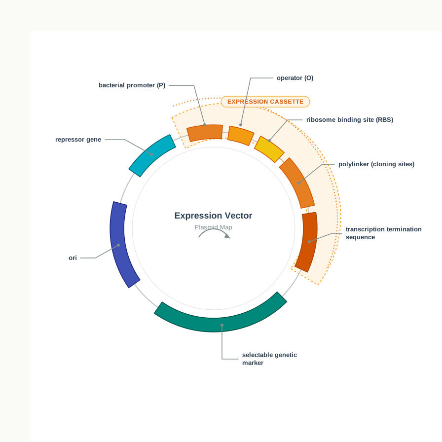
Fig. 2.19 – Elements of an expression vector.
The Parts That Make Expression Possible
These vectors contain an efficient promoter for expression of the cloned genes. A promoter is the sequence at which RNA polymerase binds to begin transcription; without one placed correctly, the cloned gene is copied along with the vector but is never read.
These promoters are generally inducible in nature, so that expression of the cloned gene can be regulated. Study Fig. 2.19 again with this in mind: the vector also carries a gene encoding a repressor that binds the operator (O) and thereby regulates the promoter (P). The repressor keeps the promoter switched off until an inducer is supplied.
Control of this kind is not a refinement but a necessity. A foreign protein made continuously is a heavy metabolic burden, and may even be toxic to the host, so the cells would grow poorly or lose the vector altogether. With an inducible promoter the culture is first grown to a high cell density with the gene switched off, and only then is expression turned on — so that a large, healthy population makes the product all at once.
Downstream of the promoter there are unique restriction sites for insertion of the gene to be expressed. Fig. 2.19 shows these gathered as a polylinker, so that the gene may be cut with whichever enzyme is convenient.
Furthermore, in order to ensure correct termination of transcription of the cloned genes, a proper transcription termination sequence is provided by the vector near the 3’ end of the gene. Without a terminator, RNA polymerase would continue past the gene into neighbouring vector sequences, producing long, untidy transcripts and wasting the cell’s resources.
The Expression Cassette
Thus, the cloned gene is inserted between a promoter towards the 5’ end (upstream) and a terminator at the 3’ end (downstream) of the cloned gene in an expression vector. This portion of the vector is called the expression cassette. Such vectors are therefore sometimes also called sandwich expression vectors — the gene is sandwiched between the two control sequences.
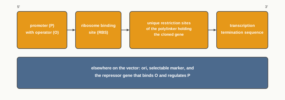
Fig. 2.20 – Order of elements along an expression cassette.
Heterologous Gene Expression
Expression of a gene by recombinant strategies depends on the source of the gene as well as the host being used. The two must be considered together, because a gene that works perfectly in its own organism may fail entirely in another.
Thus, for example, if a gene of eukaryotic origin is to be expressed in a prokaryotic host, or vice versa, this is known as heterologous gene expression, as the gene being expressed is foreign to the host.
Such heterologous expression requires that the eukaryotic gene being expressed:
does not contain intron(s), as the process of splicing is absent in the prokaryotic host; and
codes for a protein that does not require post-translational modification — for example, glycosylation — for its biological activity, that is, its functionality.
Both conditions describe machinery the bacterium simply does not possess. A bacterium has no spliceosome, so an intron-containing transcript would be translated straight through the intron, giving a nonsense protein. Equally, a bacterium cannot attach sugar chains to a protein, so a product that needs glycosylation to work would be made in full but would be biologically inactive.
The way round the first condition is to supply the gene without introns, and the standard route to that is cDNA, prepared from mature mRNA after splicing has already occurred in the eukaryotic cell.
This is not merely a theoretical restriction. Recombinant human insulin, the first recombinant protein to reach patients, is manufactured in bacterial hosts precisely because the insulin polypeptide chains require no sugar attachment to be active; a gene free of introns is used, and the product folds into a functional hormone. Human proteins that do depend on glycosylation, such as erythropoietin used in the treatment of anaemia, cannot be made this way at all, and are produced instead in cultured mammalian cells, which possess the necessary post-translational machinery. The choice of host, once again, follows from the biology of the product.
2.6 Shuttle Vectors
Vectors developed for replication in two alternative hosts, either prokaryotic or eukaryotic, are called shuttle vectors. The name describes the behaviour exactly: the same molecule can travel back and forth between two different kinds of cell, replicating in each.
These vectors are themselves constructed by recombinant DNA techniques, and many different types have been made. Some of them shuttle between two prokaryotic species; others between a prokaryote — usually E. coli — and eukaryotic cells, including yeast, plants and animals. Indeed, most of the eukaryotic vectors are shuttles. The YAC met in Section 2.4, grown as a circle in bacteria and used as a linear chromosome in yeast, is a familiar example.
Two Origins, One Active at a Time
Shuttle vectors contain two origins of replication. However, in a given host, only one origin is active at a time, as shown in Fig. 2.21. A yeast cell recognises the eukaryotic origin and ignores the bacterial one; in E. coli the reverse happens. The unused origin is simply carried along as inert DNA until the vector reaches a host that can read it.
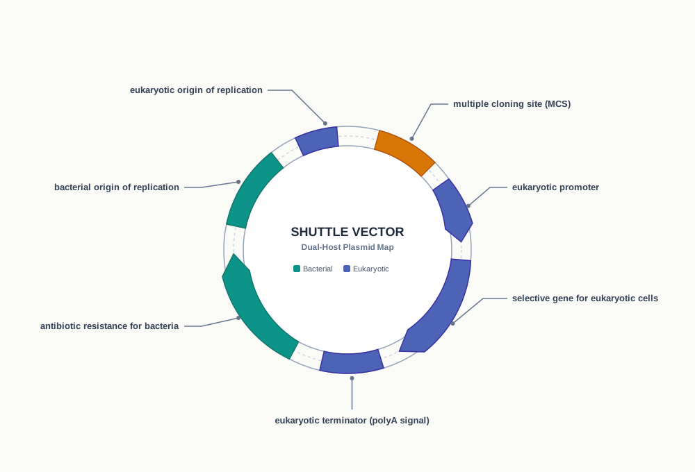
Fig. 2.21 – Shuttle vector for E. coli and yeast.
Notice in Fig. 2.21 that the duplication runs right through the vector’s design. There are two origins, and there is also a selectable marker for each host — antibiotic resistance for the bacterium, a selective gene for the eukaryotic cell — because transformants must be identifiable in whichever cell the vector currently occupies.
Why the Arrangement Is Worth the Trouble
The reason for building such vectors is practical. Cutting, ligating and multiplying vector DNA is quick and cheap in E. coli, whereas yeast, plant and animal cells grow slowly and are harder to transform. A shuttle vector lets all the construction work be done in bacteria, after which the finished recombinant molecule is moved into the eukaryotic cell where the gene is actually to be studied or expressed — with the eukaryotic promoter and terminator seen in Fig. 2.21 ready to drive that expression.
2.7 Summary
rDNA technology is a two-component system: a compatible host and a vector working as a pair. The vector provides the essential sequences required for its replication, and the compatible host provides the replication functions — the enzymes and proteins that do the copying.
A cloning vector should be small in size and must have an origin of replication (ori site), unique restriction sites, and at least one selectable marker.
Plasmids are circular, extra-chromosomal double stranded DNA (dsDNA) molecules capable of autonomous replication. Plasmids that can integrate into the host chromosome are called episomes.
On the basis of copy number, plasmids are either high copy plasmids, whose replication is free from chromosomal control and is called relaxed replication, or low copy plasmids, whose replication is under chromosomal control and is called stringent replication.
The bacteriophage lambda (λ) and M13 are the two most common phages whose genomes have been frequently used to make cloning vectors for the E. coli host.
The bacteriophage lambda, a bacterial virus that infects E. coli, has been widely used as a cloning vector.
Typical vectors coming out of the lambda genome fall into two broad classes, namely insertion vectors, which have a single cloning site, and replacement vectors, in which a central stuffer region is replaced by the insert.
M13 is a filamentous bacteriophage of E. coli, having a genome consisting of 6.4 kb long circular DNA packaged in a tubular capsid.
An example of an M13 based vector for E. coli is M13mp18, which facilitates blue/white selection of recombinants.
Cosmids are a type of hybrid (combination) vector that replicate like a plasmid but can be packaged in vitro into lambda phage coats.
A typical cosmid has replication functions, unique restriction endonuclease sites, and selective markers contributed by plasmid DNA, combined with a lambda DNA segment that includes the joined cohesive ends (cos sites).
Phasmids are true combination vectors between phage and plasmid. They are linear duplex DNAs whose ends are lambda segments containing all the genes required for a lytic infection, and whose middle segment is linearised plasmid.
Among eukaryotic host vector systems, the most common is the baker’s yeast, Saccharomyces cerevisiae, from which YACs have been derived through genetic engineering.
A YAC cloning vector consists of two copies of a yeast telomeric sequence (telomeres are the sequences at the ends of chromosomes), a yeast centromeric sequence, a yeast ARS (an autonomously replicating sequence), and appropriate selectable markers.
YACs, BACs and PACs are high capacity cloning vectors, developed because plasmids, phage vectors and cosmids cannot hold the very large DNA fragments needed for the genome analysis of higher organisms.
Expression vectors facilitate efficient expression of cloned genes. They contain inducible promoters, unique insertion sites, and termination sequences. The promoter, the cloned gene and the terminator together form the expression cassette.
In heterologous expression, the gene being expressed must lack introns, because the prokaryotic host cannot splice, and its protein product must not rely on post-translational modifications for activity.
Shuttle vectors are versatile DNA constructs that replicate in two hosts, one prokaryotic and one eukaryotic. They are made using recombinant DNA methods and carry two origins of replication, of which only one is active in a given host.
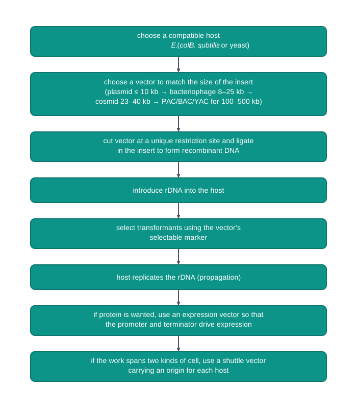
Fig. 2.22 – Summary of the gene cloning process.
2.8 Key Terms
Recombinant DNA (rDNA) technology — The technology of joining two different DNA molecules so that a gene can be isolated, propagated, characterised and manipulated.
Insert — The desired DNA molecule or target gene isolated from a source organism, which is to be carried by a vector.
Vector — A DNA molecule capable of replicating inside a host cell, used to carry an insert into that cell.
Recombinant DNA (rDNA) — The new DNA molecule formed when an insert is joined to a vector.
Host — The living organism into which recombinant DNA is introduced, and whose enzymes and proteins replicate it.
Gene cloning — The whole process of inserting a target gene into a vector, introducing it into a compatible host, and allowing the host to propagate and express it.
K12 strain — A long-established laboratory strain of E. coli that is one of the most commonly used hosts in gene cloning.
Origin of replication (ori) — The sequence on a vector at which the host’s replication machinery begins copying, making autonomous replication possible.
Unique restriction site — A restriction enzyme recognition sequence that occurs only once in a vector, so that the enzyme opens it at a single predictable point.
Selectable marker — A gene on a vector, such as \(tet^R\) or \(amp^R\), that allows cells which have taken up the vector to be identified and selected.
Transformant — A host cell that has taken up the recombinant DNA.
Plasmid — A circular, double stranded, extra-chromosomal DNA molecule capable of autonomous replication in a host cell.
Episome — A plasmid that is able to integrate into the host chromosome and be replicated along with it.
R-plasmid — A plasmid carrying genes for enzymes that deactivate antibiotics, thus conferring drug resistance on the host cell.
Col plasmid — A plasmid that produces toxins called colicins, which kill other bacteria.
F-plasmid — A plasmid carrying transfer genes whose products form a pilus, through which a copy of the plasmid can pass to another bacterial cell.
High or multi copy plasmid — A plasmid whose replication is free from the control of chromosomal replication, giving 10–30 copies per cell.
Relaxed replication — The replication of high copy number plasmids, which is independent of the control of bacterial chromosomal DNA replication.
Low or single-copy plasmid — A plasmid whose replication is under the same control as chromosomal replication, giving only one or a few copies per cell.
Stringent replication — The replication of low copy number plasmids, which is controlled along with bacterial chromosomal DNA replication.
pBR322 — A widely used plasmid cloning vector of 4,361 bp, carrying an ori, unique cloning sites and the selectable markers \(amp^R\) and \(tet^R\).
Multiple cloning site (MCS) — Also called a poly cloning site or polylinker; a short synthetic DNA fragment containing a large number of unique restriction sites.
Blue/white selection — A method of picking out recombinants using the β-galactosidase (lacZ) gene of E. coli, studied in detail in Chapter 3.
pUC19 — An improved plasmid cloning vector of 2.686 kb carrying ori, \(amp^R\) and part of lacZ with a multiple cloning site.
Bacteriophage — A virus that infects bacterial cells, and whose genome can be modified into a cloning vector.
Plaque — A clear patch of lysed bacterial cells on a bacterial lawn, produced by phage infection and used in screening recombinants.
Lambda (λ) phage — A bacteriophage of E. coli, with a head and tail and a 48,490 bp linear double stranded genome, widely used as a cloning vector.
Prophage — The λ genome after it has integrated into the host chromosome during the lysogenic pathway.
cos sequences — The 12-base single stranded cohesive ends at either end of the linear λ genome, which are complementary to each other.
cos site — The sealed cohesive end formed when the two cos sequences pair and are ligated inside the host, circularising the λ genome.
Rolling circle replication — The mode of λ replication in which a template strand rotates, producing many genome copies joined end to end.
Concatemer — The long chain of λ genomes joined end to end and separated by cos sites, produced during rolling circle replication.
Insertion vector — A λ-based vector having a single restriction enzyme site at which insert DNA is added.
Replacement vector — A λ-based vector having a pair of cloning sites flanking a non-essential central stuffer region, which is replaced by the insert.
Central stuffer region — The segment of non-essential λ DNA lying between the two cloning sites of a replacement vector.
M13 — A filamentous bacteriophage of E. coli with a circular single stranded genome of about 6.4 kb, which infects only cells harbouring the F plasmid.
Replicative form (RF) — The double stranded circular form of the M13 genome produced inside the host, and the form used for M13 vector construction.
Intergenic region — The small non-essential region of the M13 genome flanking ori, into which the insert is placed.
M13mp18 — An M13 based vector for E. coli that facilitates blue/white selection of recombinants.
Cosmid — A hybrid vector that replicates like a plasmid but can be packaged in vitro into lambda phage coats, accommodating inserts as large as 45 kbp.
Terminase — The enzyme that binds the cos junction and cleaves it during packaging of DNA into a phage head.
Phasmid (phagemid) — A true combination vector whose ends are λ DNA carrying all genes for lytic infection and whose middle region is linearised plasmid.
Intron — A non-coding stretch within a eukaryotic gene, which may be hundreds of kilobases long and which a prokaryotic host cannot remove.
Artificial chromosome — A vector built with chromosome-like parts so that it can carry huge lengths of eukaryotic DNA.
Saccharomyces cerevisiae — Baker’s yeast; the most commonly used eukaryotic host, which multiplies by budding and grows much as E. coli does.
GRAS — ‘Generally Recognised As Safe’; the category to which S. cerevisiae belongs because of its long history of safe use in the food industry.
2 µm plasmid — A double stranded circular plasmid found in S. cerevisiae, from which a number of yeast vectors have been developed.
Yeast Artificial Chromosome (YAC) — A high capacity vector containing two yeast telomeric sequences, a yeast centromeric sequence, a yeast ARS and selectable markers, carrying inserts of 200–500 kb.
Telomere — The sequence at the end of a chromosome, two copies of which are present in a YAC to protect its linear ends.
ARS (autonomously replicating sequence) — The yeast sequence at which DNA replication begins, serving as the origin of replication in a YAC.
High capacity cloning vector — A vector such as a YAC, BAC or PAC, designed to carry very large DNA inserts.
Bacterial Artificial Chromosome (BAC) — A high capacity cloning vector derived from a bacterial plasmid, taking inserts up to 300 kb.
Phage Artificial Chromosome (PAC) — A high capacity cloning vector derived from a phage, taking inserts of 100–300 kb.
Expression vector — A vector that not only carries and replicates an insert but also allows the insert to be expressed efficiently as protein.
Inducible promoter — A promoter that can be switched on when required, so that expression of the cloned gene is regulated.
Transcription termination sequence — The sequence provided near the 3’ end of the cloned gene to ensure that transcription stops correctly.
Expression cassette — The region of an expression vector in which the cloned gene lies sandwiched between an upstream promoter and a downstream terminator.
Sandwich expression vector — Another name for an expression vector, from the way the cloned gene is placed between promoter and terminator.
Heterologous gene expression — Expression of a gene in a host of a different group from its source, such as a eukaryotic gene expressed in a prokaryotic host.
Post-translational modification — Chemical finishing of a protein after translation, such as glycosylation, which prokaryotic hosts cannot perform.
Shuttle vector — A vector able to replicate in two alternative hosts, carrying two origins of replication of which only one is active in a given host.
2.9 Points to Ponder
A vector must be small enough to enter a host cell easily, yet large enough in capacity to carry a useful insert. These two demands pull in opposite directions. How far do you think a vector can be shrunk before it stops being useful, and what would be the first function you would refuse to delete?
Antibiotic resistance genes make convenient selectable markers, and they are used precisely because they work so well. Yet the same genes are the reason certain infections have become difficult to treat. Should this trouble us when such vectors are used routinely in laboratories, and what precautions would you argue for?
The λ head can package only a molecule of about 38 to 52 kbp. A physical container, in other words, decides how large a gene a scientist may clone in that system. Can you think of other places in biology where the size of a structure sets a limit on what is biologically possible?
Natural plasmids evolved for the benefit of bacteria, not of experimenters. Reflect on how much of genetic engineering consists of borrowing devices that evolution built for entirely different purposes.
A cosmid keeps only 250 bp of λ DNA, yet that fragment is enough to make the packaging machinery accept the whole molecule as a phage genome. What does this tell you about how recognition works at the molecular level — and about how little of a system may have to be copied for it to be fooled?
The K12 strain of E. coli is considered safe partly because long laboratory culture has left it unable to establish itself in the human gut. Is ‘weakened by domestication’ a sound basis for safety, or should safety be demonstrated afresh for every engineered strain?
Baker’s yeast is Generally Recognised As Safe because of centuries of use in bread and brewing. When such an organism is genetically modified, how much of that inherited trust should carry over to the new strain?
Expression vectors usually carry inducible promoters, so that the cloned gene stays switched off until the culture is ready. Consider why a cell might be harmed by making too much of a perfectly ordinary protein, and what this suggests about the cost of gene expression.
Bacteria cannot glycosylate proteins, so certain human proteins can never be produced in E. coli. Does this limitation argue for developing better bacterial hosts, or for accepting that some products simply require eukaryotic cells?
A shuttle vector carries two origins of replication, one of which is always idle. In engineering, carrying unused parts is usually considered waste. Why is it justified here, and can you think of a biological system that similarly carries capacity it uses only occasionally?
Table 2.2 shows the insert capacity of vectors rising from ten kilobases to five hundred. Each new class of vector was invented because the previous one ran out of room. What limitation of today’s methods do you think the next generation of vectors will be designed to overcome?
2.10 NCERT Questions
Describe the importance of host vector system in rDNA technology.
What are the major characteristics of a vector?
What is plasmid and what are its different types?
Discuss the strategy applied for the development of (pBR322) plasmid cloning vectors.
Briefly describe the structure of lambda bacteriophage and also discuss the role of lambda phage based vectors.
Discuss the M13 based vectors and its application.
Differentiate between cosmids and phagemids.
Why is a vector required for cloning of a gene?
A plasmid capable of getting integrated into host chromosome is called:
- Col plasmid
- Episome
- Ti plasmid
- R plasmid
Why the replication of single copy plasmid called stringent replication?
Identify the incorrect match pair from the following:
| Column A | Column B |
|---|---|
| a. Multicopy plasmid | i. Stringent replication |
| b. Col plasmid | ii. Kills bacteria |
| c. pBR322 | iii. Plasmid |
| d. Prophage | iv. Phage genome inserted into a host genome |
How can a large size eukaryotic gene insert be cloned?
Assertion: An ideal vector should have selectable marker. Reason: Selectable markers are required to screen out transformation.
- Both assertion and reason are true and the reason is the correct explanation of the assertion.
- Both assertion and reason are true but the reason is not the correct explanation of the assertion.
- Assertion is true but reason is false.
- Both assertion and reason are false.
- Assertion: Cosmid is a hybrid vector. Reason: Cosmid has properties of both plasmids and lambda phage vector.
- Both assertion and reason are true and the reason is the correct explanation of the assertion.
- Both assertion and reason are true but the reason is not the correct explanation of the assertion.
- Assertion is true but reason is false.
- Both assertion and reason are false.
2.11 Objective Questions
- The vector pBR322 has a size of:
- 2.686 kb
- 4,361 bp
- 9 kb
- 48,502 bp
- The number of copies of a high or multi copy plasmid present per cell is:
- 1
- 2–5
- 10–30
- 200–500
- Replication of low or single-copy plasmids is referred to as:
- Relaxed replication
- Rolling circle replication
- Stringent replication
- Autonomous replication
- A plasmid that produces toxins which kill other bacteria is called:
- R-plasmid
- F-plasmid
- Col plasmid
- 2 µm plasmid
- Plasmids that carry transfer genes encoding proteins which form a pilus are:
- F-plasmids
- Col plasmids
- R-plasmids
- Episomes
- A short synthetic DNA fragment containing a large number of unique restriction sites is called:
- Central stuffer region
- Multiple cloning site
- Intergenic region
- Expression cassette
- The linear λ genome inside the phage head contains:
- 6,407 base pairs
- 48,490 base pairs
- 48,502 base pairs
- 4,361 base pairs
- In its circular form, the λ phage genome is 48,502 base pairs long because:
- Twelve base pairs are synthesised afresh by the host
- The 12-nucleotide single stranded ends at both sides pair to form double stranded DNA
- A 12 bp fragment of host DNA is added at the cos site
- Rolling circle replication adds 12 base pairs to each genome
- The cos sequences at each end of the linear λ genome are:
- 6 bases long and identical
- 12 bases long and complementary to each other
- 250 bases long and complementary to each other
- 12 base pairs long and double stranded
- During multiplication of λ, the long chain of many genome copies joined end to end and separated by cos sites is called a:
- Prophage
- Polylinker
- Concatemer
- Replicative form
- For efficient packaging into the λ head, the size of the recombinant DNA must lie between:
- 6 and 20 kbp
- 23 and 40 kbp
- 38 and 52 kbp
- 100 and 300 kbp
- λ EMBL3 is an example of a:
- Insertion vector selected by blue white screening
- Replacement vector selected by plaque formation
- Insertion vector selected by plaque formation
- Shuttle vector selected by antibiotic resistance
- Among the λ vectors listed in Table 2.1, the one selected on the basis of blue white screening is:
- λgt10
- λgt11
- λ EMBL3
- M13mp18
- Bacteriophage M13 can infect only those E. coli cells that harbour:
- The R plasmid
- The Col plasmid
- The F plasmid
- The 2 µm plasmid
- The genome of bacteriophage M13 is:
- Linear double stranded DNA of about 48 kb
- Circular single stranded DNA of about 6.4 kb
- Circular double stranded DNA of about 6.4 kb
- Linear single stranded RNA of about 6.4 kb
- For construction of an M13 based vector, the insert is placed in the:
- Central stuffer region
- Intergenic region flanking ori
- Gene encoding the major coat protein
- cos site
- The enzyme that binds to and cleaves the cos junction during packaging is:
- DNA ligase
- Restriction endonuclease
- Terminase
- DNA polymerase
- The minimum amount of λ DNA sufficient to provide a functional cos junction in a cosmid is about:
- 12 bp
- 250 bp
- 2,500 bp
- 45,000 bp
- Which of the following is a linear duplex DNA whose ends are lambda phage DNA and whose middle region is linearised plasmid?
- Cosmid
- Phasmid
- Replacement vector
- Shuttle vector
- A yeast artificial chromosome does NOT contain:
- A yeast centromeric sequence
- Two copies of a yeast telomeric sequence
- A yeast ARS
- A λ cos site
- The autonomously replicating sequence (ARS) of a YAC serves the same purpose as:
- The origin of replication of a plasmid
- The selectable marker of a plasmid
- The polylinker of pUC19
- The telomere of a chromosome
- The insert size that can be cloned in a YAC is:
- ≤ 10 kb
- 23–40 kb
- 100–300 kb
- 200–500 kb
- Baker’s yeast belongs to the category of GRAS organisms because:
- It is a single celled eukaryote
- It has a long history of safe use in the food industry
- It multiplies by budding
- It harbours the 2 µm plasmid
- The portion of an expression vector in which the cloned gene lies between an upstream promoter and a downstream terminator is called the:
- Polylinker
- Expression cassette
- Intergenic region
- Replicative form
- Heterologous expression of a eukaryotic gene in a prokaryotic host requires that the gene:
- Contains introns and codes for a glycosylated protein
- Lacks introns and codes for a protein needing no post-translational modification
- Lacks a promoter of its own and contains introns
- Is carried on a low copy number plasmid
- In a shuttle vector, at any given time in a particular host:
- Both origins of replication are active
- Only one origin of replication is active
- Neither origin is active until an inducer is added
- The origins act alternately in each cell cycle
Assertion: A cloning vector must possess unique restriction sites. Reason: If a vector possesses too many restriction sites, it would be fragmented into several pieces on digestion.
Assertion: The vector pBR322 was constructed by shortening pBR313. Reason: More than half of the DNA of pBR313 was not essential for its role as a vector.
Assertion: Use of a high copy number plasmid gives stronger signals during screening of recombinants. Reason: Replication of high copy number plasmids is under the same control as replication of the bacterial chromosomal DNA.
Assertion: M13 based vectors do not kill the host cell. Reason: The single stranded genomic DNA of M13 is assembled with capsid proteins during its passage through the host cell, without causing cell lysis.
Assertion: A YAC must contain a yeast centromeric sequence. Reason: The centromere allows the artificial chromosome to be segregated properly into the daughter cells at division.
Assertion: Promoters used in expression vectors are generally inducible in nature. Reason: An inducible promoter allows the expression of the cloned gene to be regulated.
Assertion: A human protein that requires glycosylation for its activity cannot be obtained in functional form from E. coli. Reason: Prokaryotic hosts lack the machinery for post-translational modification of proteins.
Assertion: Shuttle vectors carry two origins of replication. Reason: Both origins function simultaneously in whichever host the vector occupies.
2.12 Check Your Concepts
Why is a host described as one half of a “two component system”? State clearly what the vector contributes and what the host contributes to gene cloning.
A researcher introduces a purified gene fragment directly into E. coli without ligating it into any vector. The cells are healthy and take up the DNA, yet no clone is obtained. Give two reasons for this failure.
Explain why a host strain whose restriction system is intact is unsuitable for gene cloning.
A vector is small in size, carries an ori and has a single unique restriction site, but has no selectable marker. The ligation and transformation steps work perfectly. What difficulty will the experimenter face at the next stage, and why?
Why must the restriction sites on a vector be unique? Describe what would happen to the vector if the chosen enzyme cut it at four different places.
Both E. coli and Bacillus subtilis are used as prokaryotic hosts. For a project in which the protein product must be harvested from the culture medium rather than from inside the cells, which host would you choose, and why?
Compare high copy number plasmids and low copy number plasmids with respect to control of replication, copies per cell, and yield of cloned DNA. Under what circumstance would a low copy plasmid actually be preferable?
Why is the replication of a single copy plasmid described as “stringent”, while that of a multicopy plasmid is described as “relaxed”? Base your answer on what controls each type of replication.
Classify plasmids on the basis of the genes they carry, giving the characteristic property of each type. Which of these types is most directly useful in constructing a cloning vector, and why?
An episome and an ordinary plasmid are both present in a bacterial cell. Explain how their behaviour differs during the life of the cell.
Trace the development of plasmid based vectors from pBR313 to pUC19. At each stage, state the problem that was being solved.
Draw a labelled map of pBR322 and mark on it the origin of replication, the two selectable marker genes, and any three unique cloning sites.
Why did reducing the size of pBR313 from 9 kb to the 4,361 bp of pBR322 make it a better vector? Give two distinct advantages of the smaller size.
What is a polylinker, and why is a cluster of many unique restriction sites more useful to an experimenter than a single cloning site?
State two reasons why bacteriophage vectors are considered more efficient than plasmid vectors.
Distinguish between the lytic and lysogenic cycles of λ phage. Which of the two is exploited when λ is used as a cloning vector, and why is the other one dispensable?
The linear λ genome is 48,490 bp long, while the circular form is 48,502 bp. Account for this difference of 12 base pairs.
Describe the two distinct roles played by the cos sequences in the life cycle of λ phage.
Arrange the following steps of λ multiplication in the correct sequence: packaging of one genome into an empty head; formation of a concatemer; rolling circle replication; release of phage particles by lysis; cleavage at cos sites; attachment of the tail.
Why can only a limited size range of recombinant DNA be cloned in a λ vector? Explain what happens if the recombinant molecule is much shorter than 38 kbp.
Differentiate between insertion vectors and replacement vectors. Why does a replacement vector accept a larger insert than an insertion vector?
M13 infects only those E. coli cells that carry the F plasmid. Explain the connection between the F plasmid and the ability of M13 to attach to the cell.
Why is the double stranded replicative form of M13, rather than its single stranded genome, used for vector construction?
M13 releases progeny phage without lysing the host cell. State one practical advantage that this gives the experimenter.
Why is the insert placed specifically in the intergenic region of the M13 genome and nowhere else?
A cosmid carries only about 250 bp of λ DNA, yet it can be packaged into a λ phage head. Explain how so small a fragment is sufficient.
A cosmid is packaged into a phage coat and used to infect E. coli, yet no plaques appear on the bacterial lawn. Explain why this is the expected result and not a failure of the experiment.
Differentiate between a cosmid and a phasmid with respect to shape, the λ material each carries, and the way each replicates inside the host.
A phasmid vector carrying the gene for the lambda repressor is introduced into E. coli. Predict whether it will replicate as a phage or as a plasmid, and justify your answer.
Why can an ordinary plasmid not be used to clone a complete eukaryotic gene? Give both reasons discussed in the chapter.
Name the four components of a YAC vector and state the function of each. Which of them would you expect to be unnecessary if the vector were kept permanently in circular form inside E. coli?
Why does a YAC have to be linearised before it is used in yeast, when the very same molecule is grown as a circle in bacteria?
Match the following:
| Column A | Column B |
|---|---|
| a. ARS | i. Protects the ends of the linear vector |
| b. Centromeric sequence | ii. Site at which DNA replication begins in yeast |
| c. Telomeric sequence | iii. Allows selection of transformed yeast cells |
| d. URA3 | iv. Ensures correct segregation into daughter cells |
Arrange the following vectors in increasing order of the insert size they can accommodate: cosmid, YAC, plasmid, BAC, bacteriophage.
A fragment of 30 kb is to be cloned. Using Table 2.2, name two vectors that could be used and justify why a plasmid could not.
Compare a cloning vector with an expression vector. Name three features present in an expression vector that a simple cloning vector does not need.
Why are the promoters of expression vectors usually inducible rather than constantly active? Explain what could go wrong if a foreign protein were expressed continuously from the moment of transformation.
What is the function of a transcription termination sequence in an expression vector? Predict what would happen to transcription if it were absent.
A cloned gene is inserted into an expression vector in the reverse orientation. Transformants are obtained normally but no protein is detected. Explain both observations.
State the two conditions that a eukaryotic gene must satisfy before it can be expressed in a prokaryotic host, and explain the reason behind each.
A team clones a eukaryotic gene straight from genomic DNA into an E. coli expression vector and obtains a protein of the wrong size. Identify the mistake in the protocol and suggest the correction.
Why are most eukaryotic vectors also shuttle vectors? Explain the practical advantage of building the recombinant molecule in E. coli before moving it into a eukaryotic cell.
A shuttle vector carries two origins of replication, but only one works in a given host. Explain why the idle origin is nevertheless retained on the molecule.
Draw a labelled diagram of a shuttle vector for E. coli and yeast, marking the two origins of replication, the selectable marker for each host, and the multiple cloning site.
A student proposes the following protocol: “Digest the vector with an enzyme that cuts it at three sites, ligate the insert, transform a wild bacterial strain, and select the colonies on a plain nutrient agar plate.” Identify three separate errors in this protocol and correct each one.
2.13 Case-Based Questions
- A laboratory receives a 32 kb fragment of human DNA that must be cloned intact. A junior worker begins by digesting pUC19 with EcoRI, ligating the fragment into the polylinker, and transforming E. coli. Several white colonies appear on the selection plate, but when the plasmid DNA is isolated from them, the inserts recovered are only 2–4 kb long.
- Explain why pUC19 was an unsuitable choice for this particular fragment.
- Using Table 2.2, name two vectors that could carry a 32 kb insert, and state the insert range of each.
- What does the appearance of white colonies indicate about the vector that was used, even though the experiment failed?
- A team wishes to produce a human hormone in bacteria. The hormone is a small polypeptide that is biologically active as soon as it is folded, and requires no sugar residues. The gene is first cloned from genomic DNA into an expression vector carrying an inducible promoter, a ribosome binding site, a polylinker and a terminator. The culture grows well, the promoter is induced, and a protein is produced — but it is much larger than expected and shows no hormonal activity.
- Identify the most likely reason for the incorrect size of the protein.
- What form of the gene should have been cloned instead, and why does that form solve the problem?
- State the two conditions that must be satisfied for heterologous expression of a eukaryotic gene in a prokaryotic host.
- If the hormone had required glycosylation for activity, would changing the form of the gene have helped? Justify your answer.
- Two students carry out the same cloning experiment. Student A uses a well-characterised E. coli K12 laboratory strain as host. Student B, wanting a “stronger” bacterium, isolates a wild E. coli strain from a soil sample and uses it instead. Both plate their transformation mixtures on ampicillin agar, since the vector carries \(amp^R\). Student A obtains a good number of colonies; Student B obtains almost none.
- Give the most likely explanation for Student B’s result, in terms of what a host must and must not do.
- Why is the K12 strain preferred as a cloning host, both for reliability and for safety?
- What is the purpose of plating the cells on ampicillin agar rather than on plain nutrient agar?
- A worker is constructing a genomic library in a λ replacement vector. The two essential arms of the vector together measure 31 kbp, and the recombinant molecule must be between 38 and 52 kbp for efficient packaging. In one batch the ligation is carried out badly, and many molecules are formed in which the two arms have simply joined to each other with no insert between them. When these are packaged and plated, very few plaques appear.
- Calculate the smallest and the largest insert, in kbp, that this vector can accept.
- Explain why the arms-only molecules fail to give plaques.
- Why is the packaging limit described as a physical constraint rather than a biochemical one?
- Name the region of the λ genome that is removed in a replacement vector, and state why its removal does not prevent lytic infection.
- A genome sequencing centre is preparing to analyse the genome of a crop plant. The team decides to build the first-stage library in YACs, and to keep and manipulate the vector in E. coli before transferring it into Saccharomyces cerevisiae. Before the yeast step, the circular vector is digested with BamHI.
- Why were YACs chosen in preference to cosmids for this first-stage library?
- Explain the purpose of the BamHI digestion step.
- Name the three chromosome-like components of the YAC and state the function of each.
- Why does the vector also carry a bacterial origin of replication and an antibiotic resistance gene, when the cloning is ultimately to be done in yeast?
- A food company plans to manufacture an enzyme used in bread-making by introducing the corresponding gene into Saccharomyces cerevisiae and growing the modified yeast in large fermenters. The company points out in its application that baker’s yeast is a GRAS organism with centuries of safe use, and argues that no fresh safety assessment of the modified strain should be required.
- Explain what GRAS status means and on what evidence it rests.
- Present one argument in favour of the company’s position and one argument against it.
- Why is it generally held that GRAS status attaches to the host organism rather than automatically to every modified strain derived from it?
- A student is given an M13 based vector, M13mp18, and is asked to clone a short DNA fragment into it. The student prepares single stranded phage DNA from the culture medium, attempts to digest it with a restriction enzyme and ligate the insert, and then transforms an E. coli strain that does not carry the F plasmid. No progeny phage are obtained, and no plaques appear.
- Identify two separate errors in this protocol.
- Which form of the M13 genome should have been used for vector construction, and why?
- Where in the M13 genome should the insert be placed, and what would be the consequence of placing it elsewhere?
- M13 releases progeny phage without lysing the host cell. State how this happens and one practical advantage it offers the experimenter.
2.14 Practice With Data
A single E. coli cell carrying a recombinant plasmid is inoculated into nutrient broth and grows under ideal conditions, doubling every 20 minutes. Using \(N = N_0 \times 2^n\), where \(N_0\) is the initial number of cells, \(n\) the number of generations and \(N\) the final number of cells, calculate the number of cells present after 5 hours. If each cell carries 20 copies of the plasmid, how many copies of the cloned gene are present in the culture at that time?
A high copy plasmid is maintained at 25 copies per cell and a low copy plasmid at 2 copies per cell. Two cultures of equal cell number are grown, one carrying each plasmid. Calculate the ratio of the total plasmid DNA recoverable from the two cultures, and state which type of replication each plasmid shows.
The vector pBR322 is 4,361 bp long and pUC19 is 2.686 kb long. Calculate the percentage reduction in vector size from pBR322 to pUC19. If both vectors were used to make recombinant molecules of the same total length, which would carry the larger insert, and by how many base pairs?
A single E. coli cell contains a chromosome of \(4.6 \times 10^6\) bp and 20 copies of pBR322 (4,361 bp each). Calculate the total plasmid DNA per cell in base pairs, and express it as a percentage of the total DNA in that cell.
The linear λ genome is 48,490 bp long and each end carries a 12-base single stranded cos sequence. After circularisation the genome measures 48,502 bp. Show by calculation how the second figure is obtained from the first, and state what happens to the 12 unpaired nucleotides at each end.
Approximately one-third of the 48,490 bp λ genome is dispensable for lytic infection. Calculate the approximate size of this dispensable region in kbp. Then, given that the recombinant molecule must lie between 38 and 52 kbp for efficient packaging, work out the largest insert that can be accommodated if the two essential arms together measure 32 kbp.
Study Table 2.1 and answer the following. (i) By what factor does the maximum insert size of λ EMBL3 exceed that of λgt10? (ii) Two of the three vectors are selected by plaque formation and one by blue white screening — identify them. (iii) Explain the relationship you observe between the type of vector and its maximum insert size.
Four DNA fragments are to be cloned: 8 kb, 35 kb, 120 kb and 450 kb. Using the insert sizes given in Table 2.2, select a suitable vector for each fragment. For which fragment is more than one vector suitable, and why do the ranges in the table overlap?
A genome of \(1.2 \times 10^5\) kb is to be covered by a clone library. Calculate the minimum number of clones required (i) if a cosmid with an average insert of 40 kb is used, and (ii) if a YAC with an average insert of 350 kb is used. Comment on what your two answers show about the value of high capacity cloning vectors.
A restriction enzyme with a six-base recognition sequence is expected, on average, to cut once in every \(4^6\) base pairs of random DNA. Calculate this interval. Then estimate the number of sites for such an enzyme expected in (i) pBR322 of 4,361 bp and (ii) the λ genome of 48,490 bp, and explain why this calculation supports the requirement that a cloning vector be small.
A cosmid contains 250 bp of λ DNA and accommodates an insert as large as 45 kbp. Express the λ contribution as a percentage of the total recombinant molecule, assuming the plasmid portion is 5 kbp. Comment on how so small a proportion of phage DNA is enough for the molecule to be packaged into a phage head.
In a transformation experiment, 0.1 µg of recombinant plasmid DNA is used and the following colony counts are obtained.
| Plate | Medium | Number of colonies |
|---|---|---|
| A | Nutrient agar, no antibiotic | Lawn, uncountable |
| B | Nutrient agar + ampicillin | 480 |
| C | Nutrient agar + ampicillin (no DNA added, control) | 0 |
- Calculate the transformation efficiency in transformants per microgram of DNA. (ii) Explain what plate A tells you and why it cannot be used to count transformants. (iii) State what conclusion would have to be drawn if plate C had shown 200 colonies.
- NCERT states that a DNA of more than 42 kb can be cloned by M13 bacteriophage, whose own genome is 6,407 bp. Express the insert as a multiple of the genome size, and state which region of the M13 genome the insert is placed in.