Chapter 6: Microbial Culture
In the early 1880s, in Robert Koch’s laboratory in Berlin, a young physician named Walther Hesse was stuck. He wanted to find out what kinds of bacteria were floating in city air, and for that he needed to grow them on a firm surface where each one could form its own separate patch. The jelly used in the laboratory at the time was gelatin, and gelatin kept letting him down. On warm days it softened and ran, mixing his carefully separated patches into one mess. Worse, several bacteria could digest gelatin itself, liquefying the very surface they were growing on.
He mentioned the problem at home. His wife, Angelina Fanny Hesse, who also drew the illustrations for his work, recognised it at once from her own kitchen. She set fruit jellies and puddings with agar-agar, an extract of seaweed that stayed firm even in summer heat. Walther tried it in the laboratory. The surface held, and the bacteria could not eat it.
Koch adopted agar, and it quickly became the standard base for growing microbes. More than a century later, almost every plate of bacteria grown in a hospital, a food-testing lab or a school laboratory still sits on a jelly that came out of a kitchen conversation.
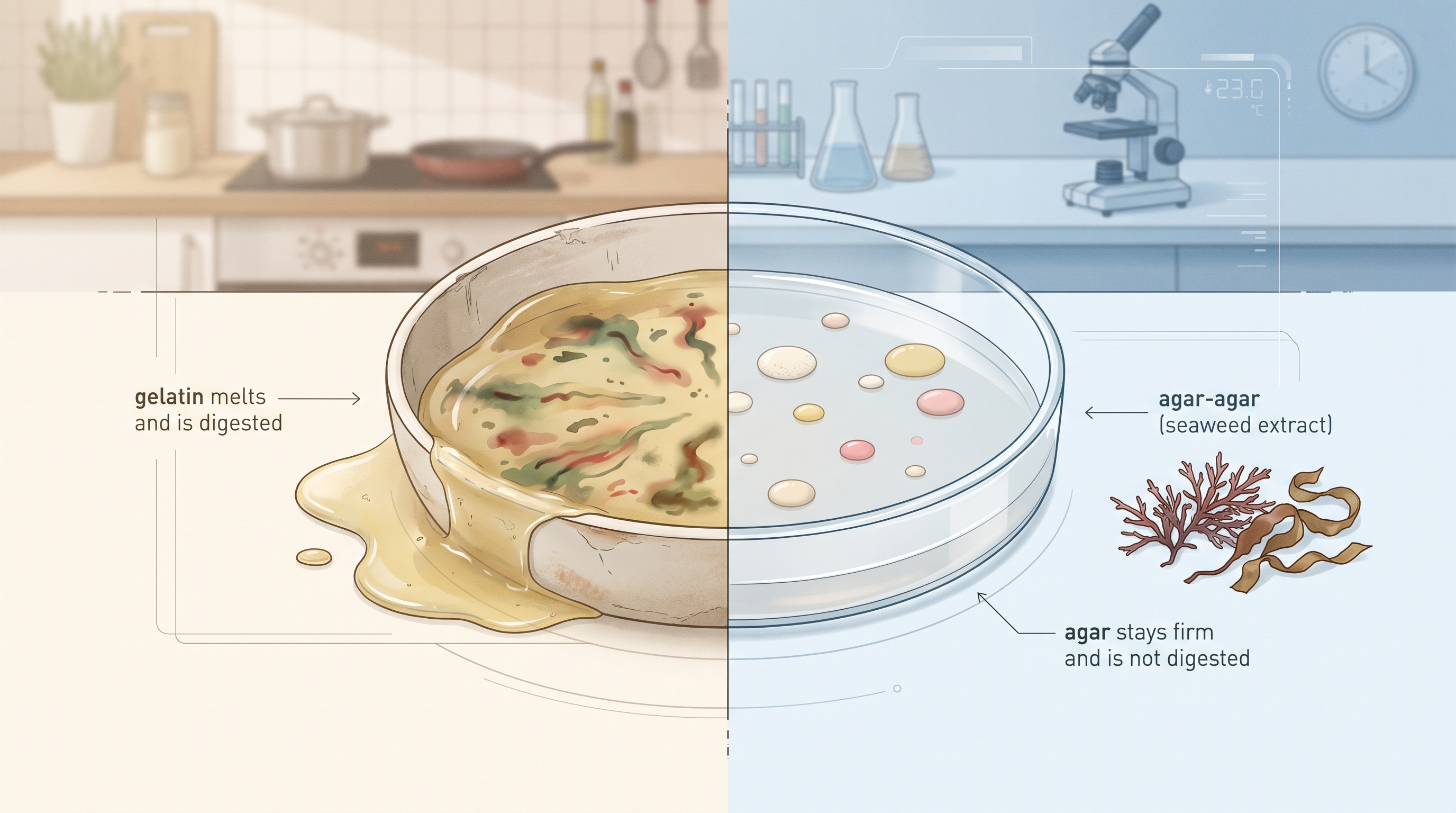
Fig. 6.0 – Agar and gelatin as culture media.
This chapter will answer each of these questions as you work through it.
6.1 Historical Perspective
The world of microorganisms is astonishingly diverse. Microbes differ from one another in structure, in the way they function, in the habitats they occupy and in the uses we have found for them. Some live in boiling springs, some in the cold of glaciers, some in soil, some in the human gut.
Microorganisms are ubiquitous in nature, which simply means that they are present everywhere — in air, in water, in soil, on the surfaces around you and on your own skin. The branch of science that studies them is microbiology: the study or science (logos) of small (micro) organisms (bios).
Microbiology today occupies a dominant position in fundamental research, in agriculture, in the pharmaceutical industry, in medicine, in environmental science, in food technology and in genetic engineering. Almost every branch of modern biotechnology rests on the ability to grow microbes reliably.
That ability is what this chapter is about. You will learn about the nutrition of microorganisms, culture media used to grow them, the sterilisation techniques that keep those media free of unwanted organisms, and the growth curve that describes how a microbial population changes with time.
Before any of this became routine, however, someone had to prove that microorganisms exist at all, and that they come from other microorganisms rather than appearing out of nowhere. That is where the story begins.
The First Sight of a Microbial World
The discovery of the microscope during the mid 1600s laid the foundation of microbiology. Without a way of magnifying what the eye cannot resolve, an entire kingdom of life simply remained invisible.
In the 1670s to 1680s, a Dutch merchant named Antonie van Leeuwenhoek developed the microscope further, examined drops of pond water, saliva and other everyday samples, and observed tiny moving organisms in them. He called these creatures ‘animalcules’, meaning ‘little animals’.
After the era of Leeuwenhoek, the development of microbiology was slow. Microscopes were rare and difficult to obtain, and there was little interest in these newly seen organisms. A discovery only grows into a science when many people can repeat it, and for a long time very few could.
The Spontaneous Generation Debate
Scientists at that time believed that microorganisms originate from lifeless matter. This idea — that living things arise on their own from non-living material, such as maggots forming out of decaying meat or microbes forming inside a broth — is called the theory of spontaneous generation.
Evidence against the idea began to appear. Lazzaro Spallanzani observed that there were no microscopic forms of life in boiled broth. His result suggested something important: if boiling removes the microbes and none reappear, then the broth itself is not producing them.
Supporters of spontaneous generation had a ready objection. They argued that boiling and sealing destroyed some ‘vital force’ in the air that living matter needed. To settle the argument, an experiment was required that allowed air to reach the broth freely while still keeping microorganisms out. That is exactly what Louis Pasteur designed.
In the middle and late 1800s, Louis Pasteur performed experiments to prove the importance of microorganisms in everyday life, and encouraged scientists to think about the role of bacteria in human illness. His famous flask experiment, shown in Fig. 6.1, is worth following step by step.
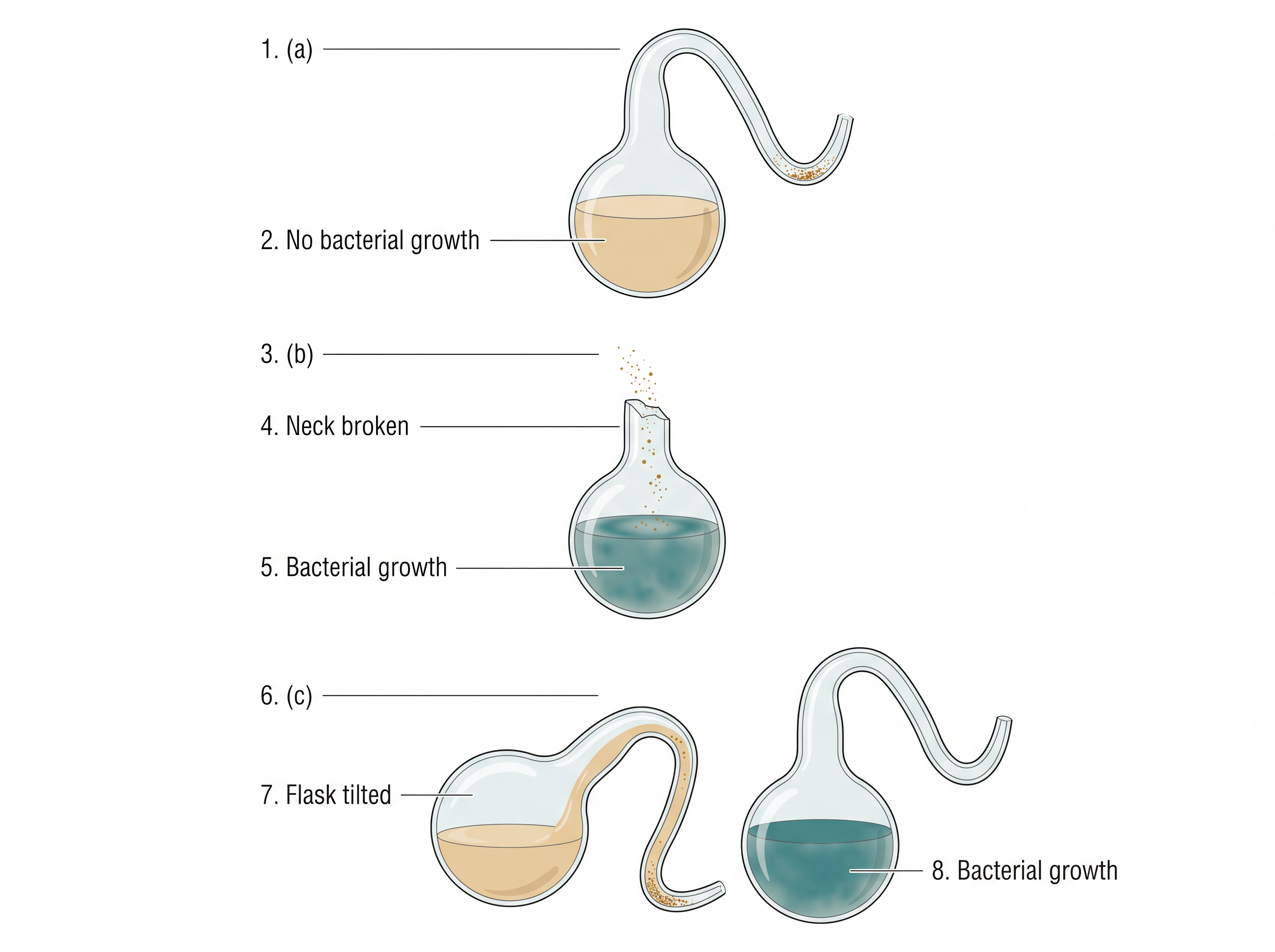
Fig. 6.1 – Pasteur’s swan-neck flask experiment.
Notice how carefully the experiment is designed. The three flasks differ in only one respect — whether or not outside dust can reach the broth. Everything else, including the broth, the boiling and the exposure to air, is identical. This is the logic of a controlled experiment, and it is the reason Pasteur’s result was accepted where earlier ones were argued away.
Koch and the Proof from Pure Cultures
Pasteur showed that microbes come from outside and can cause disease in general. The German scientist Robert Koch went one step further and linked one particular microbe to one particular disease.
Koch further proved the germ theory by injecting pure cultures of bacilli — rod-shaped bacteria — into mice, and showed that these bacilli caused anthrax. A pure culture, as you will see in Section 6.4, means a culture containing only one kind of organism, so there can be no doubt about which microbe caused the illness.
Accordingly, Koch postulated and provided a set of principles that implicated microorganisms as the causative agents of diseases. The reasoning behind his experiments is shown in Fig. 6.2, and the sequence he followed is set out in Fig. 6.3.
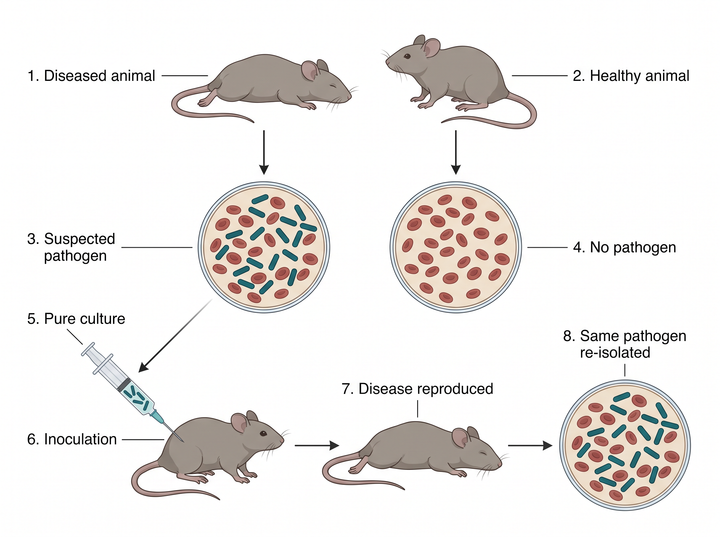
Fig. 6.2 – Koch’s experimental proof of the germ theory.

Fig. 6.3 – Koch’s experimental sequence.
The final step of that chain is the one that matters most. Re-isolating the same organism from the newly diseased animal closes the loop and rules out the possibility that something else in the injection caused the illness.
The Golden Age of Microbiology
In the course of the development of microbiology, based on the suggestions made by Pasteur and Koch, the period of the late 1800s and the early decade of the 1900s is referred to as the Golden Age of Microbiology.
During this period, the causative agents responsible for a large number of microbial diseases were discovered, one after another. Once the method was clear — isolate, culture, test, re-isolate — the discoveries followed quickly.
Early Culture Media
Discovering a microbe is one thing; keeping it alive in the laboratory is another. Through the 19th century, bacteriologists attempted to use various foods or materials to grow microbes.
In 1860, Louis Pasteur used a medium containing yeast, ash and ammonium salts. This medium contained the basic nutrients needed for microbial growth: a carbon source (for example, sugar), nitrogen (for example, ammonium salts) and vitamins (for example, ash).
Each ingredient was there for a reason, and that logic still governs media design today. Carbon supplies the framework of every organic molecule the cell builds; nitrogen is needed for proteins and nucleic acids; vitamins act as helpers in enzyme reactions. You will meet these requirements in full in Section 6.2.
In order to develop a more versatile basal medium, Ferdinand Cohn further refined Pasteur’s medium by varying the types of sugars used. Robert Koch observed that broths based on fresh beef serum or meat extracts could be used for optimal growth.
From Broth to Solid Media
The use of solid media instead of liquid medium also proved an important milestone in the development of bacteriology. In a broth, all the organisms mix freely; on a solid surface, each organism stays where it lands and grows into a separate visible patch.
In 1881, Robert Koch had recognised the difficulties of using broth media for the isolation of pure cultures and considered the use of solid media alternatives.
During the isolation of Bacillus anthracis, Koch evaluated media with coagulated egg albumin, starch paste and an aseptically cut slice of a potato, but these proved to be relatively poor at recovering the pathogenic bacteria.
After that, Koch further developed a medium with meat extract and gelatin, gelatin being derived from the collagen of skin and bones. During the course of development, agar finally replaced gelatin.
Agar provides the requisite solid support for microbiological media, but it does not provide the nutrients necessary for the bacteria to grow. That combination — firm support without nutrition of its own — is precisely what makes it valuable, because the person preparing the medium keeps complete control over what nutrients the microbes actually receive.
On the basis of his observations of the growth of bacteria on solid media, Koch coined the term ‘colony’ to describe the pure and discrete growth seen on the surface.
Media That Choose Their Microbes
Most of the media used during the 19th century were non-selective, and were designed to grow a wide range of bacteria. Such media are useful when you want to see everything that is present, but unhelpful when you want to pick out one organism from a crowded mixture.
At the beginning of the 20th century, further advances in culture media occurred, which led to the isolation of pure culture on media called selective media. Such media contained compounds which facilitate the growth of only a specific type of microorganism.
The idea is straightforward: instead of trying to separate organisms physically, you change the chemistry of the medium so that only the organism you want can flourish in it. The different families of media built on this idea are dealt with in Section 6.2.
Twentieth-Century Milestones
By the 1930s, researchers started to consider the importance of growth factors in bacterial nutrition — organic substances that certain microbes cannot make for themselves and must be given ready-made.
In the 1940s, with the invention of the electron microscope, the study and culture methods for viruses were introduced, and knowledge of viruses developed rapidly. Viruses are far too small to be resolved by a light microscope, so this was a case of a new instrument opening a new field.
By the 1950s, the mechanisms of coenzymes in bacterial growth were found to be useful. Another important development was the use of antibiotics in media, and by the 1960s antibiotics were being used in culture media as selective agents.
After World War II, further advancement of microbiology led to the discovery of antibiotics as medicines for various diseases, such as pneumonia, tuberculosis, meningitis and syphilis. During the period of the 1950s and 1960s, many vaccines against viral diseases — polio, measles, mumps and rubella — were developed. The main sequence of these milestones is collected in Fig. 6.4.
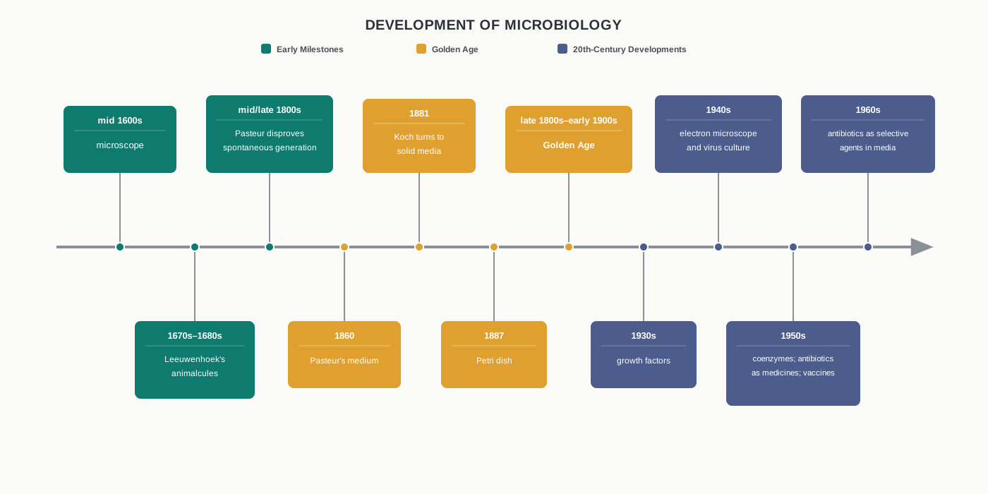
Fig. 6.4 – Milestones in microbiology.
Microbiology Today
Modern microbiology has applications in many fields: the development of pharmaceutical products, food and dairy products, the control of disease-causing microorganisms, and the industrial exploitation of microorganisms.
Microorganisms are used to produce vitamins, amino acids, enzymes and growth supplements, as well as a range of foods — fermented dairy products such as sour cream and buttermilk, pickles, breads and alcoholic beverages. The idli batter fermenting in a kitchen overnight and the curd setting in a bowl are the same science running without a laboratory.
In biotechnology, applied microbiology has a dominant role, because microorganisms are used as living factories to produce pharmaceuticals. These include the human hormone insulin, the antiviral protein interferon, numerous blood-clotting factors and clot-dissolving enzymes, as well as a number of vaccines.
For rDNA technology — recombinant DNA technology, in which DNA from two different sources is joined together — genetically engineered microorganisms are used as host vectors in order to develop genetically modified organisms (GMOs) with improved traits.
Every one of these applications depends on being able to grow the chosen microbe reliably, in a medium of known composition, free from contamination. That is why a chapter on microbial culture sits at the base of biotechnology.
Nutritional Categories of Microbes
Microorganisms can be categorised on the basis of how they obtain two things: their carbon and their energy. Keeping these two questions separate is the key to the whole classification.
Most microorganisms obtain their carbon in the form of organic molecules, derived directly or indirectly from other organisms, and are described as heterotrophs. Heterotrophs include all the fungi and protozoans, as well as most types of bacteria.
Microorganisms that obtain their carbon from carbon dioxide are called autotrophs. Autotrophs include a significant number of bacteria and almost all of the algae.
Autotrophs may further be of two categories. Chemoautotrophs obtain their energy as well as their carbon from inorganic sources, by the oxidation of inorganic molecules such as sulphur or nitrite. Photoautotrophs have photosynthetic pigments and use them to convert light energy into chemical energy.
The majority of heterotrophs obtain energy by the chemical oxidation of organic nutrient molecules, and are thus called chemoheterotrophs. Although some heterotrophs — such as the green and purple non-sulphur bacteria — obtain light energy from photosynthesis, they still get their carbon from organic nutrients, and hence are known as photoheterotrophs.
A third question can also be asked about a microbe: where does it get its electrons? A lithotroph is an organism that uses inorganic molecules, such as \(\mathrm{H_2O}\), \(\mathrm{H_2S}\) or ammonia, as a source of electrons, while an organotroph uses organic molecules for the same purpose.
These categories are not merely labels to be memorised. They tell you what to put into a culture medium. A photoautotroph will need light and a supply of carbon dioxide, and will not need sugar; a chemoheterotroph will need an organic carbon source and no light at all. Board and entrance examinations frequently test this link by giving the growth requirements of an unnamed organism and asking you to name its nutritional category, or the reverse.
6.2 Nutritional Requirements and Culture Media
A microbe in a flask is doing the same thing a microbe in soil or in the gut does: taking in raw materials, releasing energy from them, and building more of itself. If we want that to happen in the laboratory, we must supply every raw material it needs.
Microorganisms require nutrients that are essential for energy production as well as for their growth and multiplication. Some of these are needed in large quantities, others in traces, but a shortage of any one of them will stall growth just as surely as a shortage of all of them.
Macronutrients and Micronutrients
Carbon, oxygen, hydrogen, nitrogen, sulfur, phosphorus, potassium, calcium, magnesium and iron are called macronutrients, and they are required in relatively large amounts.
Among them, C, O, H, N, S and P are the components of carbohydrates, lipids, proteins and nucleic acids — in other words, they make up the actual substance of the cell. K, Ca, Mg and Fe play a variety of roles, being involved as cofactors of various enzymes.
A cofactor is a non-protein partner that an enzyme needs in order to work. This is why a metal required in milligram quantities can still halt growth completely if it is missing: without it, whole enzyme systems simply stop.
Besides macronutrients, some micronutrients or trace elements, such as Mn, Zn, Co, Mo, Ni and Cu, are also required for microbial growth in small amounts.
These micronutrients are ubiquitous in nature and already exist along with various regular media components, so they are not usually supplied separately. In practice, the tap of impurities in ordinary chemicals and extracts provides enough of them.
Components of a Culture Medium
The essential nutrients have to be supplied in the form of a nutrient medium. A culture medium is simply a prepared mixture, liquid or solid, that contains everything a chosen microbe needs to grow. The major components required for the medium are described below, in the order in which a person preparing a medium would think about them.
Carbon Source
Carbon is required for the skeleton or backbone of all organic molecules, and the carbon sources usually also provide oxygen and hydrogen atoms. Supplying carbon therefore takes care of three macronutrients at once.
In microbial culture media, a variety of carbon sources are used — for example, glucose, lactose, sucrose, starch, glycogen and cellulose, as well as mixtures of various carbohydrates such as cereal grain powders and cane molasses.
The choice is not arbitrary. A microbe can only use a carbon source it possesses the enzymes to break down, which is why the same sugar can be a feast for one organism and useless to another. Industrial media often use cane molasses simply because it is a cheap, rich mixture of sugars available in bulk.
Nitrogen, Phosphorus and Sulphur Sources
A nitrogen source is required for the biosynthesis of amino acids, nucleic acids and enzymes. The ammonium salts, urea, animal tissue extracts, amino acid mixtures and plant-tissue extracts are the main nitrogen sources used in culture media.
Phosphorus is present in nucleic acids, phospholipids, nucleotides, several cofactors and other cellular components. Almost all microorganisms use inorganic phosphate as their phosphorus source.
Sulfur is required for the biosynthesis of various amino acids, such as cysteine and methionine, and of some carbohydrates. Most microorganisms use sulfate as a source of sulfur.
Notice the pattern here: the cell can usually take a simple inorganic ion — ammonium, phosphate, sulfate — and build it into complex organic molecules itself. That is why a medium does not need to supply ready-made amino acids to every organism.
Growth Factors
Growth factors are organic compounds — for example, certain amino acids, purines, pyrimidines and vitamins — that cannot be synthesised in the cells and are required for the growth and multiplication of cells.
An organism that has lost the ability to make one of these compounds must be given it, or it will not grow at all, however rich the rest of the medium may be. Organisms with such extra requirements are described as fastidious, and they are the reason complex extracts are added to media.
Anti-foams
Anti-foams are not a nutritional component. However, media having nutritional components including starch, protein and other organic compounds, as well as products formed during cell growth, may show excessive foaming due to the agitation of the culture media.
To prevent this foam formation, some anti-foaming agents including olive oil, sunflower oil and silicones are included in the media.
Foam is a genuine problem rather than an untidy one. A thick foam layer blocks gas exchange, carries cells up into the neck of the vessel where they dry out, and can push contaminating organisms past a filter or a plug.
Water
Water is an essential component or base of any culture medium, whether liquid or solid. Every nutrient must be dissolved before a cell can take it up, and all the chemistry of the cell happens in water.
In solid culture media, the quantity of water used is comparatively less than in liquid media.
A culture medium, then, has all the nutrients, growth factors, energy source and so on required to support the growth of microorganisms.
Choosing a Medium for a Given Microbe
Though all microorganisms need sources of energy, carbon, nitrogen, phosphorus, sulfur and various minerals, the specific composition of a suitable medium will depend on the particular species that is to be grown in the laboratory.
Thus, for selecting an appropriate culture medium for a microorganism, prior knowledge of the habitat of the microorganism is useful, because its nutrient requirements are based on its natural surroundings.
Types of Media
Culture media can be sorted in three different ways: by what they are made of, by how firm they are, and by what they are used for. The full scheme is set out in Fig. 6.5. Note that the three schemes describe the same set of media from different angles, so one medium may appear in more than one category.

Fig. 6.5 – Classification of culture media.
Media Classified by Chemical Composition
On the basis of chemical composition, the culture media are classified into two types: synthetic or chemically defined media, and complex media.
Synthetic or Chemically Defined Media
A medium in which all chemical components are known is called a defined medium or synthetic medium. Every ingredient is a pure chemical, weighed out in a known amount.
Some microorganisms, specifically photolithotrophic autotrophs such as cyanobacteria, can be cultured on a medium containing \(\mathrm{CO_2}\) as a carbon source, nitrate or ammonia as a nitrogen source, along with sulfate, phosphate and a number of minerals.
A variety of heterotrophs may be cultured in a medium with glucose as a carbon source and an ammonium salt as a nitrogen source.
Defined media are widely used in research, and are prepared by mixing various components according to the requirements of the experimental microorganism. Their value lies in control: if you know exactly what went in, you can remove one ingredient and see precisely what its absence does.
M9 medium is widely used for the cultivation and maintenance of Escherichia coli (E. coli) cultures, and BG11 medium is used for the cultivation and maintenance of blue green algae (cyanobacteria).
Complex Media
Media that contain some constituents with unknown chemical composition are called complex media. Nobody can write down the exact formula of a beef extract, but that does not stop it from working.
These types of media are useful because even a single complex medium may have almost all the constituents needed to meet the nutritional requirements of a number of different microorganisms.
The undefined components in complex media are peptones, beef extract, yeast extract and so on.
Peptones are protein hydrolysates prepared by the partial proteolysis of meat, casein, gelatin and other protein sources, and serve as a source of carbon, energy and nitrogen. Partial proteolysis means the proteins are broken only part of the way down, into peptides and amino acids that cells can absorb directly.
The beef extract and yeast extract are aqueous extracts of beef and brewer’s yeast, respectively. Beef extract contains amino acids, peptides, nucleotides, organic acids, vitamins and minerals, whereas yeast extract is a rich source of vitamins, nitrogen as well as carbon compounds.
Examples of complex media are nutrient broth, tryptic soy broth and MacConkey agar. Lysogeny broth (Luria-Bertini) is a commonly used medium for culturing bacteria such as E. coli. The LB medium, first developed by Giuseppe Bertani (1951), consists of 10% tryptone/peptone, 5% yeast extract and 10% sodium chloride.
Potato dextrose agar (PDA) is used for the identification, cultivation and enumeration of various fungi including yeasts and moulds.
Media Classified by Consistency
On the basis of consistency, the culture media are of three types: liquid, solid and semi-solid. The difference between them comes down to one ingredient — how much agar is added.
Liquid Media or Broth
In liquid media, agar is not added. After inoculation and later incubation, the growth of cells becomes visible in the broth, which turns cloudy as the cell number rises.
Broths are used when large numbers of cells are wanted quickly, because the organism can spread through the whole volume rather than being confined to a surface.
Solid Media
For the surface cultivation of microorganisms, solid culture media are required. For the preparation of solid media, the liquid medium is solidified with the addition of 1.0–2.0% agar.
Agar is a sulphated polymer composed of D-galactose, 3,6-anhydro-L-galactose and D-glucuronic acid, and is generally extracted from red algae.
Solid media are used for making agar slants, slopes and agar stabs. A slant or slope is agar allowed to set at an angle in a tube, giving a larger surface for growth and long-term storage; an agar stab is a tube of set agar into which the inoculum is pushed with a straight wire.
Agar is preferred as a solidifying agent because most microorganisms cannot degrade it. Although other solidifying agents, such as silica gel, can also be used, none has replaced agar in routine work.
Semi-solid Agar Media
The semi-solid media are prepared by adding 0.5% of agar to the medium. The result is soft enough for motile organisms to move through, yet firm enough to hold its shape.
This type of medium may be selective, as it promotes the growth of one organism and may retard the growth of another organism.
Media Classified by Application and Function
On the basis of their applications and functions, the culture media are classified into selective media, differential media and enrichment media. Here the question is no longer what the medium is made of, but what job it is being asked to do.
Selective Media
A selective medium favours the growth of a particular type of microorganism. Such media provide nutrients that increase the growth of a particular type of microorganism and suppress all other microorganisms that are present in the culture.
This medium is useful for isolating a specific microorganism from a mixed population. Thus, for the isolation of cellulose-digesting bacteria, a medium containing only cellulose as a carbon and energy source is used — an organism that cannot digest cellulose finds no usable carbon at all and simply fails to grow.
Endo agar, eosin methylene blue agar and MacConkey agar media are used for the growth of E. coli and similar bacteria.
Certain media that contain bile salts or dyes, such as basic fuchsin and crystal violet, are used to suppress the growth of Gram-positive bacteria but encourage the growth of Gram-negative bacteria. The difference in the cell envelope between the two groups is what makes this possible: Gram-positive bacteria are more easily damaged by these dyes and salts.
Differential Media
Differential media are used to distinguish different groups of microorganisms based on their appearance on the medium as well as their biological characteristics, which permits the identification of microorganisms.
For example, if a mixture of bacteria is inoculated on to a blood-containing agar medium (blood agar), some of the bacteria may hemolyze — that is, destroy — the red blood cells, while others do not. Thus, this medium distinguishes between hemolytic and non-hemolytic bacteria. Therefore, blood agar medium is both a differential medium and an enriched one.
MacConkey agar is also kept under both differential and selective media. Certain indicators are present in this medium, which allow such differentiation.
For example, MacConkey’s agar contains lactose and a dye which changes colour when the pH drops. The microorganisms that ferment lactose produce an acid as the end product, which lowers the pH and produces a red colony. Microorganisms that fail to ferment lactose produce colourless colonies. This colour change is read as shown in Fig. 6.6.
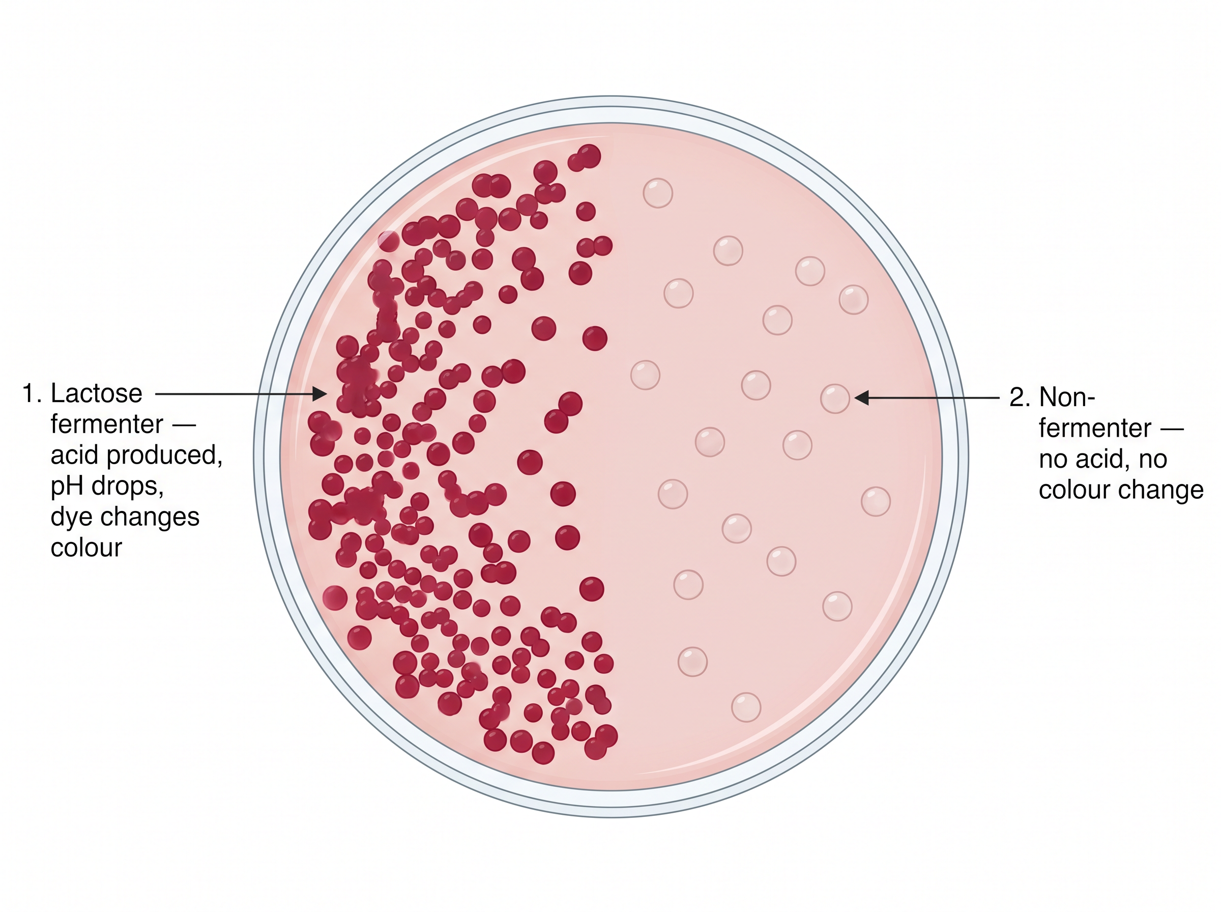
Fig. 6.6 – Differential growth on MacConkey agar.
The dye in MacConkey’s agar also inhibits the growth of Gram-positive bacteria. This medium is, therefore, both selective and differential — selective because only certain organisms grow on it, differential because those that grow can be told apart by their colour.
Enrichment Media
In an enrichment medium, the nutritional environment is adjusted in order to selectively enhance the growth of certain microorganisms within a mixed inoculum.
For example, the addition of plant and animal tissue extracts into nutrient broth and nutrient agar media provides additional nutrients, which enhance the growth of fastidious heterotrophic bacteria. Blood agar is also an enrichment medium.
The distinction from a selective medium is worth holding on to, because examinations test it often. A selective medium works mainly by holding back the organisms you do not want; an enrichment medium works by giving extra help to the organism you do want.
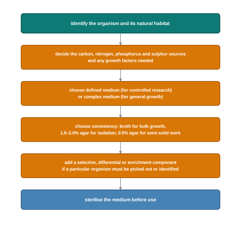
Fig. 6.7 – Selection of culture media.
6.3 Sterilisation Methods
A culture medium is nourishing for every microbe, not only for the one you intend to grow. Left to itself, it will grow whatever happens to land in it from the air, the bench or your fingers.
In any microbial study, therefore, all media, working surfaces, glassware and plasticware should be made free from contaminants by sterilisation. Without this step, every result is doubtful, because you can never be sure which organism produced it.
Two related words are often used alongside sterilisation, and examinations regularly ask you to separate them. Disinfection is the killing or removal of disease-causing microorganisms from non-living objects and surfaces, and it need not destroy spores. Sanitisation is the reduction of the microbial population to a level judged safe by public health standards, as when utensils and equipment are cleaned in a dairy or a canteen. A chemical applied to living tissue, such as skin, to prevent infection is called an antiseptic.
Usually, sterilisation can be achieved by both physical and chemical methods. In physical methods, sterilisation can be done by heat, radiation and filtration, while in chemical methods it is achieved using chemicals. Which one you choose depends almost entirely on what the article is made of and whether it can survive the treatment, as summarised in Fig. 6.8.
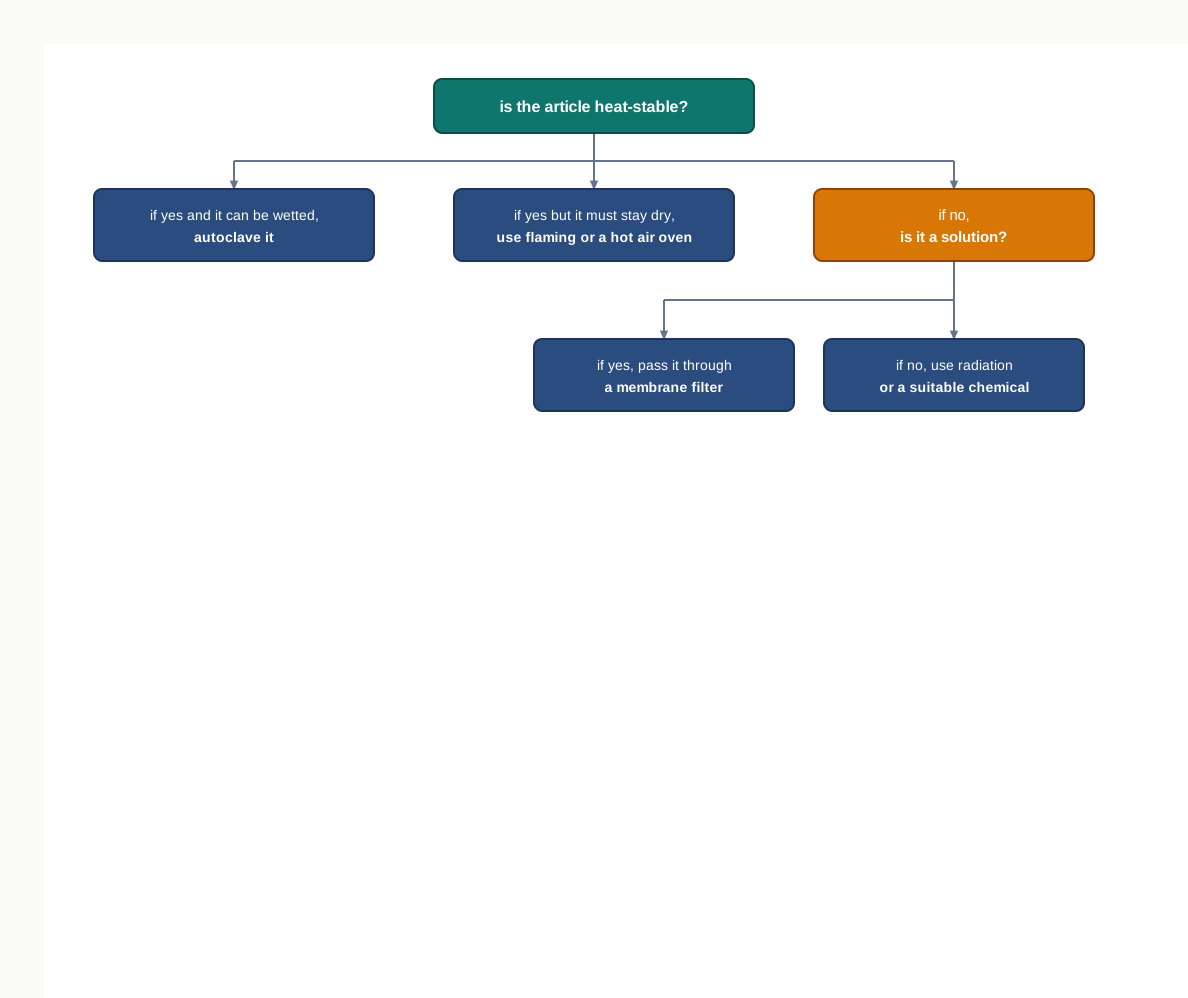
Fig. 6.8 – Selection of sterilisation methods.
Sterilisation by Heat
Sterilisation by heat is considered to be the most effective and rapid method for articles that can withstand heat.
Heat acts by the denaturation and coagulation of proteins, by exerting an oxidative effect, and by interfering with the metabolic reactions that lead to the death of cells. Denaturation means the unfolding of a protein from its working shape; once an enzyme has lost its shape, it can no longer catalyse anything, and the cell dies.
Sterilisation by heat is performed by various methods.
Boiling
Sterilisation can be done at 100°C for 30 min in a boiling water bath. Syringes, rubber goods and surgical instruments may be sterilised by this method.
Boiling is simple and needs no special equipment, but it has a limit that matters: at 100°C, resistant bacterial spores can survive. For work where nothing at all must be left alive, a higher temperature is required, and that is why the autoclave exists.
Autoclaving
Autoclaving is one of the most common methods for sterilisation in almost all laboratories. In this method, sterilisation is done by steam at a temperature higher than 100°C under pressure.
At atmospheric pressure, water boils at 100°C, but if the pressure is raised, the boiling temperature also increases. In an autoclave, the water is boiled in a closed air-tight chamber, so the steam cannot escape and the pressure builds up.
At a pressure of 15 lb per square inch (psi) inside the autoclave, the temperature is raised to 121°C. The articles inside are exposed to the steam released at this temperature, and sterilisation is achieved.
Steam is doing more work here than hot air would. When steam meets a cooler surface it condenses, releasing a large amount of heat directly onto that surface and wetting it, and moist heat destroys proteins far faster than dry heat at the same temperature.
Autoclaves are of vertical or horizontal design, and they may be jacketed or non-jacketed, as shown in Fig. 6.9. In a jacketed autoclave, the steam flows surrounding the jacket in which the articles or media are kept, and they are not directly exposed to the steam; in a non-jacketed autoclave, the articles or media are directly exposed to the steam.
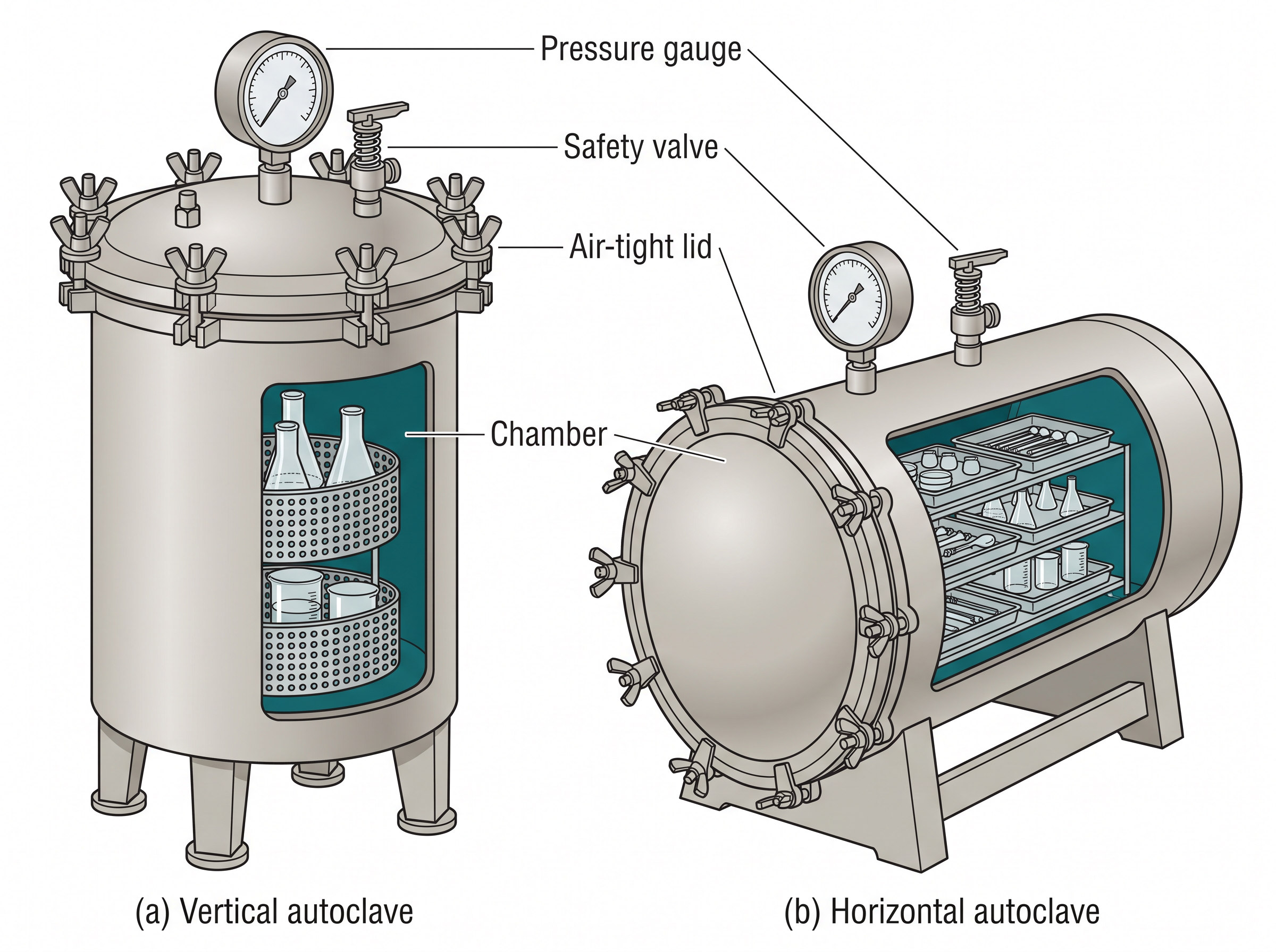
Fig. 6.9 – Vertical and horizontal autoclaves.
The holding time in autoclaving is the time for which the entire load in the autoclave is exposed and actual sterilisation takes place. The holding time depends on the size and type of the load and generally exceeds 30 min.
Note carefully that the holding time begins only after the whole load has reached the working temperature. A large flask of medium takes longer to heat through than a small one, which is why the load size decides the time.
Pasteurisation
This process is used in the food and dairy industry. Pasteurisation is the heating of milk to a particular temperature for a period of time in order to kill the pathogenic microorganisms that may be present in the milk.
It does not fully sterilise the milk, so many living organisms including spores are not destroyed. That is deliberate. Heating milk hard enough to sterilise it would spoil its taste and damage its nutrients, so the aim is to make it safe to drink rather than lifeless — which is also why pasteurised milk must still be refrigerated and does not keep indefinitely.
There are two methods of pasteurisation: the holder method (low temperature long time, LTLT, heated at 63°C for 30 min) and the flash method (high temperature short time, HTST, heated at 72°C for 15 sec) followed by quick cooling.
Other pasteurisation methods are also available, such as Ultra-High Temperature (UHT) at 140–150°C for 1–3 seconds.
Quick cooling after heating matters as much as the heating itself: it stops the milk from sitting warm, which is exactly the condition in which any surviving organisms would multiply.
Heating and Flaming
Many objects are sterilised in the absence of water by dry heat sterilisation.
Articles such as inoculation loops, straight wires, tips of forceps and searing spatulas are sterilised by holding them in a Bunsen flame till they become red hot. Red heat burns away everything organic on the metal, which is why the loop must be flamed before and after every transfer.
Some articles, such as the mouths of test tubes, flasks and glass slides, are passed through the flame a few times without allowing them to become red hot. This is enough to kill organisms sitting on the rim and to set up a rising current of hot air that keeps airborne contaminants away from the opening.
A hot air oven is also used to sterilise glassware. Dry heat has to be applied for longer and at higher temperatures than moist heat, because without water the proteins of the cell are much slower to be destroyed.
Sterilisation by Radiation
Two types of radiation, namely ionizing and non-ionizing radiation, are used for sterilisation. As radiation does not generate heat, sterilisation by radiation is termed ‘cold sterilisation’ — which is precisely why it suits plastics, foods and drugs that heat would ruin.
Non-ionizing radiations, such as UV rays, have germicidal activity in the wavelength range of 200–280 nm, with 260 nm being the most effective.
That figure is not a coincidence. DNA absorbs ultraviolet light most strongly near 260 nm, so this is the wavelength that does the most damage to the genetic material.
UV rays induce the formation of thymine–thymine dimers, which ultimately inhibit DNA replication and induce mutation in cells. A dimer is formed when two neighbouring thymine bases on the same strand become joined to each other, distorting the strand so that the replication machinery cannot read past it, as shown in Fig. 6.10.
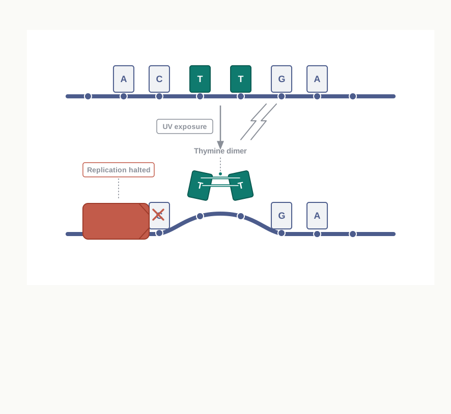
Fig. 6.10 – Formation of a thymine–thymine dimer by UV radiation.
UV radiation is useful for surface disinfection — that is, to disinfect the culture laboratory, laminar hood, operation theatres, virus laboratories and so on.
The disadvantages of UV rays include their harmful effect on skin and eyes, and the reversal of the damage due to DNA repair enzymes of bacteria. In other words, some cells mend the dimers afterwards and survive, so UV cannot be relied on for true sterilisation. It also penetrates very poorly, which is why it only disinfects exposed surfaces.
Ionizing radiations are of two types: particulate (electron beams) and electromagnetic rays (gamma rays).
Electron beams are used to sterilise syringes, gloves, dressing packs, foods and pharmaceuticals.
Electromagnetic rays have more penetrative power than electron beams, but require a longer time of exposure. These radiations damage the nucleic acid of the microorganism.
Electromagnetic radiations are used to sterilise disposable Petri dishes, plastic syringes, antibiotics, vitamins, hormones, glassware and fabrics. Because gamma rays pass right through packaging, articles can be sealed first and sterilised afterwards, so they cannot be recontaminated on the way to the user.
Sterilisation by Filtration
Filtration is used to sterilise heat-labile substances in solutions, such as serum, antibiotics, sugars and urea. Heat-labile simply means destroyed by heat: autoclaving such a solution would leave it sterile but useless.
Filtration does not kill microbes, but separates them out on the basis of size. The solution passes through, the cells are held back on the filter.
Membrane filters with pore sizes between 0.2–0.45 μm are commonly used to remove microbes from solutions. Bacterial cells are larger than these pores and cannot get through; note, however, that viruses are smaller still, so filtration of this kind does not remove them.
Air can be filtered using High Efficiency Particulate Air (HEPA) filters. HEPA filters are 99.97 percent efficient at removing particles >0.3 µm in diameter. They are usually used in biological safety cabinets, where they supply a continuous stream of microbe-free air over the working surface.
Sterilisation by Chemicals
Sterilisation or disinfection can also be achieved by certain chemicals, such as alcohols, aldehydes and heavy metals. Chemicals are the method of choice for large fixed surfaces, for skin, and for rooms — things that can be neither autoclaved nor filtered.
Alcohols sterilise the surface through cell dehydration, membrane disruption and protein coagulation.
70 percent ethyl alcohol is used as an antiseptic on skin. It may seem odd that 70 percent works better than absolute alcohol, but the water is needed: it helps the alcohol enter the cell and coagulate the proteins inside instead of merely hardening the surface.
Isopropyl alcohol can be used to disinfect surfaces and clinical thermometers.
Aldehydes, such as 40 percent formaldehyde (formalin), are used for surface disinfection and for the fumigation of rooms, chambers, operation theatres, biological safety cabinets and so on. Fumigation means filling a closed space with the vapour so that every exposed surface, including those a cloth could not reach, is treated.
Heavy metals such as mercury, silver, arsenic, zinc and copper are used as germicides. They act by causing the precipitation of proteins.
Hydrogen peroxide solution is used for the skin disinfection of wounds and ulcers, and for deodorising wound dressings.
6.4 Pure Culture Techniques
Bacteria may be cultured using either liquid or solid media, and the choice depends on what you want from the culture. Solid media are particularly useful in the isolation of bacteria and are also used for their long-term storage, while liquid (broth) cultures are used for the rapid and large-scale production of bacteria.
As you know, microorganisms in their natural habitat exist as part of complex ecosystems comprising numerous other microorganisms. A pinch of soil or a drop of pond water holds hundreds of different kinds of organism, all mixed together.
Obtaining a pure culture is important for studying a particular microbe. Unless the other organisms are removed, you cannot tell which of them produced the enzyme, the pigment or the disease you are studying — the same difficulty Robert Koch had to overcome before he could link one organism to one illness.
A culture consisting entirely of one strain of organism is called a pure culture.
The three standard ways of obtaining one — the streak plate, the pour plate and the spread plate — all rest on the same idea. Spread the cells far enough apart on a solid surface, and each single cell will grow into a separate patch of its own descendants.
The Streak Plate Method
The standard method of obtaining a pure bacterial culture is the streak plate method, shown in Fig. 6.11.
An inoculating loop is used to streak out a drop of bacterial suspension, or material from a bacterial colony, on a solid agar plate in such a way that the inoculum becomes progressively more dilute along the path of streaking. The loop is flamed between one set of streaks and the next, so that each new sector starts with fewer cells than the one before.
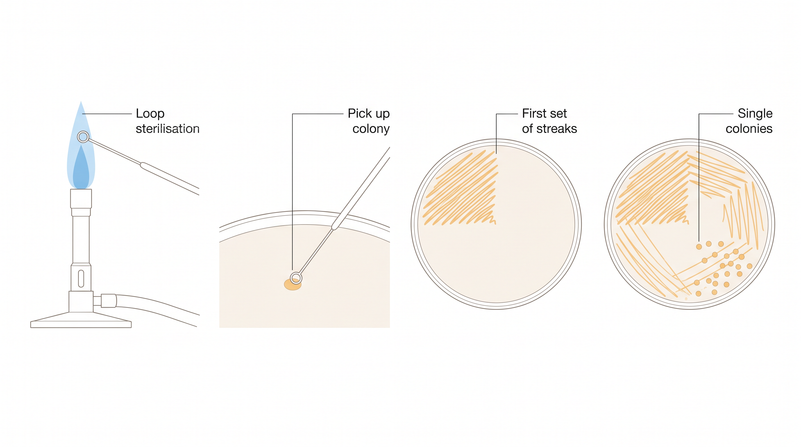
Fig. 6.11 – The streak plate method.
After proper incubation at an appropriate temperature, visible bacterial colonies develop as a result of repeated cell divisions.
Colonies are formed because all the progeny stay in the same place: movement of daughter cells is not possible on the solid surface. In other words, a clump of cells deposited together gives rise to a colony. Near the end of the streak, isolated colonies are obtained — and an isolated colony, having grown from one cell or one clump, is the starting point of a pure culture.
The Pour-Plate Method
An alternative method for the isolation of pure cultures is the pour-plate method, shown in Fig. 6.12.
In this method, the original microbial sample is diluted several times, in order to dilute the population sufficiently to obtain separate colonies upon plating. The dilution is usually carried out in steps, 1 mL of the sample being transferred into 9 mL of saline, so that each tube in the series contains one-tenth of the cells of the tube before it.
A small volume (1 mL) of each dilution is then poured on to the bottom of a Petri plate. After that, the melted medium with agar is poured on to the Petri plates and mixed gently with a circular motion, so that the cells are distributed evenly through the whole layer.
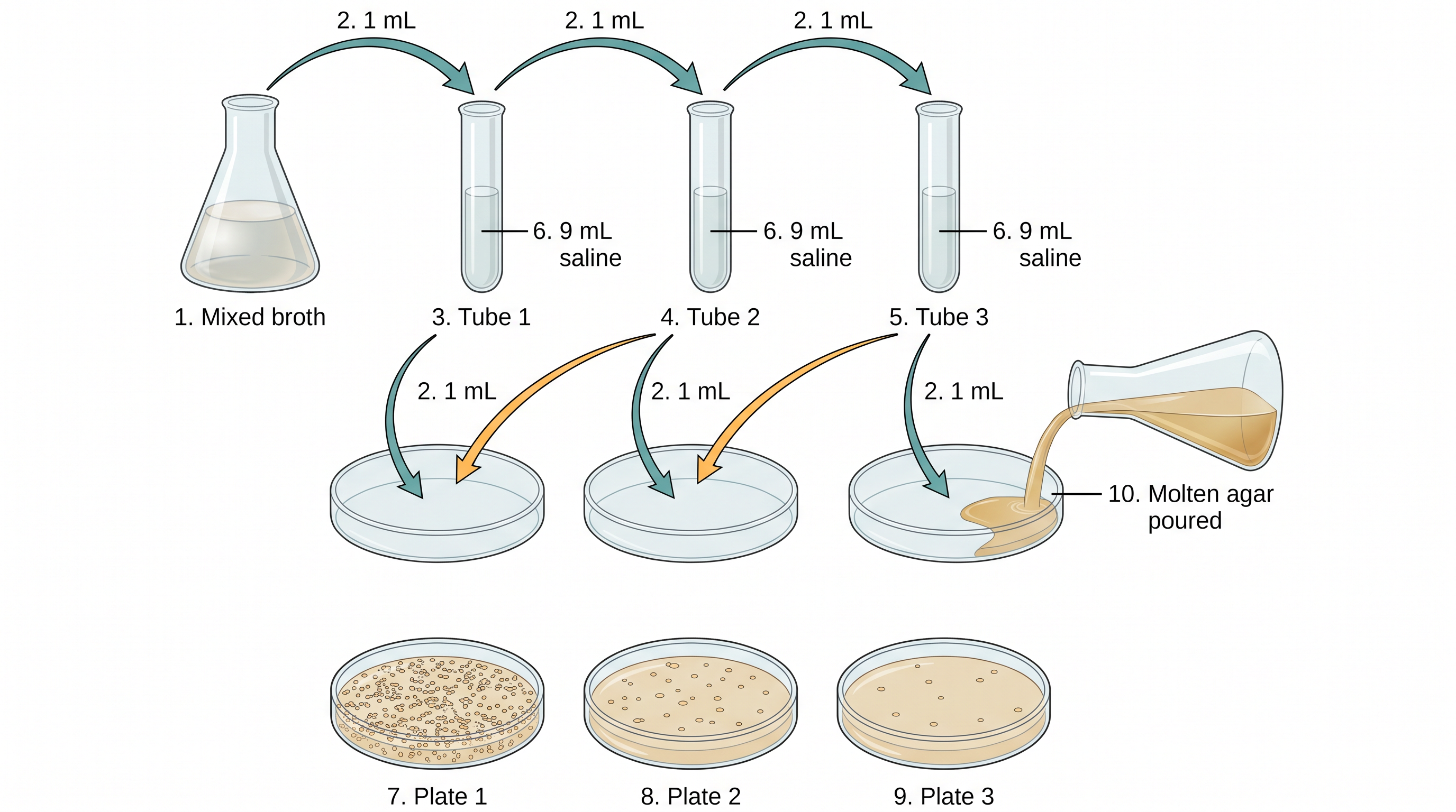
Fig. 6.12 – The pour-plate method.
Reading the three plates in Fig. 6.12 from left to right shows the point of the whole exercise: as the dilution increases, the number of colonies on the plate falls, until a plate is reached on which the colonies are separate enough to be counted and picked.
After incubation at an appropriate temperature, the isolated cells grow into colonies, which can be used to establish pure cultures. Because the cells are trapped within the agar as well as on it, colonies appear both on the surface and below it; the ones inside the medium are usually smaller and lens-shaped.
The Spread-Plate Technique
The spread-plate technique is another easy and direct way of achieving a pure culture, shown in Fig. 6.13.
In this technique, a small volume of the diluted bacterial mixture — about 0.1 mL — is transferred to the centre of a solidified agar plate and spread evenly over the surface with a sterile, L-shaped glass spreader.
The glass rod is usually sterilised by dipping it in alcohol and flaming it to burn off the alcohol. The rod must be allowed to cool before it touches the plate, or the heat will kill the very cells being spread.
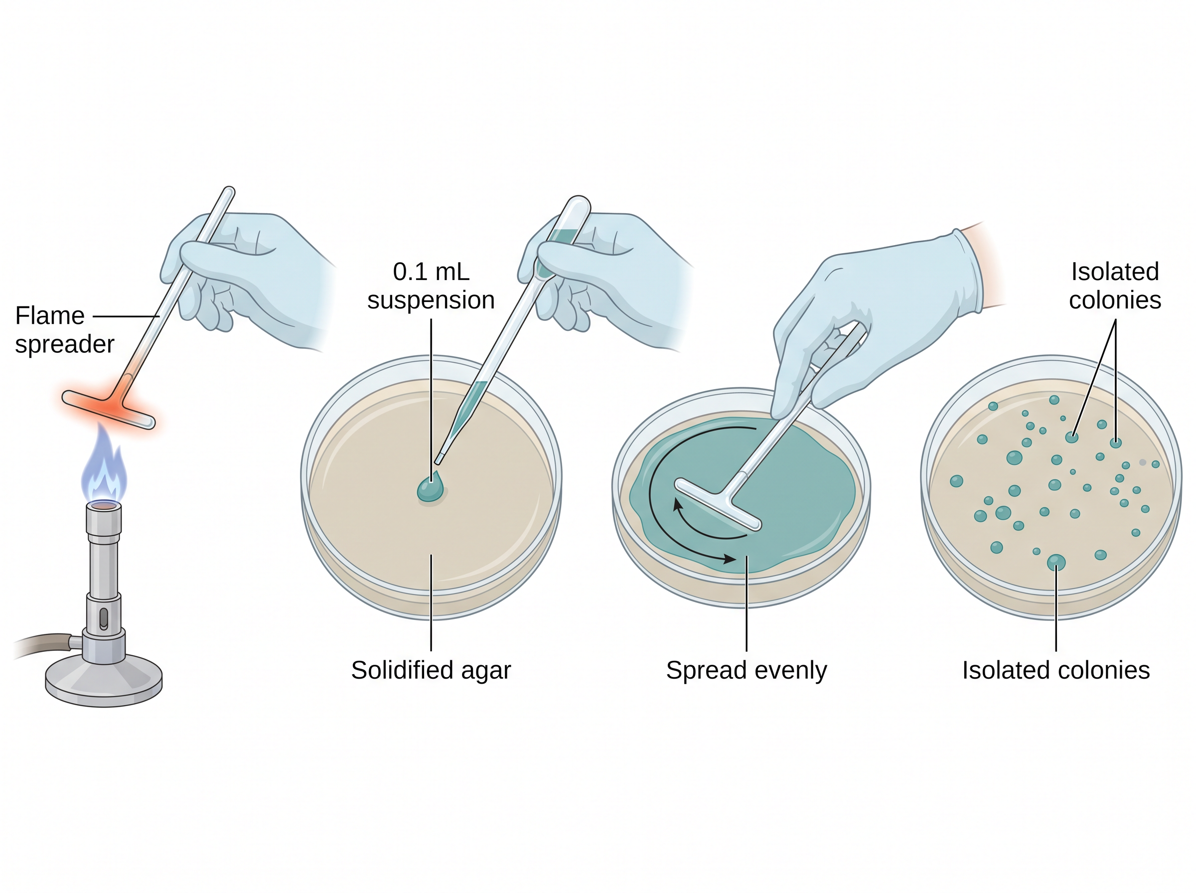
Fig. 6.13 – The spread-plate technique.
After proper incubation at an appropriate temperature, some of the dispersed cells develop into isolated colonies.
Because the agar has already set, every colony grows on the surface. This makes the colonies easy to see, to describe and to pick, and it avoids exposing the cells to the warmth of molten agar — an advantage when working with organisms that are sensitive to heat.
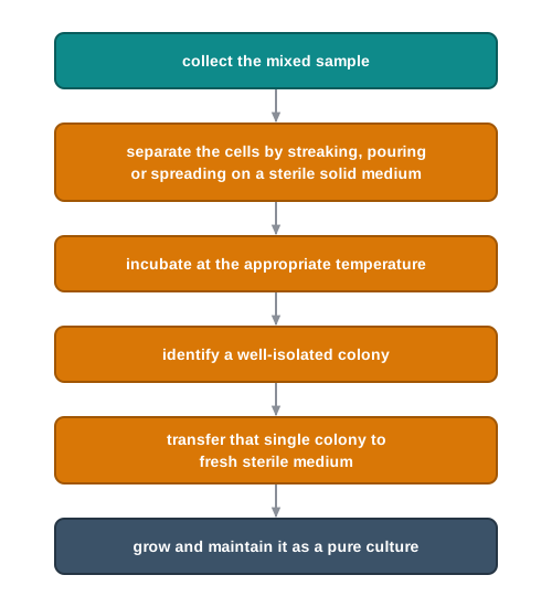
Fig. 6.14 – Sequence for obtaining a pure culture.
6.5 Factors Affecting Microbial Growth
Supplying the right nutrients is only half the task. A medium can be perfect on paper and still yield nothing if the physical conditions around it are wrong.
Microbial growth may be affected by a number of factors. The major factors are described below. Each of them acts, in the end, on the same target: the enzymes and membranes of the cell, which work well only within a limited range of conditions.
Temperature
Microorganisms are able to grow over a wide range of temperatures — far wider than the range in which our own cells can function.
For any single microorganism, however, the minimum and maximum growth temperatures are typically about 25–45°C. Below the minimum, the cell is too slow to divide; above the maximum, it is damaged.
The growth rate increases with temperature until the optimum temperature is achieved, and beyond that the growth rate declines again due to loss of enzymatic activity. This is why a temperature curve rises gently and then falls steeply, as shown in Fig. 6.15.
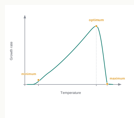
Fig. 6.15 – Effect of temperature on microbial growth rate.
Read the shape of that curve carefully, because it is a favourite in data-interpretation questions. The rise is gradual because warming speeds up chemical reactions steadily; the fall is sudden because, past a point, the enzymes begin to denature, and a denatured enzyme does not work slowly — it does not work at all.
The majority of microorganisms achieve optimal growth in the range of about 20–45°C and are called mesophiles. Most bacteria met in the laboratory, in food and in the human body belong to this group, which is why an incubator set near 37°C suits so much routine work.
Some microorganisms are capable of growing within a range of about 40–80°C, with an optimum temperature around 50–65°C, and are called thermophiles.
In addition, some are extreme thermophiles, which can tolerate temperatures above 100°C.
pH
The growth of microorganisms is strongly influenced by variation in the pH of their surroundings. pH affects the shape and charge of proteins, so a change of even one unit can slow an enzyme sharply.
Fungi can tolerate a wide range of pH variation when compared to bacteria.
Most microorganisms grow best around neutrality (pH 7). A number of bacteria prefer slightly alkaline conditions, but a few are tolerant of acidic conditions.
On the contrary, fungi generally prefer slightly acidic conditions, and therefore tend to dominate bacteria when these conditions prevail together. This single fact explains a common kitchen observation: it is mould, not bacteria, that usually appears on pickles, jams and citrus fruit, because the acidity that holds bacteria back does not trouble fungi.
The pH of growth media is adjusted to the desired value, depending upon the type of microorganism, by the addition of acid or alkali during preparation. The adjustment is made before sterilisation, since heating can itself shift the pH of a medium slightly.
Oxygen
Oxygen is present as one of the major constituents (21%) of our atmosphere. Microorganisms that are dependent on oxygen for survival are called aerobes.
Some microbes are also able to survive in the absence of oxygen, and are called anaerobes.
During the culture of aerobic organisms, oxygen must be provided. For a shallow layer of medium, such as that in a Petri dish, sufficient oxygen is available as dissolved in the surface moisture.
But in a flask of broth, aerobes will only grow in the surface layers, because oxygen dissolves poorly in water and diffuses slowly through it. Therefore, additional oxygen is provided by shaking or mechanical stirring to facilitate uniform growth. This is the reason a broth culture of an aerobe is grown on a shaker rather than standing still on a bench.
Obligate anaerobes cannot tolerate oxygen, and they are cultured in special anaerobic chambers, where oxygen is excluded from all liquid and solid media. For these organisms oxygen is not merely useless but poisonous, so it must be kept out of the medium itself and not only out of the air above it.
The facultative anaerobes can survive like aerobes in the presence of oxygen, and are also able to survive in anaerobic conditions. They are the most adaptable of the four groups, using oxygen when it is available and switching to fermentation when it is not.
Aerotolerant anaerobes are organisms that are generally anaerobic, but they are not inhibited by atmospheric oxygen, as they do not utilise it. The distinction from a facultative anaerobe is worth noting: an aerotolerant anaerobe puts up with oxygen but gains nothing from it.
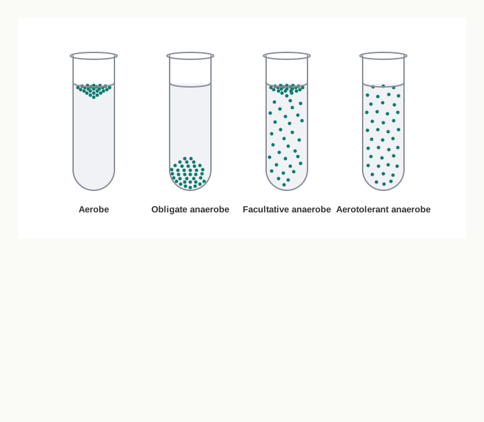
Fig. 6.16 – Oxygen requirements of bacteria in broth culture.
Carbon Dioxide
Autotrophic organisms are able to use carbon dioxide as a carbon source. When they are grown in culture, \(\mathrm{CO_2}\) is provided as bicarbonate in their growth medium, or they are incubated in a \(\mathrm{CO_2}\)-enriched atmosphere.
Bicarbonate is used because a gas dissolved in water escapes readily, whereas a salt dissolved in the medium stays where it is put and releases carbon dioxide steadily as the cells consume it.
Light
Phototrophic microorganisms require light in order to carry out photosynthesis. For them, light is not a comfort but a nutrient — it is the energy source itself.
In the laboratory, during the culturing of such microorganisms, one should ensure that light of the correct wavelength is used, since the photosynthetic pigments of a given organism absorb only certain wavelengths and will not respond to others.
6.6 The Growth Curve
When single-celled microorganisms are grown in a suitable nutrient medium with an initially limited amount of nutrients, the cells start growing in number and size at their maximum rate.
The growth of cells in an artificial medium depends on various factors, including nutritional factors and environmental factors — the very factors you met in the previous two sections. The growth of cells in a nutrient medium also depends on cell concentration, and the outcome of the whole process is cellular reproduction.
The word “growth”, in microbiology, usually means growth of the population rather than growth of an individual cell. A single bacterium does not get steadily bigger; it divides.
Unicellular organisms divide by binary fission. The cell replicates its genetic material and then divides into two identical cells, which divide into four, four into eight, and so on, leading to an exponential increase in the number of cells.
This is why microbial numbers become unmanageable so quickly. Doubling begins slowly in appearance, but after twenty doublings a single cell has become more than a million.
The growth of cultured microorganisms can be analysed by plotting the logarithm of the number of viable cells against the incubation time. This gives a curve with four distinct phases — lag phase, exponential phase, stationary phase and decline phase — as shown in Fig. 6.17.
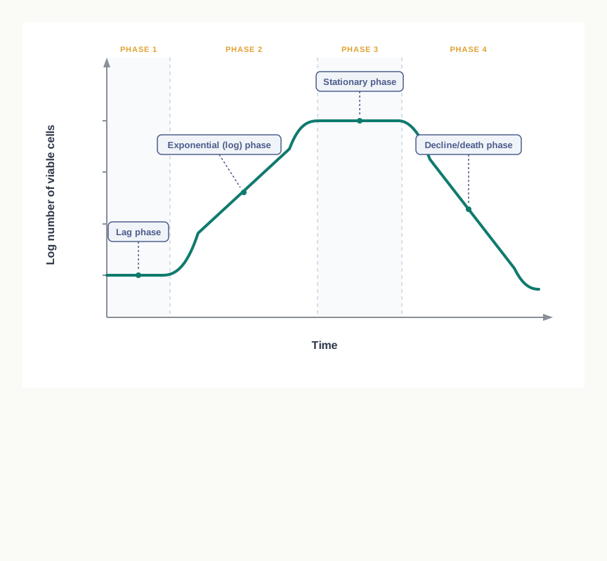
Fig. 6.17 – The microbial growth curve.
Reading the Growth Curve
Before following the phases one by one, notice two things about the axes, because questions are often set on exactly these points.
First, the y-axis plots the logarithm of the number of viable cells, not the number itself. On a logarithmic scale, a population that doubles at a constant rate gives a straight rising line, so the exponential phase appears as a straight slope rather than a sharply bending curve. A steeper slope means a faster growth rate.
Second, the count is of viable cells — those still alive and able to divide. This is what makes the stationary phase understandable: cells continue to be born and to die there, but the number alive at any moment stays the same.
Lag Phase
Initially, when microorganisms are introduced into a fresh culture medium, an increase in cell number is usually not observed immediately. This period is therefore called the lag phase.
Due to the lack of cell division during the lag phase, there is no net increase in mass. The cell is, however, far from idle: it is synthesising the new components required for cell division, such as the enzymes needed to use the nutrients available in the new medium.
The duration of the lag phase varies with the condition of the inoculated microorganisms and the nature of the medium.
This phase may be long if the inoculum is taken from an old culture or a refrigerated culture, because such cells must first repair themselves and rebuild their enzyme systems. On the contrary, when a young or growing microbial culture is inoculated into a fresh medium, the lag phase may be short or even absent, since those cells are already equipped for the conditions they meet.
Exponential Phase
During the exponential or log phase, the microorganisms are growing and dividing at the maximal rate. The microorganisms double in number at regular intervals during this phase, and the growth rate is constant.
In this phase the population will double in number during a specific time period, which is called the generation time or doubling time. For example, the doubling time for E. coli is 20 minutes.
During exponential growth, the doubling time can be expressed as:
\[t_d = \frac{0.693}{\mu}\]
where \(\mu\) is the specific growth rate, obtained from
\[\mu = 2.303\,\frac{(\log X_t - \log X_0)}{t}\]
In these expressions:
- \(X_t\) = cell concentration at time \(t\) (cells per mL)
- \(X_0\) = initial cell concentration (cells per mL)
- \(t\) = time taken (in hours, or in minutes)
- \(t_d\) = doubling time, in the same unit of time as \(t\)
- \(\mu\) = specific growth rate, with the unit of reciprocal time (h\(^{-1}\) if \(t\) is in hours)
Read the relationship between the two quantities carefully, because it is the source of most errors. The doubling time is inversely proportional to the specific growth rate: a large \(\mu\) means a short \(t_d\), and a small \(\mu\) means a long one. A fast-growing organism doubles often, so each doubling takes little time.
During this phase, the population is most uniform in terms of its chemical and physiological properties, and therefore the microorganisms of this phase are usually used for biochemical and physiological studies. Every cell is doing much the same thing at much the same rate, so a sample taken now represents the whole culture.
\[t_d = t/n \qquad (n = \text{number of generations})\]
The specific growth rate \(\mu\) is an index of the growth of microbes in a particular environment. It is defined as the rate of increase of biomass of a cell population per unit of biomass concentration. The value of \(\mu\) is characteristic for each microbe, and depends upon the temperature, pH, media composition and \(\mathrm{O_2}\) dissolved in the medium. As the value of \(\mu\) increases, the microbial culture has a lower doubling time. On the basis of the values of \(\mu\) and \(t_d\) of an organism, the media composition and the fermentation batch time are decided, so as to optimise production in commercial fermenters.
Example. If a culture contains \(10^4\) cells/mL at \(t_0\) and \(10^8\) cells/mL after 2 h, calculate the specific growth rate and the generation time of the culture.
Solution. \(X_0 = 10^4\) cells/mL, \(X_t = 10^8\) cells/mL, \(t = 2\) h
\[\text{(i)}\quad \mu = 2.303\,\frac{(\log 10^8 - \log 10^4)}{2} = 2.303\,\frac{(8-4)}{2} = 2.303\,\frac{(4)}{2} = 4.606\ \mathrm{hr^{-1}}\]
\[\text{(ii)}\quad t_d = \frac{0.693}{\mu} = \frac{0.693}{4.606} = 0.15\ \mathrm{h}\]
Stationary Phase
After a certain time in the log phase, the population growth ceases and the growth curve becomes horizontal. This depends on the overall nutrient availability and other factors, as well as on the type of microorganism being cultured.
During the stationary phase, the total number of viable microorganisms remains constant, due to the development of a balance between cell division and cell death. The flat line therefore hides a great deal of activity: cells are still dividing, but just as many are dying.
There are several factors that push microbial populations into the stationary phase. One important factor is nutrient limitation: if any essential nutrient is severely depleted, then the population growth will be slow.
Death Phase
Unfavourable environmental conditions, such as nutrient deprivation and the accumulation of toxic wastes, lead to a further decline in the number of viable cells, which is the characteristic of the decline or death phase.
During this phase, the death of a microbial population is usually logarithmic, just as the growth during the exponential phase was logarithmic. A constant proportion of the surviving cells dies in each interval of time, which on a logarithmic plot gives a straight falling line.
The accumulation of metabolic waste products may often become a factor in the decline of population growth. For example, Streptococci can produce lactic acid and other organic acids from sugar fermentation, which results in the medium becoming acidic and growth being inhibited.
Notice how this links back to the previous section: the organism is not being harmed by anything from outside. It has changed the pH of its own surroundings until those surroundings no longer suit it.
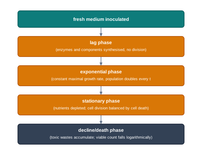
Fig. 6.18 – Fate of a batch culture.
This curve is not merely descriptive. In industry, the whole point of knowing \(\mu\) and \(t_d\) is to decide when to harvest. A product made by growing cells is collected while the culture is still in the exponential phase, whereas a product that accumulates only after growth slows is collected later — and knowing exactly where the culture sits on this curve is what makes the difference between a good batch and a wasted one.
6.7 Summary
Microbiology is the study of small life forms, that is, microorganisms. Microorganisms are ubiquitous, and the subject now underpins medicine, agriculture, food technology, environmental science and genetic engineering.
The subject grew out of a long argument. Leeuwenhoek first saw microorganisms, Spallanzani showed that boiled broth stays lifeless, and Pasteur’s flask experiments disproved spontaneous generation and established the germ theory of disease. Koch then linked a single organism to a single disease by using pure cultures, and introduced solid media, on which he named the discrete growth a colony.
Microorganisms require a source of energy, carbon, nitrogen, oxygen, iron, other minerals, micronutrients and water for growth and multiplication. Macronutrients supply the substance of the cell and act as enzyme cofactors, while micronutrients are needed only in traces and usually come in with the other ingredients.
A culture medium supplies these needs through a carbon source, nitrogen, phosphorus and sulphur sources, growth factors and water; anti-foams are added to control foaming, though they are not nutrients.
Selection of an appropriate culture medium for a microorganism depends on prior knowledge of its habitat, because its nutrient requirements reflect its natural surroundings.
Culture media can be categorised in three ways. On the basis of chemical composition they are synthetic (chemically defined) or complex. On the basis of consistency they may be liquid, solid or semi-solid. On the basis of their application and functions they may be selective, differential or enrichment media, and one medium may belong to more than one of these groups.
A pure culture contains only one strain of organism, and is essential if a particular microbe is to be studied. It is obtained by the streak plate, pour-plate or spread-plate method, each of which works by separating the cells far enough apart on a solid medium for a single cell to grow into a visible colony.
Sterilisation is of prime importance in any microbial study. It is the process in which all living microorganisms, including bacterial spores, are killed or removed.
Sterilisation can be achieved by physical methods — heat, radiation and filtration — and by chemical methods. Heat-stable articles are autoclaved at 121°C under 15 psi, heat-labile solutions are passed through membrane filters, and surfaces and rooms are treated with radiation or chemicals.
Microbial growth may be affected by a number of physical factors, such as temperature, pH, oxygen, carbon dioxide and light. Organisms are grouped by their responses to these factors — for example as mesophiles, thermophiles and extreme thermophiles, or as aerobes, obligate anaerobes, facultative anaerobes and aerotolerant anaerobes.
The growth of cultured microorganisms can be analysed by plotting the logarithm of the number of viable cells against the incubation time, which gives a curve with four distinct phases: lag, exponential, stationary and death.
In the exponential phase the population doubles at a constant rate, described by the doubling time \(t_d = \dfrac{0.693}{\mu}\), where \(\mu\) is the specific growth rate. A high specific growth rate means a short doubling time, and these two values are used to decide media composition and batch time in commercial fermenters.
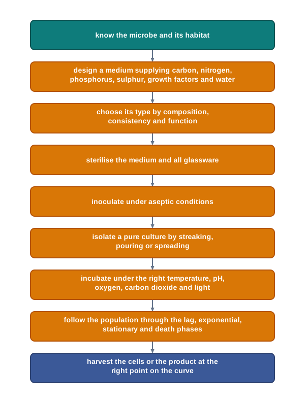
Fig. 6.19 – Overview of microbial culture.
6.8 Key Terms
Ubiquitous — Present everywhere, as microorganisms are in air, water, soil and on living surfaces.
Microbiology — The branch of science that deals with the study of microorganisms, which are too small to be seen with the unaided eye.
Animalcules — The name given by Antonie van Leeuwenhoek to the tiny moving organisms he first observed under his microscope.
Spontaneous generation — The discarded idea that living organisms can arise directly from non-living matter.
Germ theory of disease — The principle, established by Pasteur, that microorganisms are the cause of infectious diseases.
Golden Age of Microbiology — The period from the late 1800s to the early 1900s during which the causative agents of many microbial diseases were discovered.
Colony — A visible mass of microbial growth on a solid medium, formed by cells that multiply while remaining in one place.
Agar — A sulphated polymer of D-galactose, 3,6-anhydro-L-galactose and D-glucuronic acid, extracted from red algae, used to solidify culture media without adding nutrients.
Petri dish — The shallow, circular dish with a loose-fitting cover, introduced by Julius Richard Petri in 1887, in which solid media are poured.
rDNA technology — Recombinant DNA technology, in which DNA from two different sources is joined together.
Genetically modified organism (GMO) — An organism whose genetic material has been altered by genetic engineering to give it improved traits.
Heterotroph — An organism that obtains its carbon from organic molecules made by other organisms.
Autotroph — An organism that obtains its carbon from carbon dioxide.
Chemoautotroph — An autotroph that obtains its energy by the oxidation of inorganic molecules such as sulphur or nitrite.
Photoautotroph — An autotroph that uses photosynthetic pigments to convert light energy into chemical energy.
Chemoheterotroph — A heterotroph that obtains energy by the chemical oxidation of organic nutrient molecules.
Photoheterotroph — A heterotroph that obtains light energy by photosynthesis but takes its carbon from organic nutrients.
Lithotroph — An organism that uses inorganic molecules as its source of electrons.
Organotroph — An organism that uses organic molecules as its source of electrons.
Macronutrients — Elements such as C, O, H, N, S, P, K, Ca, Mg and Fe that microorganisms require in relatively large amounts.
Micronutrients (trace elements) — Elements such as Mn, Zn, Co, Mo, Ni and Cu that are required only in very small amounts.
Cofactor — A non-protein partner that an enzyme needs in order to function.
Growth factors — Organic compounds, such as certain amino acids, purines, pyrimidines and vitamins, that a cell cannot synthesise and must be supplied ready-made.
Fastidious organism — An organism with extra nutritional requirements that grows only when these are supplied, often in the form of complex extracts.
Anti-foams — Non-nutritional agents such as olive oil, sunflower oil and silicones added to prevent excessive foaming of agitated media.
Culture medium — A nutrient preparation, solid or liquid, that supplies everything a microorganism needs to grow in the laboratory.
Synthetic or chemically defined medium — A medium in which every chemical component is known and weighed out, such as M9 medium or BG11 medium.
Complex medium — A medium containing constituents of unknown chemical composition, such as peptone, beef extract or yeast extract.
Peptone — A protein hydrolysate prepared by the partial proteolysis of meat, casein, gelatin or other proteins, serving as a source of carbon, energy and nitrogen.
Beef extract — An aqueous extract of beef supplying amino acids, peptides, nucleotides, organic acids, vitamins and minerals.
Yeast extract — An aqueous extract of brewer’s yeast that is a rich source of vitamins, nitrogen and carbon compounds.
Lysogeny broth (LB medium) — A commonly used complex medium for culturing bacteria such as E. coli, first developed by Giuseppe Bertani in 1951.
Broth (liquid medium) — A culture medium to which no agar is added, in which growth appears as turbidity.
Solid medium — A medium solidified with 1.0–2.0% agar, used for surface cultivation, isolation and storage.
Semi-solid medium — A medium containing 0.5% agar, soft enough to allow movement through it, which may promote one organism while retarding another.
Selective medium — A medium that allows a particular type of microorganism to grow while suppressing the others present.
Differential medium — A medium on which different groups of microorganisms produce visibly different appearances, allowing them to be identified.
Enrichment medium — A medium whose nutritional environment is adjusted to enhance the growth of a desired organism within a mixed inoculum.
Sterilisation — The process by which all living microorganisms, including bacterial spores, are killed or removed.
Disinfection — The killing or removal of disease-causing microorganisms from non-living objects and surfaces, without necessarily destroying spores.
Sanitisation — The reduction of a microbial population to a level accepted as safe by public health standards.
Antiseptic — A chemical applied to living tissue, such as skin, to prevent infection.
Autoclaving — Sterilisation by steam under pressure, in which 15 psi raises the temperature of steam to 121°C.
Holding time — The period for which the entire load in an autoclave is held at the sterilising temperature, during which actual sterilisation occurs.
Pasteurisation — A controlled heat treatment that kills the pathogenic microorganisms in milk without fully sterilising it.
Holder method (LTLT) — Pasteurisation at low temperature for a long time, namely 63°C for 30 min.
Flash method (HTST) — Pasteurisation at high temperature for a short time, namely 72°C for 15 sec, followed by quick cooling.
Ultra-High Temperature (UHT) — Pasteurisation at 140–150°C for 1–3 seconds.
Dry heat sterilisation — Sterilisation in the absence of water, as by flaming articles red hot or using a hot air oven.
Cold sterilisation — Sterilisation by radiation, so called because radiation does not generate heat.
Thymine–thymine dimer — The joining of two adjacent thymine bases on a DNA strand by UV light, which blocks DNA replication and induces mutation.
Non-ionizing radiation — Radiation such as UV, germicidal in the range 200–280 nm and most effective at 260 nm, used for surface disinfection.
Ionizing radiation — Particulate radiation (electron beams) and electromagnetic radiation (gamma rays), which sterilise by damaging nucleic acid.
Heat-labile substance — A substance such as serum, an antibiotic, a sugar solution or urea that is destroyed by heating and must be sterilised by filtration.
Membrane filter — A filter of pore size 0.2–0.45 μm that removes microbes from a solution by size, without killing them.
HEPA filter — A High Efficiency Particulate Air filter, 99.97 percent efficient at removing particles larger than 0.3 µm, used in biological safety cabinets.
Pure culture — A culture containing only one strain of a single species, with no other organisms present.
Streak plate method — A method of obtaining a pure culture by progressively diluting the inoculum across a solid plate with a flamed loop.
Pour-plate method — A method in which diluted sample is mixed into molten agar in a plate, so that separated cells grow into colonies within and on the medium.
Spread-plate technique — A method in which about 0.1 mL of a dilute suspension is spread over a set agar surface with a sterile L-shaped glass spreader.
Serial dilution — A stepwise dilution, such as 1 mL into 9 mL of saline at each step, used to reduce cell numbers to a countable level.
Mesophiles — Microorganisms that grow best at about 20–45°C.
Thermophiles — Microorganisms that grow within about 40–80°C, with an optimum around 50–65°C.
Extreme thermophiles — Microorganisms that can tolerate temperatures above 100°C.
Aerobes — Microorganisms that depend on oxygen for survival.
Anaerobes — Microorganisms able to survive in the absence of oxygen.
Obligate anaerobes — Organisms that cannot tolerate oxygen and must be grown in anaerobic chambers.
Facultative anaerobes — Organisms that grow like aerobes when oxygen is present and can also survive without it.
Aerotolerant anaerobes — Generally anaerobic organisms that are not inhibited by atmospheric oxygen because they do not use it.
Phototrophic microorganisms — Microorganisms that require light of the correct wavelength in order to carry out photosynthesis.
Binary fission — The division of a unicellular organism into two identical cells after replication of its genetic material.
Growth curve — The curve obtained by plotting the logarithm of the number of viable cells against incubation time, showing four distinct phases.
Lag phase — The initial phase in which cell number does not increase because the cells are synthesising the components needed for division.
Exponential (log) phase — The phase in which cells grow and divide at the maximal rate, doubling at regular intervals.
Generation time (doubling time) — The time required for a microbial population or its cell mass to double during balanced growth.
Specific growth rate — The rate of increase of biomass of a cell population per unit of biomass concentration, characteristic for each microbe.
Stationary phase — The phase in which the viable count stays constant because cell division is balanced by cell death.
Death (decline) phase — The phase in which the viable count falls, usually logarithmically, due to nutrient deprivation and accumulation of toxic wastes.
6.9 Points to Ponder
Pasteur’s flasks differed from one another in only one respect. Think about why a well-designed experiment must change one thing at a time, and what would have happened to the spontaneous generation debate if he had changed the broth and the neck of the flask together.
Agar came into microbiology from a kitchen, not from a laboratory. What does this suggest about where useful scientific ideas come from, and about who deserves credit for them?
Koch could only link a particular organism to a particular disease because he could grow it in pure culture. Consider what this means for diseases caused by organisms that still cannot be grown in the laboratory.
A complex medium supports many organisms at once, while a defined medium supports very few. Reflect on when a scientist would deliberately choose the medium that grows less.
Selective media work by holding organisms back, and enrichment media by helping one along. Think about which approach you would trust more if you had to prove that a particular pathogen was present in a water sample, and why.
Pasteurisation is deliberately incomplete: it leaves organisms alive in the milk. Consider the reasoning behind choosing a treatment that is known to be less than total, and where else in daily life we accept “safe enough” instead of “completely free of risk”.
UV light damages microbial DNA, and bacterial repair enzymes can undo some of that damage. Ponder what this tells you about relying on a single method of sterilisation.
An antibiotic can be a medicine in a patient and a selective agent on a plate. Think about what the widespread use of antibiotics in both roles might mean for the rise of resistant organisms.
Extreme thermophiles were found only because someone looked in a place where life was assumed to be impossible. Where else might we still be failing to look, and what might that cost us?
In the stationary phase, cells are still dividing and still dying, yet the count does not change. Reflect on how many other flat lines in science hide two opposing processes in balance.
A culture in a closed flask eventually poisons itself with its own waste products. Consider how far this is a fair model for populations of larger organisms, including our own.
Knowing the specific growth rate lets an industry decide when to harvest a batch. Think about what is lost, in money and in material, when a fermenter is emptied a few hours too early or too late.
6.10 NCERT Questions
Describe the nutritional requirements of microorganisms.
What is culture media? Classify the culture media.
Write an account of the growth curve analysis of a microorganism.
Discuss any two methods to isolate a pure culture.
Define sterilisation, disinfection, and sanitisation.
Give a detailed account on the various methods of sterilisation.
Bacterial strains that do not require any organic supplement are called:
- Auxotroph
- Prototroph
- Heterotroph
- Chemotroph
Who was the first to develop the process of colony purification on solid media?
- Louis Pasteur
- Robert Koch
- Fannie Hesse
- Richard Petri
HTST and UHT methods belong to:
- Pasteurisation
- Isolation of pure culture
- Staining of bacteria
- Culture of bacteria
Spontaneous generation was suggested by:
- Francesco Redi
- Lazzaro Spallanzani
- Robert Koch
- Louis Pasteur
- Germ theory of disease was suggested by:
- Francesco Redi
- Lazzaro Spallanzani
- Robert Koch
- Louis Pasteur
Calculate the specific growth rate and generation time of a bacterial population in which the number of bacteria increases from \(10^4\) cells/mL to \(10^7\) cells/mL, during 4 hours of exponential growth.
Assertion: Alcohol production by a batch culture of Saccharomyces starts declining steadily even though conditions like temperature are optimum. Reason: Alcohol concentration of around 13% is toxic for yeast cells.
- Both assertion and reason are true and the reason is correct explanation of the assertion.
- Both assertion and reason are true but reason is not the correct explanation of the assertion.
- Assertion is true but reason is false.
- Both assertion and reason are false.
- Assertion: A selection medium containing Ampicillin is sterilised by autoclaving. Both ampS and ampR microbes show growth on the medium. Reason: To inhibit the growth of ampS microbe ampicillin should not have been autoclaved but sterilised by using micro-filters before adding to the medium.
- Both assertion and reason are true and reason is correct explanation of the assertion.
- Both assertion and reason are true but reason is not the correct explanation of the assertion.
- Assertion is true but reason is false.
- Both assertion and reason are false.
- Assertion: Microorganisms are able to grow and multiply over a wide range of temperatures. Reason: Extreme thermophiles can tolerate temperatures of above 100°C.
- Both assertion and reason are true and reason is the correct explanation of the assertion.
- Both assertion and reason are true but reason is not the correct explanation of the assertion.
- Assertion is true but reason is false.
- Both assertion and reason are false.
6.11 Objective Questions
- The term ‘animalcules’ was coined by:
- Lazzaro Spallanzani
- Antonie van Leeuwenhoek
- Ferdinand Cohn
- Julius Richard Petri
- Agar is generally extracted from:
- Red algae
- Brewer’s yeast
- The collagen of skin and bones
- Cane molasses
- Which of the following is NOT a constituent of agar?
- D-galactose
- 3,6-anhydro-L-galactose
- D-glucuronic acid
- D-ribose
- Semi-solid media are prepared by adding agar at a concentration of:
- 0.5%
- 1.0%
- 1.5%
- 2.0%
- Inside an autoclave, a pressure of 15 lb per square inch raises the temperature of steam to:
- 100°C
- 110°C
- 121°C
- 140°C
- UV rays show germicidal activity in the range 200–280 nm, the most effective wavelength being:
- 220 nm
- 240 nm
- 260 nm
- 280 nm
- UV radiation kills microorganisms mainly by:
- Coagulating membrane lipids
- Inducing thymine–thymine dimers that block DNA replication
- Precipitating cellular proteins
- Dehydrating the cell
- HEPA filters are 99.97 percent efficient in removing particles of diameter greater than:
- 0.1 µm
- 0.2 µm
- 0.3 µm
- 0.45 µm
- Membrane filters commonly used to remove microbes from solutions have pore sizes of:
- 0.02–0.05 μm
- 0.2–0.45 μm
- 1.0–2.0 μm
- 5.0–10.0 μm
- The holder method of pasteurisation involves heating at:
- 63°C for 30 min
- 72°C for 15 sec
- 100°C for 30 min
- 140–150°C for 1–3 sec
- Which of the following is a chemically defined (synthetic) medium?
- Nutrient broth
- MacConkey agar
- M9 medium
- Potato dextrose agar
- BG11 medium is used for the cultivation and maintenance of:
- Moulds and yeasts
- Blue green algae (cyanobacteria)
- Obligate anaerobes
- Cellulose-digesting bacteria
- Potato dextrose agar (PDA) is chiefly used for:
- Isolating Gram-negative bacteria
- Culturing cyanobacteria
- Identification, cultivation and enumeration of fungi
- Growing obligate anaerobes
- Peptones used in complex media are prepared by:
- Aqueous extraction of brewer’s yeast
- Partial proteolysis of meat, casein, gelatin and other proteins
- Acid hydrolysis of starch
- Precipitation of milk proteins with heavy metals
- A medium that suppresses Gram-positive bacteria and also distinguishes lactose fermenters from non-fermenters is:
- Blood agar
- MacConkey agar
- M9 medium
- Nutrient broth
- On MacConkey agar, an organism that ferments lactose produces:
- Colourless colonies
- Red colonies
- No growth at all
- Growth only below the surface of the medium
- Ethyl alcohol is used as an antiseptic on skin at a concentration of:
- 40 percent
- 70 percent
- 90 percent
- 100 percent
- Heavy metals such as mercury, silver and copper act as germicides by:
- Causing precipitation of proteins
- Forming thymine dimers
- Removing microorganisms by size
- Lowering the pH of the medium
- Sterilisation by radiation is called ‘cold sterilisation’ because:
- It is carried out inside a refrigerator
- Radiation does not generate heat
- It works only on frozen material
- The article must be cooled before exposure
- In the spread-plate technique, the volume of dilute bacterial suspension placed on the plate is about:
- 0.01 mL
- 0.1 mL
- 1 mL
- 10 mL
- Microorganisms that are generally anaerobic but are not inhibited by atmospheric oxygen, because they do not utilise it, are called:
- Obligate anaerobes
- Facultative anaerobes
- Aerotolerant anaerobes
- Aerobes
- Thermophiles show optimum growth at about:
- 20–45°C
- 50–65°C
- 80–100°C
- Above 100°C
- The phase of the growth curve in which the population is most uniform in its chemical and physiological properties, and which is therefore preferred for biochemical studies, is the:
- Lag phase
- Exponential phase
- Stationary phase
- Death phase
- If the specific growth rate of a culture increases, its doubling time:
- Increases in proportion
- Decreases
- Remains unchanged
- First increases and then becomes constant
- In a dilution series, transferring 1 mL of culture into 9 mL of saline produces a dilution of:
- 1 in 9
- 1 in 10
- 1 in 90
- 1 in 100
Assertion: Agar is preferred over gelatin as a solidifying agent for culture media. Reason: Most microorganisms cannot degrade agar, and agar supplies no nutrients of its own.
Assertion: An antibiotic meant to make a medium selective is added after the medium has been autoclaved and cooled. Reason: Antibiotics are heat-labile and would be destroyed by autoclaving.
Assertion: A broth culture of an aerobic bacterium is shaken or stirred instead of being left standing. Reason: Oxygen dissolves poorly in water and diffuses slowly, so without agitation aerobes grow only in the surface layers.
Assertion: The lag phase may be short or even absent in some cultures. Reason: When a young, actively growing culture is inoculated into fresh medium, its cells already possess the components required for division.
Assertion: UV radiation cannot be relied upon to sterilise an article completely. Reason: UV penetrates very poorly, and the damage it causes can be reversed by bacterial DNA repair enzymes.
Assertion: Pasteurised milk must still be kept refrigerated and does not remain fit for use indefinitely. Reason: Pasteurisation kills pathogenic microorganisms but does not destroy all living organisms, including spores.
Assertion: During the stationary phase the number of viable cells remains constant. Reason: In this phase cell division and cell death occur at balanced rates.
Assertion: The growth curve is plotted using the logarithm of the number of viable cells rather than the actual number. Reason: On a logarithmic scale, a population doubling at a constant rate gives a straight rising line whose slope indicates the growth rate.
Assertion: Blood agar is described both as a differential medium and as an enriched medium. Reason: It allows hemolytic bacteria to be distinguished from non-hemolytic ones, and it also supplies additional nutrients for fastidious organisms.
6.12 Check Your Concepts
In Pasteur’s experiment, the flask with the long twisted ‘S’-shaped neck was left open to the air, yet nothing grew in the broth. Explain how this single design feature demolished the argument that boiling destroys a ‘vital force’ needed for life to appear.
Koch could have injected material directly from a diseased animal into a healthy one. Explain why growing the organism in pure culture first was essential to his conclusion, and state what the final step of re-isolating the organism was meant to rule out.
Koch tried coagulated egg albumin, starch paste and slices of potato before settling on gelatin, and finally on agar. Discuss what agar offered that the earlier materials did not, and explain why the fact that agar supplies no nutrients is an advantage rather than a drawback.
Micronutrients such as Mn, Zn, Co, Mo, Ni and Cu are essential for microbial growth, yet they are not usually weighed out and added to a medium. Why not?
A medium contains glucose and an ammonium salt as its only carbon and nitrogen sources, and is incubated in the dark. State the nutritional category of an organism that grows well on it, and give one reason for your answer.
Compare synthetic (chemically defined) media and complex media under the headings: composition, range of organisms supported, chief advantage and one example of each.
A laboratory wishes to isolate cellulose-digesting bacteria from a soil sample containing many other bacteria. Describe the medium you would prepare, explain the principle on which it works, and state which of the three classification schemes your medium belongs to under each heading.
MacConkey agar is described as both selective and differential. Explain, with reference to its ingredients, what makes it each of these two things, and describe what a lactose fermenter and a non-fermenter would look like on the plate.
Distinguish between a selective medium and an enrichment medium in terms of how each achieves its result. Name one medium that can be placed in more than one of these categories and justify your choice.
Match the items in Column A with those in Column B.
| Column A | Column B |
|---|---|
| a. 0.5% agar | i. Sterilisation of heat-labile solutions |
| b. BG11 medium | ii. Semi-solid medium |
| c. Membrane filtration | iii. Fumigation of operation theatres |
| d. 40 percent formaldehyde | iv. Cultivation of cyanobacteria |
| e. Blood agar | v. Differential and enrichment medium |
Define sterilisation, disinfection and sanitisation, and arrange them in decreasing order of the completeness with which microorganisms are removed. Give one example of a situation in which each would be the appropriate choice.
Boiling at 100°C for 30 min and autoclaving at 121°C both use heat, yet only one of them is accepted as sterilisation. Explain the difference in outcome, and explain how raising the pressure inside a closed chamber makes the higher temperature possible.
Explain why moist heat at a given temperature destroys microorganisms more rapidly than dry heat at the same temperature.
The holding time in an autoclave is said to depend on the size and type of the load. Explain the reasoning behind this, and state what error would result if the timing were started the moment the autoclave was switched on.
A student prepares a selective medium by dissolving the ingredients, adding ampicillin, and then autoclaving the whole mixture. After incubation, both ampicillin-sensitive and ampicillin-resistant organisms grow on the plate. Identify the mistake in the protocol, explain why it produced this result, and rewrite the correct sequence of steps.
Sterilisation by radiation is called ‘cold sterilisation’. Explain the name, and compare non-ionizing and ionizing radiation under the headings: mode of action, penetrating power and typical articles treated.
Give reasons for each of the following: (i) 70 percent ethyl alcohol is used on skin rather than absolute alcohol; (ii) an inoculating loop is flamed until red hot, but the mouth of a flask is only passed through the flame; (iii) filtration through a 0.45 μm membrane cannot be relied upon to give a virus-free solution.
Arrange the following steps of the streak plate method in the correct order, and state what would go wrong if the third and fourth steps were interchanged: incubate the plate; flame the loop and turn the plate; pick up material from a colony; streak the first sector; sterilise the loop and allow it to cool; streak the next sector from the edge of the previous one.
Explain why cells deposited on a solid medium give rise to a visible colony, whereas the same cells in a broth do not. Relate your answer to the reason solid media are preferred for isolation.
Compare the pour-plate method and the spread-plate technique under the headings: state of the medium at the time of inoculation, volume of inoculum used, position of the colonies formed, and one advantage of each. State which of the two you would choose for an organism sensitive to warmth, and why.
A student prepares a spread plate and obtains a continuous film of growth over the whole surface, with no separate colonies. Suggest two distinct causes and the correction for each.
Redraw the graph of growth rate against temperature for a microorganism and label the minimum, optimum and maximum growth temperatures. Explain why the curve rises gradually up to the optimum but falls sharply beyond it.
Explain each of the following observations: (i) mould, rather than bacteria, usually appears first on a jar of pickle; (ii) an obligate anaerobe must be grown in a chamber from which oxygen is excluded from the media as well as the air; (iii) refrigeration delays food spoilage without sterilising the food.
Draw the microbial growth curve, label the four phases and the two axes, and state for each phase what is happening to the number of viable cells and why. Explain in particular why the y-axis plots the logarithm of the viable count rather than the count itself.
During the stationary phase the viable count does not change, and during the death phase it falls logarithmically. Explain what is actually happening to the cells in each of these phases, and give one example of a metabolic product that can drive a culture into decline while the temperature and pH settings of the incubator remain optimum.
6.13 Case-Based Questions
A hospital laboratory receives a stool sample from a patient with severe diarrhoea. The sample is expected to contain a mixture of harmless gut bacteria along with a possible Gram-negative pathogen. The technician streaks the sample on to a plate of MacConkey agar and incubates it. After incubation, the plate shows a set of deep red colonies and a set of colourless colonies, and no growth at all of certain organisms known to have been present in the sample.
- Explain why no growth of some organisms was seen, naming the ingredients of the medium responsible.
- Account for the difference in colour between the two sets of colonies, referring to the sugar and the indicator present.
- State, with a reason, which two categories of medium (by application and function) MacConkey agar belongs to.
- The technician now wishes to obtain a pure culture of one of the colourless colonies. Describe the method she should use and the precaution she must take at each transfer.
A small dairy processes 500 litres of milk each morning. Until now it has used the holder method, heating the milk at 63°C for 30 min. To handle a larger volume, the owner is considering switching to the flash method at 72°C for 15 sec, followed by quick cooling. He asks whether the milk could instead simply be autoclaved, since that would make it completely sterile and remove the need for refrigeration.
- Compare the two pasteurisation methods in terms of temperature, time and suitability for large volumes.
- Explain why quick cooling is an essential part of the flash method.
- Give two reasons why autoclaving is not used for milk, even though it would sterilise it completely.
- Pasteurised milk still has to be refrigerated and does not keep indefinitely. Explain why, in terms of what pasteurisation does and does not destroy.
A student prepares nutrient agar plates for a practical class. She dissolves the ingredients in water, adds agar, pours the mixture into flasks and autoclaves a large batch of six one-litre flasks together for the same holding time she normally uses for a single small flask. She then pours the plates on the open bench beside a window, allows them to set, and stores them. Two days later, before any deliberate inoculation, several plates show patches of mould and small colonies of mixed appearance.
- Identify two separate errors in this procedure and explain how each could have allowed organisms to survive or to enter.
- Explain the meaning of holding time and why it depends on the size of the load.
- Describe how and where the plates should have been poured, and name the device that supplies microbe-free air for such work.
- State what would have happened if the medium had also required a heat-labile supplement, and how such a supplement should be sterilised and added.
A fermentation unit grows a bacterium to obtain an enzyme that the cells produce while they are actively dividing. In one batch, the inoculum is taken from an old, refrigerated stock culture. The operator records the viable count at intervals and finds that the count remains unchanged for nearly four hours before it begins to rise. The count then rises steadily, levels off, and finally begins to fall. In an earlier batch, started from a young culture growing in the same medium, the count began to rise almost at once.
- Name the phase that lasted nearly four hours in the first batch and explain what the cells were doing during it.
- Explain why this phase was much shorter in the second batch.
- State the phase in which the enzyme should be harvested, and give a reason based on the physiological state of the cells in that phase.
- In the first batch the count is \(2 \times 10^{5}\) cells/mL at the start of the rising phase and \(2 \times 10^{9}\) cells/mL 4 hours later. Write the expressions you would use to obtain the specific growth rate and the doubling time, naming each symbol and its unit.
A team collects water from a hot spring where the temperature is close to 70°C, hoping to isolate a bacterium whose enzymes remain active at high temperatures. Back in the laboratory, the samples are streaked on nutrient agar plates and incubated at 37°C along with the rest of the day’s work. After two days, several colonies appear, but none of the isolates produces enzymes that survive heating.
- Identify the main error in the way the samples were handled, and state the incubation condition that should have been used instead.
- Name the group to which the organisms sought by the team belong, giving the temperature range and optimum that define this group.
- Explain, in terms of enzyme structure, why an organism from such a habitat is of interest to biotechnology.
- Suggest one further piece of information about the spring that the team should have recorded before designing the medium, and explain how it would have influenced the medium’s composition.
A research laboratory routinely adds antibiotics to its culture media as selective agents, so that only the engineered bacteria carrying a resistance marker can grow. Spent plates and leftover broth, still containing the antibiotic and the resistant organisms, are discarded into the ordinary laboratory sink at the end of each week. A visiting inspector objects to this practice.
- Explain how an antibiotic works as a selective agent in a culture medium, and why the antibiotic must be filter-sterilised rather than autoclaved.
- State the concern behind the inspector’s objection, in terms of what happens when resistant organisms and antibiotic residues reach the environment.
- Describe how the waste should be treated before disposal, naming the method and the conditions used.
- Discuss briefly whether the benefits of using antibiotics as laboratory selective agents can be reconciled with the concern about resistance, mentioning at least one argument on each side.
6.14 Practice With Data
A bacterial culture in the exponential phase contains \(10^5\) cells/mL at the start and \(10^9\) cells/mL after 5 hours. Calculate the specific growth rate and the doubling time of the culture, stating the unit of each.
An organism has a specific growth rate of \(0.462\ \mathrm{h^{-1}}\) under a given set of conditions. Calculate its doubling time in hours and in minutes, and hence the number of generations it would complete in 6 hours of uninterrupted exponential growth.
A broth culture is diluted by transferring 1 mL into 9 mL of sterile saline, and the step is repeated four times in all. From the final tube, 0.1 mL is spread over the surface of an agar plate. After incubation, 46 colonies are counted. Calculate the number of viable cells per mL in the original broth culture, and state one assumption you have made in doing so.
A culture is found to have completed 12 generations during 4 hours of balanced growth. Using \(t_d = t/n\), calculate the generation time in minutes, and then calculate the specific growth rate of the culture.
Under the conditions described in this chapter, the doubling time of E. coli is 20 minutes. A flask of medium is inoculated with \(4 \times 10^{3}\) cells/mL and the culture grows exponentially for 3 hours. Calculate the number of generations completed and the expected cell concentration at the end of this period.
The viable count of a batch culture was recorded as follows.
| Time (h) | Viable cells per mL |
|---|---|
| 0 | \(1.0 \times 10^{5}\) |
| 1 | \(1.0 \times 10^{5}\) |
| 2 | \(1.0 \times 10^{6}\) |
| 4 | \(1.0 \times 10^{8}\) |
| 6 | \(1.0 \times 10^{9}\) |
| 8 | \(1.0 \times 10^{9}\) |
| 10 | \(2.5 \times 10^{8}\) |
- Plot the logarithm of the viable count against time and mark the four phases on your graph.
- Calculate the specific growth rate over the interval from 2 h to 6 h.
- State the time interval over which the stationary phase occurs, and give the reason for the change seen after 8 h.
A technician has to prepare 750 mL of solid medium containing 1.5% agar, and 400 mL of semi-solid medium. Calculate the mass of agar required for each, and state the ratio of the agar concentrations used in the two preparations.
Air is drawn into a biological safety cabinet at a rate that carries \(4.0 \times 10^{7}\) particles of diameter greater than 0.3 µm per minute up to the HEPA filter. Using the efficiency stated in this chapter, calculate the number of such particles entering the working area of the cabinet in one hour.
Two bacteria, P and Q, are grown under identical conditions. P has a specific growth rate of \(1.386\ \mathrm{h^{-1}}\) and Q has a specific growth rate of \(0.693\ \mathrm{h^{-1}}\). Calculate the doubling time of each, state which organism reaches a given cell density first, and explain the relationship between \(\mu\) and \(t_d\) that your answer illustrates.
The growth rate of an organism was measured at a series of temperatures.
| Temperature (°C) | Relative growth rate |
|---|---|
| 15 | 0.1 |
| 25 | 0.5 |
| 35 | 0.9 |
| 40 | 1.0 |
| 45 | 0.6 |
| 50 | 0.0 |
- Identify the optimum growth temperature and the approximate maximum growth temperature from the data.
- Classify the organism as a mesophile, a thermophile or an extreme thermophile, giving your reason.
- Explain why the fall in growth rate between 40°C and 50°C is much steeper than the rise between 25°C and 40°C.