Chapter 9: Stem Cell Culture and Organ Culture
In the middle of the twentieth century, doctors faced a cruel problem. A patient with leukaemia had a bone marrow that was producing diseased blood cells instead of healthy ones. Radiation and drugs could destroy the faulty marrow, but that left the patient with no way to make blood at all — and a person cannot survive many days without fresh red cells, white cells and platelets.
An American physician, E. Donnall Thomas, working in Seattle, asked a question that sounded almost impossible at the time. If the marrow could be wiped out, could healthy marrow from another person be put back in, simply by dripping it into a vein? His early attempts, first between identical twins and later between matched donors, slowly began to work. Patients who would certainly have died went on to live for years. In 1990 Thomas shared the Nobel Prize in Physiology or Medicine for this work.
What made the transplant succeed was not the marrow tissue itself. It was a small population of unspecialised cells hidden inside it — cells that could keep copying themselves and, at the same time, give rise to every type of blood cell the body needed. Today we call them stem cells, as shown in Fig. 9.0.
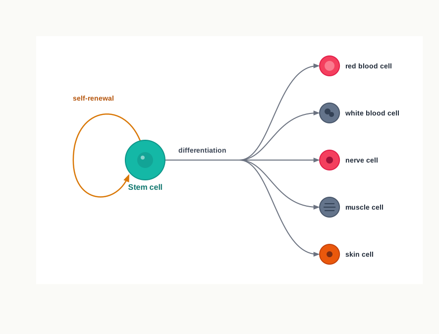
Fig. 9.0 – Stem cell self-renewal and differentiation.
Keep these five questions in mind as you read. By the end of this chapter you will be able to answer every one of them.
9.1 Stem Cell Culture
Every living thing is defined by one striking ability — it can make more of itself. In sexual reproduction, this begins with fertilisation, the fusion of the male gamete (sperm) with the female gamete (oocyte). The single cell formed by this fusion is the zygote, and the whole of the future organism is packed into it.
The zygote does not stay single for long. It divides, and the embryo forms through a series of cell divisions — first a two-celled stage, then a four-celled stage, then an eight-celled stage, and so on. At this point the daughter cells still look very similar to one another.
Later, these daughter cells begin to specialise. They differentiate into a variety of cell types, such as muscle cells, skin cells, liver cells, cardiovascular cells and epithelial cells. Differentiation is simply the process by which a general-purpose cell acquires a particular shape, a particular set of proteins, and a particular job in the body.
However, differentiation does not proceed at the same pace in every cell. Cells vary in their potential to make final cells — some become ‘mature’, which means they are fully specialised and committed to one job, while others remain ‘immature’. These ‘immature’ cells, which still hold the potential to differentiate into a wide range of specialised cell types, are called stem cells.
Two properties define them. First, they can carry out self-renewal via mitotic cell division, meaning that when a stem cell divides, the pool of stem cells is not used up. Second, they can then differentiate into a wide range of specialised cell types. A cell that only divides is not a stem cell; a cell that only differentiates is not a stem cell either. It must do both.
Historical perspective
Few areas of scientific study have attracted as much interest, inspection and debate as stem cells. The first stem cells were isolated from blood cells. Since then, scientists all around the world have been working on various types of stem cells with the aim of revolutionising regenerative medicine — the branch of medicine that tries to rebuild damaged tissue rather than merely manage its symptoms — by using the potential of stem cells to regenerate a tissue or an organ.
The most important moments in this journey are best seen as a timeline, told through the work of the pioneers of the field.
Where stem cells are found
Stem cells are non-specialised cells with an inherent property of self-renewal and potency. Potency simply means the range of cell types a stem cell is able to become. These cells are present in most of the multi-cellular organisms and are able to endure adverse conditions for long-time periods, which is one reason they can be frozen and stored for years without losing their ability to grow.
In humans, stem cells are found in the umbilical cord, the placenta, the inner cell mass of the early embryo, a few tissues of the foetus, and in some adult organs. Notice that this list runs from the very earliest stages of life right through to the adult body — the reserve of stem cells is never completely closed down.
Because of their ability to differentiate, stem cells are useful both for treating diseases and for understanding them. They can be used to:
- develop new cells in vitro to replace the damaged tissues or organs.
- study the cause of genetic defects in cells, as well as to study the cause of diseases and their treatments.
- test new lead molecules as drugs.
The third use is worth pausing on. A “lead molecule” is a chemical that looks promising as a future medicine. Testing it on human cells grown in a dish tells a company early on whether the molecule is toxic to heart or liver cells, long before it is ever given to a person.
9.1.1 Stem cell classification
Stem cells may be classified depending upon the source they are obtained from, and upon their potency, as shown in Fig. 9.1.
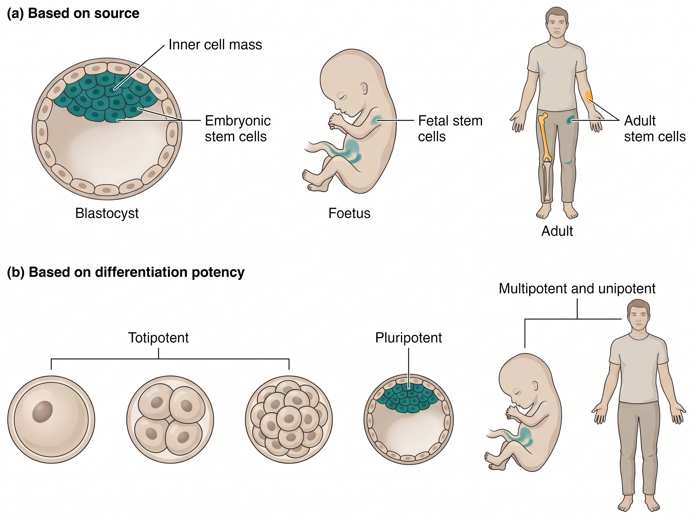
Fig. 9.1 – Classification of stem cells by source and potency.
Notice that these are two different questions being asked about the same cell. “Where did it come from?” is a question about source. “How many different things can it still become?” is a question about potency. The two classifications overlap, but they are not the same.
Classification based on source
On the basis of the source, stem cells have been categorised into three major groups — the embryonic stem cells, the fetal stem cells and the adult stem cells.
Embryonic stem cells are also known as early stem cells and are present in the inner cell mass of the blastocyst after around five days of development. The blastocyst is the hollow ball of cells that the early embryo forms; the inner cell mass is the small cluster inside it that will go on to build the body.
Adult stem cells, also called mature stem cells, are present in the umbilical cord and placenta after birth, and in mature body tissues.
As the embryonic stem cells have the property to differentiate into any tissue of the body, they are more promising for clinical applications than adult stem cells. However, the use of embryonic stem cells in humans is limited because of technical safety concerns and some ethical dilemma, since obtaining them involves the early embryo. On the contrary, there are no controversies regarding the safety and ethical issues associated with adult stem cells.
Adult stem cells were once thought to be irreversible, dedicated permanently to a specific lineage. However, they have now been shown to exhibit plasticity — that is, stem cells from one tissue can differentiate and produce cells of a completely different tissue. This single finding widened the therapeutic value of adult stem cells enormously, because it meant a patient’s own accessible tissue might be used to repair a tissue that is not accessible at all.
(a) Embryonic stem cells
Embryonic stem cells are classified as self-reproducing pluripotent cells and are possibly immortal, in the sense that a well-maintained line can be grown in the laboratory indefinitely. The human embryonic stem cells are derived from the embryo, that is, the blastocyst.
The distinctive features of embryonic stem cells are as follows:
- These cells can be isolated from the inner cell mass or epiblast of the blastocyst.
- These cells are pluripotent in nature and have the ability to give rise to all the three germ layers, i.e., ectoderm, mesoderm and endoderm.
- They have the capability of self-renewal and exhibit a stable diploid chromosome.
- They retain the characteristics of embryo founder cells even after extensive manipulation.
- Depending on the conditions of the in vitro environment, they can self-renew or differentiate into multiple tissue types.
The third feature deserves attention. A stable diploid chromosome number matters because a cell line that starts gaining or losing chromosomes in culture is no longer a faithful model of normal human cells, and would be unsafe to transplant. The fifth feature is equally important for the laboratory worker: the same cell will either keep dividing or start specialising depending entirely on what is put into the medium and what surface it is grown on.
(b) Fetal stem cells
Cells derived from a fetus that retain the ability to divide, proliferate and provide progenitor cells, and can differentiate into specialised cells, are called fetal stem cells. A progenitor cell is an intermediate cell — already partly committed, but still able to divide and produce specialised descendants.
The embryo is referred to as a fetus after the eighth week of development. Once a fetal stem cell has been harvested, it has the potential to live indefinitely in the laboratory.
Fetal stem cells can be isolated from fetal blood and bone marrow, as well as from other fetal tissues including the liver and kidney. Fetal blood is a rich source of haemopoietic stem cells (HSGs), which proliferate more rapidly than those in cord blood or adult bone marrow. That faster proliferation is exactly what makes them attractive — a transplant works better when the donated cells can repopulate the recipient’s marrow quickly.
(c) Adult stem cells
Adult stem cells are more precisely called somatic stem cells, because they can come from children or from the umbilical cord and not from adults only. They are undifferentiated multipotent or totipotent stem cells.
The adult stem cells are present all over the body after embryonic development, and they replace and regenerate damaged tissues or dying cells by cell division. These cells have two basic characteristics: the ability to self-renew without compromising with stemness, and the ability to differentiate into mature, morphologically and functionally dissimilar, tissue-specific cells. “Stemness” here means the full set of stem-cell properties — if a cell divides but loses those properties, it has not truly self-renewed.
The main role of adult stem cells in an organism is to preserve and repair the tissue in which they reside. Therefore, these cells maintain cellular homeostasis throughout the lifetime of mammals. Every day, skin is shed, blood cells are destroyed and gut lining is worn away; adult stem cells quietly balance those losses. The highest frequency of adult stem cells is present in the bone marrow, which is why marrow is the classical source for transplantation.
The embryonic stem cells are defined by their origin, but the origin of adult stem cells in some tissues is still under examination. The adult stem cells can either be hematopoietic or mesodermal in origin.
(i) Hematopoietic stem cells (HSCs)
Hematopoiesis, the process of formation of the different types of blood cells, occurs in the bone marrow. Bone marrow contains both hematopoietic and non-hematopoietic stem cells.
Hematopoietic stem cells (HSCs) give rise to different types of blood cells — RBCs, monocytes, neutrophils, acidophils, basophils, lymphocytes and platelets, as shown in Fig. 9.2. Non-hematopoietic stem cells are originated from common mesodermal precursor cells.
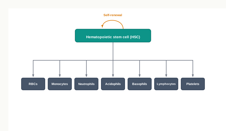
Fig. 9.2 – Lineage tree of a hematopoietic stem cell.
(ii) Mesenchymal stem cells
MSCs have the ability to self-renew, exhibit multi-lineage differentiation and are extremely proliferative. These cells can be isolated from a variety of tissues, such as bone marrow, adipose tissue and umbilical cord.
The major advantages of MSCs include their genetic stability, their ability to avoid allo-rejection, their compatibility in tissue engineering applications, and their capability of repairing many vital tissues. Allo-rejection is the immune attack launched against cells from another individual; MSCs provoke it much less strongly than most transplanted cells, which is a considerable practical advantage.
They are thought to be responsible for wound healing, growth, and replacing cells that are lost through daily wear and tear as well as through pathological conditions. MSCs are important for making and repairing skeletal tissues such as cartilage, bone and the fat found in the bone marrow. Because of these functions, they have been shown to be effective in the treatment of tissue injury and degenerative diseases.
Classification based on differentiation potency of stem cells
On the basis of the ability of self-replication and differentiation into various tissues, stem cells have been categorised into totipotent, pluripotent, multipotent and unipotent. Each of them is described below. It helps to read these four in order, because they form a descending ladder — each rung can do a little less than the one above it, as summarised in Fig. 9.3.
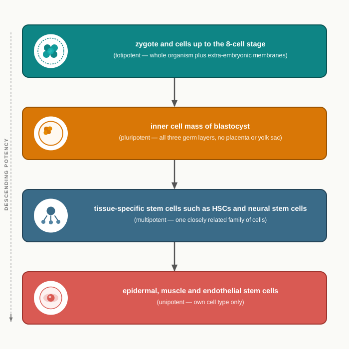
Fig. 9.3 – The descending ladder of stem cell potency.
Totipotent stem cells
Totipotent (toti = whole, potent = able to) stem cells are the most versatile form and have the utmost potential for differentiation. This permits the cells to produce both the embryo and the extra-embryonic membranes, and all the post-embryonic tissues and organs that form an entire functional organism. For example, the zygote has the potential to develop into a complete organism.
All cells of the early embryo are totipotent until the eight-cell stage; after that they begin to specialise and form the blastocyst. After nearly four days, the inner cell mass of the blastocyst becomes pluripotent.
Pluripotent stem cells
Pluripotent (pluri = several, potent = able to) stem cells have the potential to differentiate into almost all cell types of the embryo, except the cells of the extra-embryonic support tissues (placenta and yolk sac).
These cells can give rise to all the germ layers but are not able to form extra-embryonic tissues such as the placenta. As a consequence, they cannot give rise to a complete organism like totipotent stem cells can.
This division of labour is already visible in the blastocyst itself. The inner cell mass of the blastocyst gives rise to the tissues of the adult organism, while the outer layer, the trophectoderm, forms the placenta.
The pluripotent stem cells go on dividing until they begin to specialise at the gastrulation stage — the stage at which the embryo folds and sorts itself into the three germ layers. Examples comprise embryonic stem cells and the cells that are derived from the ectoderm, mesoderm and endoderm germ layers formed in the beginning stages of embryonic stem cell differentiation.
Multipotent stem cells
The multipotent cells (multi = many, potent = able to) have the ability to differentiate into a closely related family of cells. These are the plastic and more differentiated stem cells.
These multipotent stem cells can specialise into various types of cells within a specific cell lineage. For example, multipotent hematopoietic (adult) stem cells can develop into various types of blood cells, such as red blood cells, white blood cells and platelets. Similarly, neural stem cells can give rise to neurons, oligodendrocytes and astrocytes.
After differentiation, the abilities of these cells are restricted to the cells of their specific lineage. A neural stem cell will not make a blood cell, and a hematopoietic stem cell will not make a neuron under ordinary conditions.
Unipotent stem cells
A unipotent stem cell is an undifferentiated cell present in a differentiated tissue, with the property of dividing repeatedly. Unipotent stem cells have the ability to produce only cells of their own type, and they also possess the property of self-renewal that is needed for a cell to be labelled as a stem cell.
They can self-renew and differentiate into the specialised cells of that tissue — examples include epidermal stem cells, muscle stem cells and endothelial stem cells. The repetitive division in unipotent stem cells makes them a promising candidate for therapeutic use in regenerative medicine, because a tissue that constantly renews itself is exactly the kind of tissue that can be regrown.
It should be remembered that totipotent and pluripotent stem cells belong to the embryonic life of the organism, whereas multipotent and unipotent stem cells are found in adult life. In general, multipotent and unipotent stem cells are termed adult stem cells or somatic stem cells.
9.1.2 Characteristics of stem cells
Over the past several years, a great deal of attention has been paid to the differentiating ability of the adult stem cells. Plasticity of adult stem cells is the presumed ability of tissue-specific stem cells to attain the fate of cell types distinct from the tissue of origin.
Differentiation of one cell type against its fate into another cell type requires massive molecular rearrangement within the cell — whole sets of genes must be switched off and others switched on. For this, two things matter most: the competence of the responding cells, and micro-environmental cues.
Competence means the cell must be capable of responding in the first place. For example, all the types of bone marrow cells are not competent for differentiating into neurons or hepatocytes. Cues mean the correct signals must be supplied from outside. Similarly, the competent bone marrow cells do not undertake a differentiation programme against their fate unless they are exposed to an inductive microenvironment. Competence without cues, or cues without competence, produces no change of fate.
Pathways by which cells switch lineage
Some of the alternative fate-changing pathways used by somatic cells to switch lineages are trans-determination, trans-differentiation, dedifferentiation, heterogeneity, versus-gene-level pleiotropy, and fusion.
The first is trans-determination, in which a stem cell that is programmed to produce certain lineages switches to another stem cell and gives rise to the cell types of that precursor — that is, its potential is redirected.
The second pathway is trans-differentiation. In this process a differentiated cell can achieve the phenotype of another differentiated cell. Note the difference from trans-determination: here the cell that changes is already specialised, not a stem cell.
Dedifferentiation is common in lower vertebrates but rare in mammals. By the dedifferentiation process, adult stem cells revert back to the master cells from which they originate, and the master cells can then differentiate into other cell types. This is essentially a step backwards up the ladder of potency, followed by a step forwards down a different branch.
Heterogeneity is the most acceptable explanation for the plastic nature of adult stem cells. The idea is straightforward: a tissue sample may not contain one uniform kind of stem cell at all, but a mixture, so what looks like one cell type changing its fate may in fact be a second, rarer cell type in the sample doing what it always could.
One of the gold standards for plasticity of adult stem cells is its clonal level purity. Normally, the purity of stem cells is determined by the presence and absence of specific surface markers — characteristic proteins on the cell membrane that act as identity tags. When plasticity between two cells originated from the same germ layer can be explained by clonal level impurity, it is known as pleiotropic.
Fusion is an alternative mechanism proposed for the plasticity in adult stem cells. Here the donor stem cell physically fuses with a host cell, so the resulting cell carries the characteristics of both — which can be mistaken for a change of fate.
9.1.3 Applications of stem cells
The goal of any stem cell therapy is to repair a damaged tissue that cannot heal itself. Ongoing research on stem cell treatments gives hope and confidence to patients who would generally not receive a treatment that cures their disease, but only one that improves the symptoms of their chronic illness.
Stem cell therapies involve more than simply transplanting cells into the body and directing them to grow new, healthy tissue. It may also be possible to persuade the stem cells already present in the body to work overtime and produce new tissue — an approach that avoids transplantation altogether.
A number of stem cell therapeutics have been developed, but most of them are at experimental stages and are costly, with the notable exception of bone marrow transplantation. Many medical researchers predict that soon the embryonic and adult stem cells will be able to treat muscle damage, cancer, Huntington’s disease, Type 1 diabetes mellitus, Parkinson’s disease, cardiac failure, celiac disease, neurological disorders and many more conditions.
Before the application of stem cell therapeutics in clinical medicine, further research is required to understand stem cell behaviour upon transplantation, and the mechanisms of stem cell interaction with the injured or diseased micro-environment. Some clinical applications of stem cell therapies are detailed below.
Maintaining the culture
Before any of these applications is possible, the cells must be grown and maintained reliably in the laboratory. A standard routine for maintaining a cell culture, using mouse embryonic fibroblasts (MEFs) as the example, is set out in Fig. 9.4.
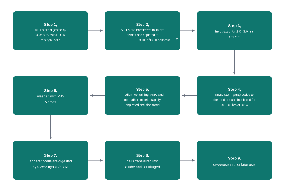
Fig. 9.4 – Protocol for the maintenance of cell culture.
The chemical details of this protocol are not arbitrary. Trypsin is a protease that breaks the protein links holding cells to the dish and to each other, while EDTA binds calcium ions, which those adhesion proteins need in order to work — the two together separate the sheet into single cells. Washing with PBS five times removes every trace of the treatment reagents, because leftover trypsin would continue digesting the cells that are meant to be kept. Incubation at 37°C is used throughout because that is the body temperature of the mammal from which the cells came.
Neurological diseases
Stem cells may become a good alternative therapeutic candidate for the treatment of neurological diseases. For example, Amyotrophic Lateral Sclerosis (ALS) is a progressive neuromuscular disease characterised by the loss of motor neurons in the brain and spinal cord. Because motor neurons are not replaced once lost, the muscles they supply gradually weaken. The investigation of ALS treatment using bone marrow stem cells and induced pluripotent stem cells (iPS cells or iPSCs) is under clinical trials.
In spinal cord injury cases, the nerve fibre bundles are damaged, which leads to paralysis. Investigation using adult stem cells to regenerate new nerve cells and to trigger the growth of severed nerve fibres is being carried out.
Stem cell therapies are also being used for eye disease treatments. In one such case an improved vision was observed with the transplantation of retinal pigment epithelial cells derived from embryonic stem cells, along with retinal stem cells. This observation brings hope for new therapeutics for eye diseases.
Wound healing or skin replacement
The skin (keratinocyte) stem cells are present in the hair follicle. These stem cells are removed after plucking a hair and are then cultured. After culture of these cells, they can be used as the equivalent of the patient’s own skin, which has less problem of rejection, as shown in Fig. 9.5. Thus, the stem cell makes it possible to develop skin from a patient’s plucked hair.
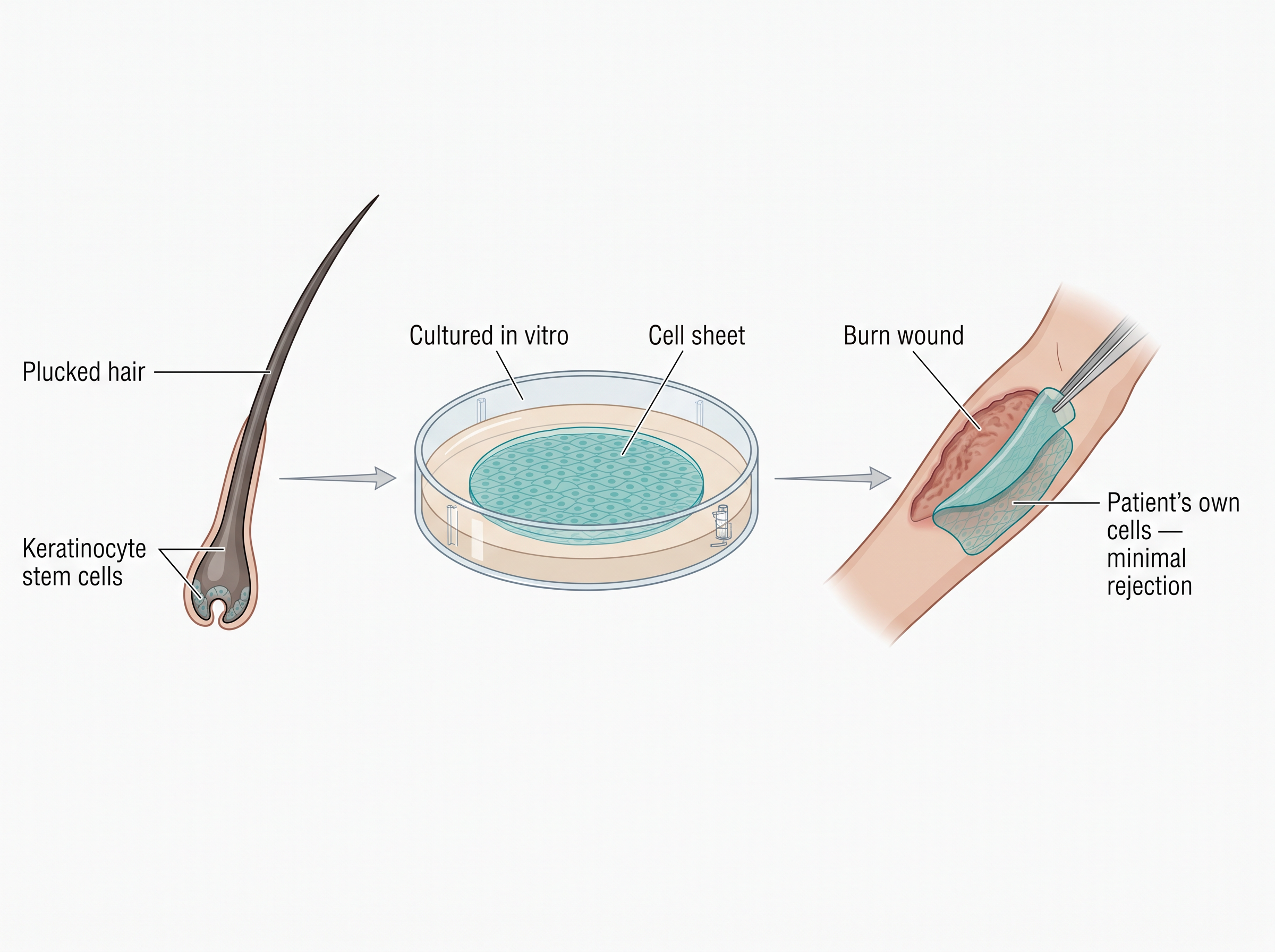
Fig. 9.5 – Culturing skin from a plucked hair.
Stem cells are a better alternative for faster healing of skin injuries, genetic disorders and burnt wounds, with lower inflammation levels compared to conventional skin grafts, which fail to restore the complete composition of the dermis that has been lost.
Cardiovascular Diseases
Cardiovascular diseases are basically characterised by ischemia and heart muscle injury, leading to hypertrophy and congestive heart failure. Ischemia means an inadequate blood supply, and heart muscle deprived of blood dies and is replaced by scar tissue that cannot contract.
The traditional and standard treatments for this are surgical procedures that repair the blocked arteries, medications that reduce fluid retention, and lifestyle changes. Notice that all three manage the consequences rather than replacing the lost muscle. With the advent of stem cells, a better therapy is under investigation that aims to re-establish the lost function of heart tissue and blood vessels.
Auto-immune Disorders
An auto-immune disorder occurs when the immune system of the body becomes over-reactive and considers its own healthy tissues as foreign and attacks them.
Type 1 Diabetes
Type I diabetes is caused by the degradation of insulin-producing pancreatic beta cells by the body’s own immune system. For treatment, insulin administration is an effective strategy, but optimisation of the doses of insulin is one of the major limitations, because the amount of insulin required depends on the glucose level, which changes through the day. Thus, its treatment with cells derived from haematopoietic stem cells is being explored, the aim being to restore a cell that senses glucose and releases insulin by itself.
Multiple Sclerosis
Multiple sclerosis is a chronic inflammatory disease that occurs in the brain or spinal cord. This disease is due to over-activation of the body’s own immune system, in which the myelin sheath of the neurons gets damaged. Since the myelin sheath is what allows a nerve impulse to travel rapidly, its loss slows or blocks conduction. For the treatment, bone marrow stem cells and neural stem cells are being investigated to regenerate the neurons with a proper myelin sheath.
Arthritis
In rheumatoid arthritis, the symptoms are chronic pain and inflammation of the joints, which are mainly due to the destruction of cartilage by the immune system. The currently used treatment strategies involve drugs that reduce the pain and inflammation. But stem cells can be differentiated into cartilage-making cells, mainly chondrocytes, so the use of stem cells could be a better alternative — one that rebuilds the damaged cartilage instead of only masking the pain.
Stem cells in the pharmaceutical industry
In pharmaceutical industries, stem cells have evolved as a suitable model to investigate the potential or mode of action of drugs, and have become an improved alternative to animal models, which carry ethical challenges of their own. In the current scenario, pharmaceutical industries emphasise the use of stem cells for drug screening.
Furthermore, the cardiomyocytes originated from human embryonic stem cells are used as a model for cardiac diseases, and the neuronal cells originated from stem cells are used as a working model for neuronal disorders. Testing a new molecule on human heart cells in a dish gives a far more direct warning of harm to the human heart than a test on an animal cell can.
Stem Cell Research Challenges
In the current research scenario, stem cell research is an emerging area with various applications, but it has some challenges yet to be resolved.
One of the major challenges is immunological rejection, in which the stem cell transplants are rejected by the recipient’s immune system. To resolve this, immunosuppressive treatments are given to recipients — but these make them prone to microbial infections, because the same immune system that would have attacked the graft also defends against microbes. However, the use of induced pluripotent stem cells (derived directly from the recipient’s own cells) may be one strategy to resolve this limitation of rejection to some extent.
The second major challenge is the behaviour of stem cells. The normal characteristics of a stem cell may have limitations for certain applications. For example, embryonic stem cells, which divide indefinitely, might induce tumour growth once transplanted — the very property that makes them useful in culture becomes a hazard in the body.
The safety of stem cells during application is also one of the challenges, as stem cells are prone to microbial infection that leads to various infectious diseases in the recipients.
9.2 Organ Culture
So far you have studied the different aspects of cell culture, which deals with the in vitro maintenance and proliferation of isolated cells using appropriate nutrient media. The cells are separated from the tissue, supplied with nutrients, and allowed to grow in a dish.
In cell culture, cell-to-cell interaction is possible, but the in vivo structural complexity is lacking, and this is the main limitation of cell culture. A liver in the body is not simply a heap of liver cells; it has layers, ducts, blood vessels and a definite architecture, and much of its behaviour depends on that architecture.
Conventional human cell cultures, which are generally used to establish differences in species, are also constrained in their depiction of in vivo responses. The reason is the lack of a suitable microenvironmental framework of the responding cell types — the cells are missing the neighbours, the matrix and the signals that would normally surround them.
Preclinical trials on animals had partial success in predicting human pathology, physiology and therapeutics. A drug that is safe in a mouse is not always safe in a human being, and this gap between the animal model and the patient has been one of the persistent problems of drug development.
To close that gap, modern culture approaches such as three-dimensional (3D) cultures, organs-on-a-chip or organoids have attempted to partially mimic the tissue microenvironment.
What organ culture means
Development of a part of an organ, or of the whole organ itself, from tissue culture techniques for research is termed organ culture. In this, the parts of an organ are explanted and grown in vitro in such a way that the anatomical relationship and the physiological functions of several tissue components are preserved in the culture on an artificial medium, so that the culture simulates its parent tissue. To explant simply means to remove a piece of living tissue from the organism and transfer it to the culture vessel.
The essential difference from cell culture is therefore not the medium or the incubator — it is that the tissue is never broken down into separate cells. Its structure is deliberately kept intact.
For success in any organ culture technique, it is crucial that the tissues must be handled with extreme care and should not be damaged or disrupted during handling. The whole purpose of the method is defeated if the architecture is crushed on the way to the dish.
Tissues should also be transported to the laboratory as fast as possible to minimise deterioration, ideally within minutes of collection. Once removed from the body, a tissue has no blood supply, so its inner cells begin to starve almost immediately.
Further, analysis of the organ cultures can be done by immunochemistry, autoradiography and histology. All three are essentially ways of looking at the tissue — its structure, the location of particular proteins, and the location of labelled molecules — rather than grinding it up for chemical measurement.
9.2.1 Characteristics of organ culture
Primarily, the structural design has to be preserved in the organ culture of the organ or tissue, and the culture has to be directed towards normal development. The important characteristics of organ culture are as follows.
Structural integrity
A major limitation in organ culture is the lack of a vascular system in the tissues. Without blood vessels, everything must reach the cells by diffusion, and this restricts the size of the culture and possibly the polarity of the cells within it. Polarity means that a cell has a distinct top and bottom — an epithelial cell, for instance, faces a cavity on one side and a basement membrane on the other, and it will not function correctly if it loses that orientation.
Overall, in the whole organ culture, some proliferation may occur on the outer cell layers, as happens in organs. These cells are unified as a single unit, whereas isolated cells in a cell culture are discrete.
Also, in organ culture the cell-to-cell communication and association are greatly preserved. Therefore, owing to the retention of the structural integrity of the native tissue, the associated cells can interchange signals via cell interaction or adhesion. This is precisely what a dispersed cell culture cannot offer, and it is the main scientific reason for accepting all the practical difficulties of organ culture.
Nutrient and gas exchange
Organ cultures are devoid of a vascular system, and this condition results in restricted gas exchange and nutrient supply to the cells. In the body, capillaries deliver oxygen and nutrients to within a fraction of a millimetre of almost every cell; in a dish, there are no capillaries at all.
When the cells are cultured as a solid mass of tissue, nutrient exchange and diffusion of gases ensue from the periphery, and this diffusion rate restricts the size of the tissue. As a result, at the central part of the tissue or organ, some amount of necrosis may happen, as shown in Fig. 9.6. Necrosis means the death of cells in a living tissue.
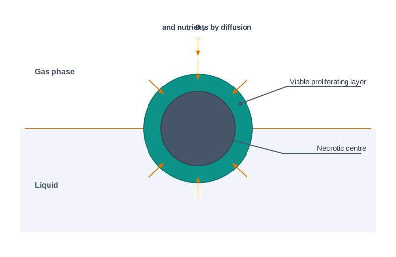
Fig. 9.6 – Organ culture at a gas–liquid interface.
Most of the time, organ cultures are exposed to high \(\\mathrm{O\_2}\) concentration to push more oxygen inward by diffusion. However, the use of high \(\\mathrm{O\_2}\) concentration is accompanied by the risk of \(\\mathrm{O\_2}\)-prompted toxicity in organs. The culture worker is therefore caught between two dangers — too little oxygen kills the centre of the tissue, and too much oxygen damages the tissue chemically.
To deal with this difficulty in organ cultures, the tissues are maintained at a gas–liquid phase to facilitate the exchange of gases while maintaining access to adequate nutrients. The tissue sits at the boundary, drawing nutrients from the liquid below and gases from the air above.
Anchorage of the tissue to a solid substrate can lead to the development of an outgrowth of cells from the tissue and resultant alterations in geometry, though this effect can be minimised by using a hydrophobic surface. A hydrophobic, water-repelling surface discourages the cells from spreading out over it, so the explant keeps its shape instead of flattening into a sheet.
The significance of maintaining organ cultures at the interface of the gaseous and liquid phases is as follows. If the liquid is retained at the optimal level, then the tissue maintains a spherical geometry. However, if the level of liquid is too superficial, the tissue will outgrow and flatten as a result of surface tension. If the level is too deep, the exchange of gases is compromised.
To enhance the permeation of oxygen in organ cultures, hyperbaric oxygen or enhanced pure oxygen content is used. Hyperbaric means oxygen supplied at a pressure above normal atmospheric pressure, which drives more of it into solution and thus deeper into the tissue.
9.2.2 Growth and differentiation
Growth refers to an increase in the number of cells in a culture, whereas differentiation refers to a change in the function of the differentiated cell. These are two different achievements, and in organ culture they pull against each other.
Growth and differentiation are related to each other in such a way that the differentiated cells may not be able to proliferate further. There is also a possibility that, irrespective of cell density, the termination of growth may promote the induction of differentiation — that is, once a cell stops dividing, it is more likely to begin specialising.
Most of the organ cultures do not grow. Instead they proliferate only on the outer layers of cells, as a consequence of the physical limitations executed by the structure and shape of the explant, which is the same diffusion limit discussed in the previous subsection.
The organ culture is tolerant to suitable cellular communications and differentiations, and also tolerant to maintaining the appropriate environment. Soluble growth factors are supplied to facilitate differentiation — since there is no blood supply to bring these signalling molecules in, they must be added to the medium instead.
Whole embryo culture
Spratt (1950s) explained the effect of metabolic inhibitors on the developing embryo in vitro. A metabolic inhibitor is a substance that blocks a particular biochemical pathway, so watching what goes wrong in the embryo afterwards reveals what that pathway was needed for.
To culture the embryo, a suitable medium has to be prepared and poured onto a watchglass, which is then placed on a small piece of moist absorbent cotton wool pad in a petridish. The moist pad keeps the humidity high inside the dish, so that the small volume of medium in the watchglass does not dry out.
To observe the chick embryo in culture, the eggs were incubated at 38°C for 40–42 hours, so that a dozen embryos could be produced. This particular window is chosen because by then the blastoderm has developed far enough to be handled and examined, but the embryo is still flat and thin enough to survive by diffusion.
The egg shell was sterilised with 70 percent ethanol, then broken into pieces and kept in 50 ml of balanced salt solution (BSS). Ethanol at 70 percent is used rather than absolute alcohol because it penetrates and kills microbial cells more effectively; sterilising the shell first prevents surface contaminants from being carried into the culture.
The vitelline membrane covering the blastoderm was removed and kept in BSS. The adherent vitelline membrane was removed with the help of a forcep. The embryo was observed under a microscope to study the developmental stage of the blastoderm.
The blastoderm was then carefully placed on the sterile absorbent cotton wool pad in the watchglass in the petridish, and the embryo culture of chick was incubated at 37.5°C for further development. The whole sequence is set out in Fig. 9.7.
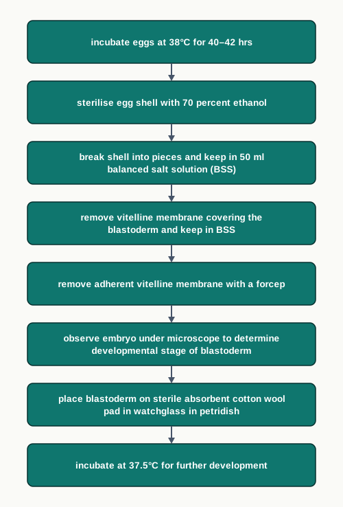
Fig. 9.7 – Whole embryo (chick) culture.
9.2.3 Types of organ culture
Histotypic culture
When any characterised cell line is proliferated or cultured at a high density in the presence of suitable soluble factors and an extracellular matrix, then such a culture is termed histotypic. The word points to the aim: a culture that resembles a tissue, built from a single, known cell type grown densely enough to behave like one.
Vascular endothelial cells can form capillary tubules in the presence of appropriate soluble factors when grown in a collagen matrix. This is a striking demonstration of the principle — given the right matrix and the right signals, the cells organise themselves into the structure they would form in the body, without being told where to go.
Cellulose sponges coated with extracellular matrix components such as collagen are another example. The sponge acts as a scaffold, giving the cells a three-dimensional space to occupy instead of a flat floor. Alternatively, cells may infiltrate the sponge and form glandular structures.
Organotypic culture
The limitation in a histotypic culture is the inability to evaluate heterologous cell interactions — that is, interactions between cells of different types, which is exactly what happens in a real organ.
When cells of different lineages are co-cultured to create a tissue-like structure, then it is termed organotypic culture, which is the easiest way to maintain a co-culture of two cell types.
For instance, co-culture of fibroblast and epithelial cell clones derived from the mammary gland allows the epithelial cells to differentiate functionally. This has been demonstrated by their ability to produce milk proteins in an optimal hormonal environment. The result is important because the epithelial cells will not do this on their own — the presence of the fibroblasts is part of what makes them behave like real mammary tissue.
The formation of characteristic structures is further validated by functional differentiation, such as the three-dimensional cords in which the fibroblast cells reorganise themselves into bundles that are further enveloped by the epithelial cells.
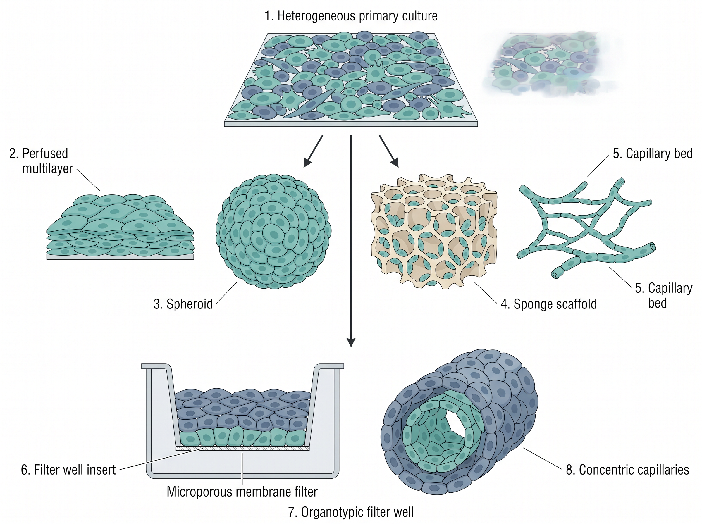
Fig. 9.8 – Histotypic and organotypic cultures.
Organoids
‘Organoids’ are self-organised 3D tissue cultures derived from stem cells. Thus the organoid is made from a combination of cells, growth factors, collagen and similar components. The key word is self-organised: the cells are not assembled into shape by the experimenter, they arrange themselves once the right conditions are provided.
An example is an artificial liver surgically implanted into the peritoneal cavity of a rat. These organoids have the potential to replace any diseased or non-functional organ, or to deliver genetically altered cells into a patient’s body.
9.2.4 Applications of organ culture
Cultured organs can be used as a substitute for organs. This may be beneficial, as the accessibility of transplantable organs is decreasing while the number of patients waiting for them is not.
One more significance of organ culture is that the organs are produced by using the patient’s own stem cells. This would permit transplantation of organs without the use of immunosuppressive drugs by patients — and, as you saw in the previous section, avoiding immunosuppression also avoids the increased risk of microbial infection that comes with it.
The organ culture helps to study the actual behaviour of a tissue in an in vitro system, as well as to understand the biochemical and functional characteristics of an organ or tissue and their comparison with those of similar organs in vivo, in an easier way.
The organ culture is also suitable for the study of hormones and their effects, either individually or in combinations with other hormones. This is difficult to do in a whole animal, where many hormones act at once and their effects cannot easily be separated. For example, the mammary gland is one of the most commonly cultured organs of the mouse.
9.2.5 Limitations of organ culture
There are some limitations of organ culture. Fundamentally, organ cultures depend on histological techniques and not so much on biochemical and molecular analyses.
The reason is that biochemical analysis is based on reproducibility, which cannot be achieved in organ culture. If two supposedly identical cultures are not truly identical, then a difference in a measured chemical value cannot safely be attributed to the treatment being tested.
Furthermore, organ cultures exhibit more difficulty in the preparation of replicates than cell cultures do. The organ culture exhibits high variations and low reproducibility. Thus, in organ culture, a fresh organ from a donor is needed for each experiment.
Various limitations of organ culture are as follows:
Since the organ cultures cannot proliferate, therefore fresh organs are required for every experiment.
In organ culture, reproducibility is less and disparities are high. This can be because of a slight variation in geometry and handling, sampling difference in organising an organ culture, and differences in the proportions of types of cell among cultures.
Organ cultures are very challenging to prepare and are also very expensive.
By this technique, the behaviour of integrated tissue only can be examined, and not of isolated cells.
Read (a) and (d) together and the trade-off at the heart of the method becomes clear. Preserving the intact tissue is what gives organ culture point its realism, and it is also what makes the cultures scarce, variable and unable to answer questions about single cells.
9.2.6 Future Prospects
One of the future challenges is to adapt the cell-based assay protocols developed for the analysis of 2D cell culture to the altered conditions of cells growing in 3D structures. An assay standardised on a flat monolayer often gives different readings in a three-dimensional tissue, because drugs and stains do not penetrate a 3D structure the way they cover a flat sheet.
3D cell cultures include multicellular spheroids, scaffold hydrogels, organoids, organs-on-chips, hanging drop, microfluidics, magnetic levitation, microtissues and 3D bioprinting. Each of these is a different answer to the same question that has run through this whole section — how to give cells a realistic three-dimensional environment while still getting nutrients and oxygen to every part of the tissue.
9.3 Summary
- Stem cells are unspecialised cells that can renew themselves by mitotic division and can also differentiate into a diverse range of cell types. A cell must do both to be called a stem cell.
- On the basis of potency, stem cells are classified into totipotent, pluripotent, multipotent and unipotent cells.
- Totipotent stem cells can differentiate into all cell types of an organism, including the extra-embryonic membranes. The zygote is the classic example.
- Pluripotent stem cells can differentiate into almost all cell types of the embryo, except the cells of the extra-embryonic support tissues such as the placenta and yolk sac. This is why they cannot form a complete organism.
- Multipotent cells can differentiate into a closely related family of cells within one lineage, as hematopoietic stem cells do for blood cells.
- Unipotent stem cells self-renew but produce only cells of their own type, such as epidermal, muscle and endothelial stem cells.
- On the basis of source, stem cells are divided into early (or embryonic) stem cells and mature (or adult) stem cells, with fetal stem cells forming a further group.
- The early stem cells, that is the embryonic stem cells, are present in the inner cell mass of a blastocyst after about five days of development, and are pluripotent.
- Adult stem cells are undifferentiated totipotent or multipotent cells found in specific mature body tissues, as well as in the umbilical cord and placenta after birth. Their frequency is highest in bone marrow.
- Plasticity is the ability of an adult stem cell to give rise to cell types unlike those of its tissue of origin. It depends on the competence of the responding cells and on inductive cues from the microenvironment.
- Stem cells have the potential to treat a number of diseases including cancer, Type 1 diabetes mellitus, Parkinson’s disease, cardiac diseases and neurological disorders, and they are also used as models for drug screening.
- The main challenges in stem cell research are immunological rejection, the infection risk that follows immunosuppression, possible tumour formation by indefinitely dividing cells, and microbial contamination.
- Development of a part of an organ, or of the whole organ itself, from tissue culture techniques is termed organ culture.
- The most important advantage of organ culture is that the whole three-dimensional structure of the organ can be recreated.
- The important characteristics of organ culture are structural integrity, nutrient and gas exchange, and growth and differentiation. Because there is no vascular system, diffusion from the periphery limits the size of the explant and may cause necrosis at the centre.
- The types of organ culture are whole embryo culture, histotypic culture and organotypic culture. Organoids are self-organised 3D cultures derived from stem cells.
- Organ culture helps to study the actual behaviour of a tissue in an in vitro system, and to understand the biochemical and functional characteristics of an organ or tissue and compare them with those of similar organs in vivo, in an easier way.
- Organ culture helps in understanding developmental biology and interaction in tissues.
- Its limitations are that the cultures do not proliferate, that fresh organs are needed for every experiment, that reproducibility is low and variation high, that preparation is difficult and expensive, and that only integrated tissue — not isolated cells — can be examined.
- The overall route from a single fertilised cell to a cultured tissue used in the laboratory is summarised in Fig. 9.9.
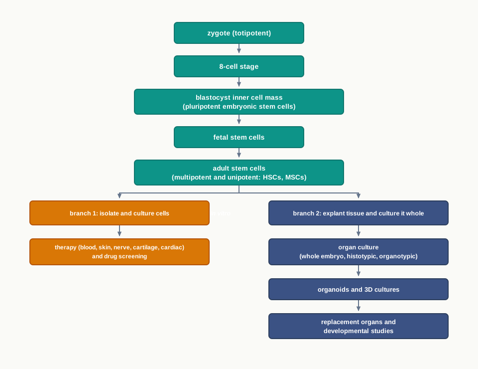
Fig. 9.9 – Stem cell and organ culture overview.
9.4 Key Terms
- Fertilisation — The fusion of the male gamete (sperm) with the female gamete (oocyte) to form a zygote.
- Zygote — The single cell formed by fertilisation, which is totipotent and can develop into a complete organism.
- Differentiation — The process by which an unspecialised cell acquires the structure and function of a particular specialised cell type.
- Stem cell — A non-specialised cell that can renew itself by mitotic division and can also differentiate into a wide range of specialised cell types.
- Self-renewal — Division of a stem cell in a way that maintains the stem cell pool instead of using it up.
- Potency — The range of cell types that a given stem cell is able to become.
- Regenerative medicine — The branch of medicine that seeks to rebuild damaged tissues and organs rather than only manage their symptoms.
- In vitro — Grown or studied outside the living body, in a culture vessel.
- In vivo — Occurring inside the living body.
- Blastocyst — The hollow ball of cells formed in early development, whose inner cell mass yields embryonic stem cells after about five days.
- Inner cell mass — The cluster of cells inside the blastocyst that gives rise to the tissues of the adult organism.
- Embryonic stem cells (ESCs) — Pluripotent, possibly immortal stem cells isolated from the inner cell mass or epiblast of the blastocyst.
- Germ layers — The three primary layers of the embryo — ectoderm, mesoderm and endoderm — from which all body tissues arise.
- Trophectoderm — The outer layer of the blastocyst, which forms the placenta.
- Gastrulation — The developmental stage at which pluripotent cells stop merely dividing and begin to specialise into the germ layers.
- Fetal stem cells — Stem cells derived from a fetus, obtained from fetal blood, bone marrow, liver or kidney, which can live indefinitely in the laboratory.
- Progenitor cell — A partly committed intermediate cell that can still divide and give rise to specialised descendants.
- Adult (somatic) stem cells — Undifferentiated multipotent or totipotent stem cells present in mature body tissues, umbilical cord and placenta, whose role is to preserve and repair the tissue in which they reside.
- Stemness — The complete set of stem-cell properties that a cell must retain when it self-renews.
- Cellular homeostasis — The steady balance between cells lost and cells replaced, maintained in tissues by adult stem cells.
- Hematopoiesis — The formation of the different types of blood cells, which occurs in the bone marrow.
- Hematopoietic stem cells (HSCs) — Bone marrow stem cells that give rise to RBCs, monocytes, neutrophils, acidophils, basophils, lymphocytes and platelets.
- Mesenchymal stem cells (MSCs) — Highly proliferative, genetically stable stem cells from bone marrow, adipose tissue and umbilical cord that repair skeletal and other vital tissues.
- Allo-rejection — The immune attack mounted against transplanted cells that have come from another individual.
- Totipotent — Able to form both the embryo and the extra-embryonic membranes, and therefore an entire functional organism.
- Pluripotent — Able to form almost all cell types of the embryo, but not the extra-embryonic support tissues such as placenta and yolk sac.
- Multipotent — Able to differentiate only into a closely related family of cells within a specific lineage.
- Unipotent — Able to self-renew but able to produce only cells of its own type.
- Plasticity — The ability of a tissue-specific adult stem cell to attain the fate of cell types distinct from its tissue of origin.
- Competence — The capability of a cell to respond to a signal and undertake a particular change of fate.
- Inductive microenvironment — The surrounding cues, factors and matrix that trigger a competent cell to change its fate.
- Trans-determination — Redirection of a stem cell’s potential, so that it switches to another stem cell type and produces that precursor’s lineages.
- Trans-differentiation — The process in which an already differentiated cell achieves the phenotype of another differentiated cell.
- Dedifferentiation — Reversion of adult stem cells to the master cells from which they originated, common in lower vertebrates but rare in mammals.
- Surface markers — Characteristic membrane proteins whose presence or absence is used to determine the purity and identity of stem cells.
- Pleiotropic — Describing plasticity between two cells of the same germ layer that is explained by clonal level impurity.
- Fusion — An alternative mechanism for apparent plasticity, in which a donor stem cell physically fuses with a host cell.
- Induced pluripotent stem cells (iPSCs) — Pluripotent cells generated by reprogramming a person’s own mature cells, which reduces the problem of immunological rejection.
- Cryopreservation — Storage of cultured cells at very low temperature so that they remain viable for later use.
- Keratinocyte stem cells — Skin stem cells present in the hair follicle, which can be cultured to produce skin equivalent to the patient’s own.
- Auto-immune disorder — A condition in which an over-reactive immune system treats the body’s own healthy tissues as foreign and attacks them.
- Ischemia — An inadequate blood supply to a tissue, which in the heart leads to muscle injury and loss of function.
- Chondrocytes — Cartilage-making cells into which stem cells can be differentiated for the treatment of arthritis.
- Organ culture — The growth of a part of an organ, or a whole organ, on an artificial medium in vitro with its anatomical relationships and physiological functions preserved.
- Explant — A piece of living tissue removed from an organism and transferred to a culture vessel.
- Necrosis — The death of cells within a living tissue, which occurs at the centre of an organ culture when diffusion cannot supply enough oxygen and nutrients.
- Polarity — The distinct orientation of a cell, such as an epithelial cell having a different upper and lower surface, which may be lost in culture.
- Gas–liquid phase (interface) — The boundary at which organ cultures are maintained so that gases are exchanged from above while nutrients are drawn from the medium below.
- Hyperbaric oxygen — Oxygen supplied above normal atmospheric pressure to improve its permeation into an organ culture.
- Balanced salt solution (BSS) — A salt solution of physiological composition used to hold and rinse tissues during dissection and transfer.
- Blastoderm — The disc of cells of the early chick embryo that is explanted and cultured in whole embryo culture.
- Vitelline membrane — The membrane covering the blastoderm, which must be removed before the embryo can be cultured.
- Histotypic culture — A high-density culture of a single characterised cell line grown with soluble factors and extracellular matrix so that it resembles a tissue.
- Organotypic culture — A co-culture of cells of different lineages that creates a tissue-like structure and allows heterologous cell interactions to be studied.
- Extracellular matrix — The network of substances such as collagen that surrounds cells and provides the scaffold and signals needed for tissue-like organisation.
- Organoid — A self-organised three-dimensional tissue culture derived from stem cells, made from a combination of cells, growth factors and collagen.
- Organs-on-a-chip — A modern culture approach that partially mimics the tissue microenvironment on a miniature device.
- 3D bioprinting — A three-dimensional culture technique in which cells and supporting material are printed layer by layer to build tissue-like structures.
9.5 Points to Ponder
- A stem cell must both renew itself and produce specialised cells. Think about what would happen to a tissue over a lifetime if every stem cell division produced two specialised daughters instead.
- The zygote is totipotent, but by the fifth day the inner cell mass is only pluripotent. Nothing has been removed from these cells. What, then, has actually changed?
- Adult stem cells were once believed to be permanently committed to one lineage. Consider what kind of evidence would be needed before a scientist could confidently claim that a cell has genuinely changed its lineage, rather than merely appearing to.
- Embryonic stem cells divide indefinitely in culture. The same property is described in this chapter both as an advantage and as a hazard. Reflect on how a single characteristic can be useful in a dish and dangerous in a patient.
- Induced pluripotent stem cells are made from a patient’s own mature cells. Think about which of the problems described in this chapter this approach solves, and which ones it leaves untouched.
- Skin can be grown from a plucked hair, but a whole liver cannot be grown the same way. What is it about an organ, as opposed to a sheet of cells, that makes the difference?
- Organ culture keeps the architecture of a tissue intact and therefore gives realistic results, yet it is also expensive, variable and hard to replicate. Consider when a researcher should accept these costs and when a simple cell culture would serve better.
- The centre of an organ culture may die while the medium around it is rich in nutrients. Think about how the body solves this same problem, and why that solution is so difficult to copy in a dish.
- In organotypic culture, mammary epithelial cells produce milk proteins only when fibroblasts are present. Reflect on what this suggests about the idea that a cell’s behaviour is written entirely in its own genes.
- Organ cultures are analysed mainly by histological methods rather than biochemical ones. Consider what kinds of scientific questions this restriction allows us to ask, and what kinds it quietly rules out.
- Stem cell therapies are described in this chapter as mostly experimental and costly. Think about who is likely to gain access to such treatments first, and what would need to change for that access to become fair.
- Bone marrow transplantation has been in routine clinical use for decades, while most other stem cell therapies remain in trials. What might explain why this one application succeeded so much earlier than the rest?
9.6 NCERT Questions
- Briefly describe the stem cells and their properties.
- Differentiate totipotent, pluripotent and multipotent stem cells.
- What are embryonic stem cells and how do they differ from adult stem cells?
- Describe some applications of stem cells.
- What parameters should be monitored during stem cell culture?
- What is organ culture?
- Describe the main characteristics of organ culture.
- Discuss the various types of organ culture.
- What are the advantages of organ culture over cell culture?
- Describe the applications of organ culture.
- Describe the various support systems used in organ culture.
- Stem cells are present in:
- unicellular organisms
- multicellular organisms
- non-living things
- viruses
- Differentiation potential of stem cells specifies:
- Stochastic differentiation
- Asymmetric replication
- Potency
- Self-renewal
- Which of the following cells is a multipotent cell?
- T-cell
- B-cell
- HSC
- Monocytes
- A stem cell is:
- a cell out of which the stem of the tree is made up of
- a part of the tissue that forms the outer layer of the skin in human beings.
- it is a cell that can divide and give rise to specialised cells.
- a type of specialised cell
- __________ can be cured with stem cells.
- Spinal cord injuries
- Type 1 diabetes
- Both (a) and (b)
- None of these
- The stem cells may be obtained from sources such as:
- Bone marrow
- Umbilical cord blood
- Adipose tissue
- All of these
- Assertion: Embryonic stem cells can give rise to different cell types. Reason: Embryonic stem cells are pluripotent.
- Both assertion and reason are true and the reason is the correct explanation of the assertion.
- Both assertion and reason are true but the reason is not the correct explanation of the assertion.
- Assertion is true but reason is false.
- Both assertion and reason are false.
- Assertion: Stem cells are undifferentiated and found in multicellular organisms, and undergo numerous mitotic cycles. Reason: Stem cells have ‘self-renewal’ feature and do not exhibit ‘cellular potency’.
- Both assertion and reason are true and the reason is the correct explanation of the assertion.
- Both assertion and reason are true but the reason is not the correct explanation of the assertion.
- Assertion is true but reason is false.
- Both assertion and reason are false.
9.7 Objective Questions
- All cells of the early embryo remain totipotent up to the:
- two-cell stage
- four-cell stage
- eight-cell stage
- gastrula stage
- Which of the following can a pluripotent stem cell NOT form?
- Ectoderm
- Mesoderm
- Endoderm
- Placenta
- Human embryonic stem cells are isolated from the:
- trophectoderm of the blastocyst
- inner cell mass or epiblast of the blastocyst
- yolk sac
- umbilical cord after birth
- The embryo is referred to as a fetus after:
- the fifth day of development
- the fourth week of development
- the eighth week of development
- the twelfth week of development
- The highest frequency of adult stem cells in the human body is found in:
- adipose tissue
- skin
- bone marrow
- liver
- Which of the following is NOT produced by hematopoietic stem cells?
- Neutrophils
- Chondrocytes
- Platelets
- Lymphocytes
- Mesenchymal stem cells are chiefly associated with the making and repair of:
- neurons and glial cells
- cartilage, bone and fat
- red blood cells and platelets
- retinal pigment epithelium
- Neural stem cells give rise to neurons, oligodendrocytes and astrocytes. On the basis of potency, they are therefore:
- totipotent
- pluripotent
- multipotent
- unipotent
- Epidermal stem cells, muscle stem cells and endothelial stem cells are examples of:
- totipotent stem cells
- pluripotent stem cells
- multipotent stem cells
- unipotent stem cells
- In the blastocyst, the placenta is formed from the:
- inner cell mass
- trophectoderm
- epiblast
- yolk sac
- The process in which an already differentiated cell achieves the phenotype of another differentiated cell is called:
- trans-determination
- trans-differentiation
- dedifferentiation
- fusion
- The purity of a stem cell population is normally determined by:
- the size of the cells
- the presence and absence of specific surface markers
- the colour of the culture medium
- the rate of oxygen consumption
- In the protocol for maintenance of cell culture, MEFs are digested to single cells using:
- 0.25% trypsin/EDTA
- 70 percent ethanol
- MMC at 10 mg/mL
- balanced salt solution
- In the same protocol, the MEFs are seeded on 10 cm dishes at a density of:
- \(8 \\times 10^{2}\)–\(1.1 \\times 10^{3}\) cells/cm²
- \(8 \\times 10^{3}\)–\(1.1 \\times 10^{4}\) cells/cm²
- \(8 \\times 10^{4}\)–\(1.1 \\times 10^{5}\) cells/cm²
- \(8 \\times 10^{6}\)–\(1.1 \\times 10^{7}\) cells/cm²
- Skin stem cells used for growing a patient’s own skin are obtained from the:
- sweat gland
- hair follicle
- dermal blood vessels
- subcutaneous fat
- In the treatment of multiple sclerosis, the structure that stem cell therapy aims to restore is the:
- motor end plate
- myelin sheath
- synaptic vesicle
- blood–brain barrier
- Which of the following is a challenge specifically created by the indefinite division of embryonic stem cells?
- Allo-rejection
- Possible tumour growth
- Microbial contamination
- Loss of polarity
- Necrosis at the centre of an organ culture is chiefly a consequence of:
- an excess of soluble growth factors
- the absence of a vascular system, so exchange occurs only by diffusion
- the use of a hydrophobic culture surface
- the removal of the vitelline membrane
- If the level of liquid in an organ culture is too superficial, the tissue will:
- remain spherical
- outgrow and flatten because of surface tension
- become completely necrotic within minutes
- detach and float
- In whole embryo culture of the chick, the eggs are first incubated at:
- 37.0°C for 24 hours
- 37.5°C for 40–42 hours
- 38°C for 40–42 hours
- 38°C for 5–7 days
- The egg shell is sterilised in whole embryo culture using:
- 70 percent ethanol
- absolute alcohol
- 0.25% trypsin
- hyperbaric oxygen
- Vascular endothelial cells forming capillary tubules in a collagen matrix in the presence of soluble factors is an example of:
- organotypic culture
- histotypic culture
- whole embryo culture
- suspension culture
- Co-culture of mammary fibroblasts with epithelial cell clones, leading to the production of milk proteins, is an example of:
- histotypic culture
- organotypic culture
- monolayer culture
- whole embryo culture
- Which of the following is a self-organised 3D tissue culture derived from stem cells?
- Explant
- Blastoderm
- Organoid
- Monolayer
- Organ cultures depend mainly on which type of analysis?
- Histological techniques
- Biochemical assays
- Molecular sequencing
- Flow cytometry of single cells
- Which of the following is NOT listed as a 3D cell culture approach in this chapter?
- Hanging drop
- Magnetic levitation
- Scaffold hydrogels
- Agar slant streaking
- Assertion: The zygote is described as totipotent, but embryonic stem cells of the inner cell mass are not. Reason: Embryonic stem cells cannot form the extra-embryonic support tissues such as the placenta and yolk sac.
- Assertion: Adult stem cells raise fewer ethical objections than embryonic stem cells. Reason: Adult stem cells are obtained from mature body tissues, umbilical cord and placenta, and not from the early embryo.
- Assertion: Immunosuppressive treatment given after a stem cell transplant makes the recipient prone to microbial infections. Reason: The same immune activity that would reject the graft also defends the body against microbes.
- Assertion: A researcher claiming plasticity of adult stem cells must first establish the clonal level purity of the starting population. Reason: Heterogeneity of the starting cells can produce an apparent change of lineage that never actually occurred.
- Assertion: Organ cultures are limited in size. Reason: Organ cultures lack a vascular system, so oxygen and nutrients reach the cells only by diffusion from the periphery.
- Assertion: Organ cultures are maintained at the interface of the gaseous and liquid phases. Reason: At the optimal liquid level the tissue keeps a spherical geometry while still exchanging gases freely.
- Assertion: Biochemical analysis is difficult to perform on organ cultures. Reason: Organ cultures show high variation and low reproducibility, and replicates are difficult to prepare.
- Assertion: Organs cultured from a patient’s own stem cells could be transplanted without immunosuppressive drugs. Reason: Cells carrying the patient’s own surface markers are not recognised as foreign by the patient’s immune system.
9.8 Check Your Concepts
- A cell in a tissue divides repeatedly but every daughter cell remains identical to the parent and never specialises. Another cell specialises perfectly but can no longer divide. Explain why neither of these qualifies as a stem cell.
- Why is the zygote able to produce a complete organism while the cells of the inner cell mass, only about five days later, cannot? Answer in terms of potency.
- Arrange the following in the correct developmental sequence and state the potency at each stage: morula, blastocyst inner cell mass, zygote, four-celled stage, adult tissue stem cells.
- Compare embryonic stem cells and adult stem cells under the headings: source, potency, ease of obtaining, clinical promise, and ethical concern.
- Embryonic stem cells are described as exhibiting a stable diploid chromosome number even after extensive manipulation. Why would a cell line that gains or loses chromosomes in culture be unsuitable for transplantation?
- Fetal blood is described as a rich source of haemopoietic stem cells that proliferate more rapidly than those of cord blood or adult bone marrow. Explain why a faster rate of proliferation is an advantage in a transplant.
- Adult stem cells are called somatic stem cells even though they can be obtained from children and from the umbilical cord. Explain why the second name is considered more precise.
- Distinguish between hematopoietic and mesenchymal stem cells with respect to origin, tissues of isolation, cells produced, and therapeutic use.
- Draw and label a diagram showing a hematopoietic stem cell giving rise to the different types of blood cells, and mark the arrow that represents self-renewal.
- “Multipotent and unipotent stem cells belong to adult life, whereas totipotent and pluripotent stem cells belong to embryonic life.” Justify this statement with one example of each.
- Match the following:
| Column A | Column B |
|---|---|
| a. Zygote | i. Unipotent |
| b. Inner cell mass of blastocyst | ii. Multipotent |
| c. Hematopoietic stem cell | iii. Totipotent |
| d. Epidermal stem cell | iv. Pluripotent |
- Two conditions must be satisfied before an adult stem cell can adopt the fate of a different tissue. Name both and explain, with the bone marrow example, what happens when only one of them is met.
- Differentiate between trans-determination and trans-differentiation. In each case, state whether the cell that changes is a stem cell or an already differentiated cell.
- Dedifferentiation is common in lower vertebrates but rare in mammals. Describe what happens during dedifferentiation and explain why it can be thought of as a step backwards followed by a step forwards along a different branch.
- A scientist reports that stem cells taken from bone marrow have produced liver cells. A reviewer asks whether heterogeneity or fusion could explain the result instead. Explain what each of these alternative explanations would mean, and what evidence would rule them out.
- In the protocol for maintenance of cell culture, the adherent cells are washed with PBS five times after the MMC-containing medium is discarded. What would go wrong if this washing step were omitted?
- Explain the role of each component in the phrase “0.25% trypsin/EDTA”, and state why the two are used together rather than separately.
- A student seeds cells at a density far below the range given in the maintenance protocol, and a second student seeds far above it. Predict the outcome in each culture and explain why a defined seeding density is specified at all.
- Explain how skin can be grown for a burn patient starting from a plucked hair, and state two advantages of this graft over a conventional skin graft.
- In Type 1 diabetes, insulin injections are already available. Explain why a stem cell-derived therapy is nevertheless being explored, in terms of what the injections cannot do.
- Why are cardiomyocytes derived from human embryonic stem cells considered a better model for testing a new drug’s effect on the heart than an animal cell model?
- The property that makes embryonic stem cells valuable in the laboratory is also the property that makes them risky in a patient. Identify the property and explain both sides of this statement.
- Explain how the use of induced pluripotent stem cells reduces the need for immunosuppressive treatment, and state which of the challenges of stem cell research it does NOT solve.
- “In cell culture the cells are discrete, whereas in organ culture they are unified as a single unit.” Explain this statement and describe one scientific question that only organ culture can answer.
- Why must a tissue meant for organ culture reach the laboratory within minutes of collection, and why must it not be crushed or disrupted during handling?
- Explain, step by step, why necrosis appears at the centre of a large organ explant even when the surrounding medium is rich in nutrients.
- Organ cultures are often exposed to a high oxygen concentration, yet high oxygen is itself described as a risk. Explain this apparent contradiction and name the technique used to improve oxygen permeation.
- Predict what will happen to an organ culture in each case: (i) the liquid level is too superficial, (ii) the liquid level is too deep, (iii) the tissue is anchored to a hydrophilic solid substrate.
- Growth and differentiation are said to pull against each other in organ culture. Explain this relationship, and state why soluble growth factors must be added to the medium.
- The following steps of whole embryo culture of the chick are jumbled. Arrange them in the correct order: place the blastoderm on the cotton wool pad in the watchglass; sterilise the egg shell with 70 percent ethanol; incubate at 37.5°C; incubate eggs at 38°C for 40–42 hours; remove the adherent vitelline membrane with a forcep; examine the blastoderm under a microscope; break the shell and keep the contents in balanced salt solution.
- In whole embryo culture, the watchglass is placed on a moist absorbent cotton wool pad rather than directly on the base of the petridish. Give the reason.
- Compare histotypic and organotypic culture under the headings: cells used, method of forming a tissue-like structure, type of interaction studied, and one example of each.
- Mammary epithelial cells produce milk proteins only when co-cultured with fibroblasts in an optimal hormonal environment. What does this tell you about the control of differentiation?
- Define an organoid and explain why the word “self-organised” is central to the definition.
- Explain why an organ produced from a patient’s own stem cells would not require immunosuppressive drugs after transplantation.
- Organ culture is described as giving realistic results but poor reproducibility. Identify the single feature of the technique that is responsible for both, and explain how it produces these opposite consequences.
- A laboratory wants to test the combined effect of two hormones on mammary tissue. Explain why organ culture is better suited to this than a study in a whole animal.
- Explain why an assay standardised on a 2D monolayer may give a different result when applied to cells growing in a 3D structure.
9.9 Case-Based Questions
A nine-year-old child is diagnosed with a disease in which the bone marrow no longer produces healthy blood cells. The doctors plan to destroy the faulty marrow and replace it with marrow from a matched donor. The parents are told that the transplant works because of a small population of cells present in the donated marrow, and that the child will be given immunosuppressive drugs for some time afterwards and kept in a protected room.
- Name the population of cells responsible for the success of the transplant and list the blood cell types they can give rise to.
- State the potency of these cells and justify your answer.
- Explain why immunosuppressive drugs are given, and why the child must then be protected from infection.
- The chapter describes most stem cell therapeutics as experimental and costly, with one notable exception. Identify the exception and comment on why this case fits it.
A patient has suffered deep burns over a large area of the body. Conventional skin grafting is difficult because there is very little undamaged skin left to harvest. The treating team decides instead to pluck hairs from the patient’s scalp, culture the cells obtained from the follicles, and grow sheets of skin in the laboratory.
- Name the type of stem cell obtained from the hair follicle and state its potency.
- Explain why a graft grown from these cells is much less likely to be rejected than skin from another person.
- State two advantages of this approach over a conventional skin graft, as described in the chapter.
- Suggest why plucking a hair is a more acceptable way of obtaining stem cells than removing a piece of bone marrow.
A research student sets up an organ culture of a small piece of mouse mammary gland. Wanting to be sure the tissue is well fed, she uses a deep layer of nutrient-rich medium so that the explant is completely submerged. She also chooses a large explant, about 4 mm thick, so that there will be plenty of tissue for sectioning. After two days, sections show a healthy rim of cells surrounding a dead central region.
- Name the condition seen in the central region and explain why it has occurred.
- Identify two mistakes in the way the culture was set up, and correct each one.
- Explain why an organ culture is normally maintained at a gas–liquid interface rather than fully submerged.
- Suggest one method mentioned in the chapter for improving oxygen permeation into an organ culture.
A school laboratory attempts a whole embryo culture of the chick, following the classical method. Fertile eggs are incubated at 38°C for 40–42 hours. One group of students, in a hurry, skips the ethanol treatment of the shell and also leaves the vitelline membrane in place over the blastoderm. Their culture becomes cloudy within a day and the embryo does not develop; the other groups obtain healthy embryos after incubation at 37.5°C.
- State the purpose of treating the egg shell with 70 percent ethanol and explain the probable cause of the cloudiness.
- Explain why the vitelline membrane must be removed before the blastoderm is placed in the watchglass.
- Why are the eggs incubated for a specific window of 40–42 hours rather than for several days?
- Name the solution in which the shell contents and the membrane are kept during the procedure, and state its purpose.
A pharmaceutical company is testing a new molecule intended to treat a heart condition. The company decides not to rely only on animal testing. Instead, it differentiates human embryonic stem cells into cardiomyocytes and exposes them to a range of concentrations of the molecule, while a second team differentiates the same cell line into neuronal cells to check for effects on the nervous system.
- Explain why human cardiomyocytes grown in culture may predict harm to the human heart better than an animal model does.
- State two other uses of stem cells in the pharmaceutical industry mentioned in the chapter.
- Name the property of embryonic stem cells that allows the same cell line to be used for both heart and nerve models.
- The team using ordinary 2D culture finds that results change when the same assay is repeated in a 3D organoid. Suggest a reason.
A national committee is reviewing an application from a laboratory that wishes to derive a new human embryonic stem cell line. The laboratory argues that the cells are pluripotent, can be expanded almost indefinitely, and could be directed into retinal pigment epithelium for patients losing their sight. Members of the public have written to the committee objecting to the source of the cells. A second laboratory has applied to work instead with induced pluripotent stem cells made from adult skin cells.
- State where human embryonic stem cells are obtained from, and explain why this is the source of the objection.
- Give one scientific and one ethical argument in favour of the first laboratory’s application.
- Explain how the second laboratory’s approach avoids the ethical objection, and state one problem it does not solve.
- Name two safeguards that such work is normally carried out under.
A technician is preparing a stock of mouse embryonic fibroblasts following the standard maintenance protocol. She digests the cells with 0.25% trypsin/EDTA, seeds them onto 10 cm dishes, incubates them at 37°C, treats them with MMC at 10 mg/mL, and then, being short of time, rinses the dish once with PBS instead of five times before the final trypsin step. The cells detach poorly and many appear damaged; the batch is discarded rather than cryopreserved.
- Explain the function of trypsin and of EDTA in the first step of this protocol.
- Explain how the shortened washing step could account for the damaged appearance of the cells.
- State the purpose of the aspiration step that immediately follows the MMC incubation.
- Name the final step of the protocol and explain why stem cells tolerate it well.
9.10 Practice With Data
- In the maintenance protocol described in this chapter, cells are seeded onto dishes of 10 cm internal diameter at a density of \(8 \\times 10^{4}\)–\(1.1 \\times 10^{5}\) cells/cm². Taking \(\\pi = 3.14\), (i) calculate the growth area of one dish in cm², (ii) calculate the minimum and the maximum number of cells needed to seed one dish, and (iii) calculate how many such dishes could be seeded at the maximum density from a suspension containing \(4.0 \\times 10^{7}\) cells in total.
- After trypsinisation, a technician obtains 8.0 mL of cell suspension containing \(2.5 \\times 10^{6}\) cells/mL. (i) Calculate the total number of cells harvested. (ii) Using the growth area calculated in the previous problem, find the volume of this suspension needed to seed one dish at the minimum density of \(8 \\times 10^{4}\) cells/cm². (iii) If each cryovial is to be frozen with \(1.0 \\times 10^{6}\) cells, how many complete vials can be prepared from the whole suspension?
- A haemocytometer count of a stem cell suspension gives 96 cells in a total counted volume of \(0.4\\ \\mu\\mathrm{L}\), after the sample was diluted 1 in 5 with the counting fluid. (i) Calculate the cell concentration in the diluted sample in cells/mL. (ii) Calculate the concentration in the original undiluted suspension. (iii) State the volume of the original suspension required to obtain \(3.0 \\times 10^{6}\) cells.
- A stock solution of MMC is supplied at 50 mg/mL. The protocol requires MMC to be added to the medium at a working concentration of 10 mg/mL. (i) Using \(C\_1V\_1 = C\_2V\_2\), calculate the volume of stock needed to prepare 12 mL of working solution. (ii) State the volume of medium that must be added to the stock. (iii) Explain what would happen to the accuracy of the final concentration if the stock were pipetted with a syringe graduated only in whole millilitres.
- A laboratory needs 500 mL of 70 percent ethanol for shell sterilisation, prepared from absolute (100 percent) ethanol and distilled water. (i) Calculate the volume of absolute ethanol required. (ii) Calculate the volume of distilled water required. (iii) The whole embryo protocol also specifies 50 ml of balanced salt solution per preparation; calculate how many preparations can be made from 1.2 L of BSS.
- A culture of mesenchymal stem cells is started with \(5.0 \\times 10^{4}\) cells and is observed to double every 24 hours under optimal conditions. The number of cells after \(n\) doublings is given by \[N = N\_0 \\times 2^{n}\] where \(N\_0\) is the initial number of cells and \(n\) is the number of doublings. (i) Calculate the number of cells present after 4 days. (ii) Calculate the number of doublings needed to exceed \(2.0 \\times 10^{7}\) cells. (iii) The observed count on day 6 is \(1.2 \\times 10^{6}\) instead of the predicted value; suggest two biological reasons for the shortfall.
- Chick eggs are placed in the incubator at 38°C at 08:00 hours on a Monday. (i) Give the earliest and the latest clock time, with the day, at which the embryos will be ready for explanting, using the 40–42 hour window given in the protocol. (ii) After explanting, the culture is transferred to 37.5°C; state the temperature difference between the two stages. (iii) Explain why the acceptable window is expressed as a range rather than a single value.
- Explants of a tissue are cultured at a gas–liquid interface, and the thickness of the healthy outer rim and the diameter of the necrotic core are measured after 48 hours.
| Explant | Diameter (mm) | Thickness of viable rim (mm) | Diameter of necrotic core (mm) |
|---|---|---|---|
| A | 1.0 | 0.5 | 0.0 |
| B | 2.0 | 0.5 | 1.0 |
| C | 3.0 | 0.5 | 2.0 |
| D | 4.0 | 0.5 | 3.0 |
- Describe the trend shown by the data. (ii) State the approximate depth to which oxygen and nutrients penetrate the tissue, and justify your answer from the table. (iii) Predict the necrotic core diameter for an explant of 5.0 mm diameter. (iv) Using this trend, state the maximum explant diameter that would show no necrosis at all, and explain why this sets a practical limit on the size of an organ culture.
- An organ culture is maintained at a liquid depth that is varied between dishes, and the shape of the explant and its central oxygen level are recorded.
| Dish | Liquid depth | Shape of explant after 24 hours | Relative central oxygen level |
|---|---|---|---|
| P | Very shallow | Flattened and spread | High |
| Q | Optimal | Spherical | Adequate |
| R | Deep | Spherical | Low |
| S | Fully submerged | Spherical | Very low |
- Explain the shape change seen in dish P in terms of surface tension. (ii) Explain the fall in central oxygen level from Q to S. (iii) State which dish represents correct practice and give two reasons. (iv) Suggest what change to the results you would expect in dish S if hyperbaric oxygen were supplied.
- A survey of five human tissues records the number of stem cells detected per \(10^{6}\) nucleated cells.
| Tissue | Stem cells per \(10^{6}\) nucleated cells |
|---|---|
| Bone marrow | 120 |
| Adipose tissue | 45 |
| Umbilical cord | 60 |
| Skeletal muscle | 15 |
| Skin (follicle-rich) | 30 |
- Express the bone marrow value as a percentage of the nucleated cells counted. (ii) Calculate the ratio of stem cell frequency in bone marrow to that in skeletal muscle. (iii) Calculate the number of nucleated cells that would have to be processed from adipose tissue to recover \(9.0 \\times 10^{3}\) stem cells. (iv) Comment on how these data relate to the statement in this chapter that the highest frequency of adult stem cells is present in the bone marrow.
- The historical timeline of human embryonic stem cells given in this chapter records the derivation of the first hESC line in 1998, the generation of human iPSCs in 2007, the first clinical trial with hiPSCs in 2009, the award of the Nobel Prize for reprogramming in 2012, and the first clinical trial with hESCs in 2014. (i) Calculate the interval, in years, between the derivation of the first hESC line and the first clinical trial using hESCs. (ii) Calculate the corresponding interval for hiPSCs, from their generation to their first clinical trial. (iii) Compare the two intervals and suggest one reason why they differ.