Chapter 3: Gene Cloning
In the early 1970s, two American scientists met at a conference in Hawaii and fell into conversation over a late-night snack. Stanley Cohen, working at Stanford, knew a great deal about small circular pieces of bacterial DNA called plasmids and how bacteria could be persuaded to take them up. Herbert Boyer, working at the University of California, San Francisco, had been studying an enzyme from Escherichia coli that cut DNA at one precise sequence and left behind short, single-stranded ends that were slightly sticky.
Neither idea alone was enough. Together, they were revolutionary. If Boyer’s enzyme could cut two different DNA molecules and leave matching sticky ends, then those ends could be joined, sealed with another enzyme, and the resulting hybrid circle slipped into a bacterial cell. Every time that bacterium divided, it would copy the foreign DNA along with its own.
Within a short time the two laboratories had done exactly that: a piece of DNA from one source, carried inside a plasmid, multiplying happily inside E. coli. A single gene had been turned into millions of identical copies — a clone.
That experiment opened the door to human insulin made in bacteria, to disease-resistant crops, to DNA testing in courtrooms, and to vaccines designed in weeks rather than years.
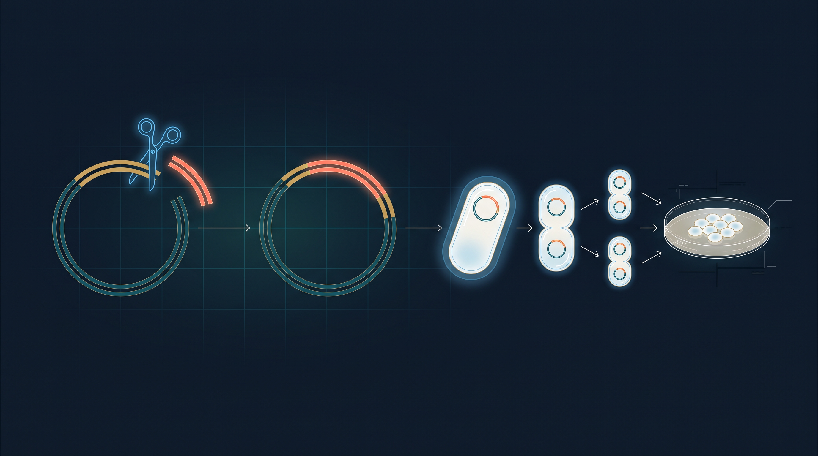
Fig. 3.0 – The core process of gene cloning.
This chapter will answer each of these questions as you work through it.
3.1 Identification of Candidate Gene
Why Clone a Gene?
Gene cloning is a routine procedure in the biotechnology industry, used mainly to obtain a gene and the product it codes for. In its traditional form, it involves transferring a DNA fragment that carries the gene of interest into a host cell with the help of a vector, so that a very large number of copies of that gene become available for study and for future use.
Three words in that sentence need to be clear before we go further. A vector is a DNA molecule that can carry a foreign piece of DNA into a living cell and replicate there on its own — a common example is a plasmid, the small circular DNA molecule found in bacteria in addition to their main chromosome. A host is the cell (usually a bacterium such as Escherichia coli) inside which the vector multiplies. A clone is a population of molecules or cells that are all identical to one another, because they were produced by copying a single original.
Advances in genetic engineering now make it possible to analyse DNA, isolate one specific gene from an entire genome (the complete set of DNA in an organism), insert it enzymatically into an autonomously replicating vector such as a plasmid to generate a recombinant DNA (rDNA) molecule, and finally introduce this rDNA into a host such as a bacterium. As the host divides, it copies the inserted gene along with its own DNA, producing a virtually unlimited number of copies, or clones, of that gene. The whole sequence is summarised in Fig. 3.1, and the rest of this chapter takes up each of these procedures in turn.
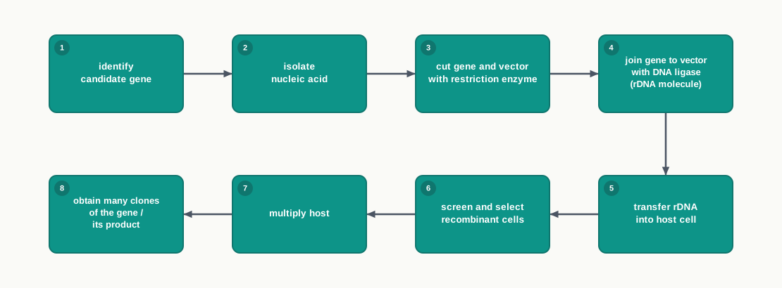
Fig. 3.1 – Overall gene cloning workflow.
What Makes a Gene a Candidate?
Over the past several decades, rDNA technology has been used to produce crops resistant to pests, diseases, herbicides and pathogens. This becomes possible when a specific gene of interest is taken from one organism and transferred into the genome of another organism, where its expression gives the desired product or activity. Bt cotton, which carries a bacterial gene that makes the plant produce an insecticidal protein against bollworm, is a familiar example of this idea working in the field.
But before any of this can happen, one hurdle must be crossed, and it is the most formidable one in the whole process: identifying the candidate gene within the genome of an organism.
Which gene deserves to be chosen depends on how significant it is in the biomedical, economic or evolutionary sense. This information comes from biochemical and physiological studies of the organism. For example, the cause of a disease (diabetes in human beings due to deficiency of insulin), or a defect in a metabolic pathway (iron deficiency leading to chlorosis in plants), or resistance to an environmental stress (salinity tolerance in plants), or resistance to infection (in both plants and animals), or economically important genes (milk protein, blood clotting factors, and so on) — all of these point to candidate genes for the improvement of human health and human needs. The insulin gene is the classic case: once physiologists had established that diabetes follows from a shortage of a single protein, the gene coding for that protein became an obvious target for cloning, and recombinant human insulin produced in bacteria is today given to patients worldwide.
Once a gene of interest has been identified, it is then explored in new sources and cloned by the procedures described in the sections that follow.
The Needle-in-a-Haystack Problem
Searching for a gene of interest is not an easy task, and a simple calculation makes this clear. A haploid human genome contains approximately 3.2 billion base pairs (bp). A gene of interest may be only 3000 to 3500 bp long — that is about one-millionth of the genome. Locating it is perhaps more difficult than looking for a needle in a haystack, because at least a needle looks different from hay, whereas the four bases of the target gene look exactly like the four bases of everything around it.
Two Routes to the Candidate Gene
A few methods have been developed to solve this problem. One of them works backwards from the protein. If the amino acid sequence of a specific polypeptide chain is known, the DNA sequence of the gene coding for it can be deduced, because each amino acid is specified by a triplet of bases in the genetic code. The deduced sequence can then be searched for, or chemically synthesised.
The second route begins from the messenger RNA (mRNA). In a cell, only the genes that are actually switched on are copied into mRNA, and a tissue that makes a particular protein in large amounts will be rich in the mRNA for that protein. So the mRNA of the desired gene is isolated from that specific tissue. A single-stranded complementary DNA (cDNA) is then synthesised on this mRNA template using the enzyme reverse transcriptase, which builds DNA from an RNA template rather than from a DNA template. This single strand is then converted into double-stranded cDNA, and that double-stranded molecule serves as the candidate gene, ready to be cloned. The steps are set out in Fig. 3.2, and the enzyme itself is discussed further in Section 3.3.
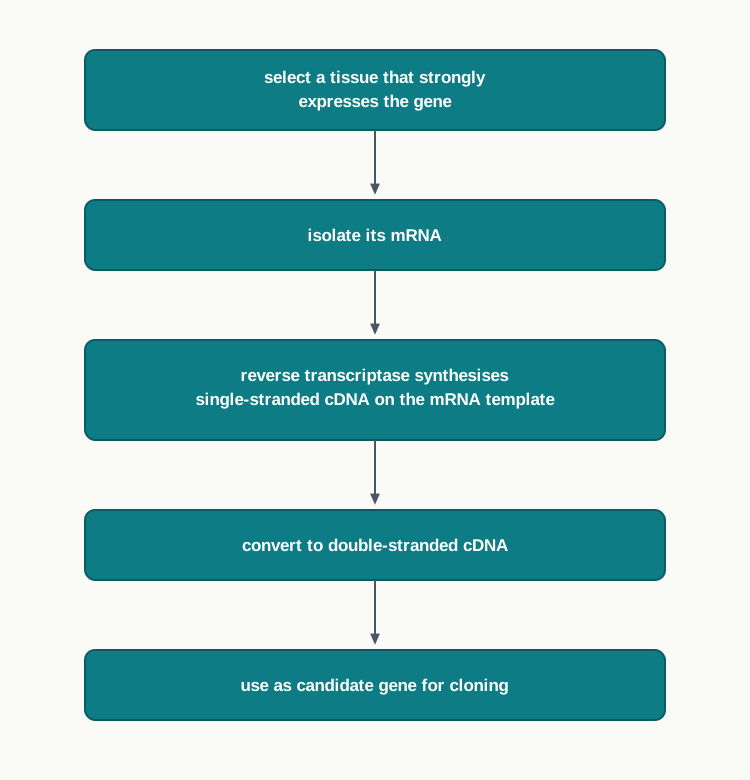
Fig. 3.2 – Obtaining a candidate gene through mRNA.
How This Section Is Tested
In board and competitive examinations, this section usually appears as short reasoning questions rather than recall: why identification is called the most formidable step, why a particular tissue is chosen as the source of mRNA, or assertion–reason items pairing a candidate gene with the evidence that made it a candidate. Being able to justify the choice of a gene, and not merely name examples, is what earns marks here.
3.2 Isolation of Nucleic Acids
The first and foremost requirement for any molecular biology experiment is the isolation of nucleic acids from organisms. Before a gene can be cut, joined, copied or transferred, it must first be brought out of the cell in a clean, unbroken form.
Two Big Challenges
Extraction of nucleic acids is faced with two big challenges. The first is related to their availability in cells: DNA and RNA are both present in very small amounts compared with other biological macromolecules such as proteins, carbohydrates and lipids. The material we want is therefore a minority component in a crowded mixture from the very beginning.
The second challenge is the enormous length of nucleic acids, which makes them susceptible to cleavage when exposed to harsh physical stress. A long DNA thread behaves rather like a fine strand of wet noodle — vigorous shaking, forceful pipetting or rough grinding will snap it into pieces.
In addition, the chemical bonds by which the different components of nucleic acids are joined with each other, and the various groups present in them, make nucleic acids vulnerable to chemical agents. Extremes of pH or reactive chemicals can attack these bonds, so every reagent used during extraction must be chosen with care.
The Four Steps of Extraction
Four important steps are involved during the extraction of nucleic acids, and each one answers a specific problem created by the step before it. The complete sequence is set out in Fig. 3.3.
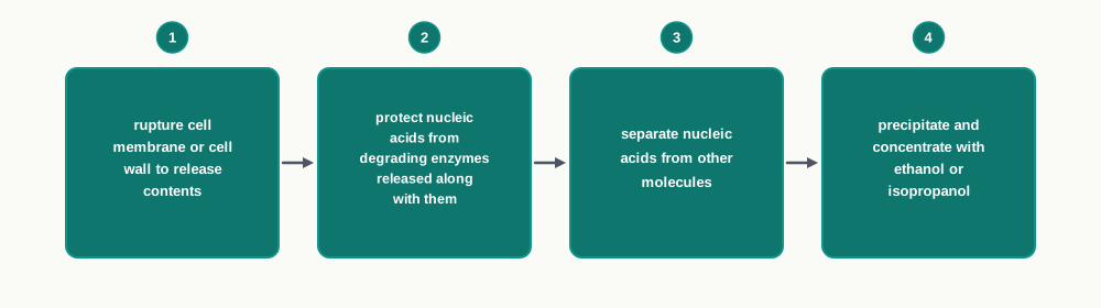
Fig. 3.3 – Four steps of nucleic acid extraction.
The first step involves the effective rupture of the cell membrane or walls, so that the nucleic acids and other cellular molecules are released. The second step involves the protection of nucleic acids from their respective degrading enzymes, which are released into the isolation medium along with other proteins.
In the third step, the nucleic acids are separated from other molecules. In the fourth and last step, the isolated nucleic acids are precipitated and concentrated by adding ethanol or isopropanol.
Notice how step two follows directly from step one. Breaking the cell open does not only release the DNA; it also releases the very enzymes that destroy DNA. Unless something is done immediately to hold those enzymes back, the first step would destroy the product of the whole exercise.
Different Cells, Different Doors
Although the chemical and physical properties of nucleic acids are similar in all organisms, the outer boundary of cells differs from one organism to another. Therefore, in order to disrupt the cell boundaries and release nucleic acids into the extraction medium, different strategies are adopted.
Animal cells have only a plasma membrane, which can be easily disrupted. On the contrary, plant cells and bacteria are protected by tough layers such as the cell wall, which need different approaches for their lysis. These approaches include homogenisation, grinding, sonication (breaking cells with high-frequency sound waves) or enzymatic treatment.
Such mechanical or enzymatic treatment ruptures the plasma membrane or cell wall so that nucleic acids get released from the cells. At the same moment, this also exposes them to nuclease enzymes — deoxyribonuclease and ribonuclease — which are released simultaneously from the same cells. This is precisely why protection cannot be postponed to a later stage.
Isolation of DNA
As bacterial cells have little structure beyond the cell wall and cell membrane, isolating DNA from them is much easier. An enzyme called lysozyme digests the peptidoglycan, the main component of the bacterial cell wall. Detergents like sodium dodecyl sulphate (SDS) are then used to lyse the cell membranes by disrupting the lipid bilayer.
Plant and animal cells are ground to release the intracellular components. Plant cells in particular are mechanically ruptured in a blender to break open their tough cell walls.
For the isolation of DNA from plant cells, cetyl trimethyl ammonium bromide (CTAB) is used as the detergent. CTAB is a cationic detergent — that is, its active part carries a positive charge, unlike SDS whose active part carries a negative charge.
Plant cells create an extra difficulty. They have a high concentration of polysaccharides and polyphenols in comparison to animal cells, and both of these pose problems during the isolation of DNA. Polysaccharides make the extract thick and sticky, while polyphenols oxidise and bind to DNA, discolouring it and interfering with later enzyme reactions.
CTAB solves the polysaccharide problem in an elegant way. The solubility of DNA and of polysaccharides in CTAB depends on the ionic strength of the solution. At low ionic strength, DNA is soluble in CTAB solution while polysaccharides are insoluble; whereas at high ionic strength, polysaccharides are soluble and DNA is insoluble. Both the molecules are therefore separated on the basis of their differential affinity to CTAB, simply by adjusting the salt concentration at the right moment. In addition, being a detergent, CTAB also lyses the cell wall.
The polyphenol problem is handled separately: addition of polyvinyl pyrrolidone (PVP) to the CTAB extraction medium neutralises phenols, so that they cannot bind to and spoil the DNA.
The soluble DNA present in the supernatant is then extracted with chloroform–isoamyl solution. The DNA present in the aqueous phase is finally precipitated using ethanol or isopropanol. In the case of animal cells, the cell membrane is simply disrupted by detergent to release the intracellular components, since there is no wall to break.
The Extraction Medium
The cells and tissues from which nucleic acids are to be extracted are broken down in a medium, either mechanically or enzymatically. This medium is usually a buffer having a mild alkaline pH with minimum ionic strength (0.05 M), containing the chelating agent ethylene diamine tetraacetic acid (EDTA). A concentration of 0.05 M means 0.05 mole of the solute is present in every litre of solution, since molarity is given by
\[M = \frac{n}{V}\]
where \(M\) is the molarity in mol L\(^{-1}\), \(n\) is the amount of solute in mol, and \(V\) is the volume of solution in litres.
Every component of this buffer has a definite job. The mild alkaline pH facilitates the reduction of electrostatic interaction between DNA and the basic proteins (histones) released during cell disruption. Histones are positively charged and cling tightly to the negatively charged DNA; loosening this grip is the first step towards obtaining protein-free DNA.
The chelating action of EDTA is equally deliberate. Chelating of divalent cations, particularly \(\mathrm{Mn^{2+}}\) and \(\mathrm{Mg^{2+}}\), prevents the action of nucleases, because these enzymes need such divalent metal ions as cofactors to work. Take the cofactor away, and the enzyme is disarmed without any need to destroy it.
Further inhibition of nuclease activity is achieved due to the alkaline pH of the buffer, which keeps these enzymes away from the pH at which they work best. In addition, chelating of divalent cations prevents the formation of their respective salts with the phosphate groups of nucleic acids, which would otherwise make the DNA harder to keep in solution.
Removing the Proteins
The next step is to separate the nucleic acids from their bound proteins. This is achieved by decreasing the interaction between proteins and nucleic acids, by exposing them to detergents such as SDS, an anionic detergent.
Exposure to SDS makes all the protein molecules anionic. Consequently, basic proteins that are positively charged and bound to negatively charged nucleic acids themselves become negatively charged, and so they dissociate from the nucleic acids — like charges repel. In addition, SDS also prevents the activities of nucleases, thereby giving additional protection to nucleic acids.
Sodium chloride is then added to the medium at high concentration. Increased salt concentration diminishes the ionic interaction between DNA and cations, thus ensuring complete dissociation of DNA–protein complexes.
Deproteinisation of the medium is achieved by exposing it to chloroform and isoamyl alcohol. These solvents are non-polar in nature; when added to the medium, which is polar in nature, and subjected to centrifugation, the mixture gives three distinct layers.
Since the density of the organic solvent mixture is higher than that of water, it forms the lower layer, which contains the denatured proteins, while the upper layer is aqueous in nature and contains the nucleic acids. Between them lies the interface.
Each solvent has a distinct role here. Chloroform causes denaturation of proteins, while isoamyl alcohol prevents the formation of foam and helps in stabilising the interface between the lower organic and the upper aqueous phase, so that the two can be cleanly separated by pipetting.
Precipitating and Storing the DNA
The nucleic acids from the aqueous phase are precipitated by the addition of ethanol to the aqueous medium. Ethanol reduces the polarity of the aqueous medium, which makes the medium non-polar, and thus the nucleic acids — which were soluble in the polar aqueous medium — become insoluble and come out of solution as visible white threads.
To remove RNA from the preparation, the enzyme ribonuclease A is added, which digests RNA into ribonucleotides. The DNA is then isolated by centrifugation and stored at low temperatures. The full sequence of tubes and phases is shown in Fig. 3.4.
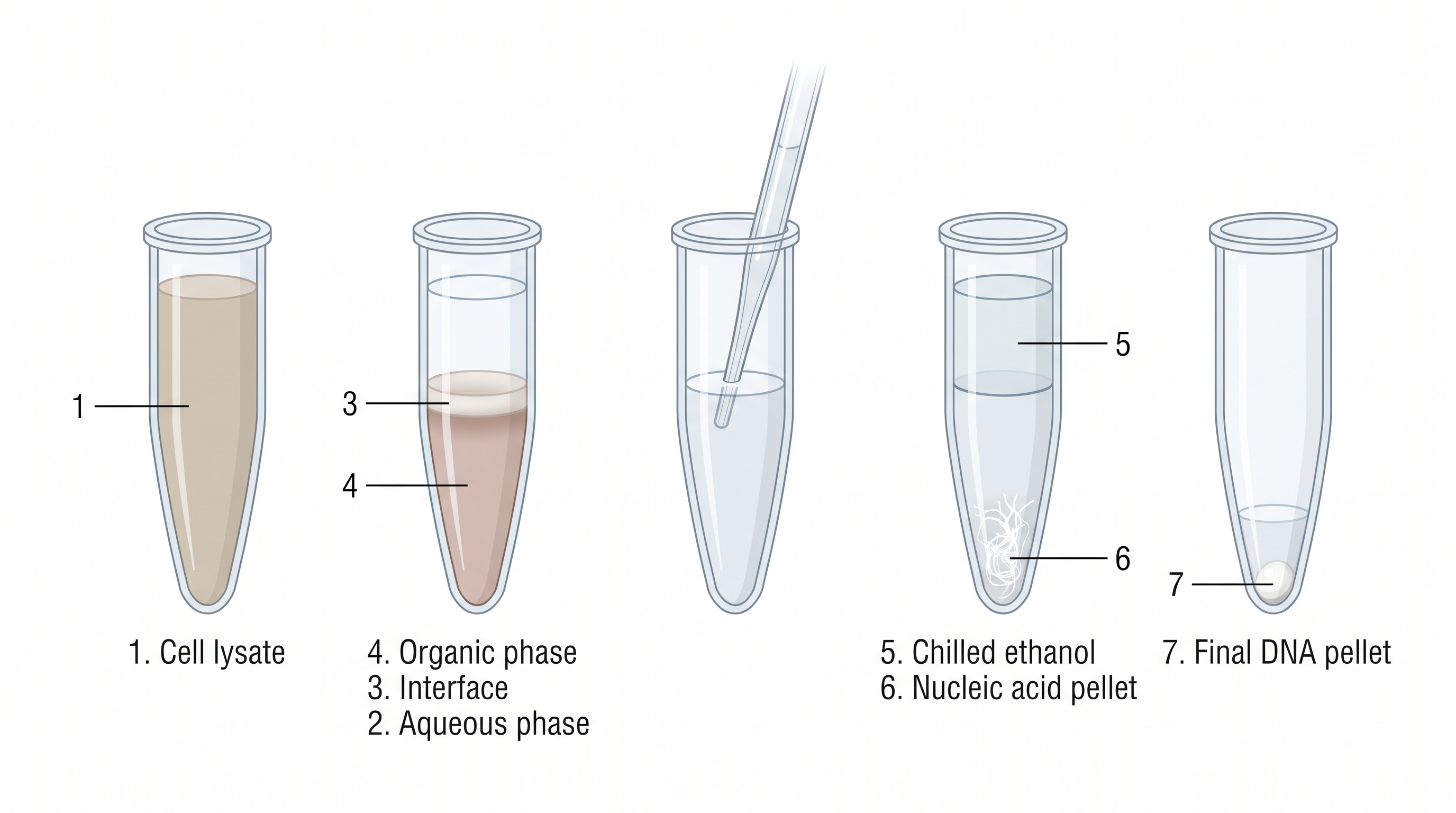
Fig. 3.4 – Steps in the isolation of DNA.
RNA Isolation
RNA is single stranded, while DNA is mostly double stranded. This single difference makes RNA both more fragile and more difficult to protect.
Ribonucleases (RNases), a group of enzymes that degrade RNA molecules, are abundant in the environment, and it is difficult to remove or destroy RNases completely. They are present on skin, on glassware and in dust, and unlike most enzymes they survive rough handling. Thus, it is often difficult to isolate intact RNA.
Total RNA is extracted from biological samples by using a specific reagent known as guanidinium isothiocyanate (GITC)–phenol–chloroform. GITC is a chaotropic reagent and acidic in nature. It disrupts hydrogen bonds and releases energy to increase entropy — that is, disorder — which reduces the hydrophobic effect of the solution, resulting in the aggregation of proteins and nucleic acids.
Phenol causes denaturation of proteins, whereas chloroform solubilises lipids. Chloroform also enhances the specific gravity of phenol with respect to water, which is what makes the organic layer settle firmly below the aqueous layer instead of mixing back into it.
When biological samples are treated with GITC solution and subjected to centrifugation, the solution gets separated into three phases: an upper aqueous phase, followed by the interface and the organic phase. Total RNA is extracted in the aqueous phase due to the acidic nature of the reagent, whereas DNA and denatured proteins are retained in the interface or the organic phase. This is the crucial separation, and it is achieved by acidity alone.
This step also inactivates the RNase enzyme that may hydrolyse RNA — so the same reagent that separates the phases also protects the product. Subsequently, RNA from the aqueous phase is precipitated with the help of isopropanol. The whole procedure is summarised in Fig. 3.5, and the phase separation itself is shown in Fig. 3.6.
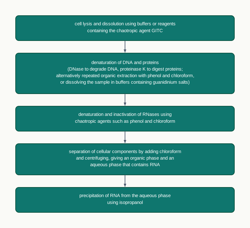
Fig. 3.5 – Isolation of RNA.
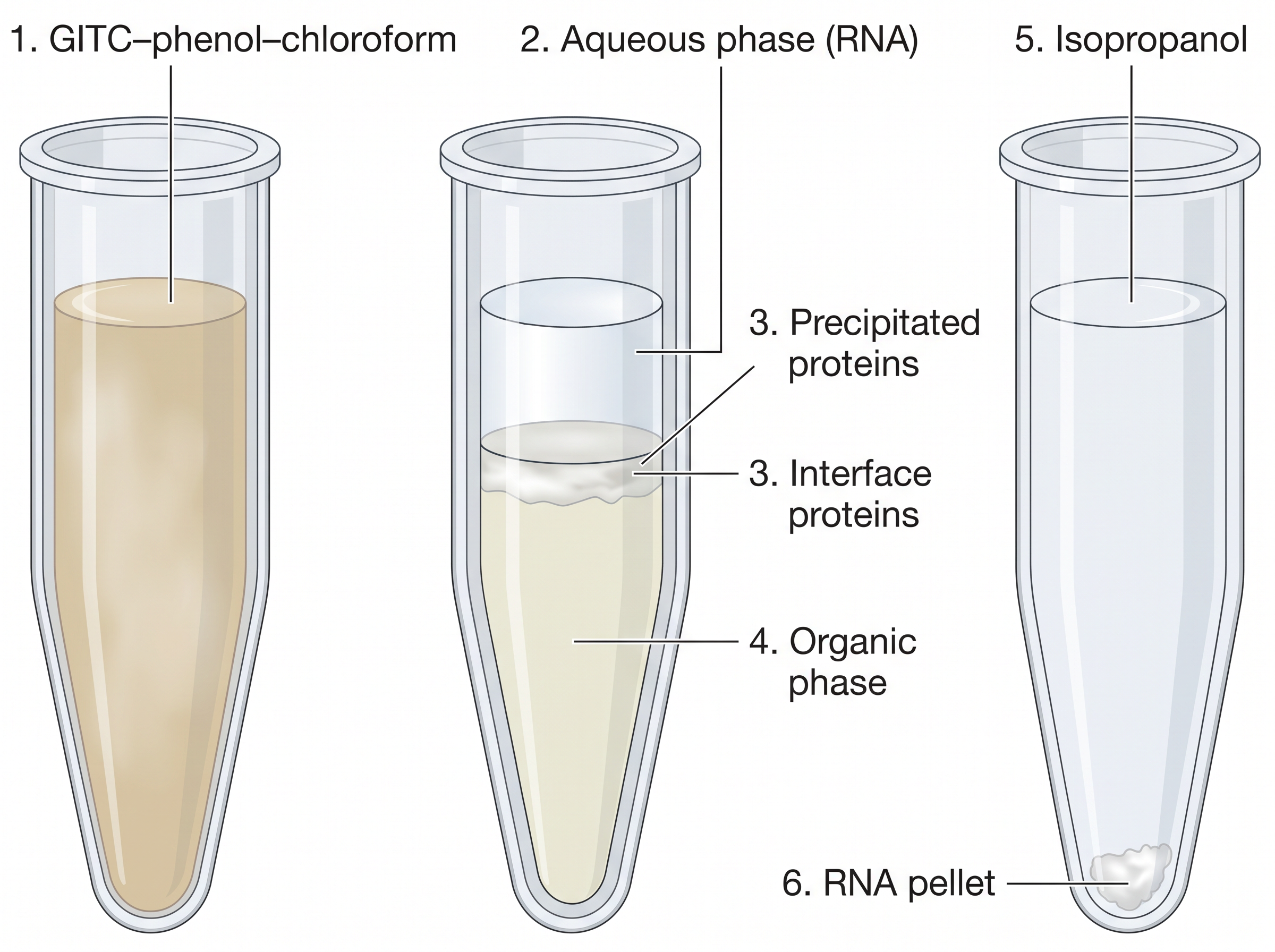
Fig. 3.6 – Phase separation in RNA extraction.
How This Section Is Tested
This section is a favourite for reason-based questions, because almost every reagent in it exists for one specific purpose. Examiners commonly ask why EDTA is added, why the buffer is mildly alkaline, what role isoamyl alcohol plays, why plant DNA isolation needs CTAB and PVP, and how RNA isolation differs from DNA isolation. Sequence-ordering questions on the four steps of extraction, and diagram-labelling on the three phases in a centrifuge tube, are also common. The safe way to prepare is to be able to name the problem that each reagent was brought in to solve.
3.3 Enzymes used for Recombinant DNA Technology
Enzymes constitute an important tool in rDNA technology. The major task in the manipulation of DNA involves cutting and ligation of the vector DNA and the gene of interest. For this, the natural abilities of different enzymes found in organisms are exploited — nothing here is invented from scratch; the biotechnologist simply borrows what cells already do.
Nine kinds of enzymes are used routinely, and it helps to see them as a toolkit in which each tool answers a different need: something to cut with, something to join with, something to copy with, and a few to tidy up the ends. The order in which the main tools are picked up during a cloning experiment is shown in Fig. 3.7.
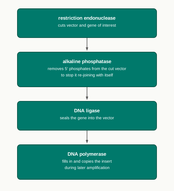
Fig. 3.7 – Order of enzyme use in building a recombinant molecule.
(i) Nucleases
Nucleases are the enzymes which cleave nucleic acids by hydrolysing the phosphodiester bond that joins the sugar residues of adjacent nucleotides. This bond is the backbone link of the polynucleotide chain, so breaking it is what actually cuts the molecule.
Some nucleases are DNA specific and are called DNases, while some are RNA specific and are called RNases. Recall from Section 3.2 that both are released as soon as a cell is broken open, which is why the extraction buffer works so hard to keep them inactive.
There are two major types of nuclease enzymes, depending on their preference for the location of the phosphodiester bonds within polynucleotide chains — DNA, RNA or a synthetic polynucleotide chain. These are exonuclease and endonuclease.
Exonuclease, as the name suggests, removes the nucleotides one at a time, that is, as mononucleotides, either from the 3′ or the 5′ ends of polynucleotide chains. Endonuclease, on the contrary, breaks internal phosphodiester bonds within a DNA or RNA molecule. The difference is illustrated in Fig. 3.8, where you should notice that the exonuclease nibbles at the free ends while the endonuclease strikes somewhere in the middle of the chain.
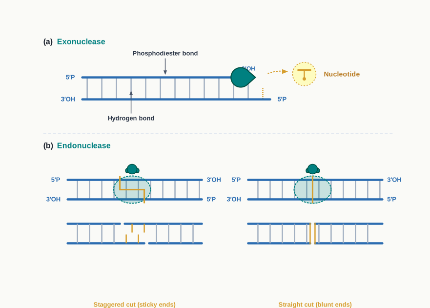
Fig. 3.8 – Action of (a) exonuclease and (b) endonuclease.
(ii) Restriction Endonuclease or Restriction Enzyme (RE)
Endonuclease enzymes that cleave DNA molecules at a specific position are called restriction endonucleases or restriction enzymes. They are mostly found in bacteria and archaea, where they provide a defence mechanism against invading bacteriophages — the bacterium cuts the incoming viral DNA to pieces before it can take over the cell. Biotechnology simply turned this defensive weapon into a precision cutting tool.
An RE recognises and binds to a specific DNA sequence called the recognition sequence or recognition site, often consisting of 4 to 8 bp.
Types of Restriction Enzymes
Restriction enzymes are categorised mainly into three groups — Type I, Type II and Type III — based on their co-factor requirement and the position of their DNA cleavage site relative to the target sequence.
Type I enzymes cleave DNA at a site that is about 1000 bp from the recognition site. They require S-adenosyl methionine (SAM), \(\mathrm{Mg^{2+}}\) and ATP, and possess DNA strand cleavage, methylase and ATPase activities.
Type II enzymes cleave within the recognition site itself and require only \(\mathrm{Mg^{2+}}\), having only DNA strand cleavage activity, as seen in Fig. 3.9. It is these Type II REs that find application in rDNA technology.
Type III enzymes cleave at sites about 24 to 26 bp away from the recognition site. They require S-adenosyl methionine (SAM), \(\mathrm{Mg^{2+}}\) and ATP, and have DNA strand cleavage and methylase activities. The three types are compared in Table 3.1.
Table 3.1: Types of Restriction Enzymes
| Cleavage site | Endonuclease and methylase function | Examples | |
|---|---|---|---|
| Type I | Random around 1000 bp away from the recognition site | Endonuclease and methylase function on a single protein molecule | EcoKI, EcoAI, CfrAI |
| Type II | Specific within the recognition site | Endonuclease and methylase are separate entities | EcoRI, BamHI, HindIII |
| Type III | Random 24–26 bp away from the recognition site | Endonuclease and methylase function on a single protein molecule | EcoP1, HinfIII, EcoP15I |
Palindromic Sequences, Sticky Ends and Blunt Ends
The recognition sequences of the widely applied Type II REs are palindromic sequences, meaning that the sequence read in the forward direction on a double-stranded DNA reads the same in the reverse direction on the complementary strand. For example, the site recognised by EcoRI is
5'-GAATTC-3' 3'-CTTAAG-5'
Read each strand in its own 5′→3′ direction and you get the same six letters, which is exactly what makes the site look identical to the enzyme from either side.
These enzymes break a specific phosphodiester bond in both strands of the DNA molecule, within the restriction sequence recognised by the enzyme, or at the site, or near the sequence. The cut generates a 5′-phosphate group at one end of the break and a 3′-hydroxyl group at the other end of the break, as shown in Fig. 3.9. These two chemical groups matter enormously, because they are precisely the two groups that DNA ligase will later join together.
Several REs cleave at different locations on the two DNA strands to produce a staggered cut, which leaves short single-stranded protruding ends called cohesive or sticky ends. Some REs produce a blunt-ended cut by cleaving both strands of DNA at the same location.
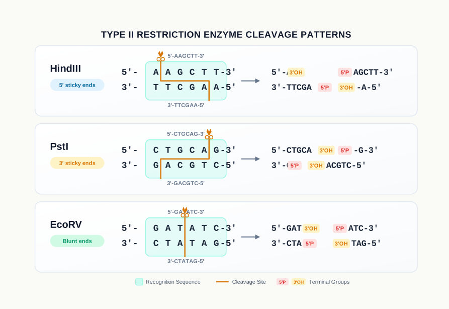
Fig. 3.9 – Type II restriction enzymes generating sticky or blunt ends.
Look carefully at the three examples in Fig. 3.9. HindIII cuts the sequence
5'-AAGCTT-3' 3'-TTCGAA-5'
between the two adenines on each strand. Because the two cuts are offset, each fragment is left with a short single-stranded projection carrying a free 5′ end — a 5′ sticky end.
PstI cuts the sequence
5'-CTGCAG-3' 3'-GACGTC-5'
between A and G on each strand, again offset, but this time the projections carry free 3′ ends — 3′ sticky ends.
EcoRV cuts the sequence
5'-GATATC-3' 3'-CTATAG-5'
exactly in the middle of both strands, at the same position. No projection is left at all, and the result is a blunt end.
Nomenclature of Restriction Enzymes
Let us now understand how these enzymes get their odd-looking names. The enzyme is named after the microorganism from which it is isolated. The first capital letter represents the genus, the second and third letters represent the species. The fourth letter specifies the strain of the microorganism, and the last Roman number represents the number of the enzyme isolated from that species, as set out in Table 3.2.
Table 3.2: Nomenclature for restriction endonucleases
| EcoRI | |
|---|---|
| Escherichia (E) | genus |
| coli (co) | specific epithet |
| strain Ry13 (R) | strain |
| first endonuclease (I) | order of identification |
| HindIII | |
| Haemophilus (H) | genus |
| influenzae (in) | specific epithet |
| strain Rd (d) | strain |
| third endonuclease (III) | order of identification |
Once you know this rule, an unfamiliar enzyme name stops being a memory burden and becomes a piece of information: BamHI, for instance, immediately tells you it was the first restriction enzyme isolated from a particular strain of Bacillus amyloliquefaciens.
(iii) DNA Ligase
The ligase enzyme facilitates the joining of DNA strands together by catalysing the formation of a phosphodiester bond in the duplex form, as shown in Fig. 3.10. If the restriction enzyme is the scissors, the ligase is the glue.
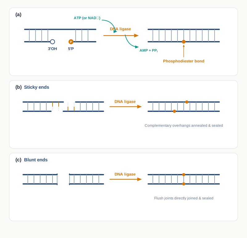
Fig. 3.10 – Ligation of DNA by ligase.
Bacterial DNA ligases, from E. coli, use the hydrolysis of NAD as their energy source, whereas ATP is the energy source for DNA ligases from bacteriophages (e.g., T4) and eukaryotic cells. Joining two DNA molecules is not spontaneous; it costs energy, and different organisms pay for it with different currencies.
The chemistry of the join is straightforward. The 5′P (5′–PO₄) group of one chain makes a covalent linkage with the 3′-OH group of the adjacent chain. These are exactly the two groups that the restriction enzyme left behind when it made the cut, which is why a restriction cut can be sealed again so cleanly.
T4 DNA ligase is used to join two DNA molecules having cohesive ends or blunt ends. E. coli DNA ligase is used to join cohesive ends. This is a small but frequently tested distinction: if an experiment involves blunt-end joining, T4 DNA ligase is the only choice among the two.
(iv) DNA Polymerases
DNA polymerases are a group of polymerases that catalyse the synthesis of a new DNA strand by using mono-deoxyribonucleoside triphosphates (dNTPs) on a template strand. A DNA polymerase enzyme synthesises the new DNA strand in the 5′→3′ direction, as shown in Fig. 3.11.
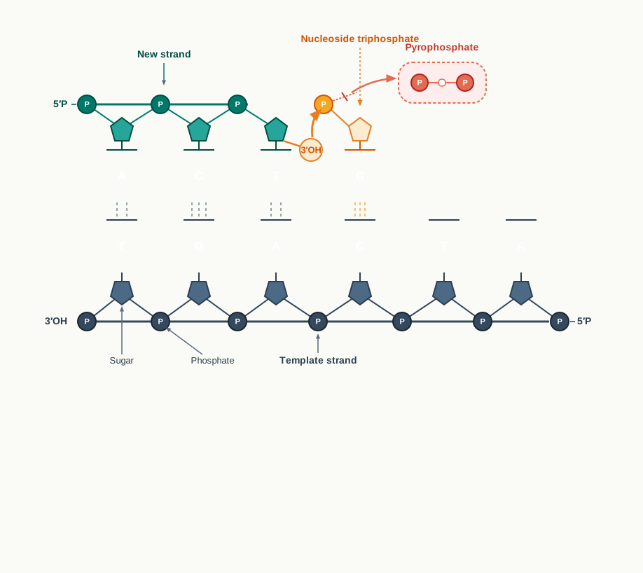
Fig. 3.11 – DNA polymerase adding nucleotides.
There is one important limitation. A DNA polymerase cannot initiate the synthesis of a new DNA strand on its own. In addition to dNTPs, it requires a primer — a short oligonucleotide carrying a free 3′-end hydroxyl group, which serves as the starting point of chain growth. The enzyme can only add to an existing 3′-OH; it cannot create one.
This single limitation shapes a great deal of biotechnology. It is the reason every PCR reaction must be supplied with designed primers, as you will see in Section 3.7.
DNA polymerase I of E. coli exhibits several other activities as well, such as 5′→3′ exonuclease and 3′→5′ exonuclease activity. The 3′→5′ exonuclease activity allows the enzyme to reverse a step and remove a wrongly added nucleotide, which is why such polymerases copy DNA with high fidelity.
(v) Alkaline Phosphatase
Alkaline phosphatase is used to remove the terminal phosphate group from the 5′ end of DNA strands.
This sounds like a small housekeeping job, but it has a practical purpose. A cut vector has sticky ends that fit each other perfectly, so the vector can simply close up again without accepting any insert. If its 5′ phosphates are stripped away by alkaline phosphatase, ligase has nothing to work with at that junction, and the vector cannot re-circularise on its own. The proportion of useful recombinant molecules therefore rises sharply.
(vi) Polynucleotide Kinase
Using polynucleotide kinase, a phosphate group can be attached to the hydroxyl (-OH) group present on the 5′ end of DNA. Polynucleotide kinase has the reverse effect of alkaline phosphatase, adding phosphate groups onto free 5′ termini.
The pair therefore works in opposite directions, and either can be chosen depending on whether an end needs to be made ready for ligation or kept out of it. Polynucleotide kinase is also the enzyme used to attach a radioactive phosphate to the 5′ end of a probe, which is how many probes used in blotting are labelled.
(vii) Terminal Deoxynucleotidyl Transferase or Terminal Transferase
This enzyme can add similar nucleotide residues to form a homopolymer tail on the 3′ end of a DNA strand. Unlike most DNA polymerases, it does not require a template.
Because it needs no template, it can be used to create artificial sticky ends. If one DNA molecule is given a tail of, say, several adenines and another a tail of several thymines, the two tails will base-pair and hold the molecules together — a useful trick when the fragments to be joined have blunt ends.
(viii) Reverse Transcriptase
Reverse transcriptase is also called RNA directed DNA polymerase and is found in many retroviruses, whose genetic material is RNA and which must convert it into DNA inside the host cell. HIV is the best known example of such a virus.
It is used to generate a complementary DNA (cDNA) strand from an RNA template, a process termed as reverse transcription, shown in Fig. 3.12. You met this enzyme in Section 3.1 as the means of obtaining a candidate gene from mRNA; it will appear again in RT-PCR and in the construction of cDNA libraries.
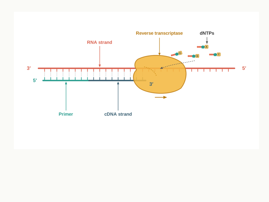
Fig. 3.12 – Reverse transcription.
(ix) Poly A Polymerase
Poly A polymerase incorporates adenine residues to the hydroxyl group at the 3′ end of RNA, as shown in Fig. 3.13. The long run of adenines it builds is the poly-A tail, which protects the mRNA from degradation and helps it to be translated efficiently.
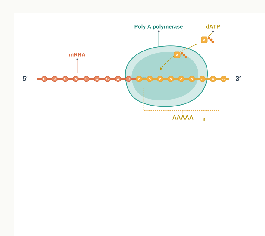
Fig. 3.13 – Addition of the poly-A tail.
How This Section Is Tested
Almost every question from this section rewards understanding of why a particular enzyme is chosen. Common forms are: matching an enzyme to its function, identifying an incorrectly matched enzyme–organism pair, deducing the ends produced when a given sequence is cut, explaining why Type II REs alone are used in rDNA technology, and assertion–reason items linking sticky ends to easier ligation. Diagram-based questions on Fig. 3.8 and Fig. 3.9 — labelling exonuclease versus endonuclease action, or marking the cut positions on a palindromic sequence — appear regularly, so practise writing out a recognition sequence on both strands with the polarity marked.
3.4 Modes of DNA Transfer
Transfer of a foreign DNA molecule to a host cell — prokaryotic or eukaryotic — from its surrounding environment is one of the basic steps in rDNA technology. Once the gene of interest has been joined to a vector, the recombinant molecule is still lying in a tube. It becomes useful only when it is safely inside a living cell that will copy it.
Nature solved this problem long before biotechnologists did. In nature, bacteria obtain foreign DNA molecules from their surroundings in three different ways: (i) transformation, (ii) transduction and (iii) conjugation. Understanding these three natural routes is worthwhile in itself, and it also explains where the laboratory methods came from.
(i) Transformation
Transformation is the genetic alteration of a cell resulting from the direct uptake of an exogenous DNA molecule from its surroundings through the cell membrane, after which the DNA gets incorporated in the recipient’s genetic material. There is no helper here — no virus, no second cell — only naked DNA and a cell willing to absorb it.
Recipient cells that have taken up the foreign DNA molecule are referred to as transformants, as shown in Fig. 3.14. Transformation occurs naturally in some species of bacteria, which are described as competent because their membranes can take up DNA from the medium.
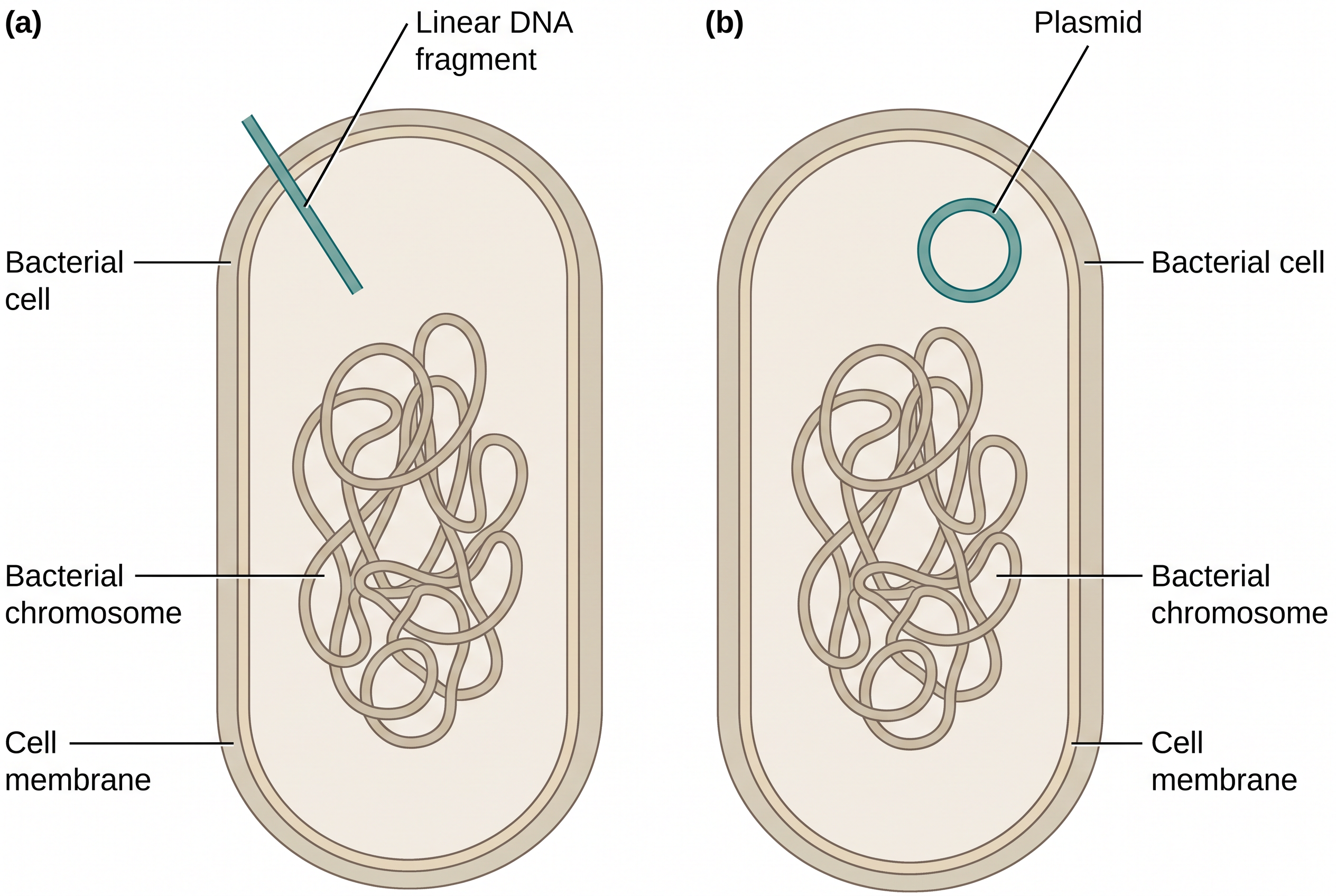
Fig. 3.14 – Transformation in bacteria.
Notice from Fig. 3.14 that the DNA taken up may be either a linear fragment or a whole plasmid. This distinction matters later, because a plasmid can replicate on its own inside the cell, whereas a linear fragment must integrate into the host’s DNA to survive.
(ii) Transduction
Viruses may also mediate the uptake of foreign DNA into the genome of a cell. Viruses that specifically infect bacterial cells are known as bacteriophages, and in transduction it is the phage that acts as the delivery van.
Bacteriophages on infecting a host follow either a lytic cycle or a lysogenic life cycle. In the lysogenic life cycle, the bacteriophage genome gets incorporated into the bacterial DNA and remains dormant there for several generations, being copied quietly along with the host chromosome.
After a period of time, when the phage genome gets excised from the host DNA, it occasionally takes small sequences of bacterial DNA with it. The excision is not always perfectly accurate, and this small inaccuracy is precisely what makes transduction possible.
The phage genome containing bacterial DNA is then packaged into phage coat proteins to form a complete, recombinant viral particle. When this phage infects another bacterial cell, the recombinant phage genome containing bacterial DNA is introduced into that bacterium, as shown in Fig. 3.15. The recipient bacterial cell is referred to as a transductant.
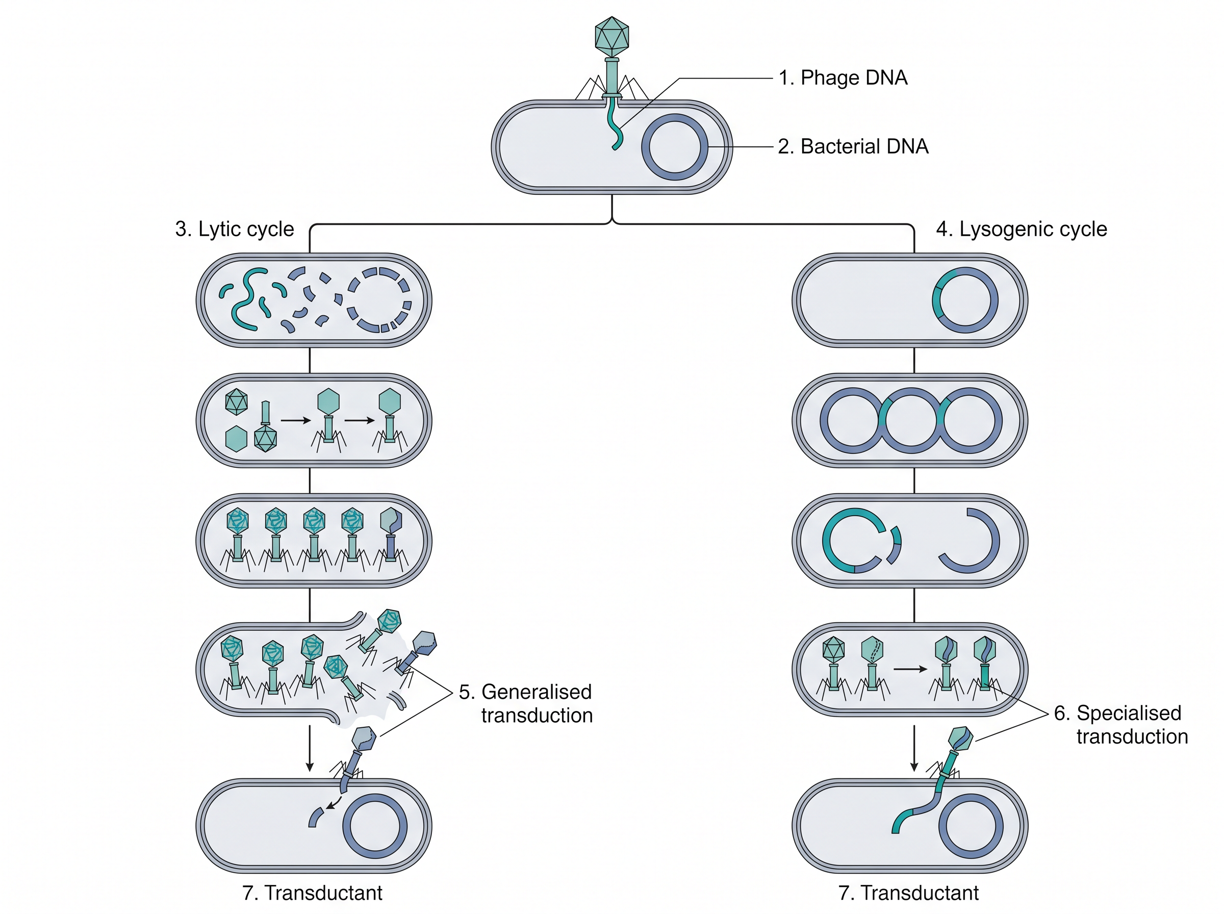
Fig. 3.15 – Transduction in bacteria.
Fig. 3.15 shows two branches. In the branch labelled generalised transduction, the bacterial chromosome is broken up during the lytic cycle and fragments of it are packaged more or less at random into new phage particles. In the branch labelled specialised transduction, the phage DNA has first integrated into the bacterial DNA in the lysogenic cycle, so the bacterial genes carried away later are only those lying next to the integration site. The sequence of events is set out in Fig. 3.16.
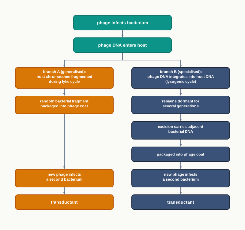
Fig. 3.16 – Pathways of transduction.
(iii) Conjugation
Conjugation is referred to as the transfer of genetic material (DNA) from one bacterium to another through cell-to-cell direct contact. The bacterial cell that transfers its DNA is called the donor cell, and the one that receives it is the recipient cell.
Conjugation is usually mediated by F plasmids that carry a DNA sequence encoding for the fertility factor, or F-factor. The F-factor forms a thin tube-like structure called a pilus, through which the donor cell makes contact with the recipient, as shown in Fig. 3.17.
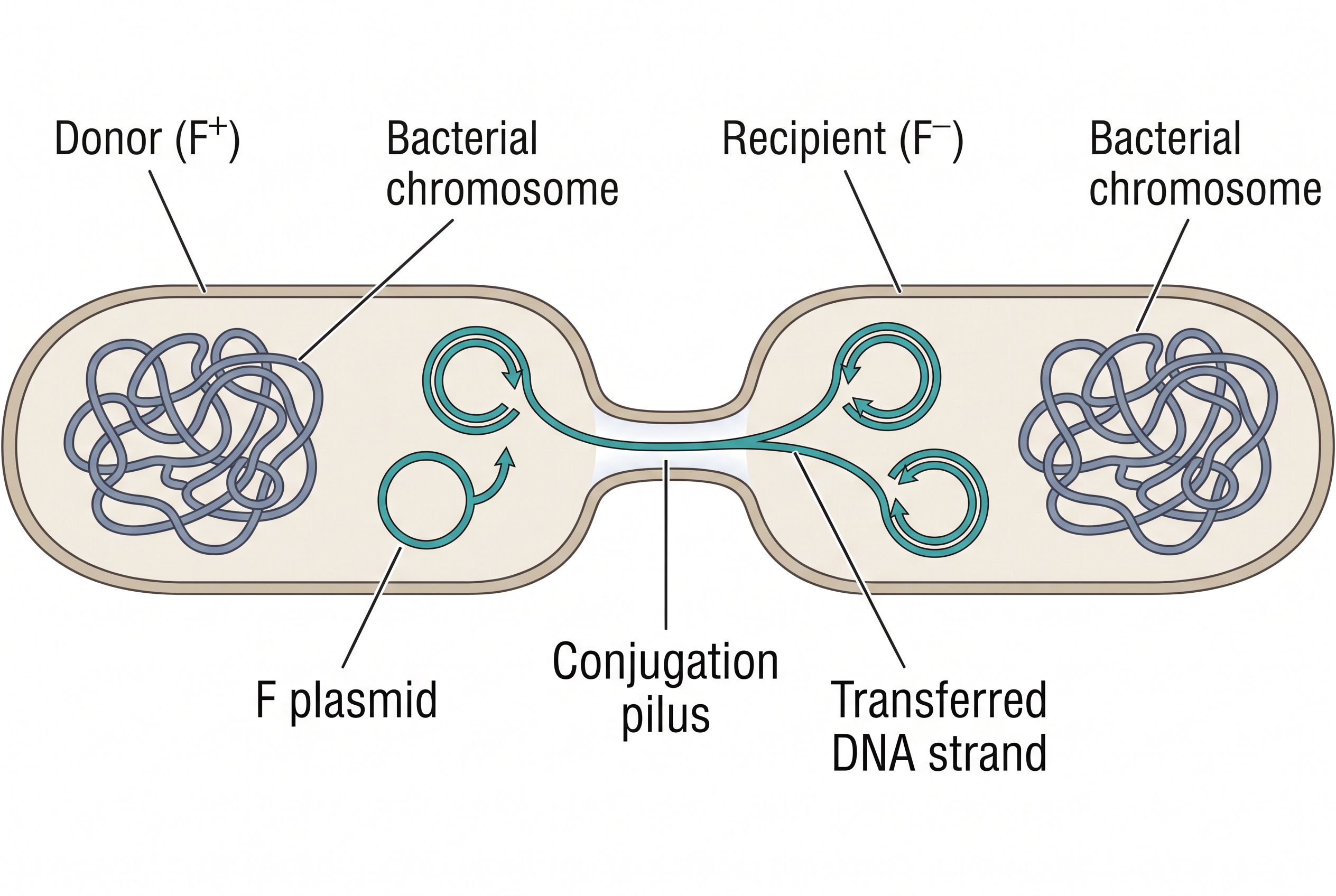
Fig. 3.17 – Bacterial conjugation.
A nick is made in one of the strands of the double-stranded F-plasmid by an enzyme called relaxase in the donor cell, and this single strand is transferred to the recipient cell through the pilus. Only one strand travels; the other stays behind in the donor.
Inside both the donor and the recipient cells, the single-stranded DNA undergoes replication to form a double-stranded F plasmid identical to the original F plasmid. This is why, as Fig. 3.17 shows, an F⁻ recipient becomes F⁺ after conjugation, and the donor loses nothing. The sequence is summarised in Fig. 3.18.
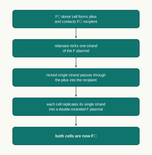
Fig. 3.18 – Steps of conjugation.
Getting rDNA into Host Cells in the Laboratory
In rDNA technology, the rDNA is introduced, or transferred, into host cells by numerous methods. These fall into two families: chemical methods (calcium chloride, lipofection, and so on) and physical methods (electroporation, microinjection and the gene gun). All five are illustrated together in Fig. 3.19.
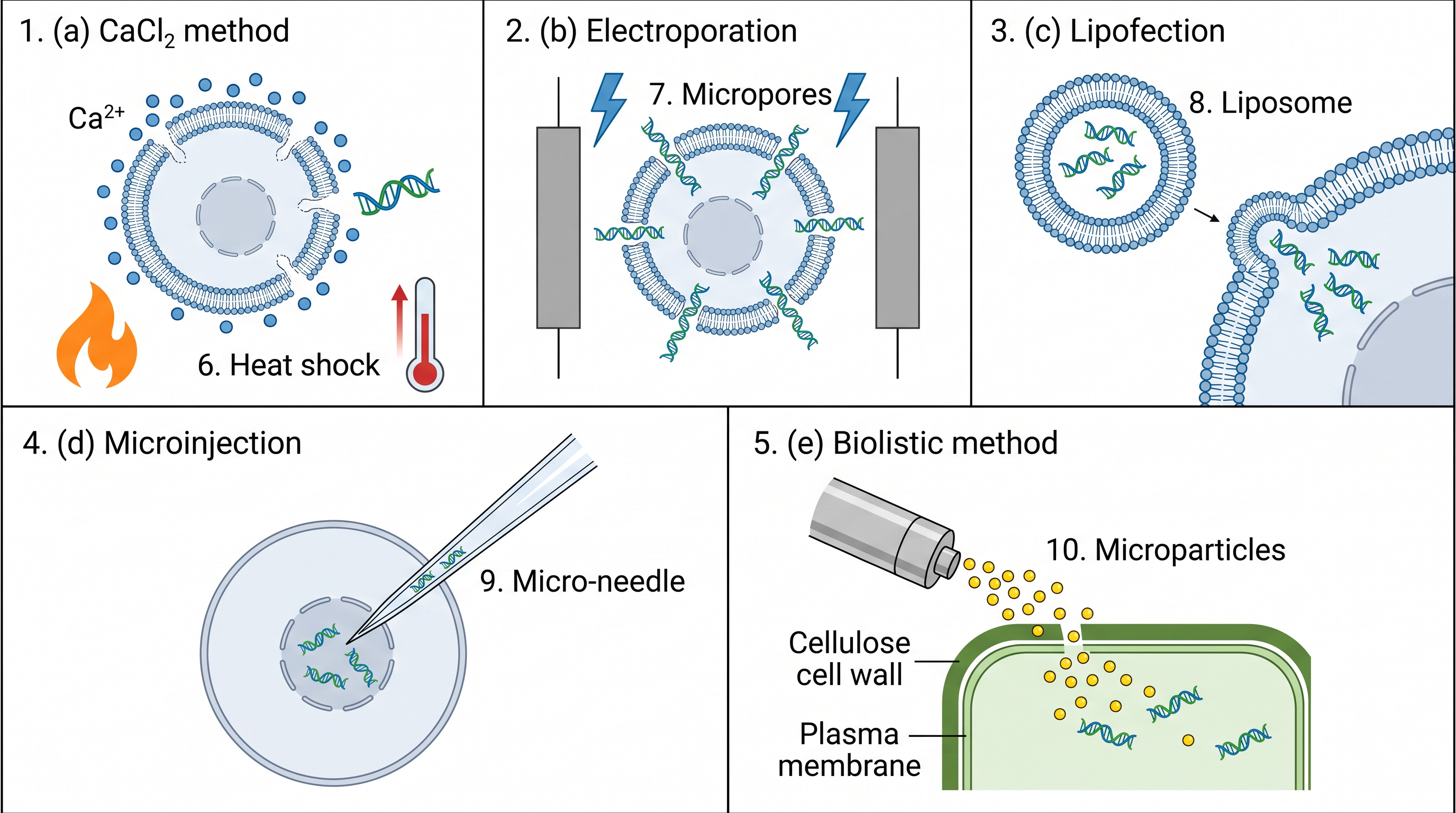
Fig. 3.19 – Methods of DNA transfer.
Calcium Chloride Method
In the calcium chloride method, the DNA to be transferred is mixed in a solution containing positively charged calcium and the negatively charged group of DNA to form a complex. The host cells then take up the foreign DNA molecule by a process of heat shock.
Electroporation
In the electroporation method, transient micropores are created on the membrane of host cells by exposing them to a mild electric current in the presence of foreign DNA molecules. The recombinant DNA molecules enter into the host cells through these transient micropores.
The word “transient” is important. The pores must open long enough for DNA to slip through, and then close again so that the cell survives and heals with the gene inside, as Fig. 3.19(b) shows.
Lipofection
Lipofection, or liposome transfection, is a technique used to inject genetic material into a cell by means of liposomes, which are vesicles that can easily merge with the cell membrane since they are both made of a phospholipid bilayer.
This is chemistry making use of a structural similarity. Because the liposome and the cell membrane are built the same way, they fuse readily, and the DNA packed inside the liposome is released directly into the cell without any pore or needle.
Microinjection
Foreign DNA molecules can be introduced directly to the nucleus of host cells using a specialised automated microinjection apparatus. A fine glass needle, guided under a microscope, delivers the DNA into a single chosen cell.
This is the most direct method of all, and also the slowest, since cells must be treated one at a time. It is chosen when a small number of very valuable cells is involved, such as fertilised eggs used to produce transgenic animals.
Biolistic or Gene Gun Method
In the biolistic method, with the help of a gene gun (also called a particle gun), microscopic particles of gold, nickel or tungsten coated with foreign DNA are bombarded into cells at high velocity, so that the foreign DNA molecule enters inside the cell, as shown in Fig. 3.19(e).
The choice of metal is not arbitrary. These metals are dense enough to carry momentum, chemically unreactive, and can be made into particles small enough to pass through a cell wall without destroying the cell. This method is especially valuable for plant cells, whose rigid cell walls defeat gentler approaches.
How This Section Is Tested
Questions here tend to test discrimination rather than recall: distinguishing transformation from transduction and conjugation, naming the recipient cell correctly in each case, or identifying which method suits a walled plant cell as against a fertilised animal egg. Reason-based questions are common — why calcium chloride is used, why the micropores in electroporation must be transient, why liposomes fuse so readily with the cell membrane. Assertion–reason items often pair a transfer method with a property of the host cell, so be ready to judge whether the stated reason really explains the assertion.
3.5 Screening and Selection
Selection of transformed bacteria carrying recombinant vectors is the most essential step for a successful cloning experiment. Here the objective is to identify the transformed cells having the recombinant vector from a mixture of non-transformed cells.
The reason this step cannot be skipped is a matter of odds. The success rate of insertion of an insert into a plasmid, and of the subsequent transfer of recombinant plasmids into bacteria, is very low. Therefore, it is difficult to select the few bacteria containing plasmids with the insert from a large population of bacteria without the insert.
Picture the plate that comes out of a transformation experiment: millions of colonies, all looking exactly alike, of which perhaps a handful carry the gene you spent weeks preparing. Screening is the art of making those few colonies announce themselves.
The Principle Behind All Selection
The method of selection of recombinant cells is based on the principle of a difference in the biological traits present in hosts with recombinant DNA compared with those without recombinant DNA. If the two kinds of cell behave identically, they cannot be told apart; so the vector is deliberately designed to make them behave differently.
Thus, the recombinant cells are distinguished from non-recombinants based on their expression or non-expression of certain traits. These traits may be antibiotic resistance, or the expression of some specific proteins such as β-galactosidase or Green Fluorescent Protein (GFP), or the dependence or independence of a nutritional requirement, such as the amino acid leucine.
A gene placed in a vector for exactly this purpose is called a selectable marker. On the basis of this principle, the selection procedure can be divided into two main types.
(i) Direct Selection of Recombinants
In this method of selection, transformed cells are distinguished from non-transformed cells based on the expression of certain traits.
For example, bacterial cells used as the host are not resistant to a particular antibiotic. But when they take up plasmids containing an antibiotic resistance gene, they become resistant to that specific antibiotic. These cells will survive and grow in a medium containing the antibiotic, whereas the host cells without the plasmid will be killed when they are exposed to the antibiotic.
The antibiotic here acts as a filter. Every cell that has not taken up a plasmid dies, and the plate is cleared of them in a single step.
(ii) Selection of Recombinants by Insertional Inactivation
This method is more efficient than the direct selection method. Here, a vector having two markers is used — either two antibiotic resistant genes, or one antibiotic resistant gene and one lacZ gene.
When the gene of interest (the insert DNA) is inserted into one of the selection marker genes in the vector, the expression of that marker gene is disrupted. The insert lands in the middle of the marker’s coding sequence and breaks the reading of that gene, so no functional product is made. This is called insertional inactivation.
The logic is neat: the marker that is lost now identifies the recombinant, while the marker that is retained identifies any cell carrying the vector at all. Two markers therefore answer two different questions with one experiment.
Using Two Antibiotic Resistance Genes
Let us use a plasmid with two antibiotic resistance genes — one for ampicillin (the ampR gene) and the other for tetracycline (the tetR gene). The target DNA, that is the insert, is inserted into the ampR gene of the plasmid, making them recombinant plasmids.
Now we have two kinds of plasmid in the tube: plasmids with the insert and plasmids without it. When this plasmid mixture is added to a culture of bacteria in the way described earlier, there will be three different populations of host bacterial cells:
- host cells without plasmids (non-transformed);
- transformed host cells with plasmids without insert;
- transformed host cells with recombinant plasmids (with insert).
Now it is essential to identify those cells that have received the recombinant plasmid. This process of screening is based on the property of resistance to ampicillin, which is lost in the host cell having recombinant plasmids. The insert gets cloned in the ampR gene, leading to insertional inactivation of the ampicillin resistance gene, as shown in Fig. 3.20.
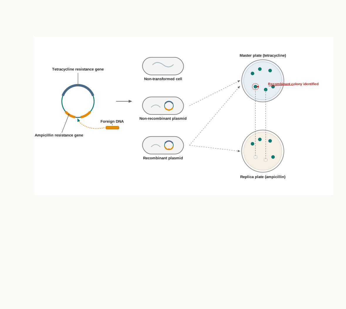
Fig. 3.20 – Selection of recombinants by insertional inactivation.
When these bacteria are plated on a medium containing tetracycline, the non-transformed cells get eliminated as they are sensitive to it. Only transformed cells, which carry a functional tetR gene, multiply and form colonies as they are resistant to it.
There will therefore be two types of colonies on this plate, which is called the master plate — one of transformed cells having a plasmid without insert (non-recombinant) and the other of transformed cells having a plasmid with insert (recombinant). At this stage the two look identical.
By using a nitrocellulose membrane, bacterial cells from the master plate colonies are then transferred and plated on a solid medium containing ampicillin. The membrane is pressed onto the master plate, picks up a few cells from every colony in their exact positions, and is then pressed onto the new plate, so the pattern of colonies is copied faithfully from one plate to the other.
Transformed cells with vectors without insert will multiply to form colonies on this second plate, called the replica plate, because their ampR gene is intact. Transformed cells with recombinant vectors will not grow, because their ampR gene has been inactivated by the insert.
Now, if we compare the master plate with the replica plate, the colonies present in the master plate and absent in the replica plate are the transformed cells with the recombinant vector containing the DNA insert of interest. Because the pattern of positions has been preserved, those missing colonies can be located and picked from the master plate, where they are still alive. The complete sequence is set out in Fig. 3.21.
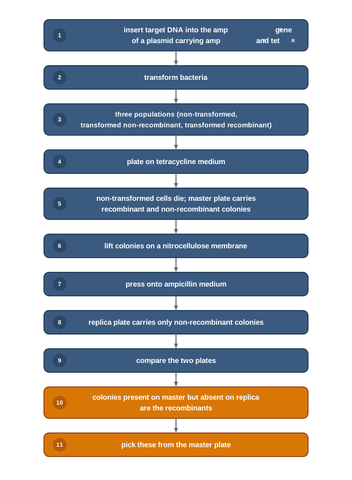
Fig. 3.21 – Insertional inactivation using two antibiotic markers.
Blue–White Selection Method
The blue–white selection method is another example of the insertional inactivation approach to selecting recombinant transformed cells. Instead of asking a colony to live or die, it asks the colony to change colour — which is faster to read and needs only one plate.
In this method, the lacZ gene present in the plasmid vector expresses the enzyme β-galactosidase. β-galactosidase cleaves a colourless chromogenic substrate called X-gal (5-Bromo-4-chloro-3-indolyl-beta-D-galactoside), an analog of lactose, to form 5-bromo-4-chloro-indoxyl. This product spontaneously dimerises to produce an insoluble blue pigment called 5,5′-dibromo-4,4′-dichloro-indigo.
When the lacZ gene in the plasmid is inactivated due to insertion of the insert DNA, the enzyme β-galactosidase is not expressed in hosts containing recombinant plasmids, as shown in Fig. 3.22. With no enzyme, X-gal is left uncut, no pigment forms, and the colony stays white.
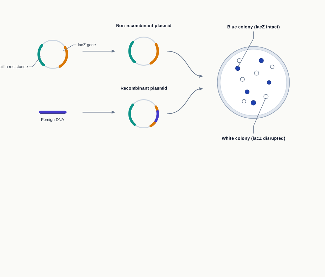
Fig. 3.22 – Blue–white selection method.
During the transformation experiment, the bacterial cells — both transformed and non-transformed — are plated on a solid medium containing ampicillin along with X-gal and IPTG (Isopropyl β-D-1-thiogalactopyranoside). IPTG is included because it switches the lacZ gene on; without an inducer, the gene would remain largely silent and even a non-recombinant colony would fail to turn blue.
The non-transformed cells get eliminated by the ampicillin, and only the transformed cells multiply and form colonies. Two types of colonies will be formed, blue and white.
The bacterial cells in blue colonies contain a vector with an uninterrupted lacZ gene, that is, no insert. Cells in white colonies, where X-gal is not hydrolysed, indicate the presence of an insert in lacZ, which disrupts the formation of an active β-galactosidase. The whole workflow is summarised in Fig. 3.23.
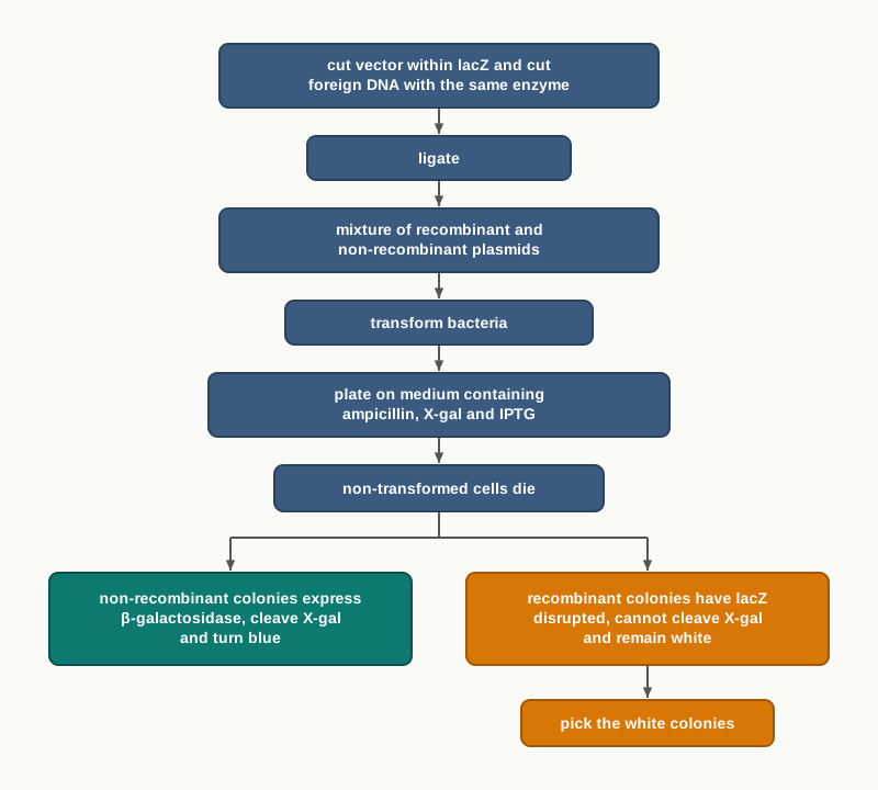
Fig. 3.23 – Steps of blue–white selection.
Screening with Green Fluorescent Protein
Alternative methods have been developed in order to screen transformed bacteria, for example using Green Fluorescent Protein (GFP). The concept is similar to that of lacZ: a DNA insert can disrupt the coding sequence within the vector and thus disrupt GFP production, resulting in non-fluorescing bacteria.
Here the read-out is glow rather than colour. Colonies carrying an intact GFP gene shine green under ultraviolet light, while recombinant colonies remain dark, and no chromogenic substrate needs to be added to the medium at all.
How This Section Is Tested
This is one of the most heavily examined sections of the chapter, and questions rarely ask for a mere description. Expect to be given a vector map and asked which colonies to pick; to explain why a colony that grows on tetracycline but not on ampicillin is the one wanted; to justify why blue–white selection is quicker than replica plating; or to correct a flawed protocol, such as one that omits IPTG or picks colonies from the replica plate. Assertion–reason items often pair insertional inactivation with the loss of a marker, so read the reason carefully and check that it explains the assertion rather than merely restating a true fact.
3.6 Blotting Techniques
Blotting techniques are widely used by scientists to separate and identify DNA, RNA and proteins from a mixture of molecules. Having cloned a gene, the next natural questions are: is my sequence really present, is it being transcribed, and is the protein actually being made? Blotting answers all three.
The Common Principle
Every blotting technique immobilises the molecule of interest on a support, which is a nitrocellulose, nylon or polyvinylidene difluoride (PVDF) membrane. The molecules are first sorted by size in a gel, and then transferred — blotted — onto this membrane, where they stay fixed in the same positions they occupied in the gel.
The choice of membrane is not casual. Both nitrocellulose and PVDF membranes are highly hydrophobic and chemically resistant to a broad range of chemicals, and they have a high affinity for binding to proteins and nucleic acids. A membrane must survive repeated washing with harsh solutions without letting go of what it holds, and these materials do exactly that.
Once proteins or nucleic acids are transferred to the membranes, they are immobilised there. A specific protein can then be detected on the membrane by using its specific antibody. Similarly, by using a specific nucleic acid probe, one can detect the desired nucleic acid on the membrane by hybridisation.
Blotting therefore uses hybridisation techniques for the identification of specific nucleic acids and genes. Hybridisation simply means that a single-stranded probe finds its complementary sequence among thousands of others and base-pairs with it — the same base-pairing rules that hold the double helix together are being used here as a search tool.
The labelled probe or antibody must finally be made visible. The detection methods used in blotting techniques are chromogenic, fluorescence, chemiluminescence or radioactive.
There are mainly three types of blotting techniques used in biotechnology — southern blotting, northern blotting and western blotting.
A Note on the Gel Step
Every blot begins with electrophoresis, so it is worth being clear about what the gel does before the transfer begins.
When you read a gel, remember that each vertical lane is one sample, and each band within it is a population of molecules of one particular size. Bands near the far end of the lane are the smallest fragments; bands that have barely moved are the largest. A known set of fragments, run alongside as a ladder, allows the size of an unknown band to be estimated by comparison.
Southern Blot Technique
The original blotting technique was invented by the British biologist Edwin Southern as a method to detect a specific sequence in DNA samples. All the later blots are variations on what he worked out.
In Southern blotting, large DNA molecules are cut into small pieces by restriction endonuclease. This is necessary because an intact chromosome is far too large to move through a gel or to be resolved into bands.
The DNA fragments are then separated on agar gel based on their size by electrophoresis. DNA from the gel is transferred onto a nitrocellulose membrane through capillary action, as shown in Fig. 3.24.
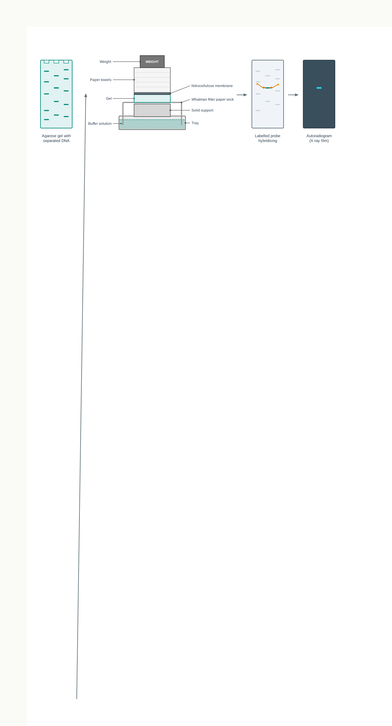
Fig. 3.24 – Capillary transfer setup for Southern blotting.
Setting Up the Capillary Transfer
The assembly deserves careful attention, because each layer has a purpose. A solid support is placed in a tray, and buffer solution is added to the tray up to half the height of the solid support. A Whatman paper strip is placed on the solid support so that it touches the buffer on two sides — these two dipping ends act as wicks, drawing buffer continuously upward.
The gel containing the DNA is kept on this Whatman strip, and a sheet of nylon or nitrocellulose membrane is placed on top of the gel. Pressure is applied evenly on the gel by placing a stack of filter papers or paper towels and a weight on top of the membrane and gel.
Buffer solution, by capillary action, moves upward through the gel and the membrane and into the filter papers above. Along with the buffer solution, the DNA moves out of the gel and onto the positively charged membrane, where the negatively charged DNA is held.
The membranes, after the transfer of nucleic acids, serve as a replica of their respective gels — every band sits in exactly the position it occupied in the gel, which is what makes the result readable.
The membrane is then baked to permanently attach or fix the transferred DNA to it, and is subsequently mixed with probes. The blot membrane is then washed to remove unhybridised probes, so that only probe molecules that have found their true partner remain.
Reading the Result
The desired DNA sequence on the membrane is subsequently detected using the probe. A probe is a single DNA strand, complementary to the sequence present in the DNA fragment to be identified. The probe is labelled with a detecting tag, which may be of a radioactive, fluorescent or chemical nature.
The labelled DNA probe anneals with its complementary strand on the membrane. The location of the target DNA fragment is then identified by visualisation on X-ray film by autoradiography.
Northern Blotting Technique
Northern blotting is used to detect specific RNA molecules in a mixture of RNA. It was developed by the American scientists J. Alwine, David Kemp and George Stark in 1977.
Like Southern blotting, it starts with the extraction of total RNA from a homogenised tissue sample or from cells. Notice that there is no restriction digestion here — RNA molecules are already short enough, and cutting them would destroy the very transcripts being looked for.
The RNA molecules are separated on an agarose gel based on their size by electrophoresis, and then transferred to a membrane where they are immobilised. A nylon membrane with a positive charge is most effective for use in northern blotting, since the negatively charged nucleic acids have a high affinity for it.
The transfer buffer used for the blotting usually contains formamide. Formamide lowers the annealing temperature of the probe–RNA interaction, thus eliminating the need for high temperatures, which could cause RNA degradation. This is a good example of a reagent chosen to protect a fragile molecule rather than to act on it.
Once the RNA has been transferred to the membrane, it is immobilised through covalent linkage to the membrane by UV light or heat. It is then mixed with radioactive probes.
The probes are specifically designed for the RNA of interest, so that they will hybridise with the RNA sequences on the blot corresponding to the sequence of interest. The blot is now washed to remove extra probes.
The labelled probe is then detected by autoradiography, in which it appears as dark bands on X-ray film, or by fluorescent labels, as shown in Fig. 3.25.
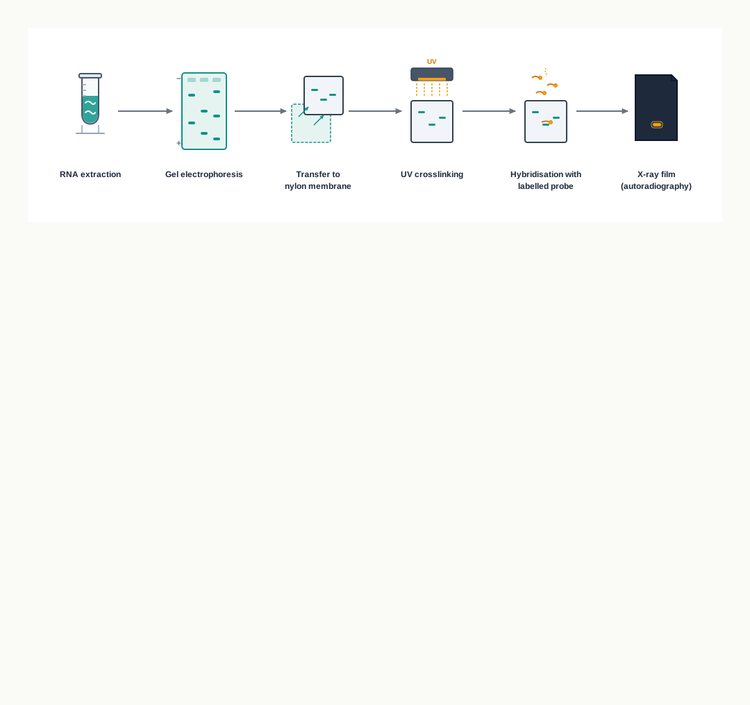
Fig. 3.25 – Identification of RNA by northern blotting.
Western Blotting Technique
The name western blotting was coined by W. Neal Burnette in 1981. It is a technique used to detect specific proteins in a sample of tissue homogenate or extract.
Proteins are first isolated from a source. They are then separated on an SDS-PAGE gel based on their electrophoretic mobility, which depends on charge, molecule size and structure of the proteins. They are transferred to a nitrocellulose membrane.
The desired protein is detected on the membrane using an antibody specific to that protein, as shown in Fig. 3.26. This is the one important difference in principle from the two nucleic acid blots: a protein has no complementary sequence to pair with, so recognition is done by an antibody rather than a probe.
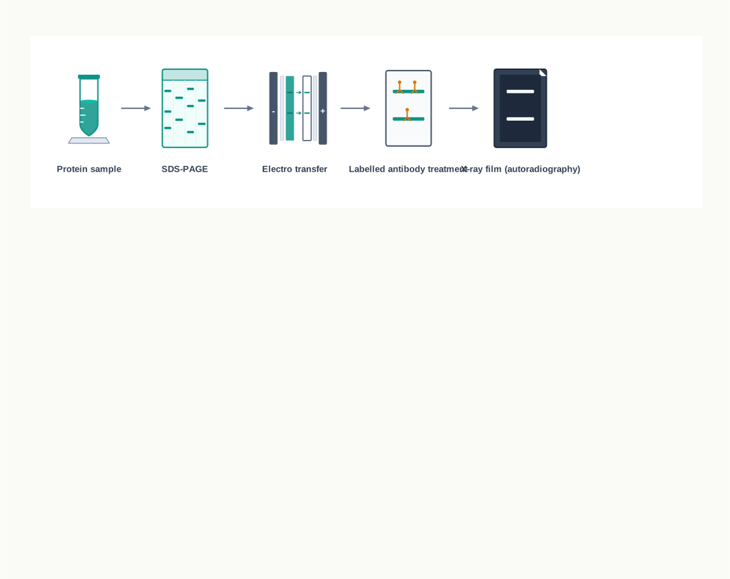
Fig. 3.26 – Identification of proteins by western blotting.
Putting the Three Together
Using all three blotting techniques, a person can identify a gene and its expression. A gene in a DNA sequence can be identified by Southern blotting; its transcripts, that is the RNA, can be identified by northern blotting; and finally the expression of the protein from mRNA, by translation, can be shown by western blotting.
In other words, the three blots follow the central dogma one step at a time, and each answers a different question about the same gene, as set out in Fig. 3.27.
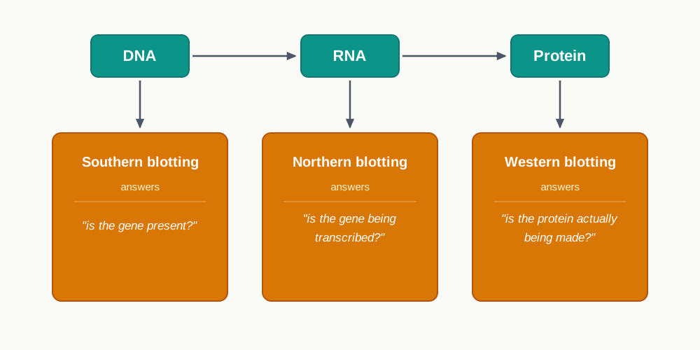
Fig. 3.27 – The three blots along the central dogma.
How This Section Is Tested
The three blots are among the most reliably examined topics in the chapter, and the commonest question is simply to differentiate them. Beyond that, expect reason-based questions — why the DNA must be cut before Southern blotting, why formamide is added in northern blotting, why an antibody rather than a probe is used in western blotting — and sequence-ordering questions on the steps of capillary transfer. Case-based questions often describe a transgenic plant or a patient sample and ask which blot should be used and what a particular result would mean, so practise matching each question about a gene to the blot that answers it.
3.7 Polymerase Chain Reaction (PCR)
Several molecular and genetic experiments require a significant amount of DNA. A single hair root, a drop of dried blood or a fragment of ancient bone may contain only a few molecules of the sequence of interest — far too little to cut, clone or analyse.
In order to generate multiple copies of DNA from a few copies, a technique was developed by Kary B. Mullis, which is known as the ‘Polymerase Chain Reaction (PCR)’. In this technique, a very small amount of DNA can be exponentially amplified to generate thousands to millions of copies.
PCR, sometimes called ‘molecular photocopying’, is often heralded as one of the most important scientific advances in molecular biology. It revolutionised the study of DNA to such an extent that its inventor, Kary B. Mullis, was awarded the Nobel Prize for Chemistry in 1993.
The Principle Behind PCR
The PCR technique is based on the same principle that cells use to replicate their DNA. Nothing unnatural is being done to the molecule; the reaction simply runs the cell’s own copying process again and again in a tube, under conditions the experimenter controls.
As the name implies, it is a chain reaction carried out in repeated cycles, which involve the process of heating and cooling called thermal cycling, carried out by a machine called a thermocycler.
This repeated heating creates one serious difficulty. The reaction requires a heat stable DNA polymerase enzyme that can make new strands of DNA on template strands at a high temperature of about 72 to 78°C — a temperature at which a human or E. coli DNA polymerase would be non-functional. An ordinary polymerase would be destroyed in the first cycle and would have to be replaced by hand before every subsequent cycle.
The DNA polymerase typically used in PCR is called Taq polymerase, an enzyme isolated from the thermophilic bacterium Thermus aquaticus, which inhabits hot springs. Because this bacterium lives at temperatures that would cook most organisms, its enzymes are built to keep working there — and that is precisely the property PCR needed.
Another enzyme, Pfu polymerase, isolated from Pyrococcus furiosus, is used widely because of its higher fidelity when copying DNA. Where accuracy of the copy matters more than speed, Pfu is preferred.
Primers: Deciding What Gets Copied
Like other DNA polymerases, Taq polymerase also requires a primer, a short sequence of nucleotides that provides a 3′-OH end to start DNA synthesis. You will recall from Section 3.3 that no DNA polymerase can begin a chain from nothing.
Two types of single-stranded synthetic deoxyoligonucleotide primers, called forward and reverse primers, are used in each PCR reaction. They are complementary to the DNA sequences in the template strands flanking the target region — that is, the region that should be copied.
They are designed from pre-existing knowledge of the sequence of the DNA template to be amplified. This is a point worth dwelling on: PCR cannot be used on a sequence about which nothing at all is known, because the two primers must be written before the reaction can be set up. The primers are what tell the reaction where to start and where to stop, and therefore what gets amplified out of an entire genome.
The Three Steps of a PCR Cycle
PCR involves three steps — denaturation, annealing and extension — as shown in Fig. 3.28.
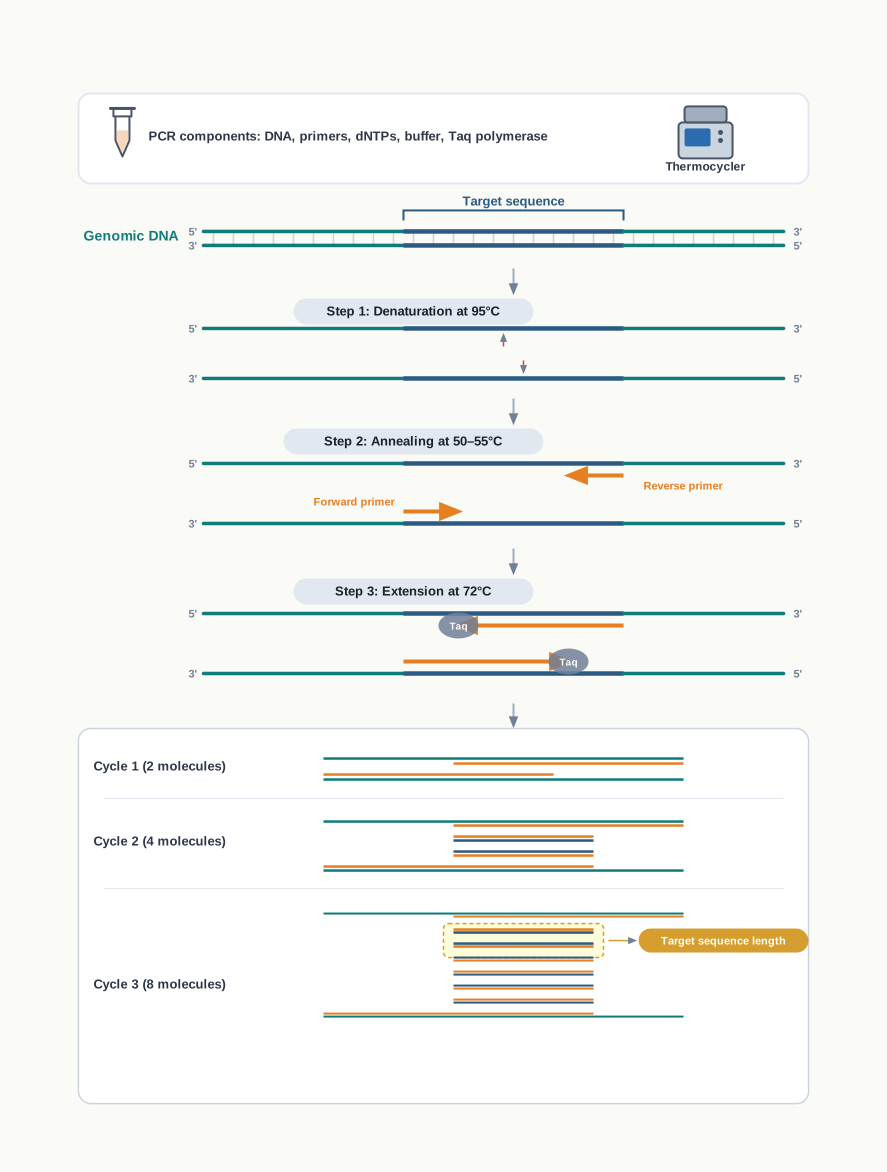
Fig. 3.28 – Steps of the polymerase chain reaction.
Denaturation. The first step is accomplished by heating the double-stranded DNA to be amplified to a temperature of about 94–95°C. At this high temperature, the hydrogen bonds that hold the two complementary strands of the DNA molecule together break down, and each strand then serves as a template for the synthesis of its new complementary strand. Note that only hydrogen bonds break; the phosphodiester backbone is untouched, which is why the strands survive intact.
Annealing. The second step is annealing, during which the temperature is lowered to around 50–55°C so that the specific primers can anneal to their respective template strands at their complementary sites and serve as the starting point for copying. The extent of lowering of temperature depends upon the length of the primer and the sequence of the primer.
Why should the temperature depend on the primer? A short primer, or one rich in A–T pairs, forms fewer hydrogen bonds and needs a lower temperature to stay bound; a longer or G–C rich primer holds on at a higher temperature. If the temperature is set too high, the primers will not bind at all; if it is set too low, they will bind at partly matching sites and the wrong stretches of DNA will be amplified.
Extension. In the third step, the temperature is raised to about 72°C, and the heat stable DNA polymerase begins adding deoxyribonucleotides — dNTPs, that is dATP, dTTP, dCTP and dGTP — onto the 3′-OH ends of the annealed primers. Thus, a new chain of DNA grows from the 5′ to 3′ direction on each template.
Copies of DNA strands formed by PCR are known as amplicons. At the end of the cycle, the temperature is raised again and the whole process is repeated, as summarised in Fig. 3.29.
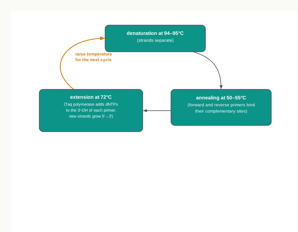
Fig. 3.29 – One cycle of the polymerase chain reaction.
Why the Numbers Grow So Fast
The number of DNA copies doubles after each cycle. Hence the number of copies at the end of each cycle would be \(2^n\), where \(n\) is the cycle number. Starting from a given number of template molecules, the same relation is written as
\[N = N\_0 \\times 2^n\]
where \(N\) is the number of copies present after \(n\) cycles, \(N\_0\) is the number of copies present at the start, and \(n\) is the number of cycles completed. Both quantities are pure counts of molecules, so the expression has no unit.
Usually, 25 to 30 cycles are carried out in a typical PCR reaction. Work out \(2^{30}\) and you will see why that is enough: a single starting molecule becomes more than a thousand million copies, and the whole run takes a few hours in a machine that needs no attention while it works.
Analysing the Product
In conventional PCR, the amplified product is analysed by gel electrophoresis at the end of the reaction, which is called end point analysis. The amount of DNA in the band on the gel plate is then estimated by measuring the intensity of the band using computer programs, and this is transferred into quantitative data. This is called semi-quantitative PCR.
The word “semi” is honest about its limitation. Band intensity at the end of a run is only a rough guide to how much template was there at the beginning, because by the last cycles the reaction has begun to run out of reagents and the neat doubling no longer holds.
Reverse Transcription PCR (RT-PCR)
If DNA material is formed from mRNA by reverse transcriptase and then used in PCR for amplification, the method is known as reverse transcription PCR (RT-PCR), shown in Fig. 3.30.
This extends the reach of PCR to RNA, which no DNA polymerase can copy directly. It matters because many viruses have RNA genomes, and because the mRNA present in a tissue is the direct evidence of which genes are switched on there.
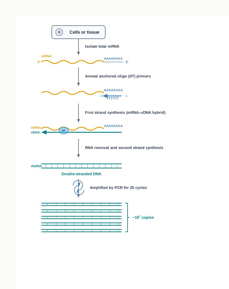
Fig. 3.30 – Steps of reverse transcription PCR.
Notice the anchored oligo (dT) primers in Fig. 3.30. These are short runs of thymines, and they work because most eukaryotic mRNAs carry a poly-A tail at their 3′ end — the tail added by poly A polymerase, which you met in Section 3.3. The oligo (dT) primer base-pairs with that tail, so it can prime the copying of almost any mRNA without knowing its sequence.
Real-Time Quantitative PCR
The latest advancement in PCR technology is real-time quantitative PCR (real-time qPCR). In this method, fluorescent markers are used which have a specific binding affinity to double-stranded DNA. When bound to dsDNA, they exhibit fluorescence behaviour.
The fluorescence emission is detected and quantitated by a detector. The amount of fluorescence emitted is directly proportional to the amount of double-stranded PCR product. Since the amount of PCR product formed can be measured after each PCR cycle, it is called real-time quantitative PCR — the reaction is watched while it happens rather than examined after it has finished.
One of the fluorescent dyes used in real-time PCR is SYBR green. The dye only binds to double-stranded DNA, which is what makes the signal a measure of product formed rather than of anything else in the tube.
The Thermocycler
The machine in which the PCR reaction is conducted is known as a thermocycler. These are automated machines having control points where one can set three gradients of temperature — for denaturation, annealing and extension — for different time periods for each step.
For a real-time qPCR thermocycler, the machine additionally has a detector to measure the emitted fluorescence. In real-time PCR, gel electrophoresis is not needed, as it is in the case of conventional PCR, because the result has already been recorded cycle by cycle.
Applications of PCR
PCR has several applications in molecular biology and rDNA technology. One of the applications of PCR is to quantify mRNA in order to assess the expression of a gene. It is also used to amplify minute DNA samples collected from crime scenes and fossils for further investigation.
In forensic work, a few cells left on a weapon or a garment can be amplified until there is enough DNA to compare against a suspect’s sample. In palaeontology, DNA recovered from preserved remains can be studied although the original molecules are few and badly damaged. In medicine, PCR detects an infection by finding the pathogen’s own nucleic acid, often before the patient has produced any antibodies at all.
How This Section Is Tested
PCR is examined more heavily than almost any other topic in this chapter. Expect to state the three steps with their temperatures in the correct order, to justify why a heat-stable polymerase is essential, to explain the role of the forward and reverse primers, and to calculate copy numbers using \(2^n\). Data-interpretation questions on the amplification curve — reading a Ct value, comparing two samples, explaining the no-template control — have become common since real-time PCR entered the syllabus. Assertion–reason items frequently pair PCR with Taq polymerase, so check whether the reason given really explains the assertion or is simply a separate true statement.
3.8 DNA Libraries
DNA molecules present in the genome of an organism are very large in terms of the number of base pairs they contain. The size of the DNA molecule present in any diploid cell from any organ of your body has around \(3 \\times 10^9\) bp.
In a genome, gene sequences are arranged randomly, and selecting or isolating a gene of interest is a big task — especially when the genomic sequences are not known. There is no index, no marking, and nothing about the appearance of a gene to distinguish it from the sequence lying next to it.
There is a further complication. Only a small portion of the genome is transcribed to give mRNA, whereas a major portion of the genome remains untranscribed. It is therefore very difficult to isolate a gene of interest, or a sequence of the genome whose location and sequence is not known.
Hence, DNA libraries are constructed by collecting DNA fragments that have been cloned into vectors, so that the specific DNA fragments of interest can be identified and isolated for further study.
The name is well chosen. A library does not make its books easier to write; it makes them easier to find. In the same way, a DNA library does not simplify the genome — it stores the genome in manageable, individually retrievable pieces, each one already inside a vector and ready to be copied.
There are basically two types of DNA libraries, genomic and cDNA libraries, which are described in the following sections.
(i) Genomic DNA Library
A genomic library is a collection of clones of small fragments of DNA that together represent the complete genome of an organism. A population of identical vectors stores the DNA inserts, each vector containing a different insert.
That last point is the whole idea in one sentence: the vectors are all alike, but no two inserts are the same, and between them the inserts cover everything the organism has.
In general, the construction of a genomic library is done as shown in Fig. 3.31. First, genomic DNA is isolated from the source. This DNA is far too large to be incorporated into a vector, and needs to be broken down into fragments of a desirable size.
Therefore, the genomic DNA is digested with a restriction enzyme to cut the DNA into fragments of a specific size. The DNA fragments are then inserted into vectors using DNA ligase to form recombinant vectors. This generates a pool of recombinant DNA molecules.
The recombinant DNA molecules are now taken up by host bacterial cells by transformation, and are then allowed to multiply in a nutrient medium to form colonies. All the host cells containing recombinant vectors together represent a genomic library.
The library so created contains representative copies of all the DNA fragments present within the genome of that organism. Every colony on the plate is a living store of one fragment, and can be grown up whenever that fragment is needed again.
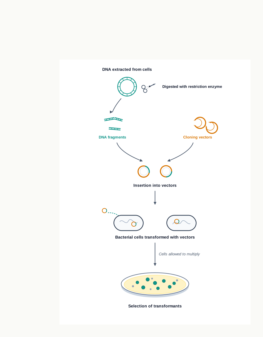
Fig. 3.31 – Construction of a genomic DNA library.
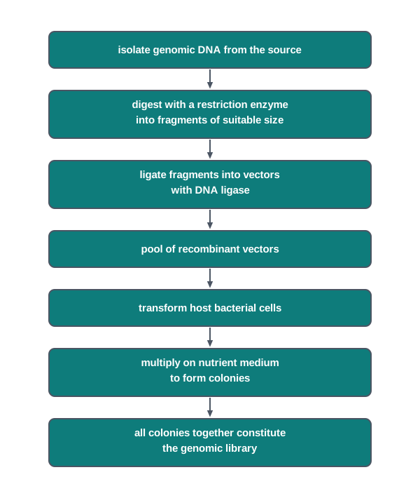
Fig. 3.32 – Workflow for constructing a genomic DNA library.
Applications of a Genomic Library
The genomic library has several applications in biotechnology. The genomic library of a species may be helpful for the complete sequencing of its genome, since the sequence of each stored fragment can be determined separately and the fragments then assembled.
One can also search such a library for many genes that are not expressed in the genome of an organism. This is a real advantage over the cDNA library described below, because a gene that is never transcribed leaves no mRNA behind and would be invisible to any method that starts from mRNA.
A genomic library is also helpful in understanding the evolution of species, since the corresponding regions of two species can be compared directly.
Finally, a genomic library can be used to compare the sequences of healthy and diseased tissues of the same organism, in order to identify genetic aberrations — the difference between the two sets tells the investigator where the fault lies.
(ii) cDNA Library
Gene expression in higher eukaryotes is tissue-specific. In specific cells, certain genes undergo moderate to high expression, while the same genes remain silent elsewhere in the body.
For example, the genes encoding insulin proteins are expressed only in the beta cells of the pancreas, while albumin encoding genes are expressed in liver cells. Every cell of the body carries both genes, but each tissue reads only the ones it needs.
Using this information, a target gene can be cloned by isolating the mRNA from a specific tissue. The tissue itself has already done the work of selection — it has transcribed the genes that matter to it, so its mRNA population is enormously enriched for exactly those genes.
The specific DNA sequences synthesised as copies from the mRNAs of a particular cell type are called cDNA (complementary DNA). Clones of such DNA copies of mRNAs are called cDNA clones. The cDNA clones of all the genes expressed in a specific cell type or tissue of an organism together represent a cDNA library.
Construction of a cDNA Library
The construction of a cDNA library involves the isolation of total mRNA from the cell type or tissue of interest.
An obstacle appears at once. mRNA, being single-stranded, cannot be cloned as such, and it is not a substrate for DNA ligase. Ligase works only on the phosphodiester backbone of DNA in duplex form, as you saw in Section 3.3.
The mRNA is therefore first converted into cDNA before insertion into a suitable vector. This is achieved using reverse transcriptase, also called RNA-dependent DNA polymerase or RTase. RTase synthesises a complementary DNA strand on the mRNA by using the mRNA as a template.
The mRNA is then removed by RNase, and the single-stranded cDNA that remains is converted into double-stranded cDNA by DNA polymerase. Only now is the molecule a proper double-stranded DNA, with the ends that a restriction enzyme and a ligase can work on.
The cDNA molecules are then cloned in an appropriate host–vector system, as shown in Fig. 3.33. The total clones of cDNA are the representative of the cDNA library of the source.
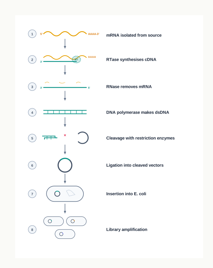
Fig. 3.33 – Construction of a cDNA library.
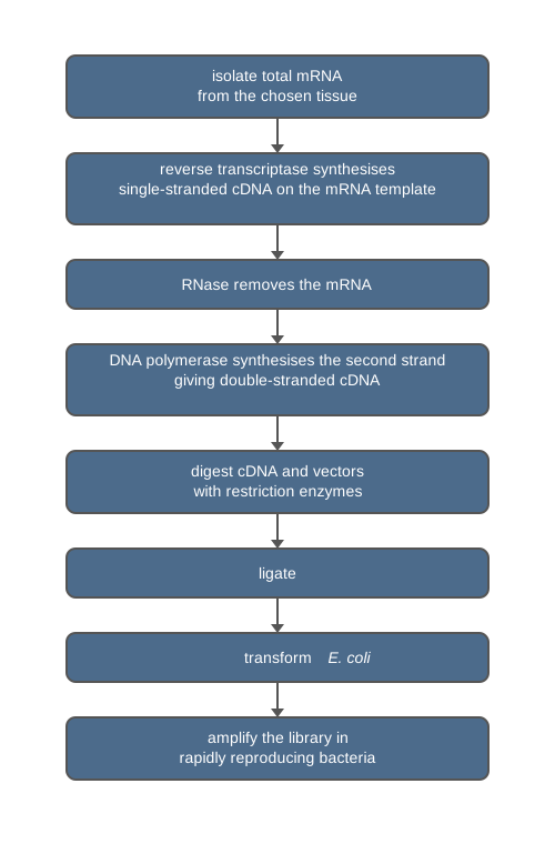
Fig. 3.34 – Workflow for constructing a cDNA library.
Since the expression of genes is different in different organs or cells of an organism, and at different physiological states, cDNA libraries prepared from different sources of the same organism may vary from each other. A liver library and a pancreas library are not two copies of the same thing; they are two different snapshots of the same genome in use.
Significance of the cDNA Library
The cDNA library has great significance in the applications of biotechnology. Its most important application is to know which genes are active in particular tissues under a particular physiological state.
It also helps us to isolate a specific gene, because a gene strongly expressed in a tissue will be well represented among its cDNA clones.
Further, using cDNA as probes, we can screen genomic libraries for a particular gene. Here the two libraries are used together: the cDNA identifies the gene, and the genomic clone it finds carries the gene together with the surrounding regions that control it.
How This Section Is Tested
The comparative account of genomic and cDNA libraries is the single most frequently asked question here, so be ready to write it as a properly organised comparison rather than as two separate descriptions. Other common forms are reason-based — why mRNA must be converted to cDNA before cloning, why cDNA libraries differ between tissues of the same organism, why a genomic library is needed to study a gene that is never expressed — and sequence-ordering questions on the construction steps in Fig. 3.31 and Fig. 3.33. Case-based questions often place you in a laboratory that must choose between the two libraries for a stated purpose, so practise justifying the choice in one or two sentences.
3.9 Summary
- Isolation of nucleic acids from different organisms is the most essential requirement for any molecular biology experiment. The process has four steps: disruption of the biological sample, protection of the nucleic acids from their degrading enzymes, separation of the nucleic acids from other molecules, and assessment of the purity and quality of the isolated nucleic acids — the nucleic acids being precipitated and concentrated with ethanol or isopropanol before this assessment.
- Various enzymes play an important role in recombinant DNA (rDNA) technology. These include nucleases, DNA ligase, alkaline phosphatase, polynucleotide kinase and poly A polymerase, among others.
- The major task in the manipulation of DNA is the cutting of the gene of interest and the vector, and the ligation of that gene into the vector DNA.
- Nucleases are the enzymes that cleave nucleic acids by hydrolysing the phosphodiester bond that joins the sugar residues of adjacent nucleotides. Two major types have been identified, based on where they act on the phosphodiester bonds of a polynucleotide chain: exonuclease and endonuclease.
- Exonuclease enzymes remove mononucleotides, one at a time, either from the 3′ or the 5′ end of the DNA molecule.
- Endonuclease enzymes that cleave DNA molecules at a specific sequence are called restriction endonucleases or restriction enzymes (REs). REs are mainly categorised into three groups — Types I, II and III — based on their cofactor requirement and the position of their DNA cleavage site relative to the target sequence. Type II REs find application in rDNA technology.
- DNA ligase joins two DNA strands together by catalysing the formation of a phosphodiester bond in the duplex form.
- DNA polymerases are a group of enzymes that catalyse the synthesis of a new DNA strand by using dNTPs on a template strand.
- Alkaline phosphatase removes the terminal phosphate group from the 5′ end of DNA strands.
- Reverse transcriptase generates a complementary DNA (cDNA) strand from an RNA template, a process called reverse transcription.
- In rDNA technology, the recombinant DNA is introduced into host cells by a number of methods, such as chemical based transfection (calcium chloride, lipofection, and so on) and physical transfection (electroporation, microinjection and biolistic methods).
- Selection of transformed bacteria is the most essential step for a successful cloning experiment, that is, to identify the transformed cells carrying the recombinant vector with the gene of interest from a mixture of transformed and non-transformed cells. These selection methods may be direct, or may work through insertional inactivation.
- In direct selection, transformed cells are distinguished from non-transformed cells on the basis of the expression of certain traits, such as resistance to antibiotics.
- In the insertional inactivation method, a vector carrying two markers is used — either two antibiotic resistance genes, or one antibiotic resistance gene and one lacZ gene.
- The blue–white selection method is another example of insertional inactivation used to select recombinant transformed cells, in which the expression of the lacZ gene can be observed directly in the bacterial colonies.
- Blotting techniques are widely used to separate and identify DNA, RNA and proteins from a mixture of molecules.
- The Southern blotting technique is used to detect a specific sequence of DNA in DNA samples.
- The Northern blotting technique is used to detect specific RNA molecules in a mixture of RNA.
- Western blotting is used to detect specific proteins in a sample of tissue homogenate or extract.
- The Polymerase Chain Reaction (PCR) is used to amplify a small amount of DNA into thousands to millions of copies, and involves three steps: denaturation, annealing and extension. The amplified product of PCR can be analysed by gel electrophoresis at the end of the reaction, which is called end point analysis.
- The latest advancement in PCR technology is real-time quantitative PCR (qPCR), in which fluorescent markers are used that have a specific binding affinity to double-stranded DNA. In qPCR, gel electrophoresis is not needed, as it is in the case of conventional PCR.
- DNA libraries are constructed by collecting DNA fragments that have been cloned into vectors, so that specific DNA fragments of interest can be identified and isolated. There are basically two types of DNA libraries — the genomic library and the cDNA library.
- A genomic library is a collection of clones of small fragments of DNA that together represent the complete genome of an organism.
- The cDNA library constitutes the cDNA clones of all the genes expressed in a specific cell type or tissue of an organism.
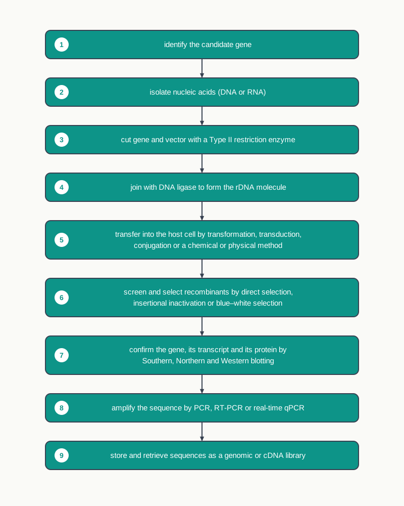
Fig. 3.35 – Overview of recombinant DNA technology.
3.10 Key Terms
- Gene cloning — The production of many identical copies of a selected gene by inserting it into a vector and allowing that vector to replicate inside a host cell.
- Vector — A DNA molecule, such as a plasmid, that carries a foreign piece of DNA into a living cell and replicates there on its own.
- Plasmid — A small circular DNA molecule found in bacteria in addition to the main chromosome, widely used as a cloning vector.
- Host cell — The cell, usually a bacterium such as Escherichia coli, inside which a vector and its insert are multiplied.
- Clone — A population of molecules or cells that are all identical, because they were produced by copying a single original.
- Genome — The complete set of DNA present in an organism.
- Recombinant DNA (rDNA) — A DNA molecule formed by joining DNA segments that do not occur together in nature, typically a vector carrying a foreign gene.
- Candidate gene — A gene selected for cloning because biochemical or physiological evidence links it to a trait, product or disorder of biomedical, economic or evolutionary importance.
- Complementary DNA (cDNA) — A DNA copy synthesised from an mRNA template, and therefore lacking the non-coding stretches present in the chromosomal gene.
- Reverse transcriptase — An RNA-directed DNA polymerase, found in many retroviruses, that synthesises a cDNA strand on an RNA template.
- Lysozyme — An enzyme that digests peptidoglycan, the main component of the bacterial cell wall, during DNA isolation from bacteria.
- Sodium dodecyl sulphate (SDS) — An anionic detergent that lyses cell membranes, makes all proteins anionic so that they dissociate from nucleic acids, and also inhibits nucleases.
- Cetyl trimethyl ammonium bromide (CTAB) — A cationic detergent used for DNA isolation from plant cells, which lyses the cell wall and separates DNA from polysaccharides according to ionic strength.
- Polyvinyl pyrrolidone (PVP) — A compound added to the CTAB extraction medium to neutralise phenols released from plant tissue.
- Chelating agent — A chemical, such as EDTA, that locks up divalent metal ions in solution and thereby removes the cofactors that nucleases require.
- Ethylene diamine tetraacetic acid (EDTA) — The chelating agent used in extraction buffers, which binds \(\\mathrm{Mg^{2+}}\) and \(\\mathrm{Mn^{2+}}\) and so protects nucleic acids from nucleases.
- Isoamyl alcohol — A solvent used with chloroform that prevents foaming and stabilises the interface between the organic and aqueous phases.
- Ribonuclease A — An enzyme added during DNA isolation to digest RNA into ribonucleotides, so that only DNA remains.
- Chaotropic agent — A reagent, such as guanidinium isothiocyanate, that disrupts hydrogen bonding and increases disorder in a solution, causing proteins and nucleic acids to aggregate.
- Guanidinium isothiocyanate (GITC) — The acidic chaotropic reagent, used with phenol and chloroform, that extracts total RNA into the aqueous phase while inactivating RNases.
- Nuclease — An enzyme that cleaves nucleic acids by hydrolysing the phosphodiester bond joining the sugar residues of adjacent nucleotides.
- Exonuclease — A nuclease that removes nucleotides one at a time, as mononucleotides, from the 3′ or 5′ end of a polynucleotide chain.
- Endonuclease — A nuclease that breaks internal phosphodiester bonds within a DNA or RNA molecule.
- Restriction endonuclease (restriction enzyme, RE) — An enzyme, found mainly in bacteria and archaea, that recognises a specific DNA sequence of about 4 to 8 bp and cleaves the DNA at or near it.
- Recognition sequence — The specific short DNA sequence, often 4 to 8 bp long, to which a restriction enzyme binds.
- Palindromic sequence — A double-stranded DNA sequence in which the order of bases read in the 5′→3′ direction is the same on both strands.
- Sticky (cohesive) ends — The short single-stranded projections left when a restriction enzyme cuts the two strands of DNA at staggered positions.
- Blunt ends — Fully base-paired ends produced when a restriction enzyme cuts both strands of DNA at the same position, leaving no projection.
- DNA ligase — An enzyme that joins two DNA fragments by forming a phosphodiester bond between the 5′-phosphate of one chain and the 3′-hydroxyl of the adjacent chain, using ATP or NAD as the energy source.
- DNA polymerase — An enzyme that synthesises a new DNA strand in the 5′→3′ direction using dNTPs on a template strand, and which cannot begin without a primer.
- Primer — A short single-stranded oligonucleotide carrying a free 3′-OH group, which provides the starting point for DNA synthesis.
- Alkaline phosphatase — An enzyme that removes the terminal phosphate group from the 5′ end of DNA strands.
- Polynucleotide kinase — An enzyme that attaches a phosphate group to the hydroxyl group at the 5′ end of DNA, the reverse of the action of alkaline phosphatase.
- Terminal deoxynucleotidyl transferase (terminal transferase) — An enzyme that adds similar nucleotide residues to form a homopolymer tail at the 3′ end of a DNA strand, without requiring a template.
- Poly A polymerase — An enzyme that incorporates adenine residues at the hydroxyl group of the 3′ end of RNA, forming the poly-A tail.
- Transformation — The genetic alteration of a cell brought about by the direct uptake of exogenous DNA from its surroundings through the cell membrane.
- Transformants — Recipient cells that have taken up a foreign DNA molecule.
- Transduction — The transfer of bacterial DNA from one bacterium to another by means of a bacteriophage that has accidentally packaged host DNA.
- Bacteriophage — A virus that specifically infects bacterial cells.
- Lysogenic life cycle — The phage life cycle in which the phage genome is incorporated into the bacterial DNA and remains dormant there for several generations.
- Transductants — Recipient bacterial cells that have received bacterial DNA through a bacteriophage.
- Conjugation — The transfer of DNA from a donor bacterium to a recipient bacterium through direct cell-to-cell contact.
- F-factor — The fertility factor encoded by the F plasmid, which enables a bacterium to act as a donor in conjugation.
- Pilus — The thin tube-like structure formed under the control of the F-factor, through which the donor cell contacts the recipient during conjugation.
- Relaxase — The donor-cell enzyme that nicks one strand of the double-stranded F plasmid before its transfer through the pilus.
- Calcium chloride method — A chemical transfer method in which positively charged calcium forms a complex with DNA, which the host cells then take up following a heat shock.
- Electroporation — A physical transfer method in which a mild electric current creates transient micropores in the host cell membrane through which DNA enters.
- Lipofection — A chemical transfer method in which DNA enclosed in liposomes enters the cell as the liposome fuses with the phospholipid bilayer of the cell membrane.
- Microinjection — A physical transfer method in which foreign DNA is introduced directly into the nucleus of a host cell using a specialised automated apparatus.
- Biolistic method — A physical transfer method in which microscopic gold, nickel or tungsten particles coated with DNA are fired into cells at high velocity by a gene gun.
- Selectable marker — A gene on a vector that confers a recognisable trait, such as antibiotic resistance or a coloured product, allowing cells carrying the vector to be identified.
- Direct selection — A method of selection in which transformed cells are distinguished from non-transformed cells by the expression of a trait such as antibiotic resistance.
- Insertional inactivation — The loss of function of a marker gene caused by the insertion of foreign DNA within it, allowing recombinant cells to be recognised by the trait they have lost.
- Master plate — The first plate, on which all transformed cells form colonies, and from which recombinant colonies are picked.
- Replica plate — The second plate, prepared by transferring cells from the master plate on a nitrocellulose membrane, on which recombinants fail to grow.
- Blue–white selection — A form of insertional inactivation in which recombinant colonies remain white because an insert within lacZ prevents the formation of active β-galactosidase, while non-recombinant colonies turn blue.
- β-galactosidase — The enzyme expressed by the lacZ gene, which cleaves X-gal to a product that dimerises to an insoluble blue pigment.
- X-gal — 5 Bromo-4-chloro-3 indolyl-beta D-galactoside, a colourless chromogenic analog of lactose used as the substrate in blue–white selection.
- IPTG — Isopropyl β-D-1-thiogalactopyranoside, included in the selection medium to switch on expression of the lacZ gene.
- Green Fluorescent Protein (GFP) — A fluorescent protein used as an alternative marker, whose production is disrupted when an insert interrupts its coding sequence, giving non-fluorescing recombinant bacteria.
- Blotting — A technique in which molecules separated in a gel are transferred to and immobilised on a membrane, where a specific molecule is identified using a labelled probe or antibody.
- Hybridisation — The pairing of a labelled single-stranded probe with its complementary sequence immobilised on a membrane.
- Probe — A short single-stranded nucleic acid carrying a radioactive, fluorescent or chemical label, used to locate a specific sequence on a blot.
- Southern blotting — The blotting technique used to detect a specific sequence of DNA in DNA samples.
- Northern blotting — The blotting technique used to detect specific RNA molecules in a mixture of RNA.
- Western blotting — The blotting technique used to detect specific proteins in a tissue homogenate or extract, using an antibody specific to the protein.
- Eastern blot — A blotting technique used to detect specific post-translational modifications of proteins.
- Autoradiography — The visualisation of a radioactively labelled probe as dark bands on X-ray film.
- Polymerase Chain Reaction (PCR) — A technique in which a small amount of DNA is exponentially amplified into thousands to millions of copies through repeated cycles of denaturation, annealing and extension.
- Thermal cycling — The repeated heating and cooling of a PCR reaction, carried out automatically by a thermocycler.
- Thermocycler — The automated machine in which a PCR reaction is conducted, with control points for setting three temperature gradients and their time periods.
- Taq polymerase — The heat-stable DNA polymerase isolated from the thermophilic bacterium Thermus aquaticus, used routinely in PCR.
- Pfu polymerase — A heat-stable DNA polymerase isolated from Pyrococcus furiosus, used widely because of its higher fidelity when copying DNA.
- Denaturation — The first step of a PCR cycle, in which heating to about 94–95°C breaks the hydrogen bonds and separates the two DNA strands.
- Annealing — The second step of a PCR cycle, in which lowering the temperature to around 50–55°C allows the primers to bind their complementary sites on the template strands.
- Extension — The third step of a PCR cycle, in which the temperature is raised to about 72°C and the heat-stable polymerase adds dNTPs to the 3′-OH ends of the annealed primers.
- Amplicon — A copy of the target DNA segment produced during a polymerase chain reaction.
- Semi-quantitative PCR — End point analysis in which the amount of DNA in a gel band is estimated from its intensity and converted into quantitative data.
- Reverse transcription PCR (RT-PCR) — A method in which mRNA is first converted into cDNA by reverse transcriptase, and that cDNA is then amplified by PCR.
- Real-time quantitative PCR (qPCR) — A method in which fluorescent markers bound to double-stranded DNA allow the amount of product to be measured after every cycle, so that gel electrophoresis is not needed.
- SYBR green — A fluorescent dye used in real-time PCR that binds only to double-stranded DNA.
- Ct value (threshold cycle) — The cycle number at which the fluorescence in a reaction crosses the threshold, which is inversely proportional to the amount of the gene of interest present in the sample.
- DNA library — A collection of DNA fragments cloned into vectors and maintained in host cells, from which a fragment of interest can later be identified and isolated.
- Genomic library — A collection of clones of small DNA fragments which together represent the complete genome of an organism.
- cDNA clones — Clones of the DNA copies made from the mRNAs of a particular cell type.
- cDNA library — The complete set of cDNA clones prepared from the mRNA of a particular cell type or tissue, representing only the genes expressed there.
3.11 Points to Ponder
- Restriction enzymes evolved in bacteria as a defence against invading bacteriophages, and DNA ligase, polymerases and reverse transcriptase all exist in cells for their own reasons. Almost every tool of gene cloning was borrowed rather than invented. What does this suggest about where the next generation of biotechnological tools is likely to come from?
- The extraction buffer used for isolating DNA contains EDTA, a mild alkaline pH and a detergent, and every one of these was added to defeat a problem created by the previous step. Trace this chain of cause and effect for yourself, and consider what it tells you about how laboratory protocols come to have the form they do.
- A cut made by a restriction enzyme can be sealed again by ligase, because the enzyme leaves behind exactly the 5′-phosphate and 3′-hydroxyl groups that ligase needs. Would gene cloning have been possible at all if restriction enzymes had left ends of some other chemical form?
- Type II restriction enzymes are useful precisely because their cutting position is predictable, while Types I and III cut at a variable distance from the recognition site. Think about how much of modern biotechnology rests on this single property of predictability.
- In blue–white selection and in replica plating alike, the recombinant cells are recognised by something they have lost rather than something they have gained. Why is a loss often easier to detect reliably than a gain, and can you think of other places in biology where absence is the clearer signal?
- Antibiotic resistance genes make selection simple and certain, but the same genes are the ones medicine is struggling to contain. Where should the line be drawn between laboratory convenience and long-term public risk, and who should draw it?
- A genomic library of an organism stays the same whatever tissue it was made from, while a cDNA library changes with tissue and even with the physiological state of that tissue. What does this difference tell you about the relationship between the genome an organism has and the organism it actually becomes?
- PCR can work from a handful of molecules. That sensitivity is what makes it valuable in forensics and diagnosis, and also what makes contamination so serious a danger. How should a laboratory design its procedures when the very strength of a technique is also its weakness?
- The Ct value in real-time PCR falls as the amount of target rises, so a low number signals a heavy viral load. Why do measurements that run in the opposite direction to intuition cause so much confusion, and how would you explain this one to someone outside science?
- The three blotting techniques follow the central dogma, each answering a different question about the same gene: is it present, is it transcribed, is it translated. Why is it not safe to assume that a gene successfully inserted into an organism will actually produce its protein?
- Cloning a human gene into a bacterium works only if the sequence used is free of the non-coding stretches present in the chromosomal gene. Consider what this single technical requirement reveals about the differences between prokaryotic and eukaryotic gene expression.
- Almost every technique in this chapter turns a natural process — bacterial transformation, phage infection, viral reverse transcription, a thermophile’s heat-stable enzyme — to a purpose its organism never had. Is there a meaningful boundary between using nature and remaking it, and where would you place it?
3.12 NCERT Questions
- Describe the methods used for isolation of DNA.
- What is the role of biological detergent in the process of isolation of nucleic acid?
- How does DNA isolation from plant tissue differ from that of bacterial cell?
- How many types of restriction enzymes (REs) are there? Can all REs be used in rDNA technology? Give justification.
- What are the challenges faced during the process of nucleic acid extraction?
- Write the role of alkaline phosphatase, DNA ligase, terminal transferase in rDNA technology.
- Describe the role of chelating agent in the process of DNA extraction.
- Briefly describe the modes of DNA transfer into the host.
- Identify the correct statement for blue–white selection method.
- A specific dye is used to stain bacterial colony.
- It is based on the expression of lacZ gene.
- The recombinant bacterial colony remains blue.
- lacZ gene is inserted in an antibiotic resistant gene.
- Identify the correctly matched pair from the following options.
- Northern blot: Detect specific sequence of DNA
- Southern blot: Detect specific sequence of RNA
- Western blot: Detect specific proteins
- Eastern blot: Detect transcriptional modifications in RNA
- Identify the incorrect matched pair from the following options.
- Taq polymerase: Thermus aquaticus
- Pfu polymerase: Pyrococcus furiosus
- HindIII: Haemophilus influenzae
- PstI: Pyrococcus stuartii
- How are recombinants screened? Describe the methods in detail.
- Differentiate between the Southern, Northern and Western blotting.
- What is PCR? Describe in detail.
- Write a comparative account of the genomic and cDNA libraries.
- Diploid human genome contains:
- 3.2 × 10⁹ base pairs
- 6.4 × 10⁸ base pairs
- 3.2 × 10⁸ base pairs
- 6.4 × 10⁹ base pairs
- Select the incorrectly matched pair from the following.
- Nucleases : Hydrolyse phosphodiester bond
- Restriction enzymes: Cleave DNA at specific sequence
- Palindromic sequence: Read same backwards and forward
- EcoRI: Type I Restriction Enzyme
- Assertion: PCR can be used to amplify very small amount of DNA using DNA modifying enzymes. Reason: PCR uses Taq Polymerase.
- Both assertion and reason are true and the reason is the correct explanation of the assertion.
- Both assertion and reason are true but the reason is not the correct explanation of the assertion.
- Assertion is true but reason is false.
- Both assertion and reason are false.
- Assertion: Foreign gene can be introduced into host bacterium by transformation techniques like electroporation. Reason: Bacteria have cell wall/membrane.
- Both assertion and reason are true and the reason is the correct explanation of the assertion.
- Both assertion and reason are true but the reason is not the correct explanation of the assertion.
- Assertion is true but reason is false.
- Both assertion and reason are false.
3.13 Objective Questions
- Which of the following is not one of the four steps involved in the extraction of nucleic acids?
- Rupture of the cell membrane or cell wall
- Protection of nucleic acids from their degrading enzymes
- Amplification of the nucleic acids by thermal cycling
- Precipitation and concentration of the isolated nucleic acids
- EDTA is included in a DNA extraction buffer chiefly because it:
- denatures histones bound to DNA
- binds divalent cations such as \(\\mathrm{Mg^{2+}}\) and \(\\mathrm{Mn^{2+}}\) required by nucleases
- reduces the polarity of the medium so that DNA precipitates
- neutralises the polyphenols released from plant tissue
- Cetyl trimethyl ammonium bromide (CTAB) is preferred for DNA isolation from plant cells because it:
- digests peptidoglycan in the cell wall
- makes all proteins anionic so that they leave the DNA
- separates DNA from polysaccharides on the basis of ionic strength and also lyses the cell wall
- inactivates ribonucleases present in the extract
- Polyvinyl pyrrolidone (PVP) is added to the CTAB extraction medium in order to:
- neutralise phenols
- chelate divalent cations
- precipitate polysaccharides
- stabilise the interface between the two phases
- After a cell lysate is treated with chloroform and isoamyl alcohol and centrifuged, the nucleic acids are recovered from the:
- lower organic layer
- interface between the two layers
- upper aqueous layer
- pellet at the bottom of the tube
- The specific function of isoamyl alcohol during deproteinisation is to:
- denature the proteins
- prevent foam formation and stabilise the interface
- increase the density of the aqueous phase
- digest the RNA present in the lysate
- In RNA isolation using GITC–phenol–chloroform, the DNA and denatured proteins are retained in the:
- upper aqueous phase only
- interface or the organic phase
- isopropanol precipitate
- filter paper stack
- An exonuclease differs from an endonuclease in that it:
- acts only on RNA
- removes mononucleotides from the 3′ or 5′ ends of a chain
- breaks hydrogen bonds rather than phosphodiester bonds
- requires S-adenosyl methionine as a cofactor
- Type II restriction enzymes are the ones used in rDNA technology because they:
- cleave about 1000 bp away from the recognition site
- cleave within the recognition site and require only \(\\mathrm{Mg^{2+}}\)
- carry endonuclease and methylase activity on a single protein molecule
- require SAM, \(\\mathrm{Mg^{2+}}\) and ATP for activity
- A restriction enzyme cuts the sequence
5'-GATATC-3'exactly in the middle of both strands. The product will have:
- 5′ sticky ends
- 3′ sticky ends
- blunt ends
- a homopolymer tail
- In the name HindIII, the Roman numeral III indicates the:
- type of the restriction enzyme
- number of base pairs in the recognition site
- strain of the microorganism
- order of identification of the enzyme from that species
- Which of the following is used as the energy source by DNA ligase obtained from E. coli?
- ATP
- NAD
- GTP
- S-adenosyl methionine
- A DNA polymerase cannot begin synthesis on its own because it requires:
- a free 5′-phosphate group
- a primer carrying a free 3′-OH group
- a methylated recognition site
- a single-stranded RNA template
- The enzyme that adds identical nucleotide residues as a homopolymer tail at the 3′ end of a DNA strand, without needing a template, is:
- polynucleotide kinase
- alkaline phosphatase
- terminal deoxynucleotidyl transferase
- poly A polymerase
- Alkaline phosphatase is used on a cut vector before ligation mainly to:
- create sticky ends on the vector
- prevent the vector from re-joining with itself
- label the 5′ end of the vector radioactively
- remove the RNA contaminating the preparation
- During bacterial conjugation, the strand of the F plasmid that passes through the pilus is first nicked by:
- lysozyme
- relaxase
- DNA ligase
- restriction endonuclease
- Gold, nickel or tungsten microparticles coated with DNA are used in the:
- calcium chloride method
- lipofection method
- biolistic method
- electroporation method
- A brief heat shock at about 42°C forms part of which method of DNA transfer?
- Microinjection
- Calcium chloride method
- Electroporation
- Biolistic method
- In the blue–white selection method, a white colony indicates that the bacterial cell contains a plasmid in which:
- the ampicillin resistance gene has been inactivated
- the lacZ gene is intact and β-galactosidase is produced
- an insert has interrupted the lacZ gene
- no plasmid has been taken up at all
- IPTG is included in the selection medium along with X-gal because it:
- supplies the colour of the blue pigment
- switches on the expression of the lacZ gene
- kills the non-transformed cells
- prevents β-galactosidase from cleaving X-gal
- In selection by insertional inactivation using a plasmid carrying amp^R and tet^R, with the insert cloned into amp^R, the master plate must contain:
- ampicillin
- tetracycline
- both ampicillin and tetracycline
- X-gal and IPTG
- A positively charged nylon membrane is most effective in northern blotting because:
- it is transparent to X-rays
- negatively charged nucleic acids have a high affinity for it
- it prevents the probe from binding non-specifically
- it withstands the high temperature of hybridisation
- Formamide is added to the transfer buffer in northern blotting because it:
- fixes the RNA covalently to the membrane
- lowers the annealing temperature of the probe–RNA interaction
- removes the unbound probe after hybridisation
- separates the RNA molecules by size
- Which sequence correctly represents the steps of one PCR cycle?
- Annealing → denaturation → extension
- Denaturation → extension → annealing
- Denaturation → annealing → extension
- Extension → annealing → denaturation
- If a PCR begins with a single target molecule and runs with perfect doubling, the number of copies present at the end of the nth cycle is given by:
- \(2n\)
- \(n^2\)
- \(2^n\)
- \(n \\times 2\)
- SYBR green is suitable as a marker in real-time PCR because it:
- binds only to single-stranded DNA
- binds only to double-stranded DNA and then fluoresces
- is cleaved by the exonuclease activity of Taq polymerase
- becomes radioactive during amplification
- Two samples are tested by real-time RT-PCR. Sample P has a Ct value of 16 and sample Q a Ct value of 32. It follows that:
- sample Q contained more target to begin with
- sample P contained more target to begin with
- both contained equal amounts of target
- no comparison is possible from Ct values
- Anchored oligo (dT) primers are used in RT-PCR because they base-pair with the:
- recognition site of a restriction enzyme
- poly-A tail at the 3′ end of the mRNA
- 5′ cap of the mRNA
- promoter region of the gene
- A gene that is never transcribed in any tissue of an organism could be recovered from:
- a cDNA library only
- a genomic library only
- either library equally well
- neither library
- cDNA libraries prepared from the liver and from the pancreas of the same individual differ from each other because:
- the genome of each tissue is different
- different restriction enzymes must be used for each tissue
- different sets of genes are expressed in the two tissues
- reverse transcriptase acts differently in the two tissues
- Assertion: The extraction buffer used for isolating DNA has a mild alkaline pH. Reason: Alkaline pH reduces the electrostatic interaction between DNA and basic proteins such as histones, and also inhibits nuclease activity.
- Assertion: Sticky ends make the joining of two DNA molecules more efficient than blunt ends do. Reason: Complementary single-stranded projections base-pair with each other and hold the two fragments together while ligase seals the backbone.
- Assertion: Type I restriction enzymes are not used in recombinant DNA technology. Reason: Type I enzymes have no DNA strand cleavage activity.
- Assertion: A bacterium is not harmed by the restriction enzyme it produces. Reason: A methylase modifies the bacterium’s own recognition sites, so the restriction enzyme no longer recognises them.
- Assertion: In selection by insertional inactivation, the recombinant colonies are picked from the master plate and not from the replica plate. Reason: Recombinant cells fail to grow on the replica plate because the marker gene into which the insert was cloned has been inactivated.
- Assertion: Southern blotting begins with digestion of the DNA sample by a restriction endonuclease. Reason: Intact chromosomal DNA is too large to be separated into bands by gel electrophoresis.
- Assertion: A probe used in blotting must be single-stranded. Reason: Only a single strand has free bases available to hydrogen-bond with the complementary sequence on the membrane.
- Assertion: PCR requires a heat-stable DNA polymerase such as Taq polymerase. Reason: Each cycle of PCR includes a denaturation step at about 94–95°C, at which an ordinary DNA polymerase would be destroyed.
- Assertion: In real-time quantitative PCR, gel electrophoresis of the product is not required. Reason: The amount of double-stranded product is measured as fluorescence after every cycle while the reaction is running.
- Assertion: mRNA cannot be inserted into a vector directly. Reason: mRNA is single-stranded and is not a substrate for DNA ligase.
3.14 Check Your Concepts
- Why is the identification of a candidate gene described as the most formidable problem in gene cloning? Support your answer with the relative size of a gene compared with the human genome.
- A researcher knows the complete amino acid sequence of a hormone but has no information about the gene that codes for it. Explain how a candidate gene could still be arrived at, and state one limitation of this approach.
- Why is a gene obtained through the mRNA route often more suitable for expression in a bacterial host than the same gene cut directly out of a eukaryotic chromosome?
- Arrange the following in the correct order and justify the position of the second step: separation of nucleic acids from other molecules; rupture of the cell membrane or wall; precipitation with ethanol; protection of nucleic acids from degrading enzymes.
- What happens if a student prepares an extraction buffer but forgets to add EDTA? Describe the appearance of the DNA finally obtained and explain why.
- Explain how a single reagent, CTAB, is able to separate DNA from polysaccharides in a plant extract. What has to be changed in the solution for this separation to work?
- Why does exposure to SDS cause basic proteins to fall away from DNA? Answer in terms of charge.
- In a phenol–chloroform–isoamyl alcohol extraction, three layers appear after centrifugation. Draw a labelled diagram of the centrifuge tube, naming each layer and stating what it contains, and explain why the organic layer lies at the bottom.
- Ribonuclease A is added deliberately during DNA isolation, although the whole procedure has been designed to keep nucleases inactive. Explain why this is not a contradiction.
- Why is it more difficult to obtain intact RNA than intact DNA? Mention two features of RNA or of RNases in your answer.
- In RNA isolation, GITC performs more than one function. List them, and explain how the acidic nature of the reagent decides where the RNA ends up after centrifugation.
- Compare and contrast the isolation of DNA and the isolation of RNA under the following heads: main lysis reagent, pH of the medium, chief enemy enzyme, and the agent used to remove the unwanted nucleic acid.
- Explain, in terms of their structure, why boiling a bacterial lysate separates plasmid DNA from chromosomal DNA.
- Match the following:
| Enzyme | Function |
|---|---|
| (i) Alkaline phosphatase | (A) Adds adenine residues to the 3′ end of RNA |
| (ii) Polynucleotide kinase | (B) Synthesises cDNA on an RNA template |
| (iii) Terminal transferase | (C) Removes the terminal phosphate from the 5′ end of DNA |
| (iv) Reverse transcriptase | (D) Adds a phosphate group to the 5′-OH end of DNA |
| (v) Poly A polymerase | (E) Adds a homopolymer tail at the 3′ end without a template |
- Why can an exonuclease do little damage to a circular plasmid, while an endonuclease can open it completely? Answer with reference to the structure of the two molecules.
- A student states that all three types of restriction enzymes could be used for gene cloning provided the right cofactors are supplied. Identify the error in this statement and explain why only one type is actually suitable.
- Write the sequence
5'-GAATTC-3'together with its complementary strand, marking the polarity of both. Show why this sequence is described as palindromic. - A vector and a gene of interest are cut with two different restriction enzymes, each producing a different sticky end. Explain why the ligation is likely to fail, and state how the protocol should be corrected.
- A restriction cut can be sealed again by DNA ligase with no loss of sequence. Explain why, referring to the chemical groups left at the cut ends.
- A laboratory has only E. coli DNA ligase available and needs to join two blunt-ended fragments. Will the experiment succeed? Justify your answer.
- Why does a DNA polymerase always need a primer, and what consequence does this single limitation have for the design of a PCR experiment?
- Compare transformation, transduction and conjugation under the heads: agent of transfer, need for cell-to-cell contact, and the name given to the recipient cell.
- Both DNA and the bacterial cell surface carry a negative charge. Explain how the calcium chloride method overcomes this difficulty, and state the purpose of the heat shock that follows.
- Why must the micropores created during electroporation be transient? What would be the fate of the host cell if they were not?
- Explain why the biolistic method is particularly suited to plant cells, and why gold or tungsten is chosen as the carrier particle.
- Microinjection treats one cell at a time and is far slower than the other methods. Give one situation in which it is nevertheless the method of choice, and explain why.
- After transformation, three populations of bacterial cells are present on the plate. Name them, and state which single plating step removes the first of the three.
- Why are the recombinant colonies picked from the master plate rather than from the replica plate? What would be the result of attempting to pick them from the replica plate?
- A student performing blue–white selection picks all the blue colonies for further culture, reasoning that the coloured colonies must be the special ones. Identify the error and explain what the two colours actually indicate.
- A blue–white selection plate contains ampicillin and X-gal but IPTG has been left out by mistake. Predict the appearance of the plate and explain the result.
- Explain why the GFP method of screening needs no substrate to be added to the medium, whereas the lacZ method does.
- Arrange the steps of Southern blotting in order: baking the membrane; capillary transfer to the membrane; hybridisation with a labelled probe; digestion with a restriction endonuclease; autoradiography; gel electrophoresis; washing away unbound probe.
- Draw and label the capillary transfer set-up used in Southern blotting, and explain the function of the Whatman paper strip and of the stack of paper towels.
- A transgenic plant is found by Southern blotting to carry the inserted gene, but the expected protein cannot be detected in its leaves. Which two blotting techniques would you use next, in what order, and what would each result tell you?
- Why is an antibody used in western blotting where a probe is used in the other two blots? Explain the difference in principle.
- Why is the transfer in western blotting carried out electrically rather than by capillary action?
- State the three steps of one PCR cycle with their temperatures, and explain what would go wrong if the annealing temperature were set considerably lower than required.
- Explain why fragments of exactly the intended length first appear only at the third cycle of a PCR, and not at the first.
- A student attempts a PCR using E. coli DNA polymerase instead of Taq polymerase. Predict the outcome after the first cycle and explain the reason.
- Why is end point analysis described as only semi-quantitative, while real-time PCR is described as quantitative?
- Study the amplification curve of a real-time PCR run. Explain what the flat baseline region represents, why the threshold line is drawn above it, and what the no-template line demonstrates.
- Explain why a low Ct value indicates a high amount of target nucleic acid in the sample. Why does this relationship confuse students so often?
- In the COVID-19 rRT-PCR test, why must the extracted RNA first be converted into cDNA before amplification? What is the role of the human RNase P primer and probe included in the kit?
- Arrange the steps of cDNA library construction in order: second strand synthesis by DNA polymerase; ligation into cleaved vectors; isolation of total mRNA; removal of mRNA by RNase; transformation of E. coli; first strand synthesis by reverse transcriptase.
- Compare a genomic library and a cDNA library under the heads: starting material, presence of non-transcribed regions, and whether the library differs from tissue to tissue in the same organism.
- A scientist wishes to study a gene that is expressed only in the beta cells of the pancreas, and also to study the region lying next to it that controls its expression. Explain how both a cDNA library and a genomic library would be used together in this work.
- Why does a genomic library remain identical whichever tissue of an organism it was prepared from, while a cDNA library does not? What does this difference reveal about gene expression?
3.15 Case-Based Questions
- A student sets out to isolate DNA from young mango leaves. The leaves are ground in a blender with a buffer containing CTAB and EDTA, but the student runs out of polyvinyl pyrrolidone and decides to proceed without it. The extract turns brown within a few minutes. After phenol–chloroform–isoamyl alcohol treatment the aqueous phase is collected and ethanol is added, but very little stringy material appears, and when the sample is later used for a restriction digestion, the enzyme fails to cut.
- Explain the chemical reason for the browning of the extract.
- Which reagent was omitted, and what job would it have done?
- Suggest one likely reason why the restriction enzyme failed to cut the isolated DNA.
- State the specific role of EDTA in the buffer the student did use.
- A laboratory wishes to clone a 2.5 kb gene into a plasmid vector. The vector carries single recognition sites for HindIII, PstI and EcoRV within its polylinker. The gene of interest is flanked on both sides by HindIII sites. After ligation and transformation, a large number of colonies grow, but on checking, most of them are found to carry a vector that has simply closed up again without any insert.
- Why was HindIII the sensible choice of enzyme for cutting both the vector and the gene of interest?
- Describe the type of ends HindIII produces and explain how they help in ligation.
- Which enzyme should have been used on the cut vector before ligation to reduce self-closure, and how does it work?
- If EcoRV had been used instead of HindIII, how would the ligation step have been affected?
- A group performs a blue–white selection experiment. The plasmid vector carries an ampicillin resistance gene and a lacZ gene with a restriction site inside it. Bacterial cells are transformed and plated on medium containing ampicillin, X-gal and IPTG. After incubation the plate shows 62 blue colonies and 18 white colonies. A second plate, prepared from the same transformation but without IPTG in the medium, shows 80 colonies, all of them white.
- Which colonies on the first plate carry the recombinant plasmid, and why?
- Explain the chemistry that produces the blue colour.
- Account for the result on the second plate and explain why it would be misleading if read at face value.
- Calculate the percentage of transformants on the first plate that carried an insert.
- A cloning experiment uses a plasmid carrying an ampicillin resistance gene (amp^R) and a tetracycline resistance gene (tet^R). The target DNA is inserted into the amp^R gene. After transformation, the cells are plated on medium containing tetracycline, and 210 colonies appear. A nitrocellulose membrane is used to transfer these colonies to a plate containing ampicillin, on which 154 colonies grow.
- Why were the cells plated on tetracycline first rather than on ampicillin?
- How many of the colonies carried recombinant plasmids, and how do you know?
- From which of the two plates must the wanted colonies be picked, and why can they not be picked from the other?
- Predict how the procedure would change if the insert had been cloned into the tet^R gene instead.
- A transgenic variety of a cereal crop has been produced by the biolistic method, using a construct that should make the plant produce an insect-resistant protein. Field trials show that the plants are still damaged by the insect pest. Southern blotting of leaf DNA using a probe for the inserted gene gives a clear band. Northern blotting of leaf RNA with the same probe gives no band at all.
- What does the Southern blot result establish about these plants?
- What does the absence of a band on the Northern blot indicate?
- Would a Western blot be expected to show the protein in these plants? Justify your answer.
- Explain why the biolistic method was chosen for a cereal crop rather than a chemical transfer method.
- During a respiratory illness outbreak, swab samples from three patients are tested by real-time reverse transcription PCR. The kit contains primers and probes for the viral N gene, the ORF1a/b gene and human RNase P. Patient A gives a Ct value of 17, patient B a Ct value of 33, and patient C shows no rise in fluorescence for the viral targets although the human RNase P signal is normal. The no-template control remains flat throughout.
- Explain why an RNA virus requires a reverse transcription step before amplification.
- Which of patients A and B had the higher amount of viral RNA at the start, and why?
- What is the purpose of including the human RNase P target in the kit, and what does patient C’s result therefore mean?
- Why is the flat no-template control essential before any of these results can be accepted?
- At a crime scene, investigators recover a few cells from the handle of a tool. The DNA recovered is far too little for direct analysis, so it is amplified by PCR before comparison with samples from two suspects. The amplified profile matches one suspect. The defence argues that the person had used the same tool at work several days earlier and that the DNA could have been deposited innocently.
- Explain how PCR makes analysis possible when only a few molecules of DNA are available, giving the relation between the number of cycles and the number of copies.
- Why must the forensic laboratory take extraordinary precautions against contamination in such a case?
- State one argument in favour of and one argument against giving heavy weight to trace DNA evidence of this kind in court.
- Name the enzyme used in the amplification and explain why an ordinary DNA polymerase could not be used.
- A research group in a coastal region is studying a wild rice variety that survives in saline soil. They wish to identify the gene responsible for salinity tolerance, and eventually to transfer it to a cultivated variety. They prepare one library from the mRNA of root tissue of plants grown in saline conditions, and another from the total genomic DNA of the same plants.
- Name the two libraries prepared and state which one would be richer in the salinity-tolerance transcript, giving a reason.
- Why would the first library differ if the same root tissue had been sampled from plants grown in ordinary soil?
- Explain how the two libraries could be used together to obtain the gene along with the region that controls its expression.
- The wild variety grows on land traditionally used by a local community. Outline the competing considerations that arise when a gene from such a source is developed commercially, and name the safeguard that applies to access of biological resources in India.
- A technician prepares total RNA from liver tissue for a gene expression study. The tissue is homogenised in a GITC–phenol–chloroform reagent and centrifuged, and three layers are obtained. The technician, working quickly, collects the interface layer instead of the upper layer, precipitates it with isopropanol, and finds that the preparation gives no signal at all on a Northern blot.
- Which layer should have been collected, and what does the interface layer chiefly contain?
- Explain why the acidic nature of the GITC reagent is essential for this separation.
- Besides separating the phases, what protective role does this reagent play during RNA isolation?
- Suggest two further precautions that must be observed while handling RNA that are unnecessary when handling DNA.
3.16 Practice With Data
- A haploid human genome contains approximately 3.2 billion bp. A candidate gene within it is 3200 bp long.
- Calculate the fraction of the haploid genome represented by this gene.
- Express this fraction as a percentage, correct to four decimal places.
- If a machine could read 1000 bp per second continuously, calculate the time in days needed to read the haploid genome once.
- A laboratory holds a 0.5 M stock solution of EDTA and needs to prepare extraction buffer.
- Using \(M\_1V\_1 = M\_2V\_2\), calculate the volume of stock required to prepare 250 mL of 25 mM EDTA.
- State the volume of water that must be added.
- The buffer is also required to have a minimum ionic strength of 0.05 M. Explain, in terms of \(M = n/V\), what this concentration means.
- A Type II restriction enzyme recognises a palindromic site 6 bp long. Assume the four bases occur at random with equal frequency.
- Show that such a site is expected once in every \(4^6\) bp, and evaluate \(4^6\).
- Estimate the number of cuts this enzyme would make in a haploid human genome of 3.2 billion bp.
- A second enzyme recognises a site of only 4 bp. Calculate the expected spacing of its sites, and state which of the two enzymes gives the larger average fragment.
- An enzyme has 5 recognition sites in a linear DNA molecule and 5 recognition sites in a circular plasmid of the same length.
- State the number of fragments produced in each case.
- Explain the reason for the difference.
- A complete digest of a plasmid gives fragments of 4.2 kb, 2.6 kb, 1.8 kb and 0.4 kb. Calculate the total size of the plasmid and the number of sites the enzyme has in it.
- A DNA ladder run on an agarose gel gives the following migration distances from the well: 10 000 bp — 11 mm; 5000 bp — 19 mm; 2000 bp — 31 mm; 1000 bp — 41 mm; 500 bp — 51 mm.
- Plot migration distance on the x-axis against the logarithm of fragment size on the y-axis.
- An unknown fragment migrates 36 mm. Estimate its size from your plot.
- A second fragment barely leaves the well. State what this indicates about its size.
- Explain why fragment size is plotted on a logarithmic scale rather than a linear one.
- In a transformation experiment, 1 µg of plasmid DNA was mixed with 200 µL of competent cells. After heat shock and recovery, 100 µL of the suspension was spread on a selective plate, and 320 colonies appeared.
- Calculate the number of transformants in the whole 200 µL suspension.
- Calculate the transformation efficiency in transformants per µg of DNA.
- A repeat experiment using 0.5 µg of the same plasmid gave 150 colonies from an identical 100 µL sample. Calculate the efficiency in this case and comment on whether the two runs agree.
- A cloning experiment uses a plasmid carrying amp^R and tet^R, with the insert cloned into the amp^R gene. The master plate on tetracycline medium carries 425 colonies. The corresponding replica plate on ampicillin medium carries 289 colonies.
- Calculate the number of recombinant colonies.
- Calculate the percentage of transformed cells that are recombinant.
- In a second trial the master plate carries 380 colonies and the replica plate 76 colonies. Compare the recombinant percentage of the two trials and state which ligation was more successful.
- A blue–white selection plate yields 96 blue colonies and 24 white colonies. No other colonies are present.
- Calculate the percentage of transformed cells carrying a recombinant plasmid.
- Explain why cells that took up no plasmid at all do not appear on this plate.
- If the ligation efficiency were improved so that 3 out of every 5 plasmids received an insert, calculate the expected numbers of blue and white colonies out of a total of 120.
- A PCR is started with a single double-stranded target molecule and runs with complete doubling at every cycle, following \(N = N\_0 \\times 2^n\).
- Calculate the number of copies present after 10, 20 and 30 cycles.
- By what factor does the yield rise when the run is extended from 20 cycles to 30?
- A typical reaction is run for 25 to 30 cycles rather than 50. Suggest, using your figures, why extending the run indefinitely is of little use.
- A PCR reaction begins with 5 template molecules and is run for 20 cycles.
- Calculate the theoretical number of amplicons produced.
- The measured yield corresponds to only about one-quarter of this figure. Calculate the observed number of copies.
- Suggest one reason, drawn from the working of the reaction, why the observed yield falls short of the theoretical value in the later cycles.
- In a real-time PCR run, four samples cross the fluorescence threshold at the following cycles: A — 15, B — 21, C — 27, D — no crossing recorded up to cycle 40.
- Arrange samples A, B and C in decreasing order of the amount of target present at the start.
- Assuming exact doubling each cycle, calculate how many times more target sample A contained than sample C.
- State what result D represents and one precaution that would confirm it is genuine.
- Explain why the relationship between Ct value and starting quantity is described as inverse.
- A genome of \(3 \\times 10^9\) bp is to be stored as a genomic library. The vector used accepts inserts of average size 20 kb.
- Calculate the minimum number of clones required to represent the genome once.
- If a different vector accepting 40 kb inserts were used, calculate the number of clones needed and comment on the advantage.
- In practice a library is prepared with several times the minimum number of clones. Give one reason for this.
- A cDNA library is prepared from pancreatic tissue. Out of 50 000 clones screened, 400 are found to carry insulin cDNA, while a library of the same size prepared from liver tissue carries none.
- Calculate the percentage of pancreatic clones representing the insulin gene.
- Explain the result obtained with the liver library, given that liver cells contain the insulin gene in their DNA.
- Predict, with a reason, what a genomic library prepared from each of the two tissues would show for the same gene.