Chapter 4: Applications of Recombinant DNA Technology
On a September morning in 1984, a British geneticist named Alec Jeffreys was working in his laboratory at the University of Leicester. He was studying a piece of human DNA, and he pulled an X-ray film out of the developing tank expecting a fairly ordinary result. What he saw instead was a messy ladder of dark bands — and each person’s ladder was different. Within minutes he realised what was in front of him: a pattern as personal as a fingerprint, but written in DNA. He called it DNA fingerprinting.
The first real test of the idea was not a crime at all. A family living in Britain was told that a boy returning from Ghana might not be the son of the woman claiming him, and the family faced separation. Ordinary blood tests could not settle it. Jeffreys compared the DNA band patterns of the mother and the boy, and the bands matched in the way only a mother and her own child’s bands can. The family stayed together.
That single discovery opened a door. Within a few decades, the same recombinant DNA toolkit was being used to put insect-resistance genes into cotton, to grow human insulin inside bacteria, to design vaccines without ever culturing a dangerous virus, and to attempt something once thought impossible — repairing a faulty gene inside a living patient.
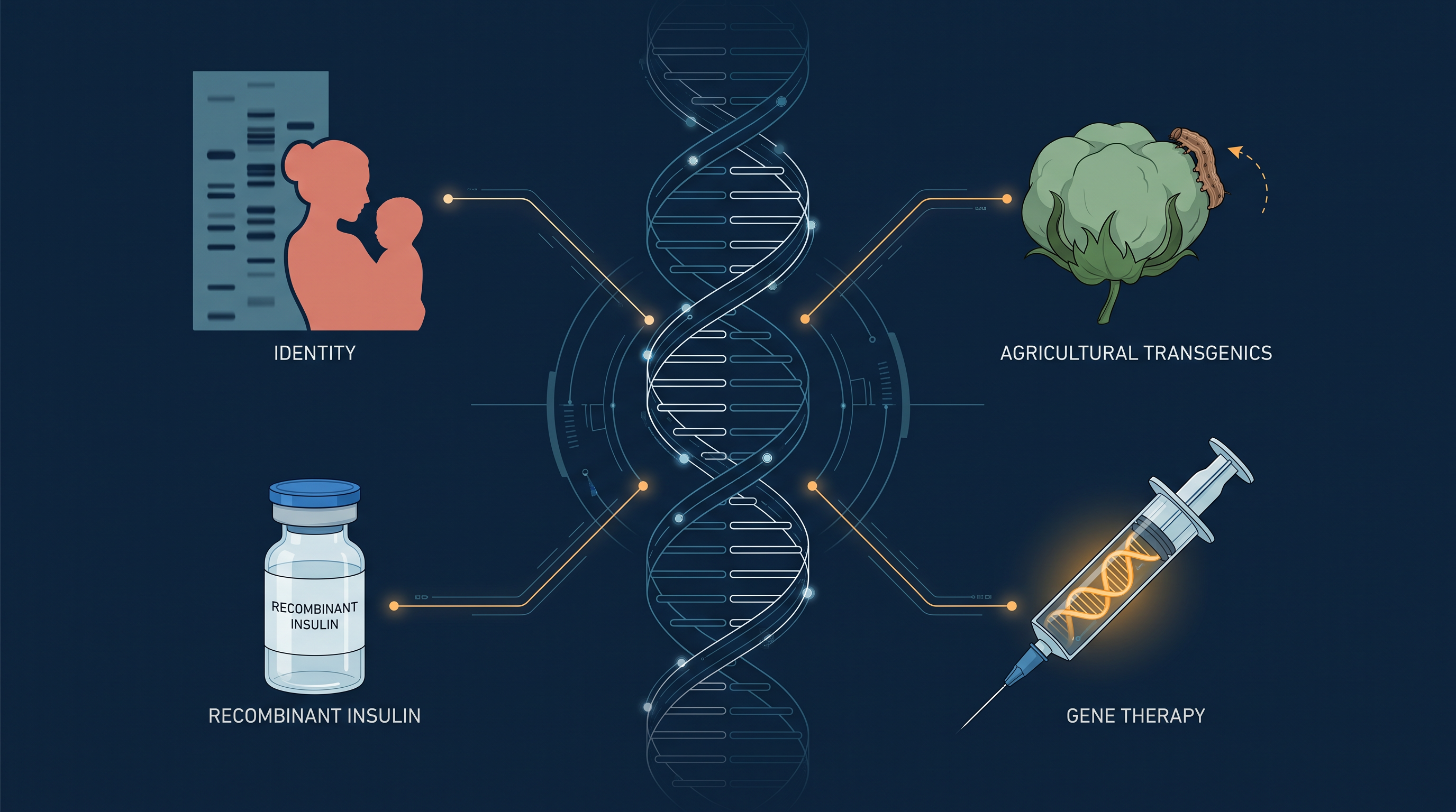
Fig. 4.0 – Applications of recombinant DNA technology.
Every one of these questions is answered somewhere in this chapter. Keep them in mind as you read — you will meet each answer in turn.
4.1 DNA Fingerprinting
Recombinant DNA (rDNA) technology has changed everyday life in ways that were unimaginable a few decades ago. It allows us to establish the identity of an individual, to introduce foreign genes into other organisms, to diagnose many diseases and treat them, and to manufacture therapeutic agents and molecules on a large scale.
This chapter takes up five of these applications one by one — DNA fingerprinting, the development of transgenic organisms, gene therapy, recombinant vaccines, and the production of therapeutic agents such as monoclonal antibodies, insulin and growth hormone. We begin with the application that turns DNA into a means of identification.
Why DNA Can Identify a Person
The chemical structure of DNA is the same in every human being. It is built from the same four nitrogenous bases — A (Adenine), G (Guanine), C (Cytosine) and T (Thymine) — joined to the same sugar-phosphate backbone. What differs from person to person is the arrangement of these bases along the DNA strands, and that arrangement is specific to an individual.
The human genome consists of \(3.2 \times 10^{9}\) bp, where “bp” stands for base pairs. That is a very large number, and yet there is very little uniqueness in the genetic makeup of humans. About 99.9% of the genome is the same among all humans. The remaining 0.1% of the genome consists of sites of inherited variations.
It is these variations in DNA that make each of us unique. If we can identify these differences, we can decide how closely two individuals are related to each other — whether they are parent and child, siblings, or complete strangers.
One obvious way of finding these differences is DNA sequencing — reading out the exact order of bases. But sequencing the DNA of every individual and comparing it, every single time an identification is needed, would not be feasible. It would be far too slow and far too expensive for routine casework.
Therefore, to study and compare the inherited variations in human DNA without sequencing, a new technique known as ‘DNA fingerprinting’ was developed by the British geneticist Sir Alec Jeffreys in 1984 at the University of Leicester.
Repeats That Vary: STRs and VNTRs
More than 90% of the human genome consists of DNA which does not code for protein. This non-coding DNA was once dismissed as useless, but it turned out to be exactly where the useful variation lies — precisely because it is not under pressure to stay constant. A change in a protein-coding gene may damage the protein; a change in the number of repeats in a non-coding stretch usually does nothing at all, so such changes accumulate freely.
Within the non-coding regions of an individual’s genome, there exist short sequences of nucleotides which are repeated a number of times one after the other. Repeats arranged end to end in this way are said to be in tandem, and such a repeated stretch is called an STR (short tandem repeat). The position on the chromosome at which such a repeat occurs is called a locus, and this locus is referred to as a VNTR (variable number tandem repeat).
VNTRs are commonly subdivided into two principal families: microsatellites, which are repeated sequences of 1 to 9 bp, and minisatellites, which are repeated sequences of 10 to 100 bp. The difference between them lies in the length of the repeating unit, not in how many times it repeats. Both types are shown schematically in Fig. 4.1.
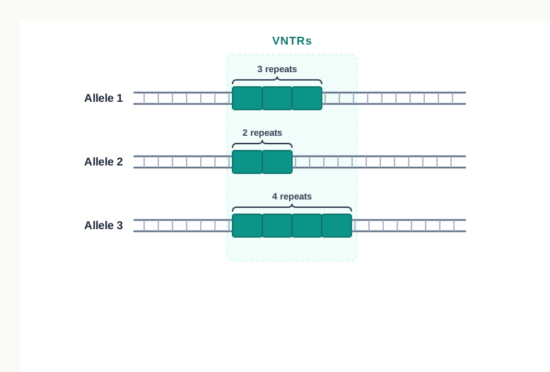
Fig. 4.1 – Schematic representation of VNTRs.
Notice in Fig. 4.1 that the repeat unit is the same at a given locus, but the number of copies is different on each of the three alleles shown. That is the whole point. These sequences show a high degree of polymorphism — that is, a high degree of variation between individuals — and this polymorphism forms the basis of DNA fingerprinting.
Furthermore, as the polymorphisms are inheritable from parent to offspring, DNA fingerprinting is the basis of paternity testing in case of disputes. A child does not invent new repeat numbers; the child receives them from the parents.
Two Copies at Every Locus
We carry two different copies of every VNTR locus, because we inherit one chromosome from the mother and one from the father. So at a single VNTR locus a person normally shows two bands — one maternal and one paternal — although the two will appear as a single band if both parents happened to pass on the same repeat number.
This is what makes paternity testing readable. Every band in a child’s profile must be traceable to one parent or the other. If a child shows a band that appears in neither claimed parent, that parent–child relationship is not supported. Fig. 4.2 shows this comparison being made for a couple and two children.
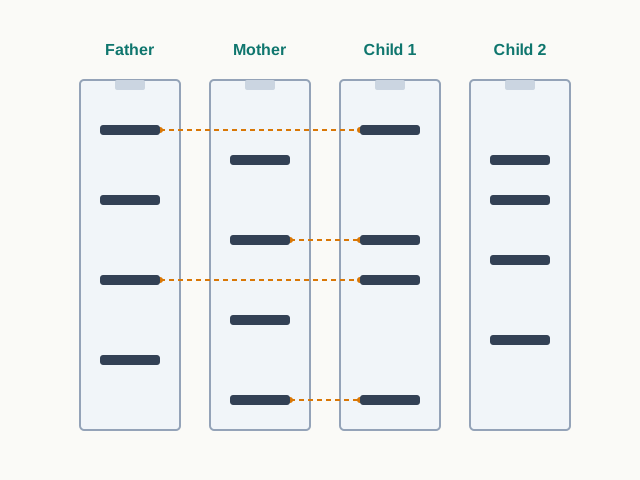
Fig. 4.2 – DNA profiling for parentage testing.
Read Fig. 4.2 the way an analyst would. Take one band in the child’s lane at a time and ask which parental lane it also appears in. For Child 1, each band is matched somewhere in the father’s lane or the mother’s lane. For Child 2, several bands have no source in either parent, and the couple can therefore be excluded as the parents of Child 2. This “band-by-band tracing” is a favourite of diagram-based board questions, so practise it on any profile you are given.
RFLP: Reading Variation Without Sequencing
In the simplest way, DNA fingerprinting can be performed using restriction digestion of DNA. Restriction enzymes are the molecular scissors introduced earlier — each one recognises a particular short sequence in DNA and cuts there, and nowhere else. If two people carry different numbers of repeats between two such cut sites, the piece of DNA released by the cutting will be of different length in the two people.
This technique is referred to as Restriction Fragment Length Polymorphism (RFLP). In RFLP, after restriction enzyme digestion of an individual’s DNA in specific regions, unique patterns are generated that are used for genetic analysis and identification (Fig. 4.2).
The logic is worth pausing on, because it explains why the whole method works without any sequencing at all. The enzyme cuts at fixed recognition sites. The distance between two such sites depends on how many repeats lie in between. More repeats means a longer fragment; fewer repeats means a shorter fragment. Fragment length therefore becomes a readable stand-in for repeat number.
From Gel to Membrane: The Actual Protocol
Conceptually, the DNA fingerprinting shown in Fig. 4.2 is correct, but in reality the identification of individual bands on a gel is difficult. A digest of human genomic DNA produces an enormous number of fragments of all sizes, which appear as a continuous smear rather than a set of clean bands. We need a way to make only the VNTR-containing fragments visible.
Therefore, hybridisation (Southern Hybridisation) using a VNTR probe is used. A probe is a labelled stretch of single-stranded DNA that finds and sticks to its complementary sequence, and nothing else. Hybridisation with VNTR probes produces a pattern of bands which are characteristic to every individual (Fig. 4.3). The steps involved in this technique are as follows.
- DNA is isolated from different samples like blood, hair, skin, semen, buccal swab, etc.
- The collected DNA sample is cut into several fragments of different sizes using one or more restriction enzymes.
- The DNA fragments are then separated by agarose gel electrophoresis. The different sized DNA pieces are separated based on size.
- The separated DNA on the gel is thus transferred to a nitrocellulose/nylon membrane. The nylon membrane is then exposed to UV radiation on a UV transilluminator for three minutes, or baked at 80°C for two hours, to permanently attach the DNA to the membrane.
- Now, Southern hybridisation is performed using VNTR Probes (the labelled stretches of single-stranded DNA used to detect the presence of complementary target sequences).
- Finally, the hybridised DNA fragments are detected.
- The patterns of DNA bands are highly specific for each individual and can be used in forensics and paternity disputes.
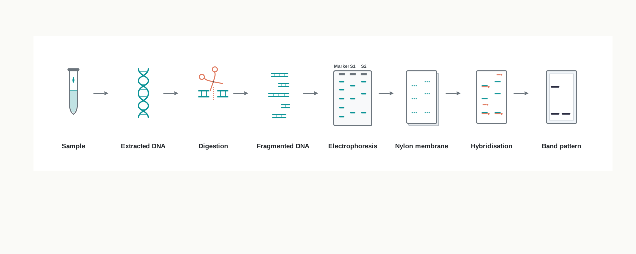
Fig. 4.3 – Laboratory steps in DNA fingerprinting.
The whole sequence is summarised in Fig. 4.4.
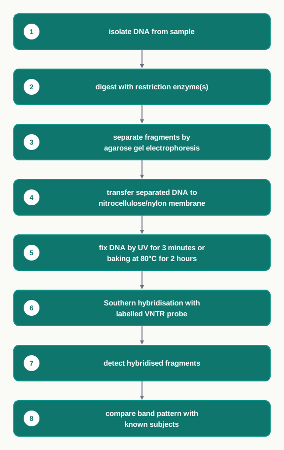
Fig. 4.4 – DNA fingerprinting workflow.
Each step exists for a reason, and it is worth knowing what would go wrong if a step were skipped or changed. If the digestion in step 2 were incomplete, fragments would not be cut to their true lengths and the bands would shift, giving a false pattern. In step 3, electrophoresis separates fragments because DNA carries a negative charge and moves towards the positive electrode through the agarose mesh; smaller fragments wriggle through faster and travel further, so distance along the gel reports size. In step 4, the fixing by UV or baking is not decoration — if the DNA were not permanently attached, it would simply wash off the membrane during the washing steps of hybridisation, and no bands would appear at all. In step 5, the probe supplies the specificity: without it we would still be staring at a smear.
Applications of DNA Fingerprinting
- VNTR patterns are used to ascertain the paternity and maternity of a person, given the fact that they are inherited from the parents. Since these patterns are very specific, even a parental VNTR pattern can be reconstructed from the known VNTR patterns of their offspring. Therefore, VNTR patterns of parent and child can be used to solve paternity and maternity cases.
- DNA isolated from tissues like blood, hair, skin, semen, etc., from the scene of a crime is used for VNTR pattern analysis as evidence. Such a pattern from the crime-scene isolate is compared with the VNTR patterns of a criminal or suspect, for establishing guilt or innocence. Hence, DNA fingerprinting helps in criminal identification and forensic studies.
- DNA fingerprinting is also used to compare DNA extracts from fossils with their modern-day counterparts, and therefore finds great significance in evolutionary biology studies.
- Preparing the DNA profile of people suffering from some particular disorder, or comparing it with a large number of people with and without the disorder, helps to identify the DNA patterns associated with that condition, and is therefore useful in studying inherited disorders.
- In addition to social security numbers, picture ID and other more routine methods, even the DNA profile (VNTR patterns) of an individual is being proposed for use as a sort of genetic barcode for personal identification.
In examinations, this section is most often tested through diagram-based questions on Fig. 4.2 (tracing a child’s bands to the parents), sequence-ordering questions on the seven steps of the protocol, and assertion–reason items that pair a statement about VNTRs with a reason about non-coding DNA. Learning the purpose of each step, rather than its position in a list, is what makes such questions straightforward.
4.2 Transgenic Organism
You must have heard about ‘Bt Cotton’ or ‘Rosie, the cow’, but have you ever wondered what these are? Are these naturally found in the environment? If not, then how have these been created? Or why have these been created at all?
Both the examples mentioned above are a transgenic plant and a transgenic animal, respectively. They have been produced by the introduction of new gene segments through the process of transgenesis, and they are not naturally found in the environment. They have been created for the benefit of human beings.
The process of insertion of a foreign gene (transgene) into the genome of an organism, and its transmission and expression in the organism’s progeny, is termed as transgenesis. Notice that two conditions are packed into that sentence. It is not enough for the foreign gene to enter the cell; it must also be passed on to the progeny and it must be expressed in them. A gene that enters a cell and is then lost, or one that sits silently in the genome without being switched on, has not produced a transgenic organism.
The organisms carrying the transgene are known as transgenic organisms or genetically modified organisms (GMOs).
4.2.1 Historical background
The first genetically modified organism was a bacterium made by Herbert Boyer and Stanley Cohen in 1973. Bacteria were the natural starting point — they grow fast, they carry plasmids that are easy to manipulate, and a single modified cell can be multiplied into billions within a day.
In the subsequent year, this was followed by the engineering of the first transgenic animal (transgenic mice) by Rudolf Jaenisch and Beatrice Mintz in 1974. Moving from a single-celled bacterium to a whole animal was a far larger step, because the transgene now had to reach every tissue of a multicellular body and be handed on to the next generation.
In 1994, the Flavr Savr tomato was released as the first genetically modified (GM) food crop approved by the US Food and Drug Administration (USFDA). With that approval, genetic modification moved out of the laboratory and onto the dinner plate, and the public debate about GM food began in earnest.
The first two entries of that timeline are worth a second look. Selecting wolves into dogs, or wild grasses into wheat, also changes the genetic makeup of a species — but slowly, indirectly, and only using variation that already existed within the species. Genetic engineering does the same thing quickly, precisely, and with genes drawn from any species at all. That difference in speed and reach, rather than the mere fact of genetic change, is what makes GMOs a separate category.
4.2.2 Production of transgenic organisms
Transgenic Plants
Transgenic or genetically modified plants are those plants whose genome is modified — by the introduction of one or more genes from another species — through genetic engineering.
The basic requirement for genetic transformation is the construction of a genetic vehicle, or vector, which carries the genes of interest flanked by the necessary regulating sequences, like a promoter or a terminator. The gene of interest by itself is only an instruction; the promoter is the switch that tells the plant’s machinery where to begin transcribing it, and the terminator tells it where to stop. Without these flanking sequences, the transferred gene would sit in the plant genome without ever being read.
The most commonly used techniques for gene transfer are of two types:
- Vector-mediated or indirect gene transfer
- Vector-less or direct gene transfer
In the first group, a living organism — a bacterium or a virus — carries the gene into the plant cell. In the second group, no biological carrier is used at all; the DNA is pushed in by physical or chemical means. The two groups have different strengths, and the choice between them depends largely on which plant is being transformed.
Bacteria-mediated Transfer
Agrobacterium tumefaciens is a rod-shaped, Gram-negative soil bacterium that is capable of naturally transferring DNA into the plant genome, and causes crown gall disease — a type of plant tumour.
Agrobacterium has the natural ability to transfer its own DNA to the plant genome randomly, where it can produce copies of itself, and therefore it is also known as the ‘Natural Genetic Engineer’. It contains a large-sized tumour inducing (Ti) plasmid. A plasmid, as you know, is a small circular DNA molecule that exists in a bacterial cell separately from the main chromosome.
A portion of the plasmid carrying the tumour inducing genes is called T-DNA, which is transferred to the plant genome upon infection. These genes encode phytohormones like auxin and cytokinins, which induce rapid cell division in the host plant cell and cause the formation of the tumour called crown gall. In other words, the bacterium forces the plant to build it a home and feed it.
This natural ability of A. tumefaciens to transfer T-DNA has been exploited to transfer genes of interest to plants and produce transgenic plants. The tumour inducing genes of the T-DNA are replaced with genes of interest, like the Bt gene for insect resistance. A Ti plasmid without tumour inducing genes is a disarmed plasmid, and this process is called disarming.
Antibiotic marker genes can be introduced along with the genes of interest, in order to differentiate between the transformed and non-transformed cells. This is necessary because Agrobacterium transfers its T-DNA to only a small fraction of the plant cells it infects, and a transformed cell looks exactly like an untransformed one under a microscope. The marker gene turns an invisible difference into a visible one.
Vectors containing modified T-DNA (recombinant disarmed T-DNA) are transferred to Agrobacterium cells, which are further used for plant transformation. Agrobacterium infection causes the T-DNA to get transferred into plant cells, and the subsequent integration of T-DNA carrying the target gene into the plant nuclear DNA.
These are further cultured on regeneration media supplemented with two things: a bacteriostatic agent, to stop further growth of Agrobacterium, and lethal concentrations of the marker antibiotic, which selectively eliminates the non-transformed plant cells. The two additions do two quite different jobs — one clears away the bacterium that has finished its task, the other clears away every plant cell that failed to take up the gene.
In a few weeks, the transformed tissue forms shoots, which are transferred onto the rooting media, where the transformed shoots make roots within two to three weeks. Plantlets are then transferred to soil. A schematic representation of Agrobacterium mediated plant transformation is given in Fig. 4.5.
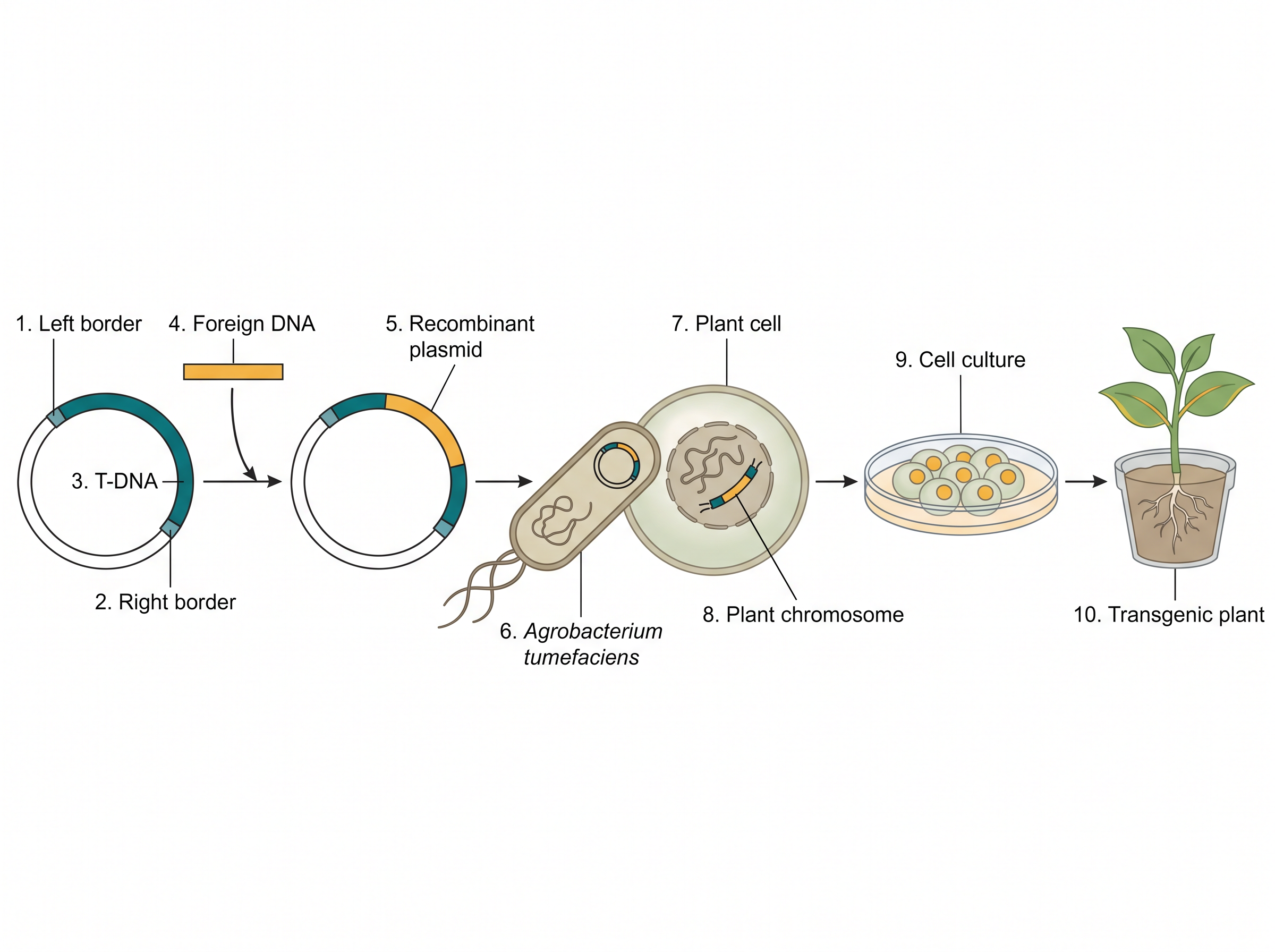
Fig. 4.5 – Agrobacterium-mediated plant transformation.
The complete sequence is set out in Fig. 4.6.
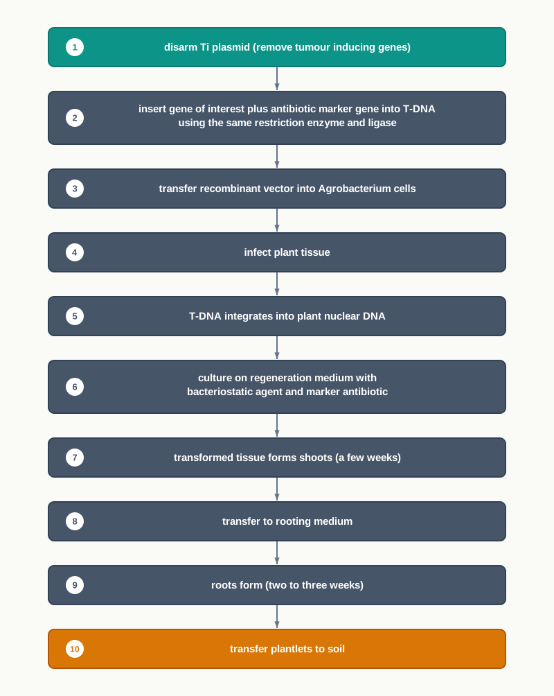
Fig. 4.6 – Steps in Agrobacterium-mediated transformation.
Plant Virus-mediated Transfer
Plant viruses, such as caulimoviruses, have the ability to enter intact plant cells and introduce their own DNA into the plant’s DNA. The use of plant viruses constitutes another method to transfer a gene of interest into plant cells. However, this is not a common method of plant transformation as compared to Agrobacterium.
The reason is practical. Viruses carry only small genomes, so the amount of foreign DNA they can accommodate is limited, and many plant viruses do not integrate their DNA stably into the host chromosome, which means the introduced gene may not be inherited by the progeny at all.
Particle Bombardment Gene Transfer
We now turn to the second group of methods. Many physical or mechanical methods have been developed to help the direct entry of DNA into plant cells, without using any biological vector.
Particle bombardment is a popular direct gene transfer method and is capable of delivering foreign DNA in both monocot and dicot plants. It is also called biolistics, a name formed from “biological ballistics”.
The DNA having the genes of interest and an antibiotic resistance marker gene is coated onto the surface of gold or tungsten particles (1 µm – 3 µm in size). These particles are accelerated to high velocity and bombarded onto the target tissue or cells using a powerful gun, which is called a particle gun or gene gun. Gold and tungsten are chosen because they are dense enough to carry momentum through the cell wall, and chemically unreactive enough not to damage the DNA riding on them.
Tissues that have been bombarded are then cultured on nutrient media containing an appropriate antibiotic. Transformed cells grow to make callus — an unorganised mass of dividing cells — and transgenic plants are then regenerated from that callus.
This method is very useful for those plant species where Agrobacterium-mediated transformation may not be very successful, for example, cereals. Since rice, wheat and maize feed most of the world, a method that works in cereals is of enormous practical importance.
The particle bombardment technique is also utilised for the transformation of the chloroplast. As a plant cell contains many chloroplasts, this method allows much higher transgene expression. Each chloroplast carries several copies of its own genome, so putting the gene there multiplies the number of templates available for transcription in a single cell.
Protoplast Transformation and Electroporation
Protoplasts are spherical naked plant cells, which are produced by the removal of cell walls. The wall is usually digested away with enzymes such as cellulase and pectinase, leaving the cell bounded only by its plasma membrane.
These naked cells are capable of taking up DNA from their surroundings, which then gets integrated into the genome of the transfected cells. Removing the wall matters because the rigid cellulose wall is the main barrier to DNA entry; once it is gone, the membrane alone can be persuaded to let DNA through.
The gene transfer process can be accelerated with the application of polyethylene glycol (PEG) and dextran sulphate. Alternatively, an electric current can also be used for gene transfer in protoplasts, and this method is known as electroporation. A brief electrical pulse opens transient pores in the plasma membrane, through which the surrounding DNA slips into the cell before the pores reseal.
Transfected protoplasts are allowed to regenerate cell walls, and then cell division begins. Transformed cells are selected on the selective nutrient medium and transgenic plants are regenerated.
Transgenic Animals
In transgenic animals, the genetic makeup has been altered by the use of various genetic engineering techniques. The most commonly used techniques for developing transgenic animals are:
- DNA pronuclear microinjection
- Embryonic stem cell-mediated gene transfer
- Retrovirus-mediated gene transfer
Notice the common thread. In every one of these methods the transgene has to be delivered very early in development — into the fertilised egg, or into cells of the early embryo. This is because the transgene must end up in every cell of the animal, including its germ cells, and the only way to achieve that is to place it in the cell (or in cells) from which the whole animal will be built.
DNA Pronuclear Microinjection
This is one of the earliest strategies used for creating transgenic mice. Just after fertilisation, the egg contains a small female pronucleus and a large male pronucleus — the two parental nuclei have arrived but have not yet fused.
In the pronuclear microinjection technique, the transgene is directly injected into the larger male pronucleus, using an extremely fine glass needle under a microscope. The larger of the two is chosen simply because it is an easier target to hit without destroying the cell.
The embryos with the injected transgene are cultured in vitro and then implanted into the uterus of foster mothers. The transgene may get integrated into the DNA of the zygote, resulting in animals that are transgenic.
The technique is simple and reliable, but the rate of success is low, as the transgene may or may not get integrated into the host DNA. Also, the site of integration of the transgene is random, which may lead to unpredictable effects. If the transgene happens to land inside an essential gene, it can disrupt that gene; if it lands in a region of tightly packed, inactive chromatin, it may never be expressed at all.
Embryonic Stem Cell-mediated Gene Transfer
Embryonic stem (ES) cells have the potential to differentiate into any other cell type of the body, including germ cells. That last point is the crucial one: if the transgene reaches the germ cells, the modification can be passed on to the next generation.
ES cells are collected from the inner cell mass region of the blastocyst stage. The r-DNA is transferred into these ES cells by electroporation — the same principle you met with plant protoplasts, applied here to animal cells.
Few ES cells take up the transgene and get transformed, so the transformed ones must be selected out. These transformed ES cells are then transferred back to the inner cell mass of a blastocyst through micro-injection. The blastocyst is implanted into the uterus of a foster mother. The offspring are then monitored for expression of the transgene.
Retrovirus-mediated Gene Transfer
The genetic material of retroviruses is RNA. Retroviral vectors are used to stably introduce a transgene into early embryos or ES cells.
The advantage of this method is that a single copy of the retrovirus carrying the transgene integrates at a particular location in the host genome. Compare this with pronuclear microinjection, where several copies may insert at an unpredictable site: a single, defined insertion makes the result far more predictable and easier to interpret. An example of such retroviruses is the lentivirus.
Fig. 4.7a shows the whole industrial version of this process applied to a goat engineered to secrete a human enzyme in its milk.
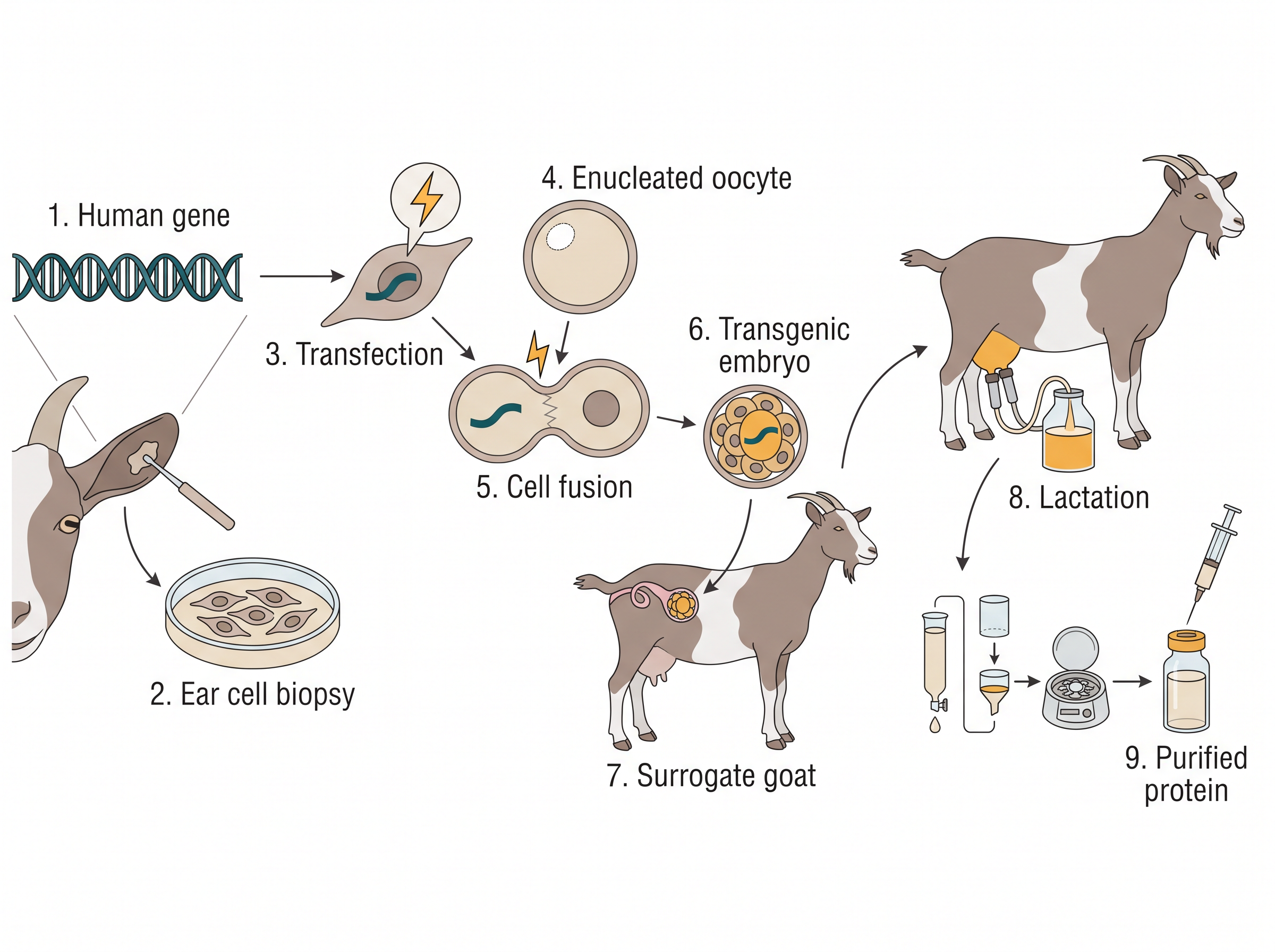
Fig. 4.7a – Production of a transgenic goat.
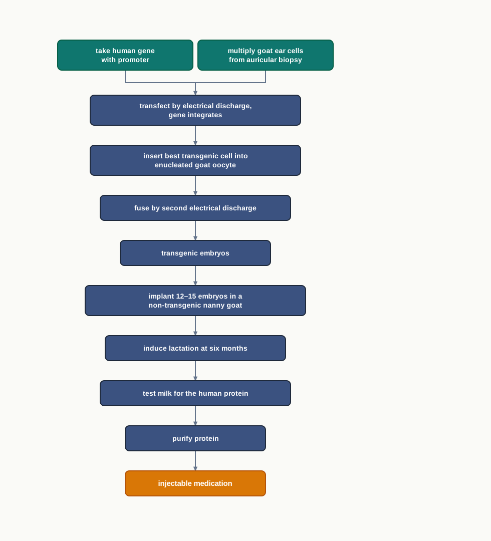
Fig. 4.8 – Production of a transgenic goat.
4.2.3 Applications of Transgenic organisms
Applications of Transgenic Plants in Agriculture
One major goal of plant transgenics has been to increase the yield of crops, and also to improve the nutritional quality of crop products, to cater to the needs of an ever increasing human population.
Crops lose yield to two broad kinds of stress. Biotic stress is due to pathogens, like bacteria, fungi, viruses or insects. Abiotic stress factors include drought, salinity and extreme temperatures. Both cause huge losses to crop productivity in terms of yield.
Biotechnological strategies have been used effectively to generate newer and more effective cultivars with increased resistance to pathogens, or which perform better in harsh environmental conditions.
The abiotic stress response reaction involves the production of stress-related osmolytes, like sugars (for example, trehalose and fructans) and amino acids (for example, betaine and proline). Certain proteins have been produced which over-express one of the above-mentioned compounds, and plants making them show better tolerance to environmental stresses. Osmolytes work by holding water inside the cell and stabilising proteins and membranes when the surrounding soil is dry or salty.
A common example of a plant offering resistance to insects is Bt cotton. This insect-resistant transgenic crop has been developed by transferring and expressing the Bt gene — also called the cry gene — from the bacterium Bacillus thuringiensis into a cotton plant.
The Bt gene encodes a toxin (Bt toxin), which is naturally produced by B. thuringiensis as an insecticidal protein to kill insects. There are several classes of cry genes, and the products of each class are harmful only to a specific group of insects and do not affect other useful insects, like butterflies and silkworms.
For example, cryIAc and cryII Ab code for a toxin that controls cotton bollworms, whereas cryIAb encodes a toxin that controls corn borers. Choosing the right cry gene for the right pest is therefore part of the design of the crop, not an afterthought.
Some of the other crops for which transgenic plants have been developed by introducing Bt genes are brinjal, maize, rice and tomato. All these transgenic crops have been proven to be effective in controlling insect pests. Many of them are currently grown commercially and have resulted in a significant increase in total yields. The most notable example in India is Bt cotton — offering resistance to the insect pest pink bollworm — which has been grown commercially since 2002.
Similarly, transgenic crops have been generated for several other traits, which result in significant enhancement of crop productivity. These traits include tolerance to herbicides; resistance to bacteria, pests, viruses or nematodes; tolerance to environmental stresses; nutrient quality; and delayed fruit ripening.
Strategies for increasing crop yield also include modifying the photosynthetic machinery of the plant, manipulating enzyme activity, increasing sugar-to-starch conversion, and improving nutritional quality — for example, increasing the expression of vitamins, such as vitamin A in plants, as in Golden Rice. Many traits have already been commercialised, and several others are being tested for their performance under field conditions.
| Crop | Source of Inserted Trait | Trait |
|---|---|---|
| Corn | Bacteria, and other species of corn | Resistance to insects; tolerance to herbicides; male corn sterility; alpha-amylase expression; increased lysine level for use in animal feed; reduction of yield-loss under water-limited conditions |
| Cotton | Bacteria | Tolerance to herbicides; resistance to insects |
| Soybean | Bacteria, corn, oats, and other species of soybean | Tolerance to herbicides; high oleic acid soybean oil; resistance to insects |
| Soybean | Mustard greens | Resistance to insects |
| Canola | Bacteria | Tolerance to herbicides; fertility restoration; male canola sterility |
| Canola | Fungus | Degradation of phytate in animal food |
| Potato | Bacteria | Resistance to insects |
| Potato | Potato virus | Resistance to potato viruses |
| Potato | Other species of potato | Lower level of reducing sugars; lower level of free asparagine; reduced black spot bruising |
| Tomato | Bacteria, potato | Delayed softening |
| Tomato | Bacteria | Resistance to insects |
| Radicchio | Bacteria | Tolerance to herbicides; male radicchio sterility |
| Alfalfa | Bacteria | Tolerance to herbicides |
| Sugar beet | Bacteria | Tolerance to herbicides |
| Rice | Bacteria | Tolerance to herbicides |
| Apple | Other species of apple | Reduced browning and bruising |
| Cantaloupe | Bacteria | Delayed ripening |
| Squash | Viruses | Resistance to viruses |
| Papaya | Viruses | Resistance to viruses |
| Flax | Mustard green | Tolerance to herbicides |
| Plum | Virus | Resistance to viruses |
| Creeping Bentgrass | Bacteria | Tolerance to herbicides |
Antisense Technology
So far, every application we have discussed adds a new ability to a plant. But sometimes the useful change is to switch something off — to stop a ripening enzyme, or to block a gene the pest needs. That is what antisense technology does.
Antisense technology is used to inhibit or silence gene expression. In this technique, the inhibition of expression of a specific gene is achieved by preventing the translation of its mRNA. Note carefully where the block acts: the gene is still transcribed, but its message is never turned into protein. There are two approaches.
The first one is known as the antisense oligonucleotide (ASO) technique. In this technique, a synthetic oligonucleotide — a short RNA or DNA molecule — which is complementary to the mRNA of the target gene, is introduced into a cell. The ASO can mediate its effect in two ways: (i) by binding of the ASO to the target mRNA, followed by RNase H mediated degradation of the mRNA; and (ii) by blocking its translation, by hindering the binding of the mRNA to ribosomes. Both routes depend on the same basic property of nucleic acids — a single strand will pair only with a strand complementary to it, which is what makes the silencing gene-specific.
The second approach is known as the RNA interference (RNAi) technique. In this technique, the expression of the target gene is inhibited by inactivating its mRNA through the RNA induced silencing complex (RISC).
A double-stranded RNA of exogenous origin (viral origin) or endogenous origin (cellular origin) is cleaved in the cytoplasm into shorter fragments by the endoribonuclease named DICER. The shorter double-stranded fragments are separated by the multiprotein complex RISC into a guide strand and a passenger strand. The passenger strand is degraded, while the guide strand, attached to RISC, binds to the target mRNA and cleaves it — and thereby the target gene is silenced (Fig. 4.9b).
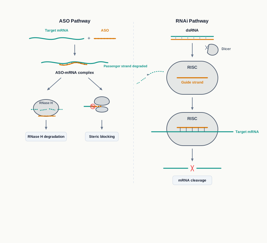
Fig. 4.9b – Mechanisms of ASO and RNA interference.
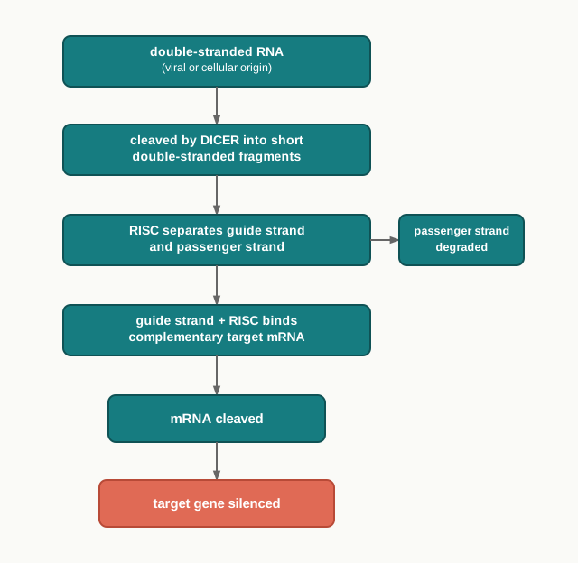
Fig. 4.10 – The RNA interference pathway.
Using a combination of Agrobacterium-mediated transformation and antisense technology, pest-resistant plants have been created. An example is provided by the tobacco plant, which is infected by the nematode Meloidogyne incognita.
Nematode-specific genes were introduced into the plant using Agrobacterium as a vector. These genes, when transcribed, produced antisense RNA, which binds to the sense RNA of the nematode — the two being complementary to each other — and thus prevents its translation into proteins that are necessary for nematode survival. This makes the transgenic plants resistant to the parasitic attack of the nematodes.
Read that example carefully, because it is unusual and it is a favourite of application-based questions: the plant is engineered to silence a gene belonging not to itself, but to the parasite that attacks it.
Molecular Farming for Industrial Production
Plants are the backbone of all life forms on earth, as they convert the energy of the sun into the chemical energy that feeds the world. Today thousands of plants are being used for food, feed and medicines, to cure acute and chronic health problems.
Advances in our understanding of plant genetics and in biotechnological tools have led to the development of a relatively new bioscience, ‘Molecular Farming’. It involves the genetic modification of a host plant through the purposeful addition of a gene or group of genes, leading to the production of new desired biomolecules.
Foreign genes can be inserted either into the nuclear genome or into the chloroplast genome. As the chloroplast has several copies of the genome, the insertion of the gene of interest into the chloroplast results in higher accumulation of the target biomolecules — the same reasoning you met while studying particle bombardment.
In this way, a large number of products have been produced in plants, which include vaccine antigens, therapeutic proteins, diagnostic reagents, nutritional products, bioplastics and industrial enzymes.
In 2012, the U.S. FDA approved the first plant-made human biologic, ‘Elelyso’. It is an enzyme that is used to treat Gaucher’s disease, and it has been produced in carrot cell cultures. Table 4.1 lists a few therapeutic molecules that have been produced in plants through genetic engineering.
Table 4.1: List of industrial or therapeutic products produced in plants through genetic engineering
| Transgenic Plant | Industrial/ Therapeutic Product(s) | Disease/ Other Usage |
|---|---|---|
| Lettuce | Hepatitis B antigen | Hepatitis B |
| Tobacco | Cancer vaccine | Non-Hodgkin’s Lymphoma |
| Carrot | Human glucocerebrosidase | Gaucher’s Disease |
| Safflower | Insulin | Diabetes |
| Rice | Human lysozyme | Anti-infection, anti-inflammatory |
Environmental Benefits
Biodegradable plastics. Plastics are non-biodegradable and are harmful to the environment. Transgenic plants can be exploited to produce biopolymers that can replace plastics.
Polyhydroxyalkanoates (PHAs) are an attractive source of non-polluting and biodegradable plastic. Production of PHAs using transgenic plants provides an economically viable alternative, because a crop grows on sunlight, soil and water rather than on petroleum feedstock. An example is the production of the bacterial polyester polyhydroxybutyrate (PHB) in transgenic sugarcane.
Phytoremediation. Phytoremediation refers to the use of plants for the removal of pollutants from the environment, especially soil. Metal-detoxifying genes from bacteria and other organisms are introduced into plants, and the transgenic plants then take up and convert the toxic elements into less toxic forms.
Mercury is a hazardous heavy metal, largely found in aquatic ecosystems. Transgenic plants have been designed to incorporate the mercuric reductase gene, which helps in the detoxification of harmful mercury into less toxic forms. Such transgenic plants act as ‘Mercury-breathing plants’, and can be used for environmental remediation.
Applications of Transgenic Animals
For industrial production. Transgenic animals are used as bioreactors for the production of normal and recombinant protein products in large quantities. This is known as Molecular Pharming. The word is deliberately spelt with “ph”, to distinguish the use of animals as living pharmaceutical factories from Molecular Farming in plants.
An early example of success in the 1990s was the transgenic ewe, Tracy, which produced high levels of the human protein α1-antitrypsin in her milk. Deficiency of α1-antitrypsin in humans causes lung diseases. The protein produced in the milk of Tracy was thought to cure the symptoms of this deficiency, but clinical trials for this recombinant protein revealed side effects in patients.
That outcome is worth remembering, because it shows something important about biotechnology: producing a human protein successfully in an animal is not the same thing as producing a safe medicine, and clinical trials exist precisely to catch the difference.
In 1997, the first transgenic cow, Rosie, was created, which could produce human α-lactalbumin-enriched milk. Human α-lactalbumin-enriched milk was found to be a more balanced product for human infants compared to natural cow milk. Table 4.2 lists a few human recombinant proteins that have been produced in transgenic animals through genetic engineering.
Table 4.2: List of human recombinant proteins produced in the milk of transgenic animals through genetic engineering
| Name | Transgenic Animal |
|---|---|
| Albumin | Cow |
| α-fetoprotein | Goat |
| Growth hormone | Goat |
| Tissue plasminogen activator | Goat |
| Coagulation factor IX | Mouse |
| Coagulation factor VIII | Rabbit |
Transgenic Animals in Research
Transgenic animals are also used as model organisms by scientists for studying normal human physiology, development and diseases.
The transgenic animals are genetically manipulated to develop symptoms of diseases, such as cancer and Alzheimer’s. This also allows researchers to understand the function of the genes involved in various diseases. An animal that develops a human-like disease on a predictable schedule is enormously useful, because the earliest stages of a disease can be studied — stages that in human patients have usually passed unnoticed long before diagnosis.
Transgenic animals are also used for toxicity testing of vaccines, drugs and chemicals before they can be used on humans.
4.2.4 Concern over GMOs
The major concern over GMOs is the potential risk to human health and to the environment.
Transgenic crops may produce proteins that may cause allergic reactions in humans. Any newly introduced protein is a protein the human diet has not encountered before, and a small number of proteins provoke allergy.
Concerns also arise over the use of viral vectors and promoters in GMOs, as these can lead to viral infections in humans.
The improved characteristics of GMOs may make them invasive for the native or wild-type species. A crop engineered to survive drought, salinity or herbicides has, by design, an advantage over its wild relatives, and such an advantage can allow it to spread where it is not wanted.
Finally, variable insertion of the transgene can lead to unpredictable or non-target effects — the same problem of random integration that limits pronuclear microinjection, appearing here as a safety question rather than a technical one.
For ethical issues related to the use of transgenic animals, India has established biosafety regulations for the production and use of GMOs. The Ministry of Environment, Forest and Climate Change has established the Genetic Engineering Appraisal Committee (GEAC), which regulates the manufacture, use, import, export and storage of genetically modified organisms and cells or hazardous microorganisms, and decides upon the validity and safety of introducing GMOs.
In examinations, this part of the chapter is commonly tested through compare-and-contrast questions (direct versus indirect gene transfer; the three methods of making transgenic animals), through questions asking you to justify a single step in a protocol (why the plasmid is disarmed, why an antibiotic marker is added, why a bacteriostatic agent is included in the regeneration medium), and through case-based questions on the Bt cotton and GMO-safety debates.
4.3 Gene Therapy
Many diseases in humans are known to be caused either due to the absence of a normal gene, or due to the presence of a defective, disease-causing gene. Can such disorders be corrected?
Until recently the honest answer was no. A patient with an inherited disorder could be given the missing protein, or treated for the symptoms, but the fault itself — a wrong instruction sitting in every cell — remained untouched. The answer to the question above lies in the approach known as ‘gene therapy’, which attempts to treat the instruction rather than only its consequences.
Gene therapy is a technique designed to repair faulty genes in humans by introducing correct genetic material inside the cells.
The obvious candidate diseases for gene therapy include the immune disorder called Severe Combined Immunodeficiency (SCID), and inherited genetic disorders such as cystic fibrosis, haemophilia and muscular dystrophy. What these conditions share is that a single known gene is at fault, and that supplying a working copy of that one gene would, in principle, be enough.
The First Attempt on a Patient
The first gene therapy was performed in 1990, in a four-year-old girl suffering from SCID caused due to a defect in the adenosine deaminase (ADA) coding gene.
Deficiency of ADA led to the inability to produce sufficient immune cells, which subsequently made the girl suffer from frequent life-threatening infections. Without functional lymphocytes, the body has almost no defence against organisms that a healthy child would clear without noticing.
A functional copy of the ADA gene was inserted into a viral vector, which was then introduced into lymphocytes collected from the patient, and these lymphocytes were then reintroduced into the patient’s bloodstream (Fig. 4.11). The immune system functioning of the patient improved, but the cure was not permanent.
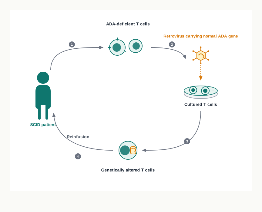
Fig. 4.11 – Gene therapy for ADA-SCID using a retroviral vector.
The reason the cure was not permanent is worth understanding, because it explains most of the limitations discussed later in this section. The gene was placed in mature lymphocytes, and mature lymphocytes have limited lifespan. As the treated cells died and were replaced by new cells from the patient’s own bone marrow — which still carried the defective gene — the benefit gradually faded, and the treatment had to be repeated.
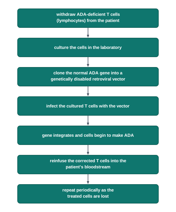
Fig. 4.12 – Steps in gene therapy for ADA-SCID.
4.3.1 Approaches for Gene Therapy
There are three main approaches for gene therapy — (i) gene replacement or gene addition, (ii) gene inhibition, and (iii) gene repair or gene editing therapies. They correspond to three different kinds of genetic fault: something missing, something harmful being made, and something written incorrectly.
Gene Replacement or Gene Addition
In gene replacement or gene addition therapy, a functional copy of the gene is delivered into the genome to replace the non-functional defective gene. The new gene carries the instructions to synthesise the protein that was not available in the cell due to the defective gene.
For example, cystic fibrosis is an inheritable disease caused due to a defective membrane protein known as the cystic fibrosis transmembrane conductance regulator (CFTR) protein. The defective protein is produced due to a mutation in the CFTR gene.
Due to the defective protein, the movement of salt and water is hampered in the body cell, causing damage to the respiratory and digestive tract (Fig. 4.13). Normally the CFTR protein moves chloride ions across the membrane, and water follows the salt. When this movement fails, the mucus lining the airways loses water and becomes thick and sticky instead of thin and mobile.
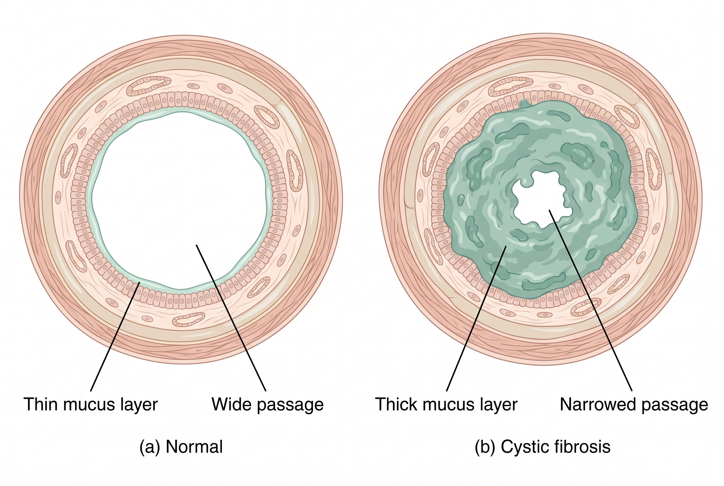
Fig. 4.13 – Air passage cross section: (a) normal and (b) in cystic fibrosis.
Compare the two panels of Fig. 4.13 carefully. In the normal passage the mucus is a thin film, and the lumen through which air travels is wide. In cystic fibrosis the mucus layer is thick and the air passage is narrowed. That single structural difference explains the breathing difficulty and the repeated chest infections that characterise the disease, because thick mucus cannot be cleared and traps bacteria.
If the mutated CFTR gene is replaced by a normal CFTR gene, the disease can be cured.
Another example is the p53 gene, which normally prevents tumour growth. Several types of cancers have been linked to defective forms of p53. Replacement of the defective form of p53 in cancer cells may cause inhibition or death of the cancer cells, thereby curing cancer.
Gene Inhibition
In the case of gene inhibition, the function of a gene which causes a disease in the cell is inactivated. Here the problem is not a missing product but an unwanted one, so adding another copy of the gene would only make matters worse.
The gene inhibition approach has been considered for disorders such as Tay-Sachs disease, phenylketonuria and colour-blindness — all of which are inherited conditions rather than acquired ones.
In gene inhibition, a gene is introduced into the cell whose product either blocks the expression of the faulty gene, or disarms the products of the faulty gene. You have already met the molecular basis of this idea in the previous section, in antisense technology and RNA interference; gene inhibition therapy is that same silencing logic applied to a human patient.
Gene Repair or Gene Editing
Finally, in the case of gene repair or gene editing, the DNA is inserted, deleted, modified or replaced in the defective gene in living cells. This is the most direct approach of the three, because it corrects the original text rather than adding a correction alongside it or silencing it.
This is achieved by a recent technology known as the CRISPR/Cas9 system — Clustered Regularly Interspaced Short Palindromic Repeats / CRISPR associated protein 9.
The CRISPR/Cas9 system was adopted from the natural defence system of bacteria or archaea against viruses that attack them. With the CRISPR system, bacteria recognise the invading virus and, with the help of the Cas9 enzyme, they cut the viral DNA into small pieces.
The technique offers an easy way to snip out mutated DNA and replace it with the correct sequence. This technique shows the possibility of treating several genetic disorders in future. For details, refer to section 5.4.2, Chapter 5.
4.3.2 Types of Gene Therapies
Gene therapies can be sorted in three different ways — by where the gene is delivered, by which kind of cell receives it, and by what strategy is used. The three classifications are summarised in Fig. 4.14.
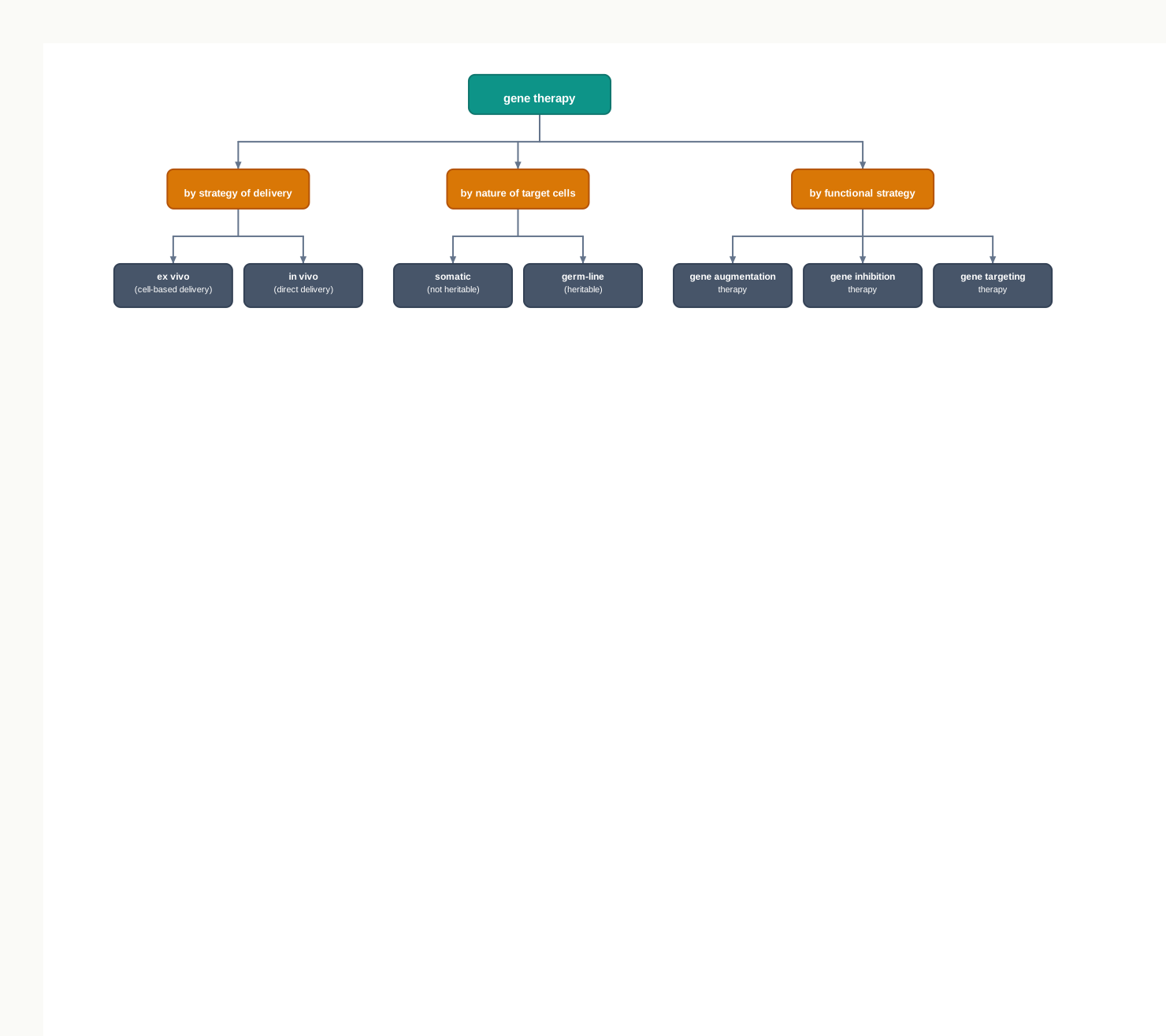
Fig. 4.14 – Classification of gene therapy.
Classification by Strategy of Gene Delivery
On the basis of the strategy used for gene delivery, gene therapy can be classified into ex vivo and in vivo (Fig. 4.15).
In ex vivo gene therapy — ex means ‘out of’ and vivo means ‘something alive’ — cells are taken out from the diseased person, grown in culture, normal genes are then introduced into these cells, and finally these transformed cells are reintroduced into the patient. This is also known as cell-based delivery.
The ex vivo approach is much simpler as compared to the in vivo approach, because it becomes easier to manipulate the cells externally (Fig. 4.15). In a culture dish the gene can be delivered under controlled conditions, and the cells that took it up can be selected and checked before being returned to the patient. The gene therapy for SCID described earlier is an ex vivo procedure.
In the case of in vivo gene therapy, normal functional genes are directly introduced into the target cells and tissues of the person with the diseased condition. This is also known as the direct delivery method.
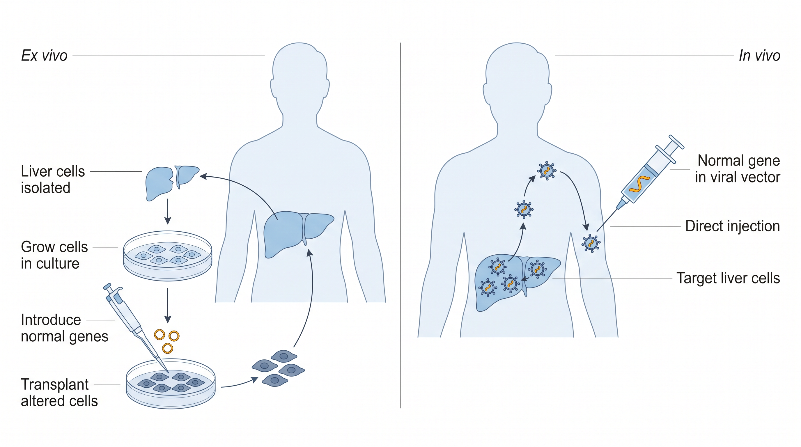
Fig. 4.15 – Ex vivo and in vivo gene therapies.
Classification by Nature of Target Cells
On the basis of the nature of the target cells or tissues, gene therapy can be classified into somatic and germ-line gene therapy.
In somatic gene therapy, functional copies of the target gene are introduced into the somatic cells of the patient. Somatic cells are all the body cells other than the germ cells, so a change made in them affects the patient alone and stops with that patient.
In germ-line gene therapy, functional genes are introduced into the germ cells (egg or sperm). The functional gene then gets integrated into the genome of the germ cells. Changes made to the germ-line are heritable.
That single word — heritable — is the reason germ-line therapy is treated so differently from somatic therapy, both scientifically and ethically. A somatic correction is a treatment given to one person. A germ-line correction is a change made to every descendant of that person, none of whom can be asked for consent.
Classification by Functional Strategy
Thus, based on the functional strategy used, gene therapy can be classified into:
- Gene augmentation therapy: Addition of a functional gene to the genome, with the aim of replacing the missing gene product.
- Gene inhibition therapy: Using antisense RNA or other inhibition techniques to block the expression of dominant-acting mutated genes.
- Gene targeting therapy: Replacing the non-functional gene with a normal gene using homologous recombination.
Notice the precision of the third one. Homologous recombination means the incoming normal gene pairs with the matching sequence already present in the chromosome and swaps itself in at exactly that place — so the correction lands where it belongs, instead of integrating at random.
Attempts to correct the function of a defective gene, as described above, could become possible only after advancements in rDNA technology, gene transfer and gene editing mechanisms. You have already studied the mechanism of the delivery of a target gene — the desired DNA sequence — either through viral (direct gene transfer) or non-viral (indirect gene transfer) methods, earlier in this chapter and also in Chapter 3.
4.3.3 Merits and Demerits of Gene Therapy
Gene therapy provides a potential cure for the treatment of devastating inherited diseases, for which conventional treatment strategies provide little hope. For a family living with such a disorder, this is the difference between managing symptoms indefinitely and correcting the cause.
Advancements in human genomics have shown that cancer is caused due to somatic aberrations in the human genome. This has led to enthusiasm among cancer researchers to use the gene therapy approach for the genetic manipulation of cancer cells, and to find a possible cure to the disease.
However, uncontrolled expression of the therapeutic gene needs to be resolved for an efficient gene therapy mechanism. A gene that is expressed too strongly, or at the wrong time, can be as damaging as one that is not expressed at all.
The introduction of the therapeutic gene into non-target cells, or its random integration at a wrong place in the host genome, is another concern. For example, if the DNA gets integrated into a tumour-suppressor gene, this can result in a tumour — the therapy would then have created exactly the kind of fault it was designed to correct.
There may be a requirement for frequent administration of the therapeutic gene, as gene therapy is short-lived in nature. This is the limitation you already saw in the 1990 SCID case.
Host rejection of the therapeutic gene, or of the viral vector employed in the process, can stimulate the host’s immune system. The immune system cannot distinguish a helpful virus from a harmful one; it recognises a foreign particle and responds accordingly.
Besides being a costly process, gene therapy is effective for single-gene defects but not for multigene disorders. Where dozens of genes each contribute a small amount to a condition, there is no single gene to correct.
4.3.4 Ethical Issues
Bioethics refers to the assessment of the risks associated with procedures, and of the moral implications of new techniques.
While genetic modification of somatic cells is approved by a large part of the scientific community, genetic therapy in germ-line cells often forms a subject for heated discussion in the field of science. Since gene therapy includes making changes to the body’s basic set of genes, it raises unique ethical considerations. Some of the ethical concerns associated with gene therapy include the following.
- The use of gene therapy for gene enhancement is a concern. There is a need for strict regulations, so that it is not used where the outcomes are unpredictable.
- Should gene therapy be allowed to enhance basic human traits like athletic ability, intelligence, and so on?
- Germ-line alterations are controversial. Though they can cure a genetic disorder in future generations, they may result in long-term side effects.
The second question is harder than it first appears, because the line between treating and enhancing is not always obvious. Restoring normal growth in a child with a hormone deficiency is treatment; making an already healthy child taller is enhancement — but the same intervention could be used for both.
Gene therapy as an approach is often criticised for promising too much but delivering very little. However, with constant effort, it is expected to become a common practice for the treatment of genetic diseases with single-gene defects.
In examinations, this section is most often tested through assertion–reason items on somatic versus germ-line therapy and on heritability, through compare-and-contrast questions on ex vivo and in vivo delivery, and through case-based questions that describe a patient and ask which approach would be appropriate and why.
4.4 Recombinant Vaccines
A preparation of killed or weakened pathogen, or of their components, given to elicit an immune response that subsequently recognises the infectious agent and fights off the disease, is known as a vaccine.
Where the Words Come From
The word vaccine was coined by Edward Jenner — from the Latin word vacca, meaning cow — who injected cowpox virus into the skin of a person to confer successful protection against smallpox. The name has stayed with us ever since, and it records the fact that the first vaccine in history was borrowed from cattle.
The process of injecting and administering vaccines is known as ‘vaccination’. The term vaccination was coined by Louis Pasteur in 1881.
How a Vaccine Works
During vaccination, the pathogen — live or inactivated — or a component of the pathogen known as an antigen, is injected into an individual with the purpose of inducing immunity.
Consequently, on subsequent exposure to the same pathogen, a quick and enhanced immune response is elicited, involving antibodies and memory immune cells. The word memory is the key. The first encounter is slow, because the body has to identify the invader and then build the machinery to attack it. The second encounter is fast, because that machinery already exists and only has to be scaled up. Vaccination arranges for the slow first encounter to happen safely, with something that cannot cause the disease.
The most common vaccines given to infants and young children are DPT (diphtheria, pertussis, tetanus) and MMR (measles, mumps, rubella).
Why Recombinant Vaccines Were Needed
Initially, vaccines were developed mostly by attenuation or inactivation of pathogens — that is, by weakening the organism or killing it, and then administering it.
These conventional vaccines raised several potential concerns. There were chances of infection in the case of a live attenuated vaccine, because a weakened organism can occasionally regain its virulence. There was the possibility of reversal of the toxoids to their toxigenic forms — a chemically inactivated toxin turning back into an active poison. There was also the problem of co-purification of undesirable components, since a preparation made from a whole organism inevitably carries along other molecules of that organism.
On top of this there was a practical difficulty: the complexity involved in obtaining sufficient quantities of purified antigenic components. Growing a dangerous pathogen in bulk, only to extract one useful protein from it, is slow, expensive and hazardous.
To avoid all of these problems, recombinant vaccines were developed using the various tools of rDNA technology. The central insight is simple and powerful: the immune system does not need the pathogen, it only needs the antigen. If the gene for that antigen can be transferred into a safe organism, the antigen can be made in quantity without ever handling the pathogen itself.
There are three main types of recombinant vaccine, discussed below — live genetically modified vaccines, recombinant subunit vaccines, and nucleic acid vaccines, the last group covering both DNA and RNA vaccines.
Live Genetically Modified Vaccines
The live pathogenic organism (bacteria or virus) is modified into a risk-free and safe non-pathogenic organism, by either the deletion or the inactivation of one or more genes. These deletion or gene-inactivated vaccines are primarily developed to weaken or attenuate the disease agent.
This is a more controlled form of attenuation than the older method. Instead of weakening an organism by repeated culture and hoping it stays weak, the genes responsible for causing disease are specifically removed or switched off, so the organism cannot easily revert.
Vector-based vaccines that carry a foreign gene from another disease agent also fall under this category of live genetically modified organisms (GMD). These, when injected into the host, induce an immune response. Examples of such vaccines include the Salmonella vaccine (for sheep and poultry) and a Pseudorabies virus vaccine (for pigs).
Foreign genes must be inserted into the genome of the vaccine vector in such a way that the vaccine remains viable. This condition is easy to state and hard to satisfy: the insertion must not disrupt any gene the vector needs in order to survive and replicate, or there will be no vaccine left to administer.
An example of vector-based vaccines includes the vaccinia virus, an enveloped virus belonging to the pox family. It has a large linear double-stranded DNA genome with around 200 genes. The genome of this virus can accommodate stretches of foreign DNA, which can be expressed alongside its own genes.
That last property is what makes vaccinia so useful. A large genome has room to spare, so foreign antigen genes can be added without displacing anything essential.
With the help of rDNA technology, vaccinia virus carrying antigenic genes of several different pathogens has been constructed, capable of simultaneously providing protection against several different diseases. Such vaccines are known as polyvalent or multivalent vaccines, based on their antigenic sites (Fig. 4.16).
Thus, for the production of the vaccinia vector to be used as a vaccine, a small DNA portion of the vaccinia virus is cloned in a plasmid vector, now known as an insertion vector. This part of vaccinia DNA is used as the recombination site in the later stage, for homologous recombination.
Notice why that small piece of vaccinia DNA is included. Homologous recombination requires matching sequences on the two DNA molecules that are to exchange material. By putting a stretch of vaccinia DNA into the plasmid, we give the plasmid a sequence that the viral genome will recognise as its own, so the foreign genes are delivered to a defined place in the viral genome instead of anywhere at random.
After this, the antigen genes of hepatitis virus, herpes simplex virus and influenza virus are inserted into this insertion vector, and a recombinant plasmid is formed. This recombinant plasmid is introduced into normal vaccinia virus host cells along with the normal vaccinia virus.
During viral DNA replication, the recombinant plasmid DNA recombines with the viral DNA, and the antigen-coding genes get incorporated into the normal vaccinia virus genome, thereby generating recombinant vaccinia virus. Consequently, vaccinia virus offers elevated levels of protection against pathogens.
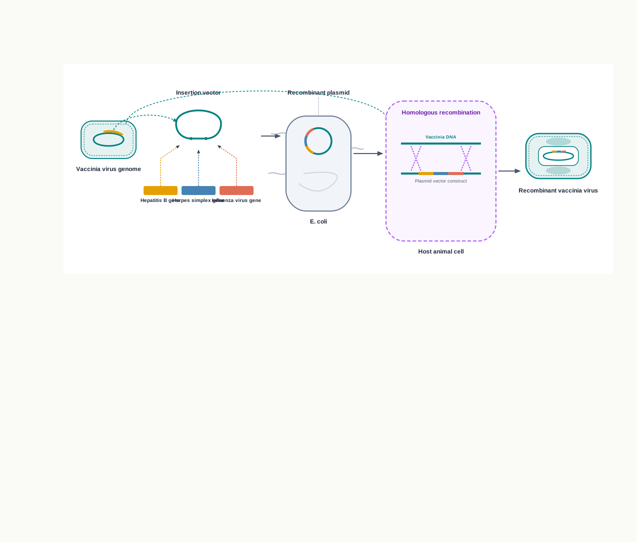
Fig. 4.16 – Production of a vector-based vaccine using vaccinia virus.
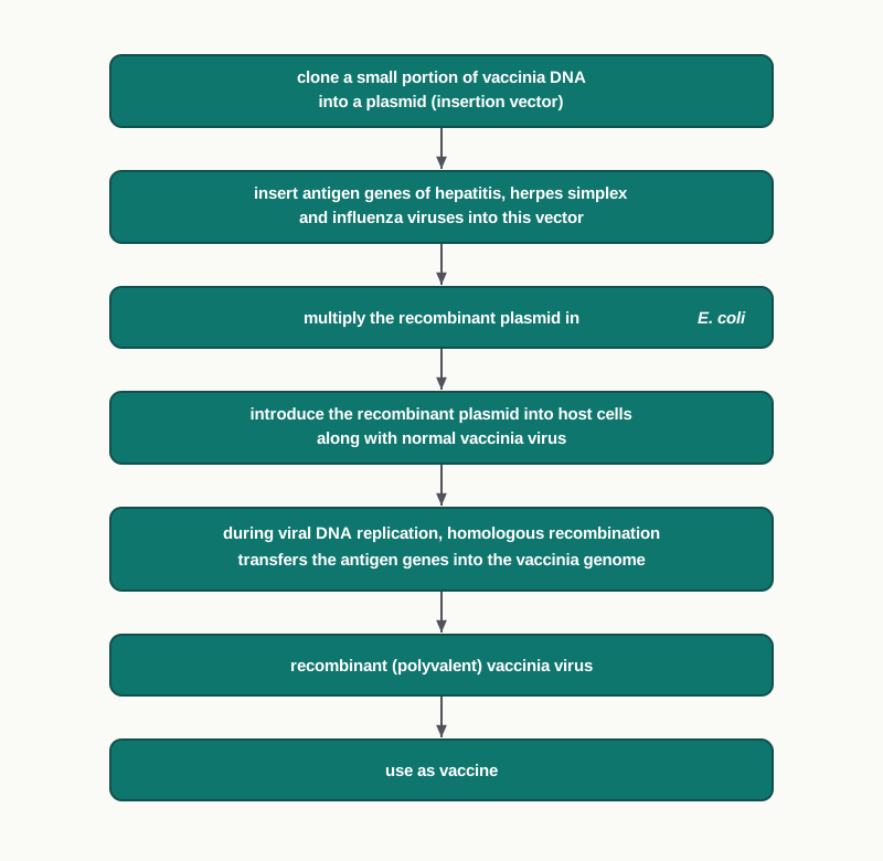
Fig. 4.17 – Producing a recombinant vaccinia vaccine.
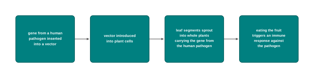
Fig. 4.18 – Edible vaccine production.
Recombinant Subunit Vaccines
Subunit vaccines contain only a part of the whole pathogenic organism. These can be either a synthetic peptide, or an expressed whole protein extracted from the pathogenic organism, or a protein expressed from cloned genes in the laboratory.
Prokaryotic systems, such as Escherichia coli, or eukaryotic systems, such as yeast, can be used to express the recombinant proteins. The choice between them matters: a bacterium grows fast and is cheap, but cannot add the sugar groups that many eukaryotic antigens need, whereas yeast can carry out such modifications and still be grown in bulk.
The advantages of these vaccines include purity, stability and safe use. Since not a single copy of the pathogen is present in the final preparation, there is nothing that could cause the disease.
One such example is the subunit vaccine developed against Hepatitis B — a widespread disease that mainly affects the liver, leading to cirrhosis, chronic hepatitis and cancer.
Hepatitis B virus consists of a core containing a viral genome (DNA), surrounded by a phospholipid envelope carrying the surface antigen HBsAg, which is the key element for the hepatitis B vaccine. Culturing hepatitis B virus and producing these surface antigens is very complicated.
Therefore, with the advent of rDNA technology, the gene coding for the HBsAg surface protein was identified and inserted into pMA56, a yeast plasmid vector, and expressed in Saccharomyces cerevisiae yeast cells.
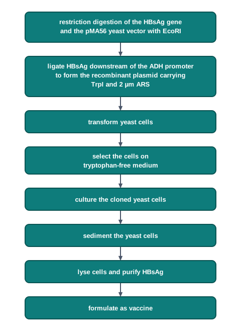
Fig. 4.19 – Production of HBsAg in yeast cells.
Two details in that protocol are worth pausing on, because they show how carefully a vector is designed. The strong ADH promoter is chosen so that the antigen is made in large amounts, not in trace quantities. The tryptophan-related selection is a clever alternative to an antibiotic: only yeast cells that have taken up the plasmid can grow on a medium lacking tryptophan, so the plate itself picks out the transformants.
DNA Vaccines
In recent years, DNA vaccines have become one of the promising advancements in vaccine technology. Here we do not supply the antigen at all — we supply the instructions, and let the patient’s own cells make the antigen.
For DNA vaccines, the genes of interest (antigens) are identified and cloned in plasmids. The DNA vaccine carrying the plasmid harbours a promoter site, a cloning site, an origin of replication, a selectable marker sequence, and a terminator sequence, like a poly-A tail.
Each of these five elements has a job. The promoter switches transcription on inside the human or animal cell; the cloning site is where the antigen gene is inserted; the origin of replication allows the plasmid to be multiplied in bacteria while it is being manufactured; the selectable marker identifies bacteria that carry the plasmid during that manufacture; and the terminator, such as a poly-A tail, ends the transcript properly and stabilises the mRNA that is produced.
During vaccination, DNA is directly injected into the muscles of an animal model system such as mice, generally using a ‘gene gun’ that is based on compressed gas, to inject the DNA into the muscle. You have already met the gene gun in the previous section as a plant transformation tool; the same principle of firing DNA into cells is used here on muscle tissue.
DNA vaccines can also be administered by nasal spray. The antigenic DNA is taken up by a few muscle cells, which — after protein expression — induce the immune system. DNA vaccines are capable of inducing both humoral and cellular immune responses.
RNA Vaccines
RNA vaccines contain mRNA. Once injected for vaccination, the mRNA is directly taken up by antigen presenting cells (APCs) and other target cells, where the mRNA is expressed as a properly folded and glycosylated protein that acts as an antigen (Fig. 4.20). These vaccines elicit both humoral and cellular immune responses against the encoded protein.
The step from a DNA vaccine to an RNA vaccine is a short one but an important one. mRNA does not have to enter the nucleus and it is never converted into DNA, so it cannot integrate into the host genome.
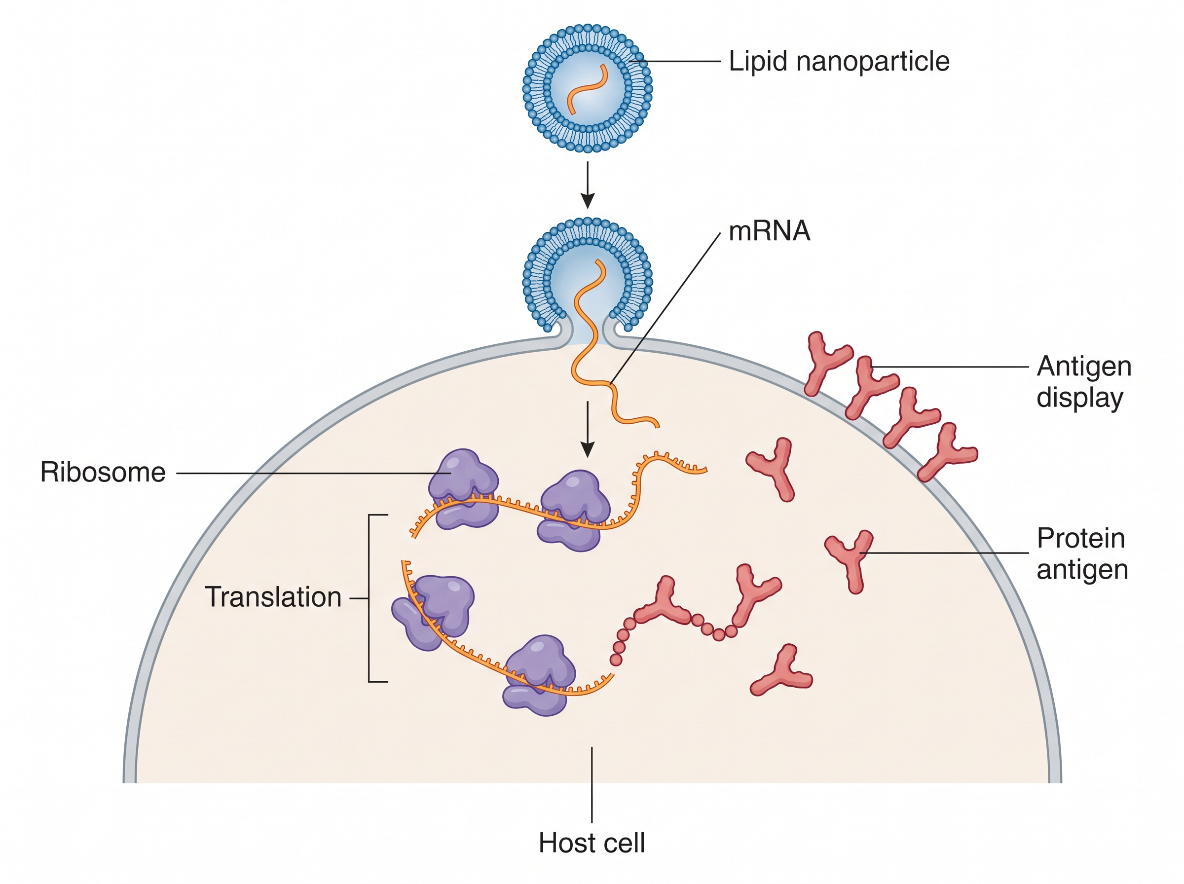
Fig. 4.20 – Mechanism of an RNA vaccine.
RNA is labile, so the injected mRNA has to be protected from rapid degradation by modifying it in a variety of ways — for example, by the addition of a 5’ cap, and by adjusting the length and structure of the 3’ poly-A tail. Therefore, RNA vaccines are formulated with specific delivery systems such as lipid nanoparticles (LNPs), which protect the RNA from degradation and increase target cellular uptake.
Thermostability of a vaccine is vital, to reduce the need for cold conditions during vaccine storage and use. Since RNA is unstable, it needs to be frozen when stored, to ensure long-term stability. This is not a small inconvenience: a vaccine that must stay frozen from factory to patient is very difficult to deliver to remote villages and to regions with unreliable electricity.
Efforts are underway to improve the thermostability of RNA vaccines. Lyophilisation, or freeze-drying, has been proposed as a way to improve their thermostability. In this direction, the formulation of RNA with thermostable lipid nanoparticles, as mentioned above, has also been suggested for improving the thermostability of RNA vaccines.
RNA vaccines are comparatively simple and have high-yield production. Their virus-free production process enables modest and scalable production facilities. As a result of cell-free production, there is less concern with respect to contaminating agents — no living cells means no cellular debris to purify away.
Another important advantage of mRNA vaccine technology is its ability to respond rapidly to emerging viral variants. This means that making modified mRNA vaccines for mutants with changed sequences is quick and simple: if the sequence of the antigen changes, only the sequence of the mRNA has to be changed, and the rest of the manufacturing process stays the same.
Thus, RNA vaccines offer several advantages over other categories of vaccines, including rapid and low-cost development, and a reduced need for optimisation and regulatory testing — because the formulation and delivery method are already optimised and licensed.
Furthermore, mRNA vaccines require a smaller amount of vaccine dose in each shot, and may not even require two doses. They can also be produced more quickly and efficiently as compared to other vaccines, in the case of new emerging variants of pathogens.
RNA vaccines are comparatively safe. There is no risk of pathogen reactivation, and RNA is degraded in vivo with no risk of antigen persistence or integration into the genome.
mRNA Vaccine Development against COVID-19
The COVID-19 pandemic demonstrated the threat of pandemics caused by novel, previously unrecognised, or mutated strains — in this case the Severe Acute Respiratory Syndrome Corona Virus-2 (SARS-CoV-2) strain, with high transmissibility.
Vaccines namely Covaxin (an inactivated whole virus vaccine) and Covishield (an adenovirus-based DNA vaccine) are used in India for protection against COVID-19. Notice that these two are built on entirely different principles — one is a conventional inactivated preparation, the other uses a viral vector to deliver the antigen gene — and both were deployed at the same time.
However, for effective control of pandemics, RNA-based vaccines have also been developed. A number of mRNA-based vaccine candidates for COVID-19 are in different phases of preclinical and clinical trials.
The pandemic made the practical advantage of mRNA technology very visible. When a new variant appears, an inactivated vaccine has to be remade from the new strain, whereas an mRNA vaccine needs only a new sequence fed into an existing manufacturing line.
In examinations, this section is commonly tested through compare-and-contrast questions (conventional versus recombinant vaccines; DNA versus RNA vaccines), through questions asking you to justify a design choice (why the vaccinia genome is suitable, why yeast rather than bacteria for HBsAg, why lipid nanoparticles are needed), and through case-based questions built around a vaccination programme or a new outbreak.
4.5 Therapeutic Agents/Molecules: Monoclonal Antibodies, Insulin and Growth Hormone
Recombinant DNA technology has revolutionised health care by facilitating the large-scale biological production of a variety of safe, pure and efficient human proteins. Compounds used for treating a disease and for improving the health of human beings are called therapeutic agents.
With the advent of rDNA technology, not only are newer therapeutic agents produced, but more efficient forms of conventionally produced therapeutics are also being manufactured. Since these recombinant therapeutics are identical to the natural human proteins — unlike those isolated from animals such as cow and pig — they do not induce immune responses and are free from the threat of any allergic response.
That single sentence contains the whole argument for the technology. An animal protein is similar to the human one, not identical, and the immune system is built to notice exactly that kind of small difference. A protein made from the human gene has the human sequence, so there is nothing for the immune system to object to.
Presently, numerous products like antibiotics, monoclonal antibodies, blood clotting factors, hormones (insulin and growth hormone), cytokines and vaccines are being produced for human welfare.
Antibodies: Polyclonal and Monoclonal
Antibodies are the protein molecules produced by B cells of the immune system, and they protect the body against invading foreign substances called antigens. In human serum, five different types of antibodies are present: IgA, IgD, IgE, IgG and IgM.
B cells, in response to a foreign particle, produce many different types of antibodies specific to different regions of the antigen. These regions on the surface of an antigen are called epitopes, and the mixed population of antibodies produced against them is designated polyclonal antibodies.
This polyclonal antibody-mediated response leads to antigen processing and degradation, and therefore has an obvious advantage for the organism in vivo. Attacking a pathogen at many points at once is exactly what a body needs.
However, due to their heterogenic nature, the efficacy of polyclonal antibodies for in vitro use is restricted. A diagnostic test needs to give the same answer every time, and a mixture of antibodies of many different specificities cannot guarantee that.
On the contrary, antibodies specific to a particular region or epitope of an antigen, known as monoclonal antibodies (MAbs), can be very well exploited for in vitro purposes, like therapeutic and diagnostic applications.
Hybridoma Technology
Earlier, following a brief exposure of an animal to an antigen, B cells were isolated and cultured for producing monoclonal antibodies. However, B cell culturing is very tricky and complicated, and the monoclonal antibodies produced were short lived. The reason is simple: a normal plasma cell has a definite lifespan and stops dividing, so the supply of antibody stops with it.
An invention made by George Kohler and Cesar Milstein (1975) provided a way out, through which the large-scale production of monoclonal antibodies became possible using hybridoma technology. In this technique, the short-lived antibody-secreting normal plasma cell is fused with immortal myeloma cells with the help of Polyethylene Glycol (PEG), leading to the formation of hybrid cells called hybridoma.
Hybridoma cells acquire immortal growth, a typical attribute of a myeloma cell, besides the antibody production property bestowed by the B plasma cell. Each parent contributes exactly the property the other one lacks — that is the whole design of the technique.
Fusion, however, is not selective. A treated dish contains fused hybridomas, but also unfused myeloma cells, unfused B cells, and myeloma–myeloma or B–B fusions. A way is needed to let only the true hybridomas survive, and this is provided by the culture medium itself.
Myeloma cells are genetically engineered such that they cannot use hypoxanthine, aminopterin and thymidine (HAT medium) as a source for nucleic acid biosynthesis, and will die in culture. On the contrary, B cells are able to survive in HAT medium, but they have a definite life span and die after that. Only B cells that have fused with these engineered myeloma cells will survive in culture when grown in HAT medium, as shown in Fig. 4.21.
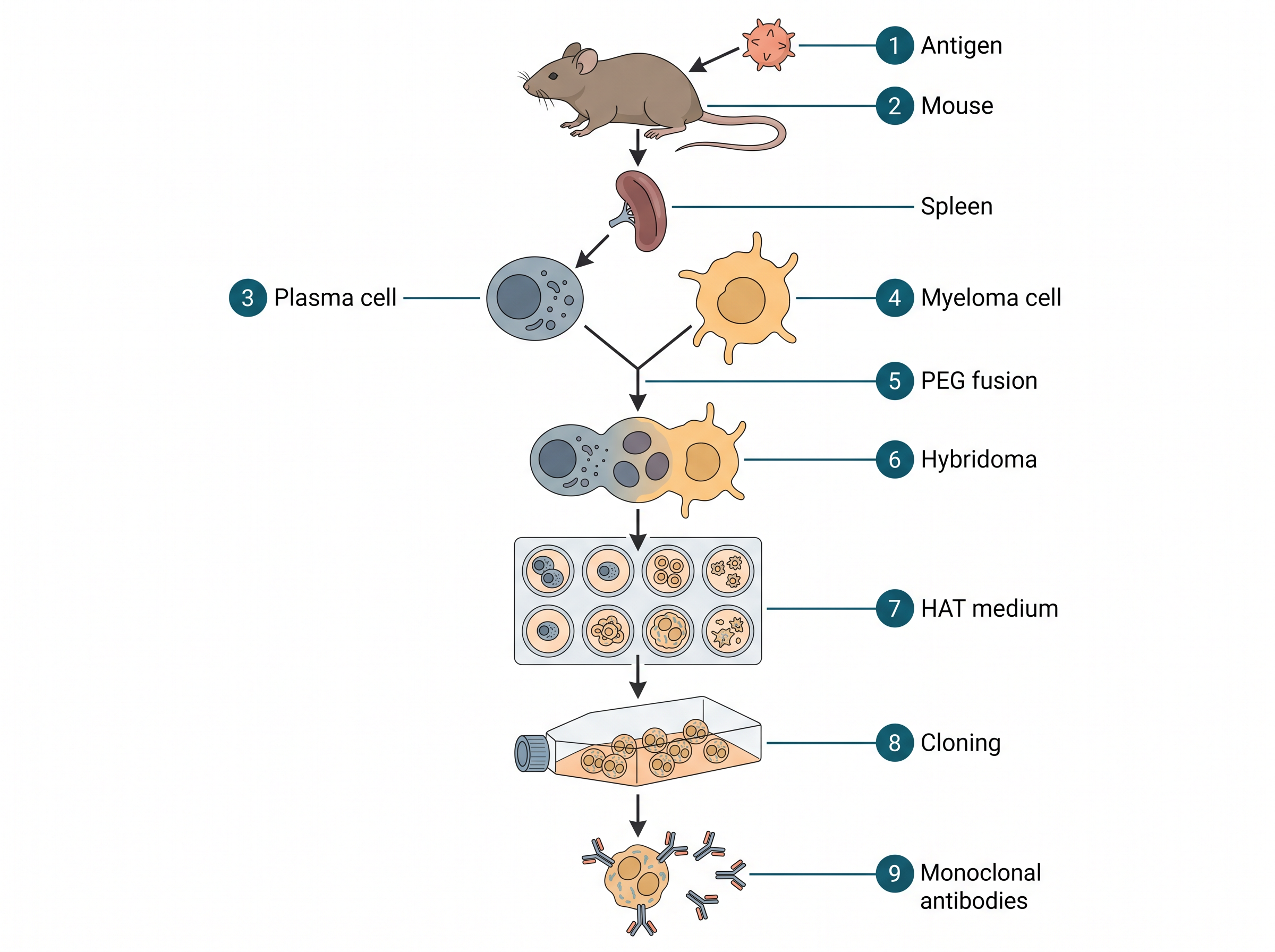
Fig. 4.21 – Production of monoclonal antibodies.
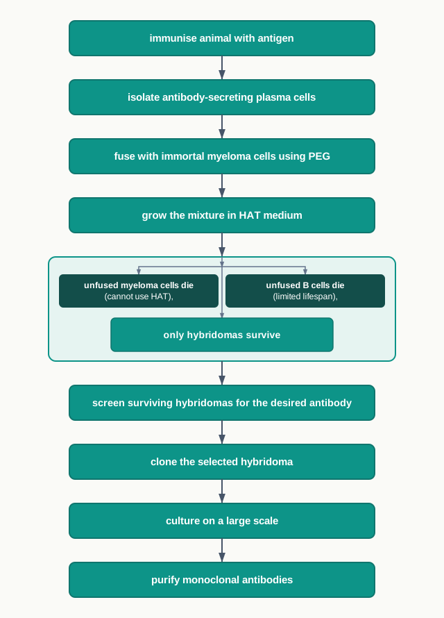
Fig. 4.22 – Hybridoma technology.
With the advances in rDNA technology, it is possible to develop mouse antibodies carrying a few human segments, known as chimeric or humanised antibodies, possessing higher effectiveness and activity.
The reason for humanising them follows directly from the opening idea of this section. A mouse antibody injected into a person is itself a foreign protein, and the patient’s immune system will eventually attack it. Replacing as much of the mouse sequence as possible with human sequence reduces that reaction, so the antibody survives longer and works better.
Applications of Monoclonal Antibodies
Monoclonal antibodies have revolutionised the diagnosis of various diseases and are used in a number of commercially available diagnostic kits, like pregnancy detection kits.
They are also used in the diagnostic imaging of cancer, wherein tumour-specific monoclonal antibodies can help in differentiating cancerous and non-cancerous cells.
In recent years, monoclonal antibodies are employed as very specific targeted therapeutic agents. They are coupled or attached with toxins or drugs and carried to the target tissues for efficient action. This is often described as a “magic bullet” strategy: the antibody supplies the address, and the attached drug or toxin does the damage — only where it is delivered, and not throughout the body as an ordinary drug would.
Insulin
Insulin, a hormone secreted by the beta cells of the pancreas, acts on different types of cells resulting in the uptake of glucose from blood. Insulin also stimulates the conversion of glucose to glycogen (glycogenesis) in the target cells.
A disease caused by the deficiency of insulin hormone, and characterised by the presence of sugar in urine, is Diabetes mellitus.
The Structure of Insulin
Structurally, insulin consists of two chains: an A chain of 21 amino acids and a B chain of 30 amino acids, interconnected with disulphide bonds, as shown in Fig. 4.23.
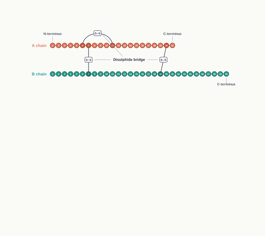
Fig. 4.23 – Primary structure of human insulin.
When you look at Fig. 4.23, note where the linkages are. The two chains are not a single continuous polypeptide; they are two separate chains stitched together by disulphide bonds between cysteine residues. That structural fact becomes very important later, when the hormone has to be manufactured in bacteria.
After the Banting and Best experiments, insulin was purified from the pancreas of pigs and cows. Human insulin, porcine insulin (from pig) and bovine insulin (from cow) are very similar, except for one or three amino acids. But these minor differences in the chemical structure can lead to an allergic response in some diabetic patients. As a result, the use of human insulin for treatment is more suitable as compared to animal insulin.
How the Body Makes Insulin
Physiological insulin first gets translated into a primary protein called preproinsulin, composed of 110 amino acids.
In the endoplasmic reticulum, first the signal peptide fragment is cleaved off from preproinsulin, thus turning preproinsulin into proinsulin, composed of 86 amino acids. The signal peptide is the address label that directs the growing chain into the endoplasmic reticulum; once it has done its job, it is no longer needed.
Proinsulin carries three distinct peptide chains: the A-chain, the B-chain, and the C-chain, which is composed of 31 amino acids. In the next step, inside the endoplasmic reticulum, the C-chain and an additional four connecting amino acids are removed to produce biologically active insulin of 51 amino acid residues, as shown in Fig. 4.24.
You can check the arithmetic for yourself, and it is worth doing because it makes the whole pathway memorable:
\[110 - 86 = 24 \\text{ amino acids removed as the signal peptide}\]
\[86 - (31 + 4) = 51 \\text{ amino acids in active insulin}\]
\[21 + 30 = 51 \\text{ amino acids, that is, the A chain plus the B chain}\]
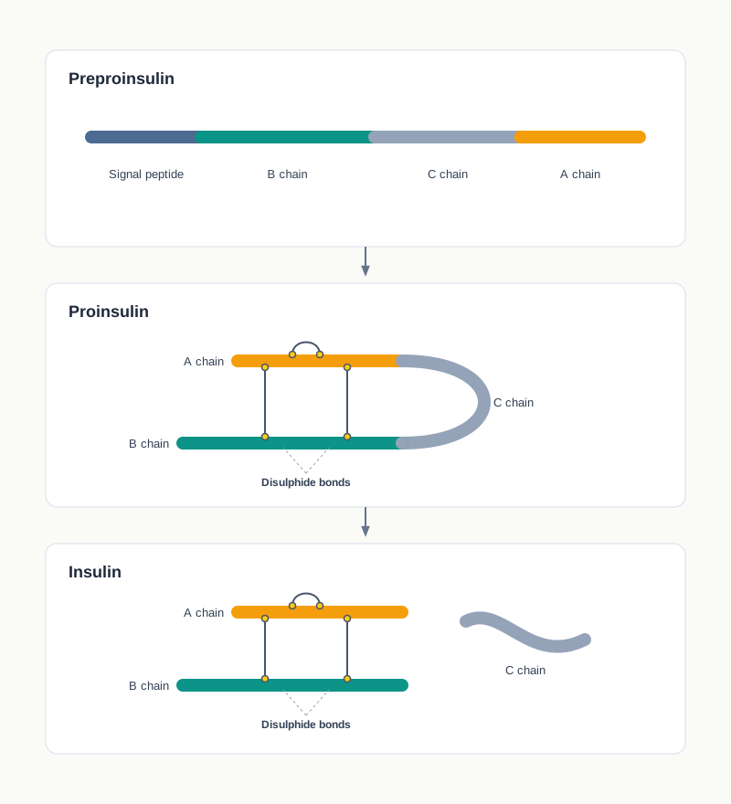
Fig. 4.24 – Conversion of preproinsulin to active insulin.
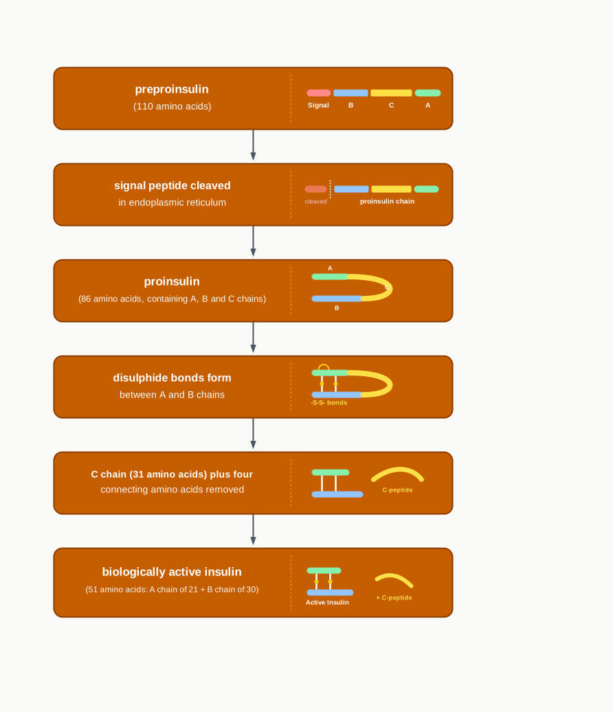
Fig. 4.25 – Natural processing of insulin.
Producing Insulin by rDNA Technology
In the late 1970s, biochemists exploited various tools of rDNA technology for the production of insulin.
The isolated insulin gene from a gene library was inserted into a bacterial plasmid of E. coli. A promoter site derived from the lac operon was inserted next to the insulin gene, so that the bacterium’s own transcription machinery would switch the gene on strongly.
The gene expression produced a fusion protein, that is, the insulin molecule fused with the enzyme β-galactosidase (β-gal), which was later cleaved to produce the human insulin. Fusing the product to a bacterial protein that E. coli normally makes in large amounts serves two purposes: the cell expresses it readily, and the fusion protects the small insulin chain from being destroyed by bacterial enzymes.
Because the processing of preproinsulin was not feasible in bacteria, scientists sidestepped this difficulty by cloning separately the genes for the A and B insulin chains in bacteria. This is an important point of understanding. A bacterium has no endoplasmic reticulum and none of the cutting enzymes that trim preproinsulin to proinsulin and then to insulin, so it cannot perform the natural processing. Rather than fight this, the scientists simply made the two finished chains separately and joined them afterwards.
The fusion protein, β-gal-insulin, was extracted and treated with cyanogen bromide (CNBr) to separate the β-gal fragment from the A and B chains. Subsequently, the disulfide bonds were again re-established between the A and B chain to reconstruct the final product, insulin, as shown in Fig. 4.26.
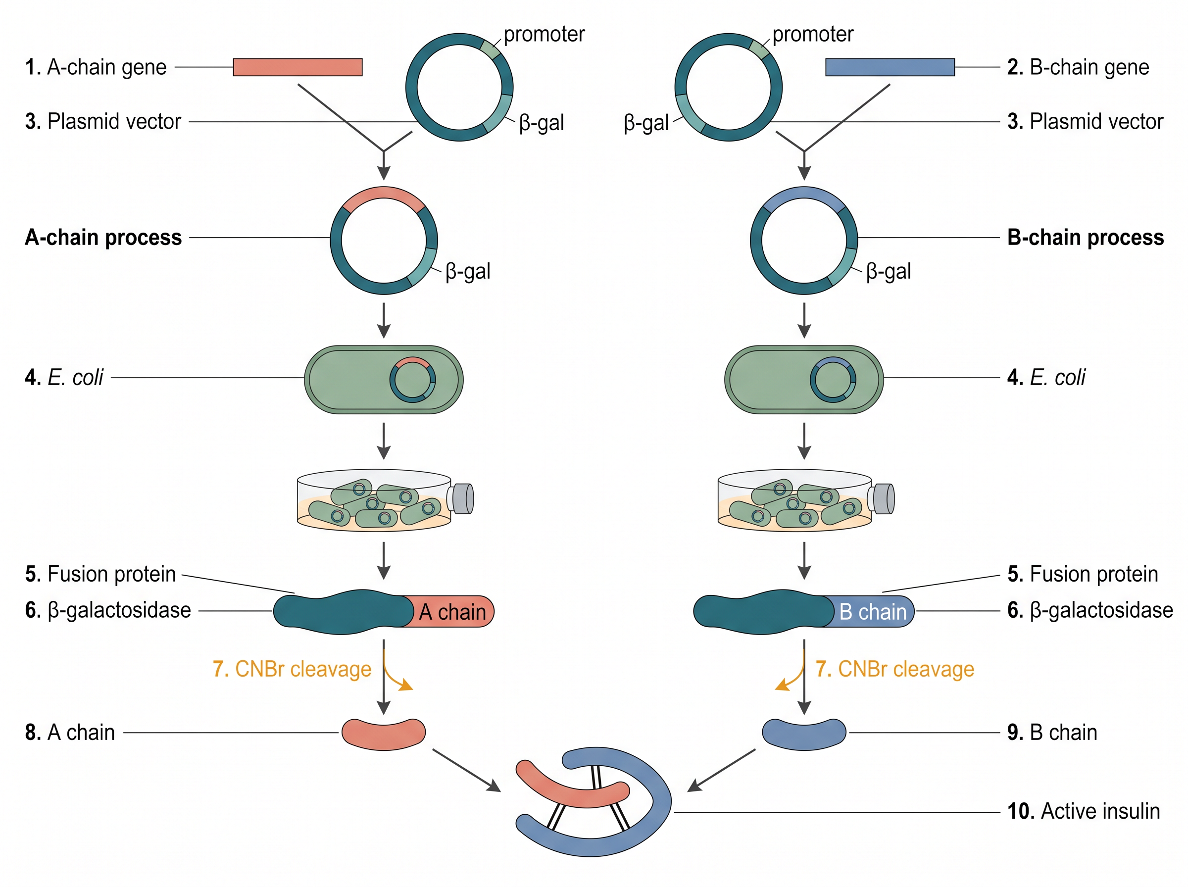
Fig. 4.26 – Steps in producing genetically engineered insulin.
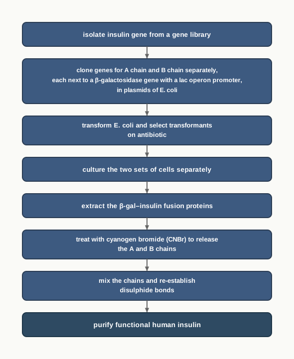
Fig. 4.27 – Recombinant insulin production.
The first genetically engineered human insulin, marketed as Humulin (Fig. 4.28), was manufactured in 1982. It was more effective as compared to pig- and cow-derived insulin, and the correct sequence was completely free from any allergic response.
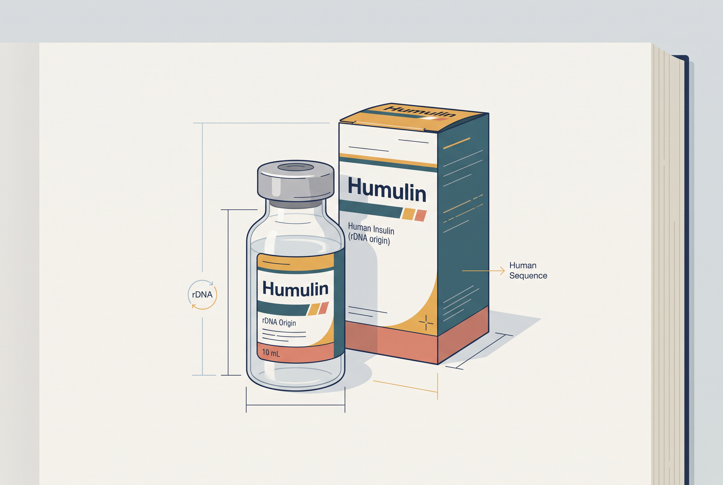
Fig. 4.28 – A vial and carton of Humulin.
Human Growth Hormone
Another important hormone secreted by the pituitary gland, human growth hormone (HGH), was produced using rDNA technology.
HGH, containing 191 amino acids, is a peptide hormone and promotes body growth by increasing the metabolism of amino acids, lipids and carbohydrates, during childhood as well as adulthood.
Childhood deficiency of HGH leads to Dwarfism. Dwarfism is characterised by retarded body growth, chubby face, added fat deposition at the waist and short height of about four feet, while the deficiency in an adult leads to average intelligence but with an abnormal body proportion, as shown in Fig. 4.29.
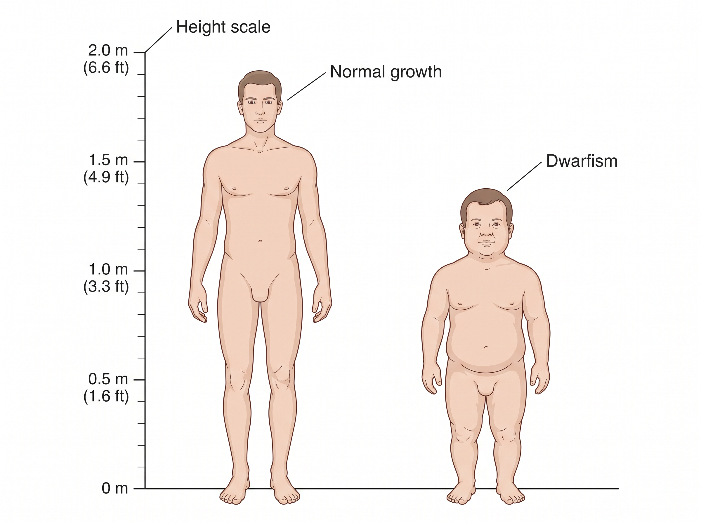
Fig. 4.29 – Effects of human growth hormone deficiency.
Since bone growth is not fully complete at an early stage, dwarfism can be treated at this stage by providing HGH shots. The timing matters biologically: once the growth plates of the long bones close, no amount of hormone will increase height, so the treatment has a window.
Why a Recombinant Source Was Needed
To obtain sufficient HGH quantity for one year using conventional treatment, the pituitary glands from more than 80 cadavers were required. Not only was this method expensive, but it also carried a risk of developing disease from the infected brain tissue of cadavers.
rDNA technology has enabled the production of the entire HGH. The production process for HGH is similar to that for human insulin production.
Constructing the HGH Gene
Though the natural HGH molecule consists of 191 amino acids, during its production in the body an intermediate molecule having an extra 26 amino acids is encoded as a signal peptide. This signal peptide chain is finally cut free during secretion.
Consequently, to synthesise the gene for HGH, the HGH-encoding mRNA was used as a template, and a complementary DNA (cDNA) molecule was synthesised. Using mRNA as the starting point is deliberate: the mRNA of a eukaryotic cell has already had its introns removed, so the cDNA made from it contains an uninterrupted coding sequence that a bacterium can translate correctly.
However, the bacterium fails to remove the signal peptide from the intermediate HGH molecule. A bacterium has no reason to recognise a human secretory signal, so if the signal peptide sequence were left in the gene, the product would carry 26 extra amino acids and would not be the natural hormone.
The nucleotide sequence coding for the signal peptide was therefore removed from the HGH gene. Since no known restriction enzyme can make this cut exactly at the required position, the restriction enzyme EcoRI was used to remove the base sequences for the signal peptide (26 amino acids) plus an additional 24 amino acids — a total of 50 amino acids — from the cDNA molecule.
The extra 24 amino acids’ base sequence that had been removed was then chemically synthesised and put back on the cDNA molecule, to generate the complete human growth hormone gene, as shown in Fig. 4.30.
This is a beautiful piece of problem-solving, and it is worth stating plainly. The enzyme could not cut where the scientists wanted, so they let it cut where it could, threw away too much on purpose, and then rebuilt the missing piece by chemical synthesis. The result is a gene that begins exactly at the first codon of mature HGH.
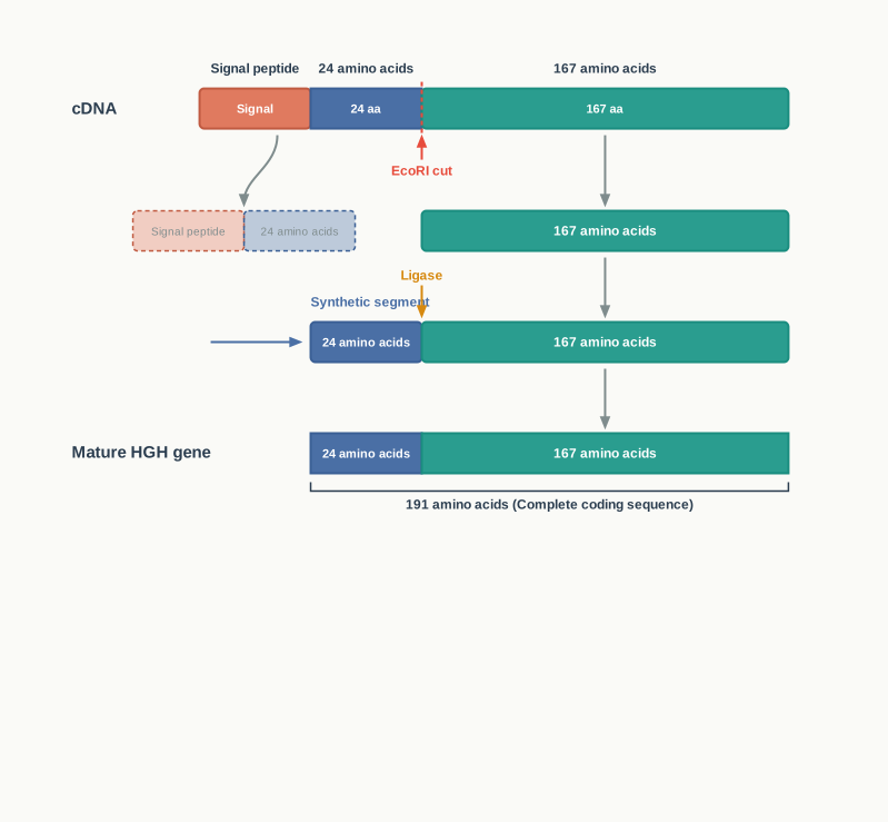
Fig. 4.30 – Assembly of the recombinant HGH gene.
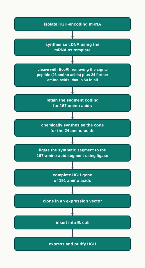
Fig. 4.31 – Construction and expression of the HGH gene.
In the absence of the signal peptide sequence, the human growth hormone gene was inserted into bacterial cells for hormone production. In 1985, genetically engineered human growth hormone was produced and marketed as Humatrope and Protropin.
Taken together, monoclonal antibodies, recombinant insulin and recombinant growth hormone show the same principle solved in three different ways: identify the human protein that is needed, obtain the gene that codes for it, and get a well-understood cell — a hybridoma, a bacterium — to make it in quantity and in the exact human form.
In examinations, this section is most often tested through the HAT-medium reasoning in hybridoma technology, the amino acid arithmetic of preproinsulin, proinsulin and insulin, the reason for cloning the A and B chains separately, the EcoRI problem in HGH gene construction, and assertion–reason items linking recombinant therapeutics to the absence of allergic response.
4.6 Summary
- To study and compare the inherited variations in human DNA without sequencing it, a technique known as ‘DNA fingerprinting’ was developed by Sir Alec Jeffreys in 1984 at the University of Leicester.
- In DNA fingerprinting, stretches of mini-satellite DNA known as Variable Number Tandem Repeats (VNTR), which are arranged tandemly, are exploited using the technique referred to as Restriction Fragment Length Polymorphism (RFLP).
- The process of inserting a foreign gene (transgene) into the genome of an organism, and its transmission and expression in the organism’s progeny, is termed transgenesis. The organisms carrying the transgene are known as transgenic organisms.
- Transgenic plants are also called genetically modified plants. Their genome is modified by the introduction of one or more genes from another species, through genetic engineering techniques.
- The basic requirement for genetic transformation is the construction of a genetic vehicle, which carries the genes of interest flanked by necessary regulating sequences such as a promoter or a terminator. The most commonly used techniques for gene transfer are of two types: vector-mediated or indirect gene transfer, and vector-less or direct gene transfer.
- Vector-mediated or indirect gene transfer includes transformation using Agrobacterium tumefaciens, in planta transformation, and plant virus-mediated transfer. Vector-less or direct gene transfer includes particle bombardment, protoplast transformation and microinjection.
- Transgenic animals are animals whose genetic makeup has been transformed by the use of various genetic engineering techniques, such as DNA pronuclear microinjection, embryonic stem cell-mediated gene transfer and retrovirus-mediated gene transfer.
- Transgenic plants have been developed with improved agronomic traits in crop plants and their products — for example, resistance to biotic and abiotic stresses, better nutrient quality and delayed fruit ripening.
- Transgenic plants are used in Molecular Farming for the large-scale production of industrial and therapeutic products.
- Transgenic animals have also been used in Molecular Pharming for the large-scale production of proteins such as α1-antitrypsin and human α-lactalbumin.
- There are a number of concerns related to the use of GMOs, both for human health and for the environment. The Genetic Engineering Appraisal Committee (GEAC), established by the Ministry of Environment, Forest and Climate Change, regulates the manufacture, use, import and export of hazardous microorganisms.
- Gene therapy is a technique designed to repair faulty genes in humans by introducing correct genetic material inside the cells. There are three main approaches for gene therapy: (i) gene replacement or gene addition, (ii) gene inhibition, and (iii) gene repair or gene editing.
- Since gene therapy includes making changes to the body’s basic set of genes, it raises unique ethical considerations.
- A preparation of a killed or weakened pathogen, or of its components, given to elicit an immune response that subsequently recognises the infectious agent and confers protection against the disease, is known as a ‘vaccine’.
- To avoid several potential concerns raised by conventional vaccines — such as the reversal of toxoids to their toxigenic forms — recombinant vaccines were developed using the various tools of rDNA technology.
- There are three main types of recombinant vaccines: live genetically modified vaccines, recombinant subunit vaccines, and RNA/DNA vaccines.
- rDNA technology enables health care by facilitating the large-scale biological production of a variety of safe, pure and efficient therapeutic agents, such as drugs — monoclonal antibodies — and human proteins, for example insulin and HGH.
- With the advances in rDNA technology, it is possible to develop mouse antibodies carrying a few human segments, known as chimeric or humanised antibodies, which possess higher efficacy and activity.
- In the late 1970s, biochemists exploited various tools of rDNA technology for the production of insulin. They isolated the insulin gene from a gene library and then inserted this gene into a bacterial plasmid of E. coli.
- The first genetically engineered human insulin, marketed as Humulin, was manufactured in 1982. Its successful production proved, without any ambiguity, the possibility of genetically engineering diverse biological organisms to produce human proteins for medicinal and therapeutic use.
- DNA technologists can now produce Human Growth Hormone entirely using rDNA technology. In 1985, genetically engineered human growth hormone was produced and marketed as Humatrope and Protropin.
The chapter as a whole follows one repeating logic, summarised in Fig. 4.32: identify the gene or DNA region of interest, move it into a chosen host using a suitable delivery method, select the cells that have received it, and harvest or apply what the host now produces.
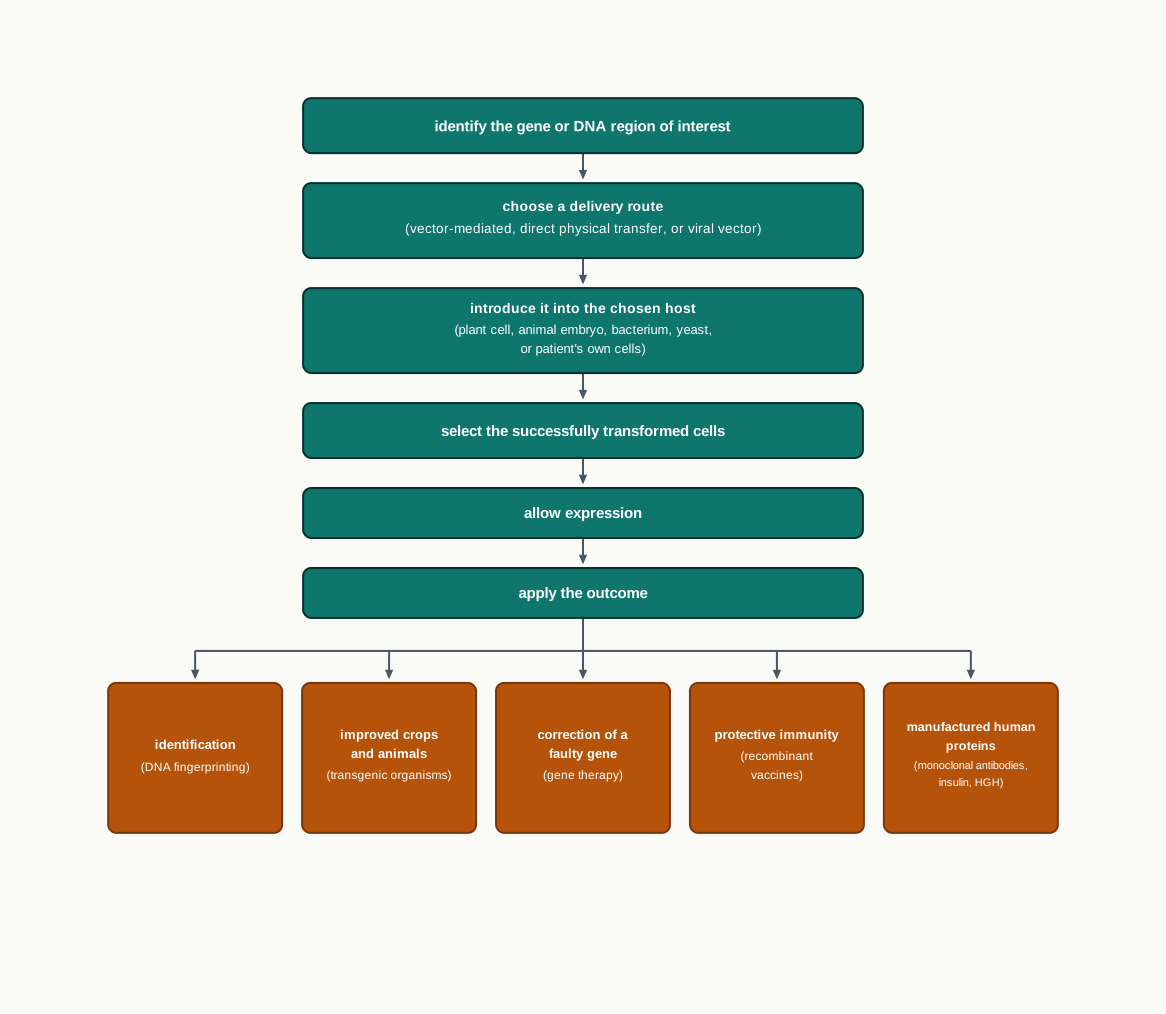
Fig. 4.32 – The common logic of biotechnology applications.
4.7 Key Terms
- Recombinant DNA (rDNA) technology — The set of techniques by which DNA from different sources is joined and introduced into a host cell so that the new gene is expressed there.
- Base pair (bp) — A pair of complementary nitrogenous bases holding the two strands of DNA together; the unit in which DNA length is measured.
- DNA fingerprinting — A technique that compares the inherited variations in an individual’s DNA, without sequencing it, to produce a band pattern characteristic of that individual.
- Short tandem repeat (STR) — A short nucleotide sequence repeated a number of times one after the other in the non-coding DNA of the genome.
- Locus — The specific position on a chromosome at which a particular gene or repeat sequence is located.
- Variable number tandem repeat (VNTR) — A locus at which a short sequence is repeated in tandem, the number of repeats differing from individual to individual.
- Microsatellites — A family of VNTRs in which the repeating unit is 1 to 9 bp long.
- Minisatellites — A family of VNTRs in which the repeating unit is 10 to 100 bp long.
- Polymorphism — The existence of many different forms of the same DNA locus in a population, here arising from differences in the number of tandem repeats.
- Restriction enzyme — An enzyme that recognises a specific short sequence in DNA and cuts the DNA at or near that site.
- Restriction Fragment Length Polymorphism (RFLP) — The variation between individuals in the length of DNA fragments produced when the same DNA region is cut with the same restriction enzyme.
- Agarose gel electrophoresis — A method that separates DNA fragments by size by making them migrate through an agarose gel under an electric field.
- Southern hybridisation — The detection of specific DNA fragments on a membrane by allowing a labelled complementary probe to bind to them.
- Probe — A labelled stretch of single-stranded DNA used to detect the presence of a complementary target sequence.
- Polymerase Chain Reaction (PCR) — A technique that amplifies DNA, used here to increase the sensitivity of DNA fingerprinting when very little DNA is available.
- Transgene — A foreign gene that is inserted into the genome of another organism.
- Transgenesis — The insertion of a transgene into an organism’s genome, together with its transmission to and expression in the organism’s progeny.
- Transgenic organism (GMO) — An organism that carries a transgene in in its genome; also called a genetically modified organism.
- Vector — The genetic vehicle that carries the gene of interest, flanked by regulating sequences, into a host cell.
- Promoter — The regulating sequence placed before a gene that tells the host’s machinery where to begin transcription.
- Terminator — The regulating sequence that marks where transcription of a gene should stop.
- Ti plasmid — The large tumour-inducing plasmid of Agrobacterium tumefaciens, a portion of which is naturally transferred to the plant genome.
- T-DNA — The portion of the Ti plasmid that is transferred into the plant genome upon infection.
- Crown gall — The plant tumour formed when T-DNA genes make the host cell overproduce auxin and cytokinins.
- Disarming — Removal of the tumour-inducing genes from the T-DNA, so that the plasmid can carry a gene of interest without causing crown gall.
- Marker gene — A gene, usually for antibiotic resistance, introduced along with the gene of interest so that transformed cells can be distinguished from non-transformed ones.
- Particle bombardment (biolistics) — Direct gene transfer in which DNA-coated gold or tungsten particles are fired into cells at high velocity.
- Gene gun (particle gun) — The device used to accelerate DNA-coated metal particles into target tissue.
- Callus — The unorganised mass of dividing cells formed in culture, from which a whole transgenic plant can be regenerated.
- Protoplast — A spherical naked plant cell produced by the removal of its cell wall.
- Electroporation — The use of a brief electric current to make a cell membrane temporarily permeable, so that DNA can enter.
- DNA pronuclear microinjection — Injection of a transgene directly into the larger male pronucleus of a just-fertilised egg.
- Embryonic stem (ES) cells — Cells from the inner cell mass of the blastocyst that can differentiate into any cell type of the body, including germ cells.
- Chimera — An organism composed of at least two genetically distinct cell lineages originating from different zygotes.
- Retroviral vector — A vector derived from an RNA virus, used to insert a single stable copy of a transgene at a particular location in the host genome.
- Bt gene (cry gene) — The gene from Bacillus thuringiensis that encodes an insecticidal protein, transferred into crops such as cotton.
- Bt toxin — The insecticidal protein encoded by the Bt gene, each class of which is harmful only to a specific group of insects.
- Osmolytes — Stress-related compounds such as trehalose, fructans, betaine and proline, whose over-production improves tolerance to abiotic stress.
- Antisense technology — The silencing of a gene by preventing the translation of its mRNA.
- Antisense oligonucleotide (ASO) — A short synthetic RNA or DNA molecule complementary to a target mRNA, which causes its degradation or blocks its translation.
- RNA interference (RNAi) — Gene silencing in which double-stranded RNA is processed so that a guide strand directs RISC to cleave the target mRNA.
- DICER — The endoribonuclease that cleaves double-stranded RNA into shorter fragments in the cytoplasm.
- RISC (RNA induced silencing complex) — The multiprotein complex that separates guide and passenger strands and cleaves the target mRNA.
- Molecular Farming — Genetic modification of a host plant so that it produces a desired biomolecule such as a therapeutic protein or vaccine antigen.
- Polyhydroxyalkanoates (PHAs) — Biodegradable, non-polluting biopolymers that can replace conventional plastics.
- Phytoremediation — The use of plants to remove pollutants from the environment, especially soil, by converting them into less toxic forms.
- Molecular Pharming — The use of transgenic animals as living bioreactors for the large-scale production of recombinant proteins, usually in their milk.
- Genetic Engineering Appraisal Committee (GEAC) — The Indian body, under the Ministry of Environment, Forest and Climate Change, that regulates the manufacture, use, import, export and storage of GMOs.
- Gene therapy — A technique designed to repair faulty genes in humans by introducing correct genetic material inside the cells.
- Severe Combined Immunodeficiency (SCID) — An immune disorder, one form of which is caused by a defect in the adenosine deaminase (ADA) gene, and the first disease treated by gene therapy.
- Adenosine deaminase (ADA) — The enzyme whose deficiency prevents the production of sufficient immune cells in one form of SCID.
- CFTR protein — The cystic fibrosis transmembrane conductance regulator, a membrane protein whose mutation hampers salt and water movement and causes cystic fibrosis.
- p53 gene — A gene that normally prevents tumour growth, defective forms of which are linked to several cancers.
- CRISPR/Cas9 system — A gene-editing technology adopted from the natural antiviral defence of bacteria and archaea, used to snip out mutated DNA and replace it with the correct sequence.
- Ex vivo gene therapy — Gene therapy in which cells are removed from the patient, corrected in culture and returned; also called cell-based delivery.
- In vivo gene therapy — Gene therapy in which functional genes are introduced directly into the target cells and tissues of the patient; also called direct delivery.
- Somatic gene therapy — Introduction of functional gene copies into the somatic cells of a patient, producing a change that is not heritable.
- Germ-line gene therapy — Introduction of functional genes into germ cells, producing a change that is heritable.
- Gene augmentation therapy — Addition of a functional gene to the genome to replace a missing gene product.
- Gene inhibition therapy — Use of antisense RNA or other inhibition techniques to block the expression of dominant-acting mutated genes.
- Gene targeting therapy — Replacement of a non-functional gene with a normal gene using homologous recombination.
- Bioethics — The assessment of the risks associated with a procedure and of the moral implications of a new technique.
- Vaccine — A preparation of a killed or weakened pathogen, or of its components, given to elicit an immune response that protects against later infection.
- Vaccination — The process of injecting and administering vaccines, a term coined by Louis Pasteur in 1881.
- Antigen — A foreign substance, or a component of a pathogen, that induces an immune response.
- Live genetically modified vaccine — A vaccine in which a pathogen is made non-pathogenic by deleting or inactivating one or more of its genes.
- Vaccinia virus — A large enveloped DNA virus of the pox family, used as a vector that can carry antigen genes of several pathogens at once.
- Insertion vector — A plasmid carrying a small cloned portion of vaccinia DNA, used as the site of homologous recombination during recombinant vaccine production.
- Polyvalent (multivalent) vaccine — A vaccine carrying the antigens of several different pathogens, giving simultaneous protection against several diseases.
- Edible vaccine — A vaccine produced in a plant and administered by eating the plant material, which stimulates the mucosal immune system.
- Recombinant subunit vaccine — A vaccine containing only a part of the pathogen, such as an expressed antigenic protein, rather than the whole organism.
- HBsAg — The hepatitis B virus surface antigen, the key element of the hepatitis B vaccine, expressed in yeast from the pMA56 vector.
- DNA vaccine — A plasmid carrying an antigen gene, injected so that host cells express the antigen and induce an immune response.
- RNA vaccine — A vaccine containing mRNA that is taken up by cells and translated into an antigenic protein.
- Lipid nanoparticles (LNPs) — Delivery particles that protect mRNA from degradation and increase its uptake by target cells.
- Lyophilisation — Freeze-drying, proposed as a way of improving the thermostability of RNA vaccines.
- Therapeutic agents — Compounds used for treating a disease and for improving human health.
- Antibody — A protein molecule produced by B cells that protects the body against invading antigens.
- Epitope — The particular region on the surface of an antigen to which an antibody binds.
- Polyclonal antibodies — The mixed population of antibodies produced by different B cell clones against different epitopes of the same antigen.
- Monoclonal antibodies (MAbs) — Identical antibodies produced by a single clone of cells, all directed against one particular epitope.
- Hybridoma — A hybrid cell formed by fusing an antibody-secreting plasma cell with an immortal myeloma cell.
- Hybridoma technology — The technique of producing monoclonal antibodies on a large scale by fusing plasma cells with myeloma cells using PEG.
- HAT medium — A medium containing hypoxanthine, aminopterin and thymidine, in which only fused hybridoma cells survive.
- Chimeric (humanised) antibodies — Mouse antibodies carrying a few human segments, which are more effective and more active in patients.
- Insulin — A hormone secreted by the beta cells of the pancreas that promotes glucose uptake from blood and its conversion to glycogen.
- Diabetes mellitus — A disease caused by insulin deficiency, characterised by the presence of sugar in urine.
- Preproinsulin — The 110-amino-acid primary translation product of the insulin gene, carrying a signal peptide.
- Proinsulin — The 86-amino-acid molecule formed after the signal peptide is removed, containing the A, B and C chains.
- Signal peptide — The short leading sequence that directs a newly made protein into the endoplasmic reticulum and is later cut away.
- Fusion protein — A protein made as a single unit with another protein, such as insulin fused to β-galactosidase, and later cleaved apart.
- Cyanogen bromide (CNBr) — The chemical used to separate the β-galactosidase fragment from the insulin A and B chains.
- Humulin — The first genetically engineered human insulin, manufactured in 1982.
- Human growth hormone (HGH) — A 191-amino-acid peptide hormone from the pituitary gland that promotes body growth.
- Dwarfism — The condition resulting from childhood HGH deficiency, marked by retarded growth, chubby face, fat deposition at the waist and short stature.
- cDNA — A DNA molecule synthesised using mRNA as the template, and therefore free of introns.
- Humatrope and Protropin — The genetically engineered human growth hormone products marketed from 1985.
4.8 Points to Ponder
- About 99.9% of the human genome is identical in all of us, and identification depends entirely on the remaining 0.1%. Reflect on what it means that our sense of individuality rests on so small a fraction of our DNA — and on the fact that the variable part lies mostly in DNA that codes for nothing at all.
- A DNA fingerprint can exclude a person with near certainty, but a match is always expressed as a probability. Think about what this asymmetry means when DNA evidence is presented in a court of law, and how confidently such evidence should be described to people who are not scientists.
- Agrobacterium tumefaciens was transferring its own genes into plants long before human beings knew what a gene was. Consider what else the phrase ‘genetic engineering’ might mean once we accept that nature has been doing it for millions of years.
- Selective breeding has been reshaping crops and animals for nearly ten thousand years, while transgenesis has been possible for about fifty. Ponder whether the two are different in kind, or only in speed and precision — and whether that distinction should matter to how each is regulated.
- The cry gene protects cotton from bollworms without harming butterflies and silkworms, because each class of toxin acts on one specific group of insects. Reflect on why this specificity is the strongest scientific argument in favour of Bt crops, and on what would follow if a pest population evolved resistance to that single toxin.
- Molecular farming can turn a field of a crop into a factory for a human medicine. Think about what safeguards would be needed to make sure such a crop never enters the food chain, and who should be responsible for enforcing them.
- Transgenic plants that carry the mercuric reductase gene can pull mercury out of contaminated soil, but the metal itself is an element and cannot be destroyed. Consider where the mercury actually goes, and what should be done with the plants afterwards.
- The first gene therapy improved a child’s immune function but did not cure her permanently. Reflect on how a partial, temporary success should be judged — as a failure of the technique, or as the necessary first step of a new kind of medicine.
- Somatic gene therapy affects only the patient; germ-line therapy affects every generation that follows. Ponder who could give consent on behalf of people who do not yet exist, and whether the permanence of a cure is only an advantage.
- The same editing technology that could remove a disease-causing allele could also be used to select for height, athletic ability or intelligence. Think about where the line between treatment and enhancement should be drawn, and whether that line can be defined scientifically at all.
- Edward Jenner’s cowpox experiment, and the recombinant vaccines of today, rest on the same biological principle separated by two centuries of technique. Reflect on what has actually changed in vaccine science, and what has not.
- An mRNA vaccine can be redesigned within days when a pathogen mutates, but it must be kept frozen. Consider what a technology’s practical requirements — cold chains, cost, distribution — mean for the countries and communities that can actually use it.
- Hybridoma technology begins by immunising an animal, and monoclonal antibodies are among the most expensive medicines made. Ponder how the benefits of such a medicine should be weighed against the costs it imposes, both on laboratory animals and on patients who cannot afford it.
- Before 1982, insulin came from the pancreases of pigs and cows, and growth hormone from the pituitary glands of cadavers. Reflect on how completely the source of a medicine can change while the medicine itself stays the same, and on what other treatments might one day be transformed in the same way.
- Every application in this chapter follows one pattern: find the gene, move it into a suitable host, select the cells that took it up, and use what they make. Think about which step in that sequence remains the hardest, and why progress in biotechnology so often depends on improving delivery rather than on understanding genes.
4.9 NCERT Questions
- What do you mean by DNA fingerprinting? Explain it through RFLP.
- What are GMOs? Describe the method of development of transgenic plants.
- Differentiate between direct and indirect method of gene transfer. Name one indirect method suitable for gene transfer in dicot plants.
- What is molecular pharming? Give applications of transgenic animals in molecular pharming.
- Differentiate between gene gun and gene therapy.
- Give the procedure of development of recombinant subunit vaccines.
- Write a short note on DNA vaccines.
- Describe the advantages of monoclonal antibodies developed by rDNA technology over that developed by Hybridoma technology.
- Briefly describe the development of Humulin through rDNA technology.
- Write a short note on Humatrope and Protropin.
- Briefly describe the applications of rDNA technology in crop improvement.
- List the ethical issues related to the use of transgenic animals.
- What is the role of vaccinia virus in the development of recombinant vaccine?
- Write a short note on recombinant therapeutic agents.
- Write a short note on humanised antibodies.
- Assertion: In hybridoma technology, B cells are fused with myeloma cells. Reason: Myeloma cells are immortal.
- Both assertion and reason are true and the reason is the correct explanation of the assertion.
- Both assertion and reason are true but the reason is not the correct explanation of the assertion.
- Assertion is true but reason is false.
- Both assertion and reason are false.
- Assertion: In Humulin, polypeptide A and polypeptide B are linked with disulfide bridges. Reason: C peptide is removed from proinsulin to form biologically active insulin.
- Both assertion and reason are true and the reason is the correct explanation of the assertion.
- Both assertion and reason are true but the reason is not the correct explanation of the assertion.
- Assertion is true but reason is false.
- Both assertion and reason are false.
- DNA fingerprinting depends on identifying specific:
- Coding sequences
- Non-coding sequences
- mRNA
- Promoter
- Short stretch of DNA used to identify complementary sequences in a sample is called:
- Probe
- Marker
- VNTR
- Minisatellite
- Variable number tandem repeat (VNTR) are:
- Repetitive coding short DNA sequences
- Non-repetitive non-coding short DNA sequences
- Repetitive non-coding short DNA sequences
- Non-repetitive coding short DNA sequences
- Cry genes or Bt genes are obtained from:
- Cotton pest
- Tobacco plant
- Bacillus thuringiensis
- E. coli
- When gene therapy is done in somatic cells, it is ______.
- non-heritable
- heritable
- rarely heritable
- not related to heritability
- In gene augmentation therapy, genetic material is ______.
- modified
- replaced
- suppressed
- removed
- Germ cell therapy is used for ____________.
- RBC
- Stomach cells
- Egg cells
- Bone marrow cells
- For the first time, from which animal was material isolated for vaccination?
- Cat
- Cow
- Goat
- Horse
- Vaccination was invented by:
- Jenner
- Pasteur
- Watson
- Crick
- For the production of insulin by rDNA technology, which bacterium was used?
- Saccharomyces
- Rhizobium
- Escherichia
- Mycobacterium
- Genetically engineered insulin is called __________.
- Humulin
- Promulin
- Bovulin
- Proculin
- Monoclonal antibodies are produced by _________.
- Mutations
- Transfection
- Hybridoma technology
- RNA interference
4.10 Objective Questions
- The proportion of the human genome that does not code for protein is:
- about 0.1%
- about 10%
- about 50%
- more than 90%
- A VNTR locus in which the repeating unit is 4 bp long would be classified as a:
- microsatellite
- minisatellite
- probe
- restriction site
- In DNA fingerprinting, DNA transferred to a nylon membrane is fixed permanently by:
- exposure to UV radiation for three minutes or baking at 80°C for two hours
- exposure to UV radiation for two hours or baking at 3°C for eighty minutes
- treatment with a restriction enzyme for three minutes
- hybridisation with a labelled probe for two hours
- In the DNA fingerprinting protocol, the step that immediately follows agarose gel electrophoresis is:
- restriction digestion of the sample
- transfer of the separated DNA to a nitrocellulose or nylon membrane
- hybridisation with a labelled VNTR probe
- comparison of the band pattern with known subjects
- PCR is used along with DNA fingerprinting mainly because it:
- cuts DNA at specific recognition sites
- separates DNA fragments according to size
- amplifies DNA irrespective of the amount present
- attaches DNA permanently to the membrane
- The portion of the Ti plasmid that is transferred into the plant genome upon infection is called:
- T-DNA
- cDNA
- the origin of replication
- the terminator
- The tumour-inducing genes of the T-DNA cause crown gall because they encode enzymes for the synthesis of:
- auxin and cytokinins
- gibberellin and ethylene
- abscisic acid and betaine
- trehalose and proline
- In particle bombardment, the DNA of interest is coated on to particles of:
- silica or calcium carbonate, 1 mm – 3 mm in size
- gold or tungsten, 1 µm – 3 µm in size
- gold or silver, 10 µm – 30 µm in size
- agarose or nitrocellulose, 1 µm – 3 µm in size
- Gene transfer into a plant cell whose wall has been removed can be promoted by:
- a bacteriostatic agent and a marker antibiotic
- hypoxanthine, aminopterin and thymidine
- polyethylene glycol and dextran sulphate
- cyanogen bromide and β-galactosidase
- In DNA pronuclear microinjection, the transgene is injected into the:
- smaller female pronucleus
- larger male pronucleus
- inner cell mass of the blastocyst
- nucleus of a myeloma cell
- Embryonic stem cells used for making transgenic animals are collected from the:
- germ cells of the adult
- inner cell mass region of the blastocyst
- male pronucleus of the fertilised egg
- mammary tissue of a foster mother
- An example of a retrovirus used as a vector for stable transgene introduction is:
- vaccinia virus
- caulimovirus
- lentivirus
- adenovirus
- The class of cry gene that encodes a toxin controlling corn borers is:
- cryIAc
- cryII Ab
- cryIAb
- all three equally
- Chloroplast transformation allows much higher transgene expression because:
- chloroplasts lack a genome of their own
- a plant cell contains many chloroplasts, each with several genome copies
- the chloroplast has no promoter requirement
- the chloroplast genome is not inherited
- In the RNAi pathway, double-stranded RNA is cleaved into shorter fragments in the cytoplasm by:
- RISC
- RNase H
- DICER
- EcoRI
- In RNA interference, the passenger strand is:
- retained by RISC and used to find the target mRNA
- degraded, while the guide strand is retained
- exported to the nucleus
- translated into an inhibitory protein
- Elelyso, the first plant-made human biologic approved in 2012, was produced in:
- tobacco leaves
- banana fruit
- carrot cell cultures
- safflower seeds
- Transgenic plants carrying the mercuric reductase gene are useful for:
- molecular pharming
- phytoremediation
- delayed fruit ripening
- production of biodegradable plastics
- The first transgenic cow, Rosie, created in 1997, produced milk enriched in:
- α1-antitrypsin
- human α-lactalbumin
- tissue plasminogen activator
- coagulation factor VIII
- In India, the body that regulates the manufacture, use, import, export and storage of genetically modified organisms is the:
- Genetic Engineering Appraisal Committee
- Centre for DNA Fingerprinting and Diagnostics
- US Food and Drug Administration
- Asilomar Committee on Recombinant DNA
- The first gene therapy, performed in 1990, treated a patient whose disease was caused by a defect in the gene coding for:
- the CFTR protein
- adenosine deaminase
- p53
- α1-antitrypsin
- Cystic fibrosis results from a mutation that affects a protein controlling the movement of:
- salt and water
- glucose and glycogen
- amino acids and lipids
- auxin and cytokinins
- Gene therapy in which cells are removed from the patient, corrected in culture and returned is known as:
- in vivo or direct delivery
- ex vivo or cell-based delivery
- germ-line therapy
- gene targeting therapy
- Replacement of a non-functional gene with a normal gene using homologous recombination is called:
- gene augmentation therapy
- gene inhibition therapy
- gene targeting therapy
- gene editing therapy
- A vaccine carrying the antigens of several different pathogens at once is described as:
- attenuated
- polyvalent or multivalent
- subunit
- edible
- The gene coding for the HBsAg surface protein was inserted into the yeast plasmid vector:
- pBR322
- pMA56
- Ti plasmid
- pUC18
- In an RNA vaccine, lipid nanoparticles are used mainly to:
- inactivate the pathogen before injection
- protect the RNA from degradation and increase cellular uptake
- provide the promoter and terminator sequences
- select the transformed cells
- The five classes of antibodies present in human serum are:
- IgA, IgB, IgC, IgD and IgE
- IgA, IgD, IgE, IgG and IgM
- IgD, IgE, IgG, IgM and IgP
- IgA, IgE, IgG, IgM and IgT
- Proinsulin consists of 86 amino acids and contains:
- only the A and B chains
- the A, B and C chains
- the A chain and the signal peptide
- the B chain and the signal peptide
- During recombinant insulin production, the β-galactosidase fragment is separated from the A and B chains by treatment with:
- polyethylene glycol
- cyanogen bromide
- aminopterin
- EcoRI
- During construction of the HGH gene, EcoRI was used to remove base sequences coding for a total of:
- 24 amino acids
- 26 amino acids
- 50 amino acids
- 167 amino acids
- Assertion: VNTR loci show a high degree of polymorphism between individuals. Reason: VNTRs lie within the non-coding regions of the genome, where changes in repeat number are generally not harmful.
- Assertion: In DNA fingerprinting, hybridisation with a labelled VNTR probe is performed instead of simply examining the stained gel. Reason: Individual bands of digested human genomic DNA are difficult to identify on a gel.
- Assertion: The Ti plasmid must be disarmed before it is used to produce a transgenic plant. Reason: The T-DNA of a natural Ti plasmid carries genes that induce crown gall.
- Assertion: Particle bombardment is preferred for the transformation of cereals. Reason: Agrobacterium-mediated transformation may not be very successful in such plant species.
- Assertion: A marker antibiotic is added to the regeneration medium after Agrobacterium infection. Reason: The antibiotic stops the further growth of Agrobacterium in the culture.
- Assertion: The improvement produced by the first gene therapy for SCID was not permanent. Reason: Gene therapy is short-lived in nature and may require frequent administration of the therapeutic gene.
- Assertion: Germ-line gene therapy raises far greater ethical concern than somatic gene therapy. Reason: Changes made to the germ line are heritable.
- Assertion: RNA vaccines need to be frozen for long-term storage. Reason: RNA is labile, and its stability has to be improved by modifications such as a 5’ cap and a 3’ polyA tail.
- Assertion: When a fusion mixture is grown in HAT medium, only hybridoma cells survive. Reason: Myeloma cells are engineered so that they cannot use hypoxanthine, aminopterin and thymidine for nucleic acid biosynthesis, while unfused B cells have a definite life span.
- Assertion: The genes for the A and B chains of insulin were cloned separately in bacteria. Reason: The processing of preproinsulin is not feasible in bacteria.
- Assertion: During HGH gene construction, the base sequence coding for 24 amino acids had to be chemically synthesised and added back. Reason: No known restriction enzyme could cut exactly at the first codon of mature HGH, so EcoRI removed more of the cDNA than was required.
4.11 Check Your Concepts
- About 99.9% of the human genome is identical in all human beings, yet DNA fingerprinting can distinguish one individual from another. Explain how both these statements can be true at the same time.
- Why are the variable sequences used in DNA fingerprinting found mainly in the non-coding regions of the genome, and not among the protein-coding genes? Reason it out in terms of what happens to a change in each kind of sequence.
- A student writes that “a person shows one band for each VNTR locus examined.” Correct this statement and explain the biological reason for your correction.
- Explain, step by step, how a difference in the number of tandem repeats between two individuals becomes a difference in the length of a restriction fragment.
- In a DNA fingerprinting experiment, the technician forgets to expose the nylon membrane to UV radiation and does not bake it either. Predict the result of the experiment and justify your prediction.
- Why is Southern hybridisation with a labelled VNTR probe necessary, when the DNA fragments have already been separated on the gel? What exactly does the probe add to the procedure?
- Arrange the following steps of DNA fingerprinting in the correct order: hybridisation with a labelled VNTR probe; agarose gel electrophoresis; isolation of DNA from the sample; comparison of the band pattern with known subjects; digestion with restriction enzymes; detection of hybridised fragments; transfer of DNA to a nylon membrane.
- Study Fig. 4.33. Explain the reasoning an analyst uses, band by band, to conclude that the couple are the parents of Child 1 but not of Child 2.
- Compare and contrast microsatellites and minisatellites under the headings: length of repeating unit, location in the genome, and role in DNA fingerprinting.
- Why is Agrobacterium tumefaciens described as a ‘Natural Genetic Engineer’? What does the bacterium gain from this ability in nature?
- What would happen if a Ti plasmid were used as a vector without first being disarmed? Explain why the resulting tissue could not be used to raise a transgenic crop.
- The regeneration medium used after Agrobacterium infection contains both a bacteriostatic agent and a lethal concentration of a marker antibiotic. State the purpose of each, and predict what would go wrong if either one were left out.
- Arrange the following in the correct sequence for Agrobacterium-mediated transformation: transfer of plantlets to soil; disarming of the Ti plasmid; formation of shoots on regeneration medium; insertion of the gene of interest into T-DNA; infection of plant tissue; transfer of the recombinant vector into Agrobacterium; transfer of shoots to rooting medium.
- Why are gold and tungsten chosen for particle bombardment, and why must the particles be only 1 µm – 3 µm in size? Answer in terms of what the particle has to achieve inside the tissue.
- Explain why particle bombardment of the chloroplast gives much higher transgene expression than insertion of the same gene into the nuclear genome.
- Differentiate between vector-mediated and vector-less gene transfer, giving two examples of each, and state one situation in which the vector-less method is clearly preferable.
- In pronuclear microinjection, why is the transgene injected into the male pronucleus rather than the female pronucleus, and why is the injection done at the single-cell stage rather than later?
- Compare the three methods of producing transgenic animals — pronuclear microinjection, ES cell-mediated transfer and retrovirus-mediated transfer — with respect to the site of integration and the copy number of the transgene. Which would you choose if predictability of integration were the deciding factor, and why?
- Bt cotton controls bollworms but does not harm butterflies and silkworms. Explain the basis of this selectivity, and state why it is a scientific advantage over spraying a chemical insecticide.
- A farmer asks why a Bt variety developed for corn borers cannot simply be planted to control cotton bollworms. Answer the farmer, using the names of the relevant cry genes.
- Explain how the over-production of osmolytes such as trehalose, betaine or proline helps a transgenic plant survive drought and salinity.
- Distinguish between the antisense oligonucleotide approach and the RNA interference approach, under the headings: molecule introduced, enzyme or complex involved, and the way the target mRNA is finally stopped.
- In the nematode-resistant tobacco plant, the transgenic plant is not itself toxic, yet the nematode cannot survive on it. Explain the mechanism, and state why the antisense RNA affects the parasite rather than the plant.
- Differentiate between Molecular Farming and Molecular Pharming, giving one example of a product from each.
- Why is milk the preferred destination for a human protein produced in a transgenic animal? Give two reasons.
- List any three concerns raised over GMOs, and for each one, suggest how it might be reduced or tested for before a crop is released.
- The first gene therapy improved the patient’s immune function but the cure was not permanent. Explain the cellular reason for this, and state what would have to be corrected instead for the benefit to last.
- Compare ex vivo and in vivo gene therapy. Why is the ex vivo approach described as simpler, even though it involves removing cells from the patient?
- Explain why a change made by somatic gene therapy dies with the patient, while a change made by germ-line gene therapy does not.
- Gene therapy is described as effective for single-gene defects but not for multigene disorders. Explain the reasoning behind this limitation.
- One of the demerits of gene therapy is random integration of the therapeutic gene. Explain, with an example, how such an integration event could produce a new disease in a patient being treated for an old one.
- Why does a vaccine protect a person even though it contains no substance that can kill the pathogen? Answer in terms of what the immune system retains after vaccination.
- State two potential concerns raised by conventional vaccines that recombinant vaccines were designed to avoid, and explain how the recombinant approach avoids each of them.
- Compare live genetically modified vaccines, recombinant subunit vaccines and DNA vaccines under the headings: what is actually administered, and where the antigen is finally produced.
- Why is the vaccinia virus particularly suitable as a vector for a polyvalent vaccine? Refer to the size and nature of its genome in your answer.
- Culturing the hepatitis B virus to obtain surface antigen is very complicated. Explain how rDNA technology sidesteps this problem entirely in the production of the hepatitis B subunit vaccine.
- Banana is described as the best plant for oral vaccine production. Give three reasons, and state one practical difficulty you would expect with an edible vaccine that an injected vaccine does not have.
- Why are RNA vaccines particularly well suited to responding to a newly emerged variant of a virus? Contrast this with what would be needed to update an inactivated whole-virus vaccine.
- Polyclonal antibodies are advantageous in vivo but of restricted use in vitro, while monoclonal antibodies are the opposite. Explain the reason for this difference in terms of epitopes.
- Explain why a hybridoma survives in HAT medium while neither of its parent cells can survive there on its own. Set out what each parent contributes.
- Arrange the following steps of hybridoma technology in order: cloning of the selected hybridoma; immunisation of the animal with the antigen; purification of monoclonal antibodies; fusion of plasma cells with myeloma cells using PEG; screening of hybridomas for antibody production; growth of the fusion mixture in HAT medium.
- Why are chimeric or humanised antibodies more effective in patients than ordinary mouse monoclonal antibodies? Relate your answer to the general reason recombinant human proteins are preferred over animal-derived ones.
- Label and explain the three stages shown in Fig. 4.24, stating for each stage the number of amino acids present and which chains are present.
- Explain why the genes for the A and B chains of insulin were cloned separately in bacteria instead of cloning the gene for preproinsulin.
- What is the purpose of fusing the insulin polypeptides to β-galactosidase, and what would be lost if the insulin chains were expressed on their own?
- A student proposes the following protocol for making recombinant insulin: “Clone the preproinsulin gene into E. coli, culture the bacteria, and purify the insulin secreted into the medium.” Identify two things that are wrong with this protocol and correct them.
- Why was mRNA, rather than genomic DNA, used as the starting material for constructing the human growth hormone gene?
- During HGH gene construction, EcoRI removed the coding sequence for 50 amino acids although only 26 needed to be removed. Explain why this was done deliberately, and how the missing sequence was restored.
- Explain why a bacterium cannot be relied upon to remove the signal peptide from an intermediate human hormone, and what the consequence would be if the signal peptide sequence were left in the cloned gene.
- Compare cadaver-derived human growth hormone with recombinant human growth hormone under the headings: source, quantity available, cost and safety.
- Match the items in Column A with those in Column B.
| Column A | Column B |
|---|---|
| a. DICER | i. Fusion of plasma cell with myeloma cell |
| b. Polyethylene glycol | ii. Cleaves double-stranded RNA into short fragments |
| c. Cyanogen bromide | iii. Removal of the signal peptide sequence from HGH cDNA |
| d. EcoRI | iv. Separates β-galactosidase from the insulin chains |
- Match the product in Column A with the organism in which it is produced, given in Column B.
| Column A | Column B |
|---|---|
| a. Human glucocerebrosidase | i. Safflower |
| b. Hepatitis B antigen | ii. Goat |
| c. Insulin (plant-produced) | iii. Carrot |
| d. Tissue plasminogen activator | iv. Lettuce |
- Every application in this chapter — fingerprinting, transgenesis, gene therapy, recombinant vaccines and recombinant therapeutics — depends on being able to select the small number of cells or fragments that carry what is wanted. Take any three of these applications and describe the selection step used in each.
4.12 Case-Based Questions
- A forensic laboratory receives three samples in a burglary case: a bloodstain lifted from a broken window, a buccal swab from Suspect 1, and a buccal swab from Suspect 2. DNA is extracted from all three, digested with the same restriction enzyme, run on a single agarose gel, blotted and hybridised with a labelled VNTR probe. The developed film shows that the bloodstain lane and the Suspect 2 lane have identical band positions, while the Suspect 1 lane differs at three of the four bands. The quantity of DNA recovered from the bloodstain was extremely small, and the analyst had to treat it before beginning the procedure.
- Name the treatment the analyst is most likely to have used on the bloodstain DNA, and state the property of that technique that makes it suitable here.
- Explain why the bands of Suspect 1 differ in position from those of the bloodstain, in terms of what is present at a VNTR locus.
- Of the two conclusions available — that Suspect 1 is excluded, and that Suspect 2 is the source — which can be stated with greater certainty, and why?
- State one reason why the same gel could not have been read directly after electrophoresis, without blotting and hybridisation.
- A couple approaches a diagnostic centre with a dispute over the parentage of a child. Profiles are prepared at four VNTR loci for the mother, the alleged father and the child. At three loci, every band in the child’s profile can be matched to a band in the mother’s profile or in the alleged father’s profile. At the fourth locus, the child shows two bands, one of which matches the mother, while the other matches neither adult.
- Explain why a child normally shows two bands at a VNTR locus.
- What conclusion does the fourth locus permit about the alleged father, and why is a single mismatch treated so seriously?
- Name two other applications of VNTR pattern analysis mentioned in this chapter, apart from parentage testing.
- A student in a plant tissue culture laboratory is asked to produce insect-resistant tomato plants. She inserts the gene of interest and an antibiotic resistance gene into the T-DNA of a Ti plasmid, transfers the recombinant vector into Agrobacterium, and infects tomato leaf discs. She then transfers the leaf discs to a plain nutrient medium containing neither a bacteriostatic agent nor the marker antibiotic. Three weeks later she reports that the culture is overgrown, that shoots have formed from many leaf discs, and that when she tests ten regenerated plants, only one carries the transgene.
- Identify the two omissions in her protocol and explain what each omitted component was supposed to do.
- Account separately for the overgrowth of the culture and for the very low proportion of transgenic plants among the regenerants.
- She had also forgotten to disarm the Ti plasmid. Predict what she would have observed in the infected tissue as a result.
- Name one physical method she could have used instead if the tomato variety had proved difficult to transform using Agrobacterium.
- A cotton-growing district adopts a Bt variety carrying cryIAc, and reports for the first four seasons a marked fall in bollworm damage and in the number of insecticide sprays. Beekeepers and silk farmers in the same district record no change in their yields. In the seventh season, several farmers report that bollworm damage has returned in fields where the same variety has been grown continuously.
- Name the bacterium from which the cry gene was obtained, and state what the gene encodes.
- Explain, in terms of the classes of cry genes, why the beekeepers and silk farmers were unaffected.
- Suggest a biological reason for the return of bollworm damage after several seasons of continuous cultivation of the same variety.
- State one trait, other than insect resistance, for which transgenic crops have been developed.
- A four-year-old child is diagnosed with an immune disorder caused by the absence of a functional enzyme in the lymphocytes. The child suffers repeated life-threatening infections. The treating team removes lymphocytes from the child, cultures them, introduces a functional copy of the gene using a genetically disabled retroviral vector, and returns the corrected cells to the child’s bloodstream. The child’s immune function improves markedly, but eleven months later the improvement has faded and the infusion has to be repeated.
- Name the disorder and the enzyme involved.
- Classify the therapy used, with reasons, as ex vivo or in vivo, and as somatic or germ-line.
- Explain why the benefit faded and the treatment had to be repeated.
- State one risk associated with the use of a viral vector in such a procedure.
- A regulatory panel is reviewing an application to permit the germ-line correction of a severe single-gene disorder in human embryos. The applicants argue that the correction would remove the disease allele from the family line permanently, sparing every future generation. Members of the panel raise objections concerning consent, unpredictable long-term effects, and the possibility that the same technique could later be applied to traits unrelated to disease.
- State the essential biological difference between somatic and germ-line gene therapy that lies behind this entire debate.
- Present the strongest argument for the applicants’ position, and the strongest argument against it, in one or two sentences each.
- Explain what is meant by ‘gene enhancement’, and why the panel treats it as a separate concern from treatment.
- Name the gene-editing technology that has made such precise correction technically conceivable, and state the natural role it plays in bacteria.
- During an outbreak of a rapidly mutating respiratory virus, a public health department must choose between three candidate vaccines: an inactivated whole-virus preparation, a recombinant subunit vaccine expressing a single surface antigen in yeast, and an mRNA vaccine formulated in lipid nanoparticles. The department serves both a well-equipped city hospital and remote rural centres with unreliable electricity supply. Midway through the programme, a new variant of the virus emerges with an altered surface protein.
- Explain how each of the three vaccines delivers its antigen to the immune system.
- Which of the three could be redesigned most rapidly to match the new variant, and why?
- Identify the vaccine that would present the greatest difficulty in the rural centres, and state the property of the molecule responsible for that difficulty.
- Name one measure being explored to improve the thermostability of that vaccine.
- A biotechnology company wishes to manufacture a diagnostic kit that detects a single protein present only in a particular tumour. Its first attempt uses serum antibodies collected from an immunised animal, and the kit gives inconsistent results from batch to batch. The company then immunises a mouse with the tumour protein, isolates its plasma cells, fuses them with engineered myeloma cells using polyethylene glycol, and grows the mixture in HAT medium. Surviving colonies are screened, and one is selected and cloned.
- Explain why the first attempt gave inconsistent results, using the terms polyclonal and epitope.
- Name the hybrid cell produced by the fusion, and state the property contributed by each parent cell.
- Explain why only the hybrid cells survive in HAT medium.
- If the company later wished to use the same antibody as a treatment rather than as a diagnostic reagent, what modification of the antibody would be required, and why?
- In a pharmaceutical unit, two separate fermenters of E. coli are grown. One carries a plasmid with the gene for insulin chain A and the other with the gene for insulin chain B, each placed next to a β-galactosidase gene under a lac operon promoter, along with an antibiotic resistance gene. After harvest, the protein from each fermenter is extracted and treated with cyanogen bromide, the released chains are mixed under controlled conditions, and the final product is purified and tested. A quality check confirms the product contains 51 amino acid residues.
- Explain why the two chains are produced in separate cultures rather than as a single precursor molecule.
- State the purpose of the β-galactosidase gene, the lac operon promoter and the antibiotic resistance gene in this construct.
- What is achieved by the cyanogen bromide treatment, and what must happen after the two chains are mixed?
- Show, using the numbers given in the chapter, how a molecule of 51 amino acid residues is arrived at from a precursor of 110 amino acids.
4.13 Practice With Data
- The human genome consists of \(3.2 \\times 10^{9}\) bp, of which about 99.9% is identical between any two human beings, and more than 90% of the whole genome is non-coding.
- Calculate the number of base pairs at which two unrelated individuals may differ.
- Calculate the number of base pairs of the genome that do not code for protein, taking the non-coding fraction as 90%.
- Comment on why the answer to (1), though a very small fraction of the genome, is more than sufficient for individual identification.
- At a minisatellite VNTR locus, the repeating unit is 25 bp long. The DNA between the two flanking restriction sites also contains a constant 150 bp that does not form part of the repeat array. Individual P carries 6 repeats on one allele and 9 repeats on the other. Individual Q carries 9 repeats on both alleles.
- Calculate the length of each restriction fragment produced by Individual P at this locus.
- Calculate the length of the fragment produced by Individual Q at this locus.
- State how many bands each individual will show at this locus after hybridisation, and explain the difference.
- On the gel, which of P’s two fragments will travel further from the well, and why?
- A paternity test is carried out at four VNTR loci. The fragment sizes (in bp) detected in each individual are given below.
| Locus | Mother | Alleged father | Child |
|---|---|---|---|
| L1 | 400, 550 | 480, 620 | 400, 620 |
| L2 | 300, 300 | 350, 410 | 300, 410 |
| L3 | 720, 860 | 640, 720 | 860, 640 |
| L4 | 500, 590 | 450, 530 | 500, 470 |
- For each locus, identify which band of the child is maternal and which is paternal, wherever this is possible.
- Identify the locus at which the child’s profile cannot be explained by these two adults, and state the band responsible.
- State the conclusion supported by this data set, and explain why a single locus is enough to reach it.
- Explain why the mother shows only one band at locus L2.
- During agarose gel electrophoresis, a marker lane gives the following distances of migration from the well.
| Fragment size (bp) | Distance migrated (mm) |
|---|---|
| 2000 | 12 |
| 1500 | 18 |
| 1000 | 26 |
| 700 | 33 |
| 500 | 40 |
| 300 | 52 |
- Plot fragment size against distance migrated and describe the shape of the relationship in words.
- A sample fragment migrates 30 mm from the well. Estimate its size from your graph.
- Explain, in terms of the agarose mesh, why the smaller fragments travel further.
- If the gel had been run for twice as long, state what would happen to the distances, and whether the estimated size of the sample fragment would change.
- In a biolistics experiment, 3,000 explants of a cereal crop are bombarded with DNA-coated gold particles carrying a gene of interest together with an antibiotic resistance marker gene. On antibiotic-containing medium, 45 explants survive and form callus. Of these, 36 regenerate into whole plants, and molecular testing shows that 33 of the regenerated plants express the transgene.
- Calculate the percentage of bombarded explants that survived selection.
- Calculate the percentage of surviving explants that regenerated into whole plants.
- Calculate the overall percentage of bombarded explants that produced a transgene-expressing plant.
- Suggest why 3 of the regenerated plants did not express the transgene, even though they survived on the antibiotic medium.
- A leaf mesophyll cell contains 90 chloroplasts, and each chloroplast contains 65 copies of the chloroplast genome. The same cell is diploid, and a transgene inserted into the nuclear genome occupies a single site on one chromosome of a homologous pair.
- Calculate the number of copies of the transgene in the cell if it is inserted into the chloroplast genome.
- State the number of copies present if the same transgene is inserted at a single nuclear site on one homologue.
- Express the chloroplast copy number as a multiple of the nuclear copy number.
- Use your answer to explain why chloroplast transformation allows much higher transgene expression.
- In a programme to produce a transgenic goat secreting a human protein in its milk, 15 embryos are implanted in each of 12 non-transgenic nanny goats. Of the 180 embryos implanted, 21 result in live births, and 6 of the kids are confirmed to carry and express the transgene when lactation is induced at six months of age.
- Calculate the percentage of implanted embryos that resulted in live births.
- Calculate the percentage of live-born kids that were confirmed transgenic.
- Calculate the overall percentage of implanted embryos that yielded a confirmed transgenic animal.
- Using your answers, comment on why the production of transgenic animals is described as an expensive process.
- Preproinsulin contains 110 amino acids, proinsulin contains 86 amino acids, and biologically active insulin contains 51 amino acid residues. The C-chain contains 31 amino acids, and four additional connecting amino acids are removed along with it. The A chain has 21 amino acids and the B chain has 30.
- Calculate the length of the signal peptide removed from preproinsulin.
- Show by calculation that the removal of the C-chain and the four connecting amino acids from proinsulin leaves 51 residues.
- Verify that the A chain and B chain together account for all 51 residues.
- Express the number of amino acids lost during the whole conversion, from preproinsulin to insulin, as a percentage of the original 110.
- Natural HGH consists of 191 amino acids. Its precursor carries an additional signal peptide of 26 amino acids. During gene construction, EcoRI removed the coding sequence for the signal peptide together with a further 24 amino acids, and the retained fragment coded for 167 amino acids. Assume three nucleotides per codon and ignore start and stop codons.
- Show by calculation that the retained fragment and the resynthesised segment together account for the full 191 amino acids.
- Calculate the number of nucleotides removed by the EcoRI cleavage.
- Calculate the minimum number of nucleotides that had to be chemically synthesised and ligated back.
- Calculate the total number of nucleotides in the complete coding sequence of mature HGH.
- Under the conventional method, the pituitary glands of more than 80 cadavers were required to obtain enough HGH to treat one patient for one year. A regional hospital has 26 children who require treatment, and each child needs treatment for an average of 5 years.
- Calculate the minimum number of cadaver pituitary glands that would be required to complete the treatment of all 26 children by the conventional method.
- State two reasons, one economic and one medical, why this method was abandoned.
- By 1993, 24 cases of Creutzfeldt-Jacob syndrome had been identified in French recipients of cadaver-derived HGH. Explain what this figure indicates about the risk carried by the conventional method.
- State the year in which genetically engineered human growth hormone was marketed, and name the two products.
- After a fusion experiment in hybridoma technology, 9,600 wells are seeded with the fusion mixture and grown in HAT medium. Colonies appear in 288 wells. On screening, 42 of these colonies are found to secrete antibody against the target antigen, and of these, 7 are specific for the required epitope.
- Calculate the percentage of seeded wells in which colonies survived the HAT medium.
- Calculate the percentage of surviving colonies that secreted antibody against the target antigen.
- Calculate the overall percentage of seeded wells that yielded an antibody of the required specificity.
- Explain why colonies appeared in only a small fraction of the wells, referring to the fate of each type of unfused cell.
- The table shows the number of insecticide sprays applied against bollworm, and the percentage of bolls damaged, in a district over eight seasons. A Bt cotton variety was introduced in Season 3.
| Season | Sprays per season | Bolls damaged (%) |
|---|---|---|
| 1 | 9 | 34 |
| 2 | 10 | 31 |
| 3 | 4 | 9 |
| 4 | 3 | 6 |
| 5 | 3 | 7 |
| 6 | 4 | 12 |
| 7 | 6 | 21 |
| 8 | 8 | 29 |
- Calculate the percentage reduction in bolls damaged between Season 2 and Season 4.
- Describe the trend in both columns from Season 5 onwards.
- Suggest a biological explanation for the trend you have described, and state what it implies about relying on a single cry gene season after season.
- Suggest one measure that farmers could adopt to slow the development of this trend.