Chapter 8: Animal Cell Culture
In the years after the Second World War, polio was one of the most feared diseases in the world. Every summer, hospitals filled with children who had suddenly lost the use of their legs, and some could no longer breathe on their own. Scientists knew the cause was a virus. The problem was that a virus cannot be grown the way bacteria are grown. A virus multiplies only inside a living cell — and at that time the only way to obtain poliovirus was to grow it inside the nervous tissue of laboratory animals. That method was slow, expensive, and gave material unsuitable for a safe vaccine.
The breakthrough came when John Enders, working with Thomas Weller and Frederick Robbins in Boston, showed that poliovirus could be multiplied in ordinary human cells kept alive in glass vessels — outside any animal body, in nutrient fluid on a laboratory bench. For this work they received the Nobel Prize in Physiology or Medicine in 1954. Suddenly the virus could be produced in large, controlled quantities, and a vaccine became possible.
The quiet hero of that story was not the virus and not the vaccine. It was the flask of living human cells that had been persuaded to keep growing outside the body. That flask is what this chapter is about.
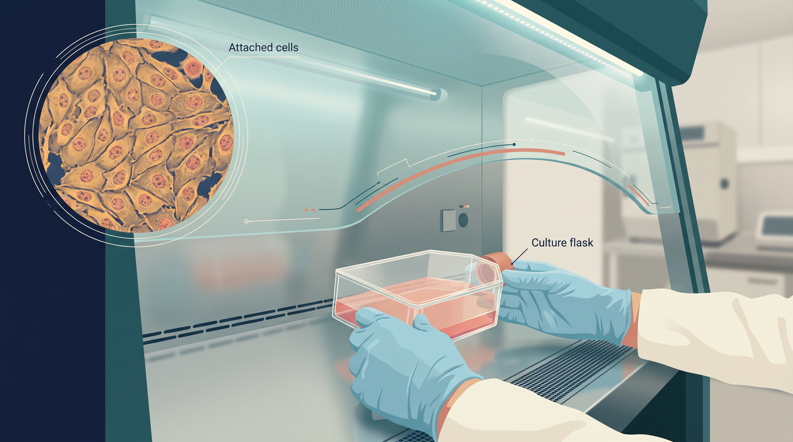
Fig. 8.0 – Animal cell culture in vitro.
Read on — every one of these questions is answered in this chapter, and you will find each answer marked at the point where it appears.
8.1 Historical Perspective
What Animal Cell Culture Means
Animal cell culture is the maintenance and multiplication of animal cells in vitro — that is, in glassware or plasticware on a laboratory bench rather than inside a living body. Cells removed from an animal will go on living and dividing outside the organism, provided they are supplied with the right nutrients and the right growth conditions.
The general process of growing cells under laboratory conditions is called cell culture. It is carried out in vitro, as opposed to in vivo, which means within the living organism. The two terms are worth fixing firmly in your mind, because almost every statement in this chapter depends on the difference between a cell inside the body and the same cell in a flask, as shown in Fig. 8.1.
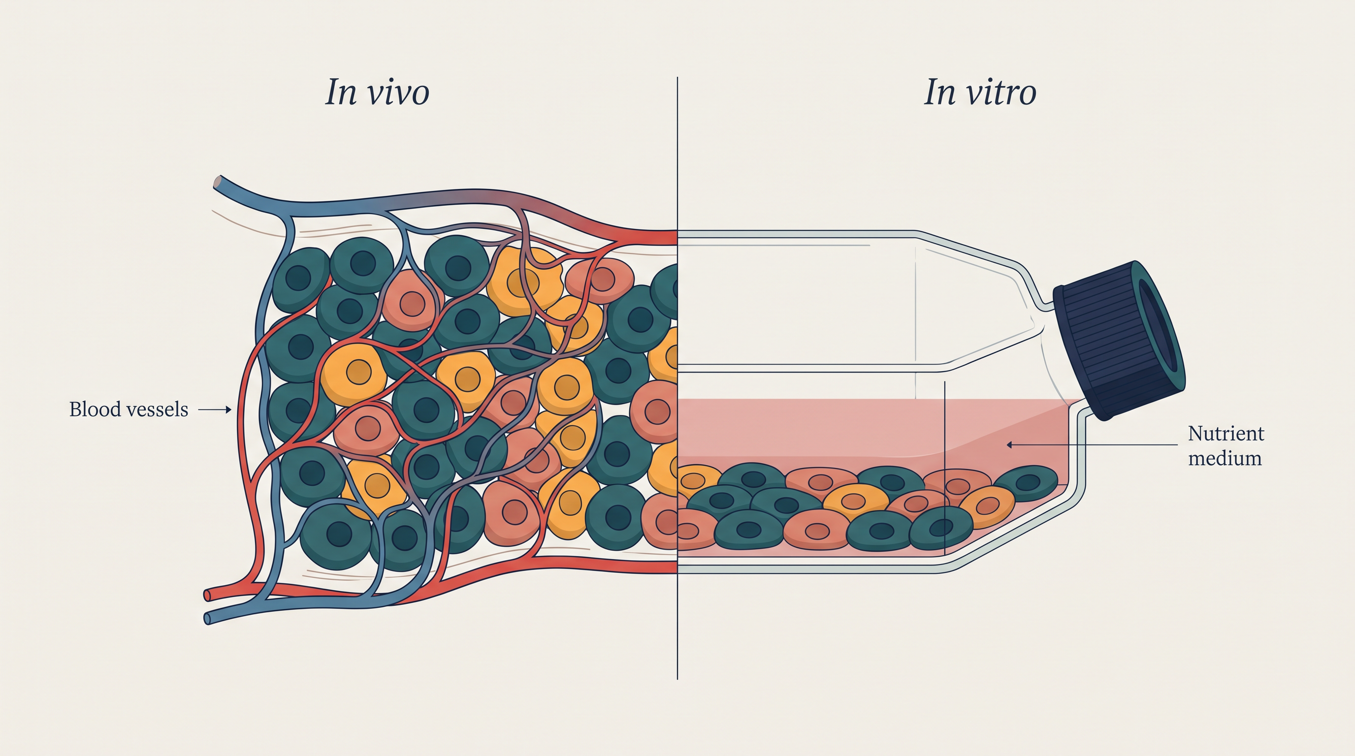
Fig. 8.1 – In vivo versus in vitro cell environments.
Animal cell culture therefore deals with three linked activities: the isolation of cells from animal tissue, the surgical removal of tissues or organs from an animal, and the placement of those cells into a suitable environment — the medium — so that they survive and proliferate. The whole sequence is summarised in Fig. 8.2.

Fig. 8.2 – Basic sequence of animal cell culture.
Clones and the Slow Pace of Animal Cells
When a whole population of cells is derived from one single parent cell, the population is called a clone. Because every member of that population has arisen by repeated division of one ancestor, all the cells in a clonal population are genetically identical to one another.
Animal cells grow slowly. A typical animal cell needs about 18 to 24 hours to complete one division. This slowness is not merely inconvenient — it is the single biggest practical danger in a cell culture laboratory.
Consider what happens if even a few bacteria enter a flask. Bacteria divide in tens of minutes, animal cells in tens of hours. Within a day or two, a small number of bacteria will have outgrown and overwhelmed a far larger population of animal cells, using up the nutrients and altering the pH of the medium. This is exactly why animal cell culture is described as highly vulnerable to contamination, and why every operation described later in this chapter is carried out aseptically.
What the Cells Need
For cells to grow optimally outside the body, four essential requirements must be met. The temperature must be regulated. A proper substrate must be available for the cells to attach to, sitting in an appropriate growth medium. And an incubator must maintain the correct pH and a defined osmolality — osmolality being a measure of the total concentration of dissolved particles in the medium, which decides whether water tends to move into the cell or out of it. Each of these requirements is dealt with in detail in Section 8.3.
Once these conditions can be held steady, a great deal becomes possible. Cell culture allows scientists to study how cell proliferation is regulated, how cells differentiate, and how they form particular products — all under controlled conditions where one factor at a time can be varied. This is why the technique has gained such a dominant position in many branches of life science research.
Today cell culture has emerged as a working tool in molecular genetics, in immunological analysis, in surgery, in bioengineering and in the pharmaceutical industry. Very little of modern medical biotechnology would exist without it.
The Rise of a Routine Technique
Animal cell culture became a routine laboratory technique in the 1950s. The turning point came when George Gey established the first human cell line, called HeLa, from the cervical cancer of a patient named Henrietta Lacks. A cell line is a cell population that can be maintained and grown on in the laboratory over many transfers; HeLa was the first human one, and it led to several important discoveries in the medical sciences.
The need for cell culture — especially on a large scale — became truly apparent with the need for viral vaccines. Viruses cannot be grown in a nutrient broth the way bacteria can, because a virus multiplies only inside a living host cell. Large quantities of cultured animal cells are therefore the only practical factory for producing large quantities of virus.
What Cell Culture Made Possible
Since the 1950s, cell culture technologies have spread into many areas of work. They are used to assess the efficacy and the toxicity of new drugs — that is, whether a candidate drug works and whether it harms cells — and they are used in the manufacture of vaccines and of biopharmaceuticals, which are medicines produced by living cells rather than by chemical synthesis.
As the technology developed, a wide variety of culture media were designed. The culture medium is not merely food. It supports cell survival and proliferation, and it also supports normal cellular functions, so that the cultured cell continues to behave in a way that resembles its behaviour in the body.
An important step forward came with the discovery of growth factors — proteins that signal a cell to grow and divide. Nerve growth factor, epidermal growth factor, insulin-like growth factor, fibroblast growth factor (FGF), platelet-derived growth factor, and transforming growth factor (TGF) were discovered one after another, and adding them to the medium led to increased cellular proliferation.
This discovery had a further consequence. If the specific growth-promoting molecules could be identified and added deliberately, then the crude biological fluid that had been supplying them — serum — might no longer be indispensable. Accordingly, in 1976, the development of serum-free media was accelerated. You will meet serum-free media again in Section 8.2.
| Name | Year | Breakthrough |
|---|---|---|
| Sydney Ringer | 1882 | Balanced salt solution with a composition similar to that of body fluids and kept frog hearts after dissection and removal from the body |
| Roux | 1885 | Medullary plate of chick embryo in warm saline |
| Jolly | 1903 | In vitro cell survival and cell division of salamander leucocytes |
| Ross Harrison | 1907 | Published experiments showing frog embryo nerve fibre growth in vitro |
| Lewis and Lewis | 1911 | Cultured connective tissue cells for extended periods and showed heart muscle tissue contractility over 2–3 months; the first liquid media consisted of sea water, serum, embryo extracts, salts and peptides |
| Alexis Carrel | 1912 | Aseptic techniques to tissue culture. Use of trypsin, embryo extracts/animal serum |
| Rous and Jones | 1913 | Use of antibiotics: penicillin/streptomycin |
| — | 1916 | Use of laminar air-flow cabinets |
| — | 1940 | Trypsinization was used to produce homogenous cell types; tissue culture media |
| Katherine Sanford, et al. | 1940s–50s | Were the first to clone mouse L-cells. Tumor cells could give rise to continuous cell lines. Used non-malignant rodent cell culture to study the effects of carcinogens/viruses |
| Margaret Gey and George Gey | 1948 | Observed contact inhibition among fibroblasts — the beginning of quantitative cell culture experimentation |
| Abercrombie and Heaysma | 1952 | Polio virus in human E-cells; production of polio vaccine |
| Enders, et al. | 1954 | Human cell lines for the production of vaccines — human and veterinary |
| Hayflick and Moorhead | 1955 | Described the finite lifespan of normal human diploid cells |
| — | 1961 | Published the methods for maintaining differentiated cells (of tumor origin) |
| Harry Eagle | 1962 | Developed defined media |
| — | 1970 | Described attachment factors and feeder layers |
| Buonassisi, et al. | 1962 | Studied the differentiation of normal myoblasts in vitro |
| Littlefield | 1964 | HAT selection |
| David Yaffe | 1968 | Human foetal lung fibroblasts |
| Kohler and Milstein | 1975 | First hybridoma capable of screening a monoclonal antibody |
Read down that table and a pattern appears. The early entries are about keeping tissue alive at all; the middle entries are about keeping it clean — aseptic technique, antibiotics, laminar air-flow cabinets; and the later entries are about control — defined media, selection systems, and cells designed to make a specific product. Each stage had to be solved before the next became possible.
8.2 Culture Media
The most significant and critical stage in cell culture is the selection of a suitable growth medium in which the cells will be cultured in vitro. Everything else — the incubator, the flask, the technique — can be perfect, and the culture will still fail if the medium is wrong for those particular cells.
There is no single best medium. The appropriate choice depends on two things: the kind of cells to be cultured, and what the culture is required to do. A culture set up simply to increase cell numbers has different requirements from one meant to make the cells differentiate, and both differ again from a culture intended to produce a desired product such as a pharmaceutical compound.
What a Typical Medium Contains
A typical culture medium contains a complement of vitamins, amino acids, glucose, inorganic salts, serum — which acts as the source of growth factors — and hormones, as summarised in Fig. 8.3. Amino acids are the raw material for protein synthesis, glucose is the energy source, and vitamins act mainly as precursors of coenzymes.
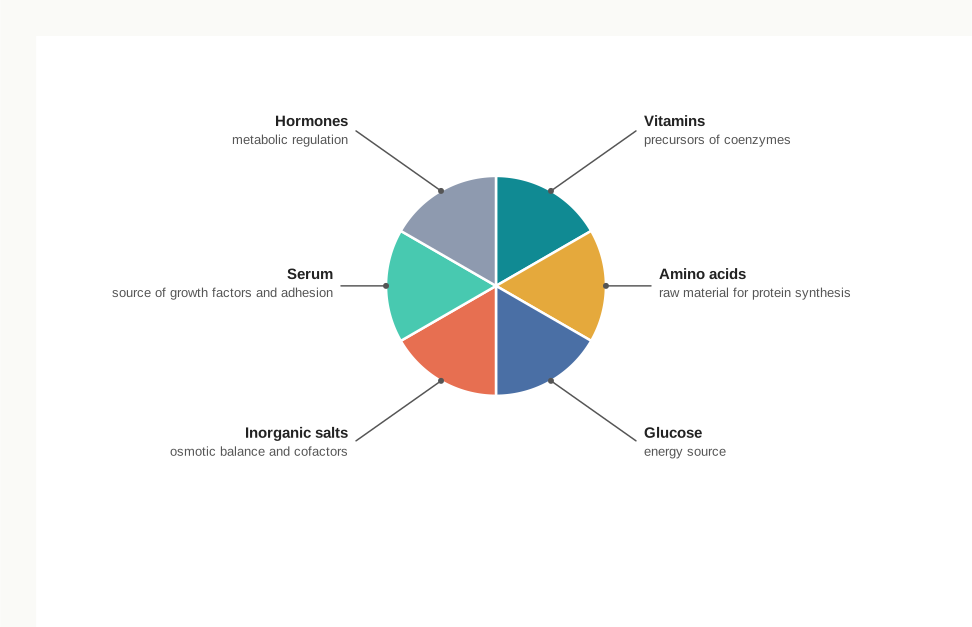
Fig. 8.3 – Composition of a typical culture medium.
The medium has a second job besides nutrition. It also helps to maintain the pH and the osmolality of the fluid surrounding the cells, holding both close to the values the cells would experience inside the body.
Natural Media and Synthetic Media
All media fall into two broad categories. Natural media consist of natural biological substances — plasma, serum and tissue extract. Artificial or synthetic media are composed of a basal medium together with supplements such as hormones, growth factors and serum. The full classification, with the sub-types of each, is set out in Fig. 8.4.
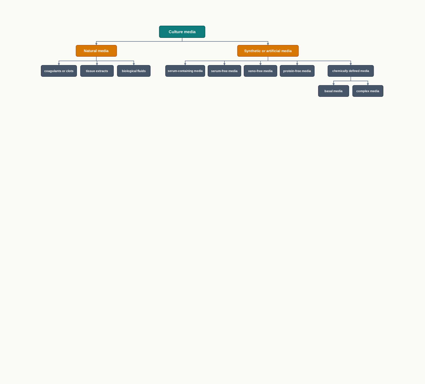
Fig. 8.4 – Classification of culture media.
The distinction matters because of reproducibility. In a natural medium, the exact composition of the biological fluid is not known and varies from one batch to the next; in a synthetic medium, the scientist decides what goes in.
| Category | Definition | Type | Description | Disadvantages/Advantages |
|---|---|---|---|---|
| Natural media | Consisting of natural biological substances, such as plasma, serum, and embryo extract | Coagulant or clots | Plasma separated from heparinised blood, serum and fibrinogen | The greatest disadvantage of natural media is poor reproducibility and reduced uniformity because the exact composition of these natural media are not known |
| Tissue extracts | Extracts of chicken embryos, liver, spleen, and bone marrow | |||
| Biological fluids | Plasma, serum, lymph, amniotic fluid, and pleural fluid | |||
| Synthetic media or Artificial media | Composed of a basal medium and supplements, such as serum, growth factors, and hormones | Serum-containing media | Human, bovine, equine, or other serum is used as a supplement | The quality of serum varies from batch to batch and deteriorates within one year. Therefore, every batch of serum needs fresh testing |
| Serum-free media | Crude protein fractions, such as bovine serum albumin or α- or β-globulin, are used as supplements | It has the ability to make a medium selective for a particular cell type, since each cell type appears to require a different recipe | ||
| Xeno-free media | Human-source components, such as human serum albumin, are used as supplements but animal components are not allowed as supplements | |||
| Protein-free media | Undefined components, such as peptide fractions (protein hydrolysates), are used as supplements | |||
| Chemically defined media — Basal media | Phosphate buffered saline (PBS), Dulbecco’s phosphate buffered saline (DPBS), Hank’s balanced salt solution (HBSS), Earle’s balanced salt solution (EBSS) | Balanced salt solution (BSS) is composed of inorganic salts that maintain the physiological pH and osmotic pressure. The physiological role played by the inorganic ions is to maintain the membrane potential | ||
| Chemically defined media — Complex media | Eagle’s Minimum Essential Medium (EMEM); Dulbecco’s Minimal Essential Medium; Roswell Park Memorial Institute (RPMI-1640); Ham’s Nutrient Mixtures (Ham’s F-12) | They also work as cofactors in enzyme reaction and in cell attachment. Na⁺, K⁺, Mg²⁺, Ca²⁺, Cl⁻, SO₄²⁻, PO₄³⁻ and HCO₃⁻ are the inorganic molecules involved. To maintain the osmolality, concentration of NaCl is generally adjusted |
Notice the logic running down the right-hand column of that table. The inorganic ions listed for the complex media are not there only to balance osmotic pressure. They also serve as cofactors in enzyme reactions and take part in cell attachment, and it is the inorganic ions that maintain the membrane potential of the cell. Where the osmolality of the finished medium needs correcting, the concentration of NaCl is the quantity that is generally adjusted.
Media Supplements
As you have seen, the culture medium contains a combination of amino acids, salts, glucose and vitamins, supplemented with other nutrients. The exact requirement for each of these components depends on the cell line that is to be cultured. This is why there is no universal recipe, and why an extensive number of media formulations is commercially available.
Some additional components are required which are present neither in the basal medium nor in serum — hormones, growth factors and signalling substances. These are added because they help in the proliferation of the cells and in maintaining normal cell metabolism. Without them, a cell may survive but will not behave normally.
Serum — The Most Important Supplement
Serum is one of the most important components of cell culture media. It is the pale fluid that remains after blood has clotted and the clot and cells have been removed, and it is remarkably rich in the substances a cell needs.
Serum is considered a good source of amino acids, proteins, vitamins, carbohydrates, lipids, hormones and growth factors. It also provides several binding proteins, such as albumin and transferrin, which can carry other molecules into the cell — transferrin, for example, carries iron, which the cell cannot easily take up on its own.
Serum does one further thing that no simple nutrient mixture can do. It supplies adhesion factors, which help the cells to stick to the substratum before they begin to divide. Many animal cells will not divide at all unless they have first attached to a surface, so this function is as essential as any nutrient. Fetal bovine serum is the serum most commonly used to support the growth of cells in culture.
Why Serum Is Also a Problem
For all its usefulness, serum brings four serious disadvantages, and every one of them follows from the fact that it is an undefined natural product.
- Serum contains an insufficient amount of cell-specific growth factors, and so it cannot be used as a stand-alone medium for culture.
- It may also contain cytotoxic compounds and some growth-inhibiting factors, which will cause inhibition of the growth and proliferation of the cultured cells.
- Serum may carry a high risk of contaminants, such as various viruses, fungi and mycoplasma. Mycoplasma are especially troublesome because they are very small bacteria that pass through ordinary filters and cause no visible cloudiness in the medium.
- The serum in the culture medium may interfere with the purification and isolation of cell culture products, including pharmaceutical compounds. Serum is itself a mixture of proteins, so separating a wanted protein from it is difficult, and additional approaches have to be applied for the isolation of cell culture products.
Taken together, these four problems explain the direction the field has taken. The more valuable the product being made, the stronger the reason to move away from serum-containing media towards serum-free, xeno-free, protein-free and chemically defined formulations.
Antibiotics in the Medium
Antibiotics are not essential for the growth of cells — a perfectly aseptic culture needs none. Even so, the use of antibiotics such as penicillin and streptomycin is common in culture medium, to control the growth of contaminants such as bacteria and fungus.
Antibiotics should be thought of as a second line of defence, not a substitute for sterile technique. They cannot rescue a badly handled culture, and constant use can allow resistant organisms and hidden mycoplasma infections to persist unnoticed.
8.3 Physical Environment for Culturing Animal Cells
Culturing animal cells under in vitro conditions means recreating, in a flask, the conditions the cell would have enjoyed inside the body. This involves simulating suitable micro-environments — nutritional, physical and hormonal — so that the cells grow optimally.
In practice this means controlling six things: the temperature, the osmolality, the pH, the gaseous requirements, a supporting surface for the cells to grow on, and the protection of the cells from physical, chemical and mechanical stresses. Section 8.2 dealt with the nutritional side of this list. The physical side is taken up here, and the supporting surface is discussed further in Section 8.5.
Cells are more delicate than they look. Rough pipetting, a sudden temperature change, or a badly chosen detergent residue on glassware will damage a monolayer of cells as effectively as any missing nutrient.
Temperature
CO₂ incubators are used to grow mammalian cells at 37°C, because this temperature simulates the body temperature of Homo sapiens. Moreover, most of the cells used in culture have been derived from warm-blooded animals, which will grow optimally at this temperature.
The choice is not arbitrary. Enzymes have an optimum temperature, and the enzymes of a mammalian cell have evolved to work best at the temperature of the body from which the cell came. Even a few degrees above 37°C begins to denature proteins; a few degrees below, and metabolism and division slow down markedly.
Osmolality
Osmolality is the total concentration of dissolved particles in a solution, and it has a substantial effect on cell growth and function. Its central role is to maintain the integrity of the cell membranes.
The reason is straightforward. An animal cell has no cell wall, so nothing resists a net movement of water across its membrane. If the outside osmotic pressure becomes lower or higher than the pressure maintained inside the cell, the cell will swell or shrink accordingly. A badly swollen cell may burst; a shrunken cell cannot function normally.
The osmolality of the medium being used is determined by the formulation of the medium. Both glucose and salt are the major donors to the osmolality of the medium, even though amino acids also contribute significantly. Generally, all commercial media are formulated so as to have a final osmolality of approximately 300 mOsmol. Osmolality can be measured by an instrument called an osmometer.
Buffering Systems
For optimum culture conditions, the regulation of pH is critical. Growing cells continuously release carbon dioxide and acidic products of metabolism into the medium, so a culture left to itself becomes steadily more acidic. A buffer is needed to absorb these changes. The regulation of pH is achieved by one of the following two buffering systems.
(A) Natural Buffering System
In a natural buffering system, gaseous CO₂ balances with the CO₂/HCO₃⁻ content of the culture medium. The concentrations of CO₂, of HCO₃⁻ and the pH are all interrelated. By increasing the external or exogenous CO₂, the pH will be reduced, making the medium acidic.
This is the chemistry of the bicarbonate buffer of blood, borrowed for use in a flask. Dissolved CO₂ combines with water to give carbonic acid, which dissociates to release hydrogen ions:
\[CO_2 + H_2O \rightleftharpoons H_2CO_3 \rightleftharpoons HCO_3^- + H^+\]
Here \(CO_2\) is carbon dioxide, \(H_2O\) is water, \(H_2CO_3\) is carbonic acid, \(HCO_3^-\) is the bicarbonate ion and \(H^+\) is the hydrogen ion whose concentration decides the pH. More CO₂ pushed into the medium means more \(H^+\), and therefore a lower pH.
The direction of that shift is shown in Fig. 8.5.
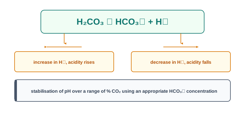
Fig. 8.5 – Bicarbonate buffering equilibrium in culture medium.
Because the buffer depends on dissolved CO₂, the gas above the medium must be controlled. Culture media with a natural buffering system need to be maintained in air with 5–10 per cent CO₂, which is usually maintained by a CO₂ incubator. If such a culture is taken out and left in ordinary room air, which contains only about 0.04 per cent CO₂, dissolved CO₂ escapes from the medium, the equilibrium shifts to the left, \(H^+\) is consumed and the medium turns alkaline.
The natural buffering system has two clear practical merits: it is low in cost and it is non-toxic to the cells.
There is also a way of reducing the dependence on the gas cylinder. The presence of pyruvate in the medium results in increased endogenous production of CO₂ by the cells themselves — “endogenous” meaning produced from within, as pyruvate is oxidised in respiration. The use of pyruvate is therefore advantageous, because it means less dependency on the exogenous supply of CO₂.
(B) Chemical Buffering System
In the chemical buffering system, a zwitter ion — a molecule carrying both a positive and a negative charge at the same time — is used in place of the bicarbonate system. The compound used is 4-(2-hydroxyethyl)-1-piperazineethanesulfonic acid, abbreviated as HEPES.
HEPES has a superior buffering capacity in the pH range 7.2–7.4, which is precisely the range in which animal cells must be held. Its great practical advantage is that with HEPES, a controlled gaseous atmosphere is not required, so cultures can be handled outside a CO₂ incubator without the pH drifting.
It has two drawbacks. HEPES is relatively expensive, and at higher concentrations it is toxic for some cell types.
Phenol Red — Watching pH by Colour
Phenol red is generally used in commercial media as a pH indicator. It allows the pH change caused by the metabolic activities of the cells during growth to be monitored effectively — and monitored simply by looking at the flask, with no instrument at all.
Because of the presence of phenol red, the medium changes its colour as the pH changes. At acidic pH levels, phenol red turns the medium yellow, while at alkaline pH levels the medium turns pink. At the optimal pH of 7.4, the medium is bright red. The structure of phenol red and its behaviour as an indicator are shown in Fig. 8.6.
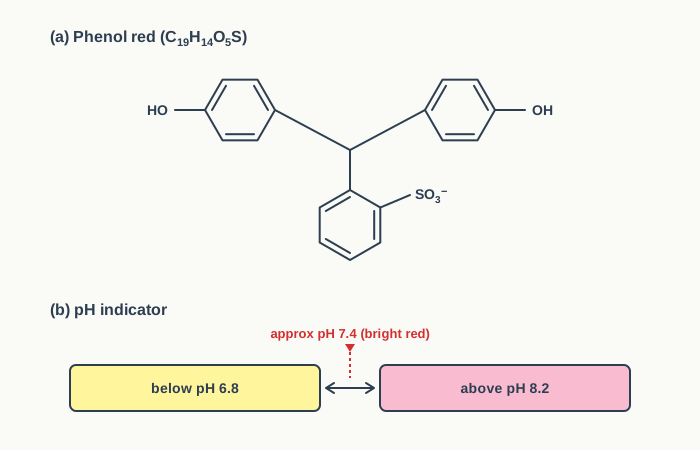
Fig. 8.6 – Phenol red structure and pH colour scale.
Read that colour scale as a working instrument. Below pH 6.8 the medium reads yellow, above pH 8.2 it reads pink, and the bright red in between is the target. The daily colour check is the fastest health report a culture can give, and it needs nothing but a pair of eyes.
The typical sequence in a healthy, busy culture is set out in Fig. 8.7.
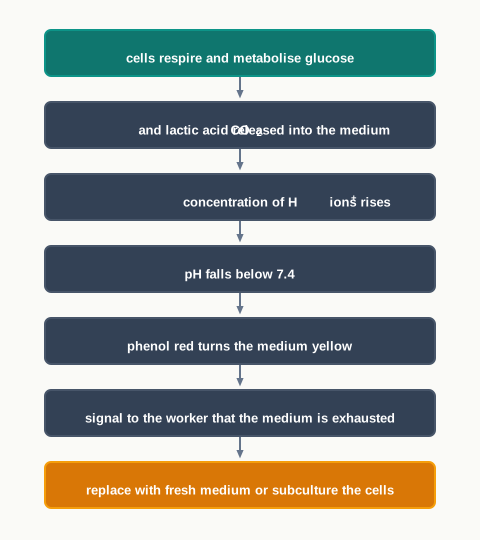
Fig. 8.7 – Sequence causing phenol red to turn yellow.
8.4 Equipment Used for Cell Culture
Culture Vessels and the Daily Check
Cultures should be maintained and examined daily. The first thing to look at is the colour of the medium, and the next is the appearance of the cells themselves — their morphology, and the density at which they are growing.
Cells are grown in culture flasks, Petri dishes or multi-well plates of several sizes and shapes, shown in Fig. 8.8. The choice depends on the scale of the work: a Petri dish for a small experiment, a flask for routine growth, and a multi-well plate when many parallel treatments must be tested side by side on the same batch of cells. A cell scraper is used to lift attached cells mechanically from the growth surface.
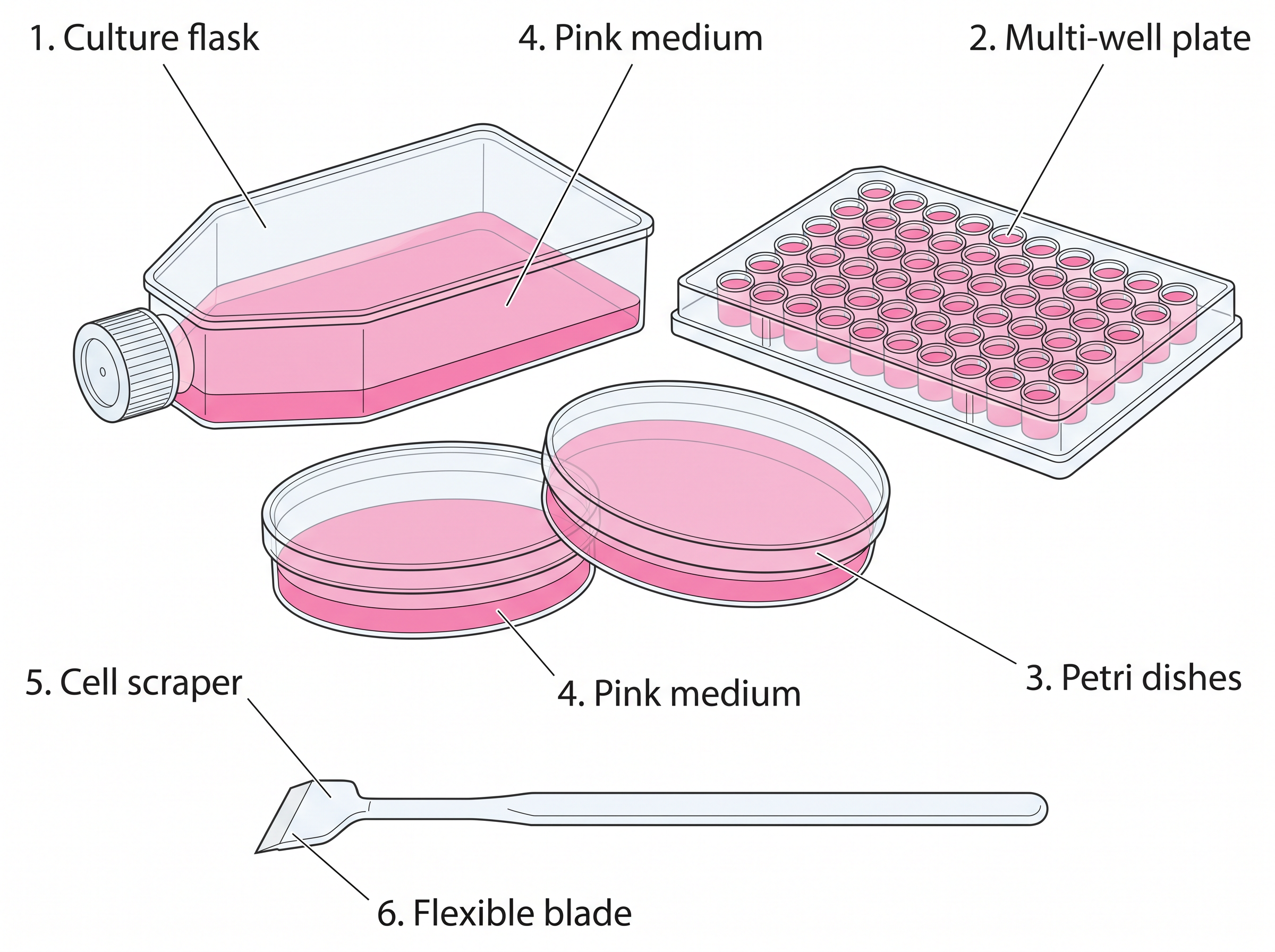
Fig. 8.8 – Culture vessels and a cell scraper.
These vessels are kept in an incubator at an appropriate temperature, humidity and gas mixture — typically 37°C, 95 per cent humidity and 5 per cent CO₂ for mammalian cells.
Culture conditions differ extensively for each cell type. This is worth taking seriously, because variations in the conditions used for any particular cell type may result in the expression of altered phenotypes — the cells stay alive, but they no longer behave, look or produce as they should. An experiment run on such cells will give data that describe the culture conditions rather than the biology under study.
An ideal animal cell culture laboratory should have equipment such as a laminar hood, a CO₂ incubator, an inverted microscope, an autoclave and a centrifuge. These are discussed below.
Laminar Flow Hood or Biosafety Cabinets
The entire work of animal tissue or cell culture should be done aseptically in the laminar hood. Given how slowly animal cells divide, this is not a precaution that can be relaxed on a busy day.
A laminar flow cabinet provides controlled filtered air, in which the levels of particulates, microbes and contamination of all kinds are regulated and kept to a minimum. This is achieved by constant air filtration through industrial-grade HEPA (High Efficiency Particulate Air) filters.
The word “laminar” is the key to the design. The filtered air is made to move in smooth, parallel layers rather than in swirls, so that any particle released in the working space is carried steadily away instead of being mixed back over the open flask.
Laminar hoods operate by drawing air under negative pressure into the top of the unit. This air initially passes through a pre-filter, which traps fungi, bacteria and dust particles. The air passed through this pre-filter is then continuously fed by fans or blowers through the HEPA filters into the enclosed cabinet, creating a sterile work area for the operator. The complete air path is traced in Fig. 8.9.
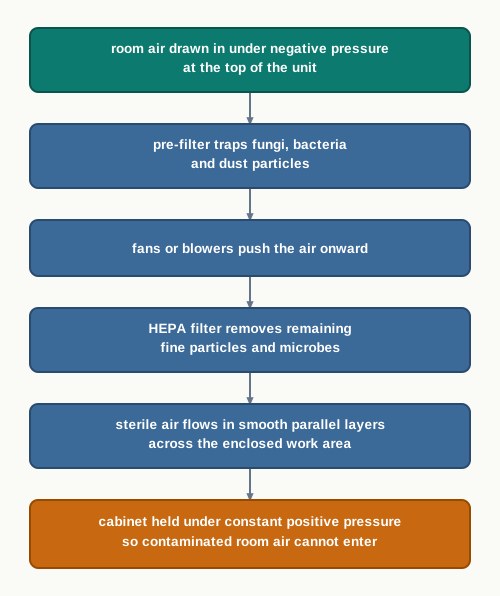
Fig. 8.9 – Air path through a laminar flow hood.
The laminar flow chamber is usually enclosed on both sides and kept under constant positive pressure. The reason for the positive pressure is important: with the pressure inside the cabinet slightly higher than the pressure of the room, air can only leak outwards, so contaminated room air is prevented from flowing in.
Vertical and Horizontal Flow
In a vertical laminar flow hood, shown in Fig. 8.10(b), the sterilised air is passed vertically downwards onto the work area — a down flow. This design is considered to be safer, because the air does not blow directly towards the person carrying out the experiments.
In the horizontal flow hood, shown in Fig. 8.10(a), air is directed towards the person doing the experiments. Hence horizontal or crossflow hoods are not popular, as they offer no protection to the operator from the potential hazards of some of the cultured cells.
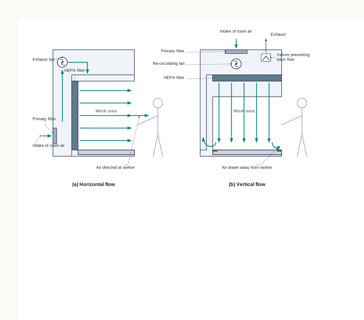
Fig. 8.10 – Horizontal and vertical laminar flow hoods.
Notice what this comparison really turns on. Both designs protect the culture equally well, because in both the air reaching the work surface has passed through a HEPA filter. They differ in whom they protect afterwards. In the horizontal design the used air leaves across the bench and onto the operator; in the vertical design it is drawn away downwards and exhausted.
CO₂ Incubators
Incubators for cell culture work depend on an external supply of CO₂, so as to maintain a fixed level of CO₂ inside the incubator. This is the gas supply on which the natural bicarbonate buffering system of the medium depends.
To maintain a sterile handling area and general cleanliness, the CO₂ cylinder should be kept outside the laboratory, and the gas should be piped through into the incubator. A heavy cylinder standing inside the culture room would be both a physical hazard and a source of dust.
The essential functions of the incubator are to maintain four things: the sterility of the chamber, a constant temperature, an atmosphere with a 5–10 per cent level of CO₂, and a high relative humidity of about 95 per cent, as shown in Fig. 8.11.
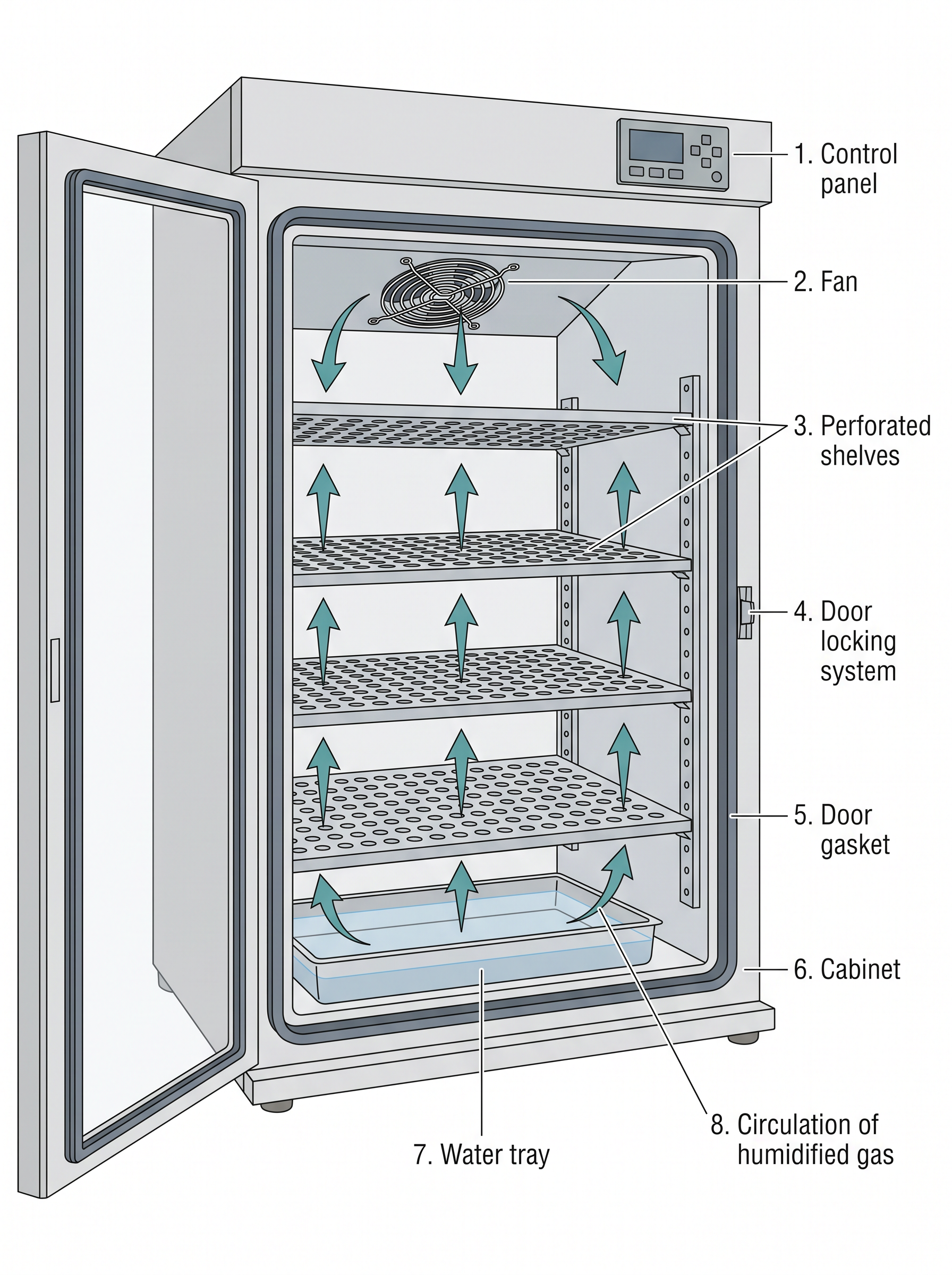
Fig. 8.11 – Sectional view of a CO₂ incubator.
The high humidity has a specific purpose. A culture flask holds only a few millilitres of medium; in dry warm air the water would evaporate steadily, concentrating the salts and driving the osmolality far above the 300 mOsmol at which the medium was formulated. Keeping the chamber near saturation prevents this.
The medium inside is further buffered with sodium bicarbonate and carbonic acid, and the pH must be strictly maintained. The incubator and the medium therefore work as one system: the gas phase set by the incubator and the bicarbonate dissolved in the medium together fix the pH the cells experience.
Inverted Microscope
A cell culture laboratory essentially requires a good quality inverted microscope with phase-contrast optics, and preferably with a photographic facility, as shown in Fig. 8.12.
In an inverted microscope the objective lenses sit below the stage and look upwards, while the light source is above. This arrangement exists for a simple physical reason: the cells grow attached to the bottom of a flask that is filled with medium and has tall walls, so an ordinary upright microscope could never bring its objective close enough to focus on them. Phase-contrast optics are needed because living, unstained cells are almost transparent, and phase contrast converts the small differences in refractive index within a cell into visible differences in brightness.
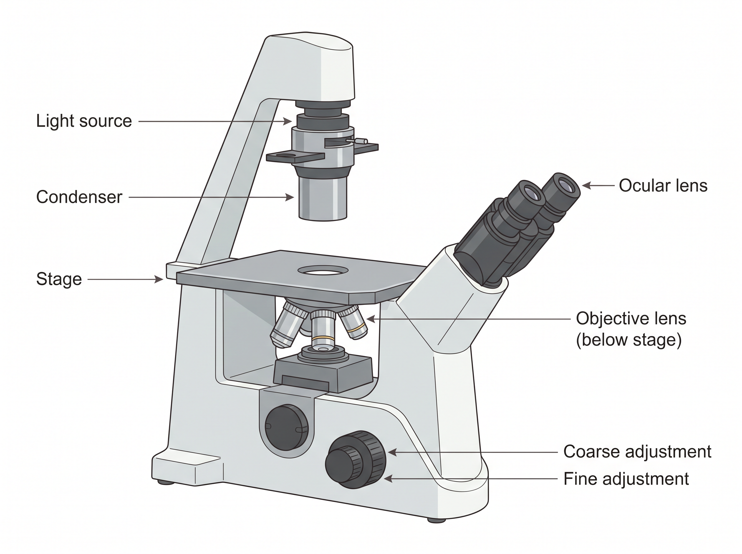
Fig. 8.12 – Parts of an inverted microscope.
A great deal can be read from the cells themselves. The morphology of the cell, its granularity, its degree of spreading, membrane blebbing, the proportion of multi-nucleate cells and the extent of vacuolation can all be monitored regularly, and together these features reveal the signs of stress in cells.
This is possible because cell morphology is sensitive to alterations in culture conditions, so problems can be detected easily and early. Early indications of a microbial contamination can also be spotted with a good phase-contrast microscope, long before the medium turns cloudy.
Regular checking of cultures under the microscope can help to avoid the loss of irreplaceable material, by allowing a problem to be noticed at an early stage. There is a second reason to check as well: experiments done on unhealthy cells may give variable or erroneous results, which is a far more dangerous outcome than an obviously failed culture, because the data look usable.
Choosing the Optics
When choosing a microscope, select the long or extra-long working distance condenser, so that flasks and even roller bottles can be viewed. A short working distance condenser simply cannot reach across a tall vessel.
Normally a 20x magnification objective is sufficient. Their depth of field is often too low to obtain a sharp image of all but the most flattened cells, which is why higher powers are less useful for routine checking than students often expect. A good, low-power, wide-field objective is extremely useful for scanning culture colonies, since it shows a large area at once and makes uneven growth or a patch of contamination easy to spot.
Autoclave and Centrifuge
Two further pieces of equipment complete the basic laboratory.
The autoclave sterilises glassware, instruments, and heat-stable solutions before they are taken into the hood. Media containing serum, vitamins and other heat-sensitive components cannot be autoclaved and are sterilised by filtration instead.
The centrifuge is used to spin cell suspensions so that the cells collect as a pellet at the bottom of the tube and the old medium, containing enzymes and waste, can be poured off from above. You will meet this step again in Section 8.5, where cells are collected and resuspended in fresh medium during subculture.
8.5 Types of Animal Cell Cultures and Cell Lines
The animal cell cultures and cell lines are broadly classified as primary and secondary cell cultures and cell lines. The distinction rests on a single question: has the culture been transferred to a fresh vessel since it was taken from the animal, or not?
(i) Primary Cell Cultures
A primary cell culture is obtained directly from the host tissue. The cells dissociated from the parental or host tissues are grown in a suitable culture medium in a container, and the culture so obtained is called a primary cell culture.
This type of culture contains mostly heterogeneous cells — that is, a mixture of the different cell types that were present together in the original tissue — and these cells are similar to their parents. Such cultures are called primary cell lines. The word “heterogeneous” is worth holding on to, because it is the chief scientific limitation of a primary culture: what you observe is the average behaviour of a mixed population.
Their great advantage is the other side of the same coin. Because these cells have not yet been altered by repeated transfer, they still closely resemble the tissue they came from, which makes them the most realistic material available for study.
Preparing a Primary Culture
For the preparation of primary cultures and cell lines, a tissue is first dissected under sterile conditions. This is followed by disaggregation — the separation of the tissue into small pieces or into individual cells — which can be done mechanically or enzymatically.
In the mechanical route, the tissue can simply be chopped and the pieces attached to a dish. Cells will now grow out from the fragment and may be used directly for culture. The fragment of tissue itself is called an explant, and it may be transferred to a fresh dish so that the same piece yields cells again.
In the enzymatic route, primary cultures are made by disaggregating the tissue with the help of enzymes, such as trypsin or collagenase. These are chosen for a specific structural reason. Collagenase digests the collagen of the extracellular matrix that holds the tissue together, while trypsin, a protease, cleaves the protein links binding one cell to another and to the matrix. The resulting cell suspension is then allowed to adhere and spread out on the substrate.
Whichever route is used, the cells that emerge may grow as adherent cultures, attached to the surface, or may remain in suspension in the medium. Both possibilities are described later in this section. The two routes are compared in Fig. 8.13.
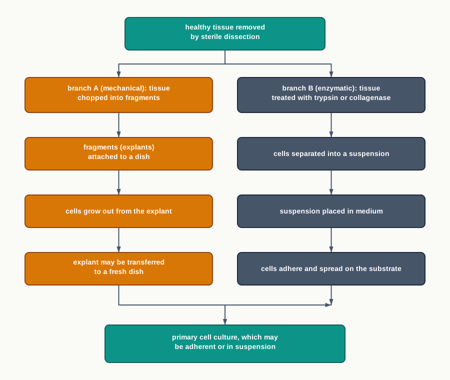
Fig. 8.13 – Preparation of a primary culture.
(ii) Secondary Cell Culture
When a primary culture is sub-cultured or passaged, the resulting culture is called a secondary cell culture. During sub-culturing, a portion of the cells is transferred to a new vessel having fresh growth medium, for the continual growth of the cells.
Sub-culturing of primary cells leads to the generation of secondary cell lines. Something interesting happens in the process. During the passage, the cells with the highest growth capacity come to predominate, and this leads to genotypic and phenotypic uniformity in the population — the mixed primary population gradually becomes a population of one dominant type.
This uniformity is both the strength and the weakness of a secondary cell line. It makes results reproducible, because every flask contains essentially the same kind of cell. But as the cells are sub-cultured at regular intervals, they may become different from the original cell of the tissue, so what is being studied slowly drifts away from what was taken from the animal.
On the basis of the culture life span, cell lines are categorised into two types.
(a) Finite Cell Lines
The cell lines with a limited number of cell divisions and having a limited life span are called finite cell lines. As the cells pass several times, they lose their ability to proliferate — a state described as senescence. Cell lines derived from primary cultures of normal cells are finite cell lines.
(b) Continuous Cell Lines
Some cells of a finite cell line undergo transformation and resume the ability to divide indefinitely, thereby becoming a continuous cell line. This type of transformation or mutation can occur spontaneously, or it can be chemically or virally induced.
Cell cultures prepared in this way can be sub-cultured and grown indefinitely as permanent cell lines, and they are immortal and tumorigenic — “tumorigenic” meaning capable of producing tumours when introduced into a suitable animal. The HeLa line you met in Section 8.1 is the classic example of such a line.
Continuous cell lines behave differently in the flask as well. They are less adherent and can also grow in suspension. They become fast growing, are less demanding for nutrients, and are able to grow up to a higher cell density than finite cell lines.
Every one of these properties makes them convenient for industrial production — and every one of them is also a warning. A cell that divides without limit, ignores its neighbours and needs little encouragement to grow is behaving, in these respects, like a cancer cell.
Adherent Cells
Adherent cells are anchorage dependent. They proliferate as a monolayer, and they require attachment to a solid or semi-solid substrate before they can proliferate at all.
These cells adhere to the culture vessel with the help of the extracellular matrix. They are usually derived from tissues that are immobile and present in the network of connective tissue — fibroblasts and epithelial cell types are the standard examples.
As growth continues, the bottom of the culture vessel becomes covered with a continuous layer of cells one cell in thickness, and such cultures are known as monolayer cultures. A majority of continuous cell lines grow as monolayers. Because they form single layers, such cells can be transferred directly to a coverslip to be examined under the microscope, which makes them convenient for microscopy and staining work. The difference from suspension growth is shown in Fig. 8.14.
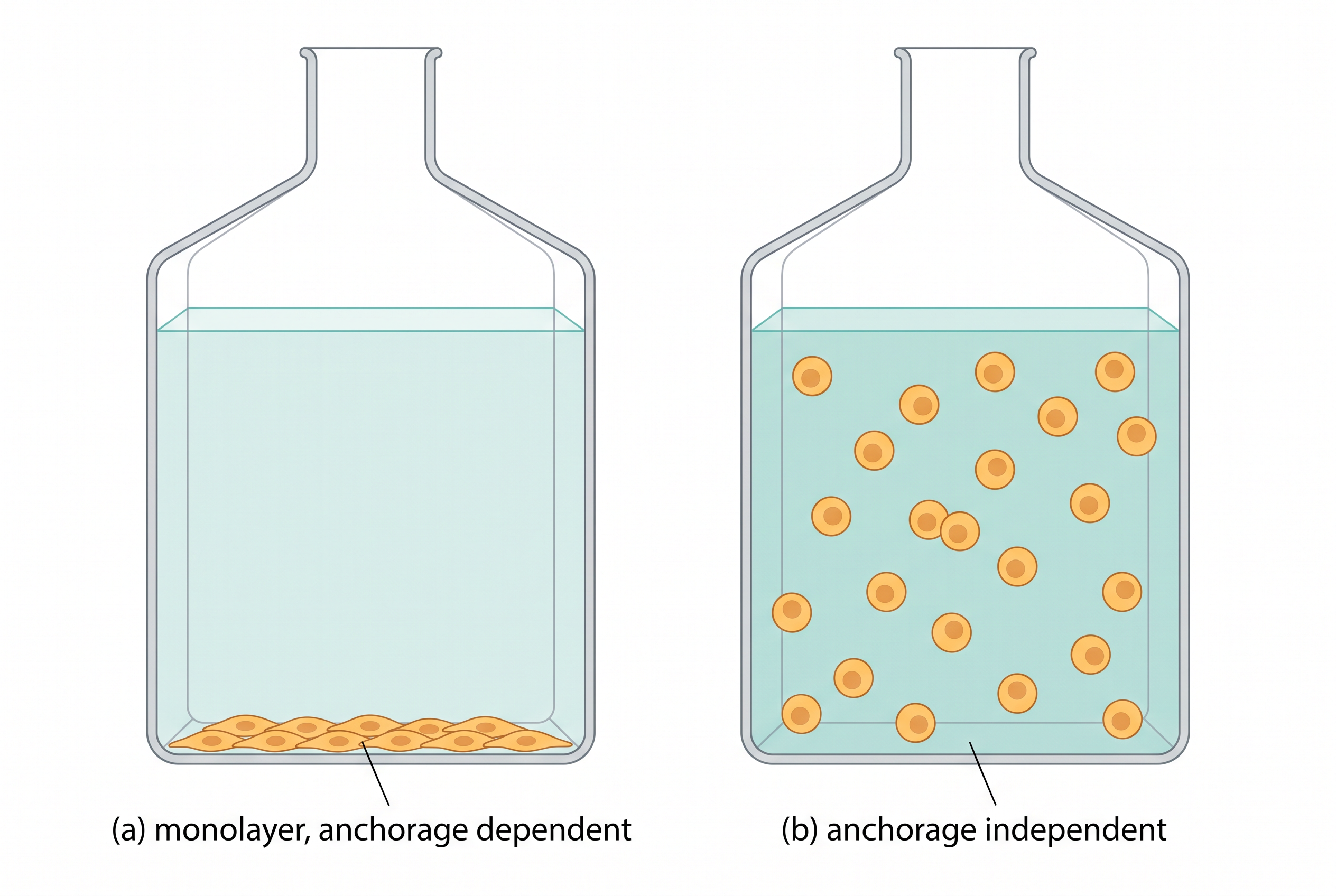
Fig. 8.14 – (a) Adherent cells and (b) suspension cells.
Suspension Cells
Suspension cells, also called anchorage-independent or non-adherent cells, are not attached to the surface of the culture vessels and float freely in the culture medium.
Haematopoietic stem cells — derived from blood, spleen and bone marrow — and tumour cells are the examples of suspension cells. The pattern makes biological sense: cells that circulate freely in the body have no need of a fixed anchorage in a flask either.
These cells grow faster, as they do not require frequent replacement of the culture medium, and they are homogeneous in nature, since every cell in the vessel meets the same conditions rather than competing for a place on a surface.
Subculture
After the isolation of cells and their subsequent culture, the cells proliferate under appropriate conditions and adhere to the available substrate until they reach confluence — the stage at which the growth surface is fully covered.
After a few days the cells form a monolayer and then overgrow, becoming crowded. This crowding is unfavourable to their growth and leads to cell death: nutrients run short, acidic waste accumulates, and there is no space left in which to divide.
At this point the cells need sub-culturing or passaging. This is the process of using an enzyme, for example trypsin, to enable the cells to detach from the old medium. The enzyme is then removed and the cells are transferred to a fresh flask after centrifugation and the addition of fresh medium.
The procedure is commonly termed splitting, because the cells of one flask are equally split into two flasks for culturing. This is also why a subculture both rescues the crowded cells and expands the stock.
A passage number refers specifically to the number of times a cell line has been sub-cultured. It is recorded carefully, because the properties of a line drift with repeated passage, and results obtained at a low passage number may not match those obtained at a high one.
For the passaging of adherent cells, all the cells are first detached or dissociated from the surface of the culture vessel. This can be done by enzymatic activity, using trypsin or trypsin with EDTA, or by mechanical means such as a cell scraper, shaking or vigorous pipetting. After that they are transferred into fresh medium. The complete sequence is shown in Fig. 8.15 and set out step by step in Fig. 8.16.
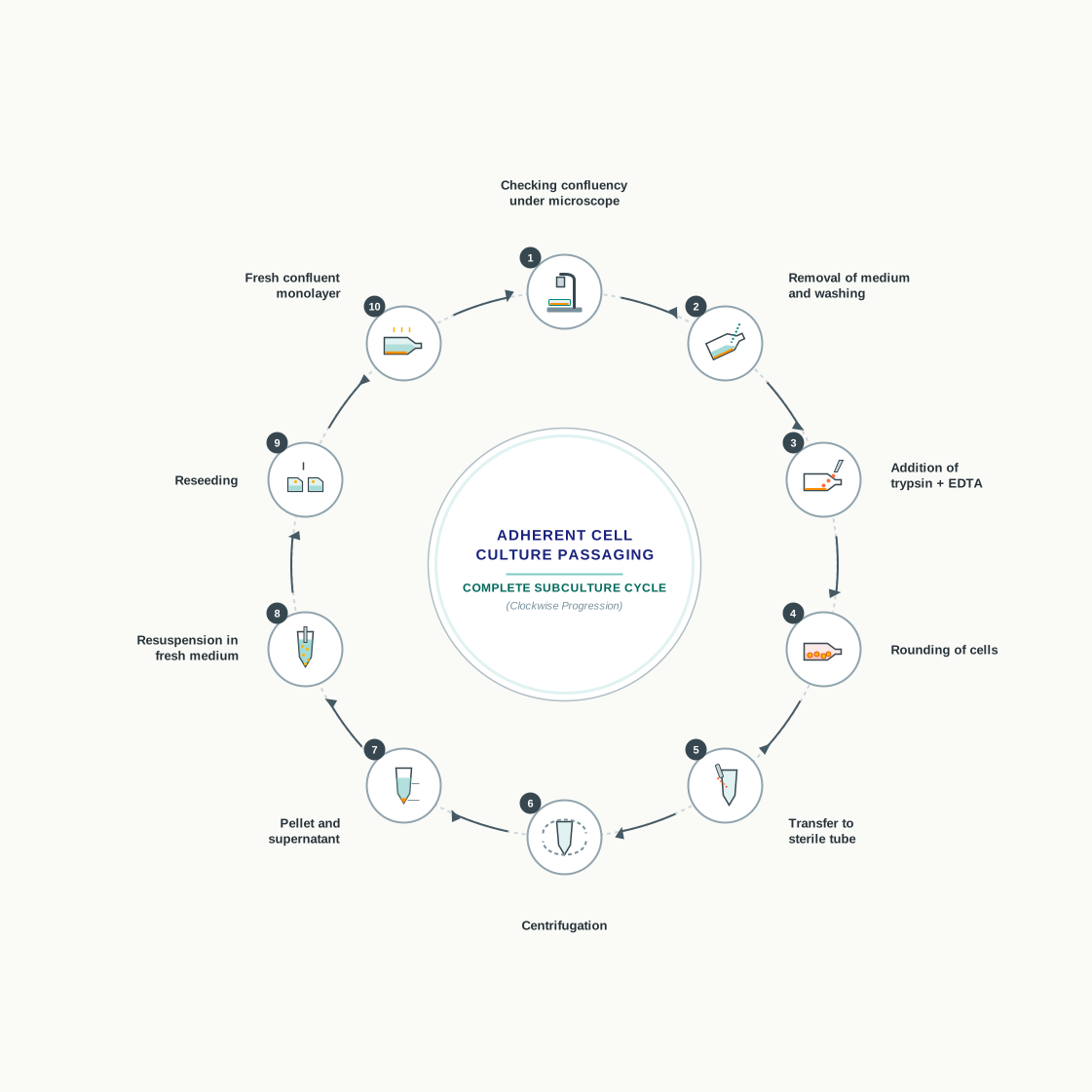
Fig. 8.15 – Steps involved in subculture or passage.
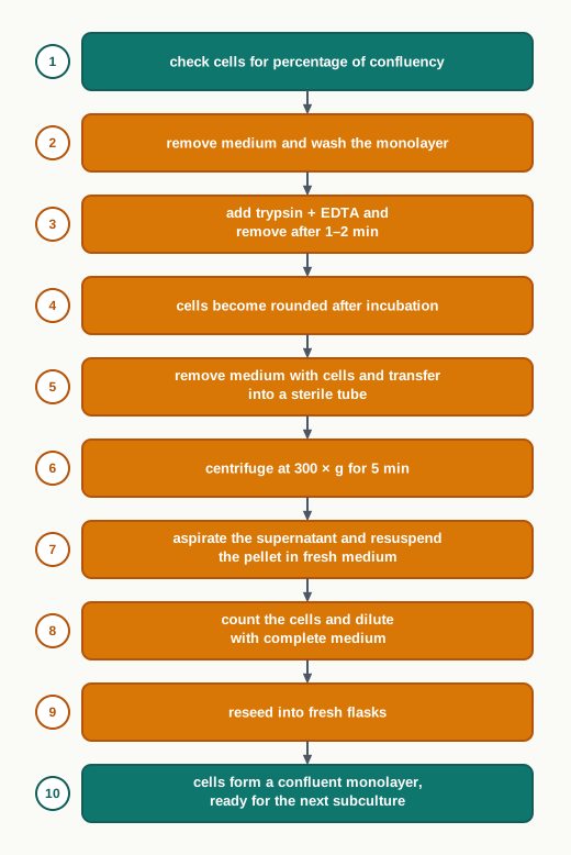
Fig. 8.16 – Steps in subculture or passage.
Two details in that sequence deserve attention. The monolayer is washed before trypsin is added because serum in the old medium contains inhibitors that would block the trypsin. And the trypsin is removed after only 1–2 minutes because trypsin, left too long, will digest the cells’ own surface proteins and damage them; the enzyme is meant to loosen the cells, not to dissolve them. EDTA is included because it chelates — that is, binds and removes — the calcium and magnesium ions that the cell adhesion molecules need, so the cells release their grip more readily.
Cryopreservation
A laboratory cannot keep every cell line growing all the time. Cells are therefore stored frozen, and revived when needed. Very low temperatures, of –180 to –196°C, are required to store cells.
Liquid nitrogen is used for freezing cells at these low temperatures, because the formation of ice crystals is reduced below –130°C. This single fact explains the whole design of the procedure.
Freezing can cause cell mortality through four kinds of damage: the damage caused by the formation of ice crystals, alterations in electrolyte concentration, dehydration, and pH changes. Ice crystals are the most destructive of these, because growing crystals physically pierce membranes from within.
To nullify the effects of freezing, cryoprotective agents are added — either glycerol or dimethyl sulfoxide (DMSO). These substances enter the cell, lower the freezing point and reduce the size of the ice crystals that form. Typically, the freezing medium contains 90 per cent serum and 10 per cent DMSO.
Freezing and Thawing Correctly
The condition of the cells matters. Preferably, healthy cells growing in the log phase — the phase of active, exponential division — should be used, and the medium should be replaced 24 hours before freezing so that the cells enter storage well fed and in fresh medium.
The cells should be cooled slowly from room temperature to –80°C. Slow cooling is deliberate: it allows the water to move out of the cells before they freeze, so that most of the ice forms outside the cells rather than inside them.
Thawing follows the opposite rule. Frozen cells should be thawed as rapidly as possible, by placing the vial containing them into a 37°C water bath with moderate shaking. Rapid warming is used to minimise ice and crystal formation, which may damage the cells — during slow warming, small ice crystals grow into large damaging ones. After thawing, the cells can be transferred directly into a tissue culture vessel containing suitable medium for further growth.
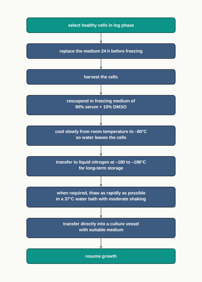
Fig. 8.17 – Cryopreservation and revival.
8.6 Cell Viability Determination
The measurement of cell viability — that is, the determination of which cells are living — plays a vital role in cell culture. A flask may look full and yet contain a large proportion of dead cells, and the difference cannot be judged by eye.
Cell viability measurements can be applied to evaluate the live status of cultured cells. This is required to determine the effectiveness of a pesticide or an insecticide, to evaluate the damage caused by toxins, and to evaluate the potential of a drug.
Notice the logic that runs through these three uses. In each case something is added to the cells and the question asked afterwards is the same: how many cells are still alive? A pesticide that kills more insect cells is more effective; a toxin that kills more cells is more damaging; an anticancer drug that kills more tumour cells at a lower dose is more promising. Viability is the measured outcome in all of them.
Usually, the cell viability assay or test should be performed at regular intervals to check whether the cells are alive or dead. There are two types of viability assay.
(a) Dye Exclusion Viability Assays
In this process, a dye or stain is used which enters the cell and usually intercalates with the DNA in the nucleus — that is, it slips in between the stacked base pairs of the DNA and stays there.
The internalisation of the dye into the cell indicates a loss of cell membrane integrity, and this loss is what causes cell death. In other words, live cells exclude the dye, while dead cells allow the dye to enter. It is from this behaviour that the method takes its name: the dye-exclusion assay.
The principle rests on one property of a healthy cell. An intact plasma membrane is selective, and these dyes are not among the substances it will let through. Once the membrane is damaged, that selectivity is gone and the dye floods in. A stained cell is therefore a cell whose membrane has failed.
Some examples of these dyes or stains are trypan blue, shown in Fig. 8.18, propidium iodide, 7-aminoactinomycin D (7-AAD) and acridine orange.
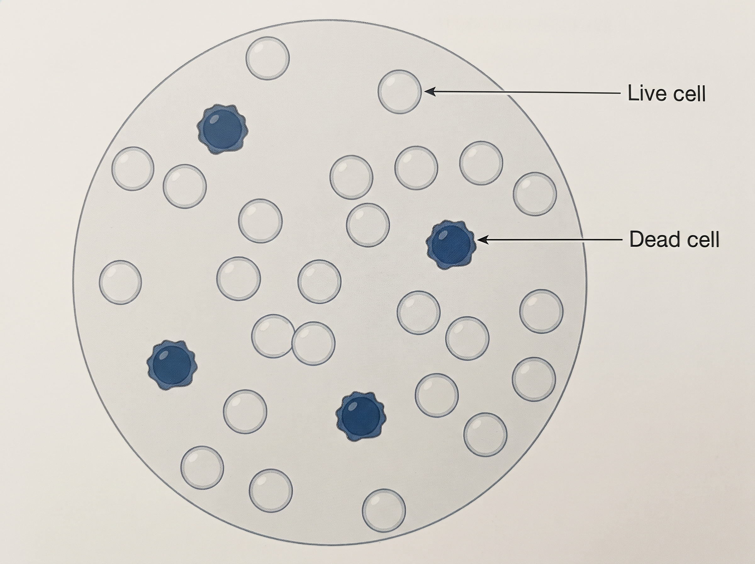
Fig. 8.18 – Trypan blue exclusion assay.
Read Fig. 8.18 the way a worker at the microscope would. The dark blue cells have taken up the trypan blue, so their membranes have failed and they are dead. The clear, unstained cells have kept the dye out and are alive. Counting both classes in a ruled counting chamber, called a haemocytometer, gives the viability of the culture:
\[\\text{Percentage viability} = \\frac{\\text{Number of unstained (live) cells}}{\\text{Total number of cells counted}} \\times 100\]
Here the numerator is the count of clear cells and the denominator is the count of clear plus blue cells in the same field. The result is expressed as a percentage and has no unit.
(b) Metabolic Viability Assays
Metabolic viability assays rely on the ability of cells to execute a specific biochemical reaction. The product of that reaction can be measured by various means, including absorbance, fluorescence or luminescence methods, as seen in Fig. 8.19.
The reasoning here is different from that of the dye exclusion assays, and the difference is worth stating plainly. A dye exclusion assay asks whether the membrane is intact. A metabolic assay asks whether the cell’s enzymes are still working — that is, whether the cell is not merely unbroken but functioning.
These methods are developed to estimate the cell number, based on the cellular content of an enzyme or a substrate and the subsequent extraction of the dye that is formed.
The MTT Assay
One such method is the MTT assay. In it, a yellow-coloured, water-soluble salt — 3-(4,5-dimethylthiazole-2-yl)-2,5-diphenyltetrazolium bromide, abbreviated as MTT — reacts with the mitochondrial dehydrogenase enzymes of live cells.
These enzymes reduce the yellow salt to purple-coloured insoluble formazan crystals. The crystals are precipitated and are then dissolved in DMSO, giving a clear purple solution whose intensity can be measured in a colorimeter or plate reader.
Two facts make this a quantitative measurement rather than a mere colour change. First, dehydrogenase content is consistent among the cells of a specific type — every cell of that type contributes about the same amount of enzyme. Second, the amount of formazan reduced is proportional to the cell number. The deeper the purple, the more living cells were present in that well.
Only living cells contribute, because the reduction depends on active mitochondrial dehydrogenase; a dead cell has no working enzyme and produces no formazan.
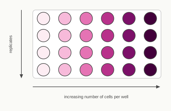
Fig. 8.19 – Multi-well plate in a metabolic viability assay.
Fig. 8.19 is worth reading carefully, because a plate of this kind is how real data in this field look. Each row is a replicate — the same condition repeated, so that a chance error in one well does not mislead. Moving from left to right across a row, the number of cells per well increases, and the colour deepens correspondingly. Reading down a column checks the reliability of the method: wells in the same column should match closely, and a well that is obviously out of step signals a pipetting error rather than a biological effect.
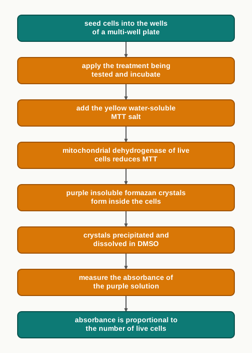
Fig. 8.20 – The MTT assay.
8.7 Advantages of Animal Cell Culture
Everything described so far in this chapter — the media, the incubators, the hoods, the passaging and the viability assays — exists to make one thing possible: the study of animal cells apart from the animal. It is worth asking plainly what is gained by doing so, and what is lost.
What Animal Cell Culture Offers
The advantages of animal cell culture are as follows.
- Cells can be grown under a controlled physico-chemical environment. The pH, the temperature, the osmolality, the oxygen and carbon dioxide tension and the concentration of every nutrient can be set by the experimenter and held steady. Inside a living body none of these can be fixed, so a result obtained in culture can be attributed to the one factor that was deliberately changed.
- Cells can proliferate into a homogenous genetic population. As you saw in Section 8.5, repeated passage produces genotypic and phenotypic uniformity, and a clone derived from a single parent cell is genetically identical throughout. Uniform material gives reproducible results.
- Cells are available in adequate numbers to conduct chemical studies. Biochemical analysis needs a large quantity of material of one kind, and a culture can supply it on demand — something no dissection of a tissue can easily do.
- Biopharmaceuticals can be produced easily. Cultured animal cells serve as living factories for therapeutic proteins, an application taken up in detail in Section 8.8.
- Ethical clearance is not required. Once a cell line is established, experiments on it do not involve procedures on a living animal, so the ethical approvals required for animal experimentation do not apply to routine work with that line.
The Limitations
However, there are a few limitations, and both are serious enough that no laboratory relies on culture alone.
- It is a highly sensitive technique, and even small changes can reduce the productivity. A slight drift in pH, a batch of serum of poorer quality, a few degrees of temperature or a short delay in subculture will alter the yield. This is the practical cost of working with cells that have been removed from the body’s own regulatory systems.
- It may not be a holistic representation of the in vivo phenotype or genotype. A cell in culture is a cell in isolation. It no longer receives the full range of signals from neighbouring tissues, hormones, nerves, blood supply and the immune system, and long-term passage may change it further. What it does in a flask is therefore not, by itself, proof of what it would do in the body.
8.8 Applications of Animal Cell Culture
Animal cell culture has diverse applications in various areas. Some of the most important are listed below.
- As a model system for the study of interactions between cells and disease-causing agents, as well as their drugs.
- As a convenient and economic tool to study virus research.
- As a useful technique for vaccine production on a large scale.
- As a production house for various medically important protein pharmaceuticals. The important ones are listed in Table 8.3.
Cultured Cells as a Model System
A model system is a simplified stand-in for the real situation. When a bacterium, a virus or a toxin is added to a dish of cultured cells, the interaction between the agent and the cell can be watched directly, and a candidate drug can then be added to see whether it prevents the damage. None of this is easy to observe inside a living body.
The value of cultured cells in virus research follows from a fact met earlier in this chapter: a virus multiplies only inside a living host cell. Cultured cells provide those host cells conveniently and economically, in whatever quantity is required, without the expense and variability of infecting animals.
The same property makes large-scale vaccine production possible. Virus grown in cultured cells can be harvested, then inactivated or weakened, and used to make vaccines in quantities that would be unattainable by any other route.
Cells as Production Houses
The largest modern application is the manufacture of protein pharmaceuticals. These are medicines that are themselves proteins, and proteins cannot simply be synthesised in a chemical plant like an ordinary drug.
The reason is worth understanding, because it explains why animal cells are used at all. Many human therapeutic proteins are glycoproteins — proteins carrying specific chains of sugar residues attached after the protein is made. Bacteria cannot carry out this glycosylation, so a protein made in bacteria may be inactive, unstable, or rejected by the patient’s immune system. Animal cells perform these modifications correctly, and that is why they are chosen despite growing slowly and demanding expensive media.
Table 8.3: Pharmaceutical proteins and their therapeutic use
| Proteins | Animal cells used | Applications |
|---|---|---|
| Follicle Stimulating Hormone (FSH) | CHO cells | Infertility |
| Human Growth Hormone (HGH) | CHO cells | GH deficiency |
| Erythropoietin (EPO) | CHO cells | Anemia |
| Factor VIII | CHO cells | Hemophilia A |
| Factor IX | CHO cells | Hemophilia B |
| Interleukin 2 (IL-2) | CHO cells | Cancer therapy |
| Tissue Plasminogen Activator (tPA) | CHO cells | Stroke |
| Monoclonal antibodies (mAbs) | Hybridoma cells | Cancer therapy & Autoimmune diseases |
* CHO — Chinese Hamster Ovary
Read down the middle column of Table 8.3 and one line stands out. Seven of the eight products are made in CHO cells — a continuous cell line derived from the ovary of the Chinese hamster, which grows reliably in suspension, tolerates large-scale culture and glycosylates human proteins in a way the human body accepts. Only the monoclonal antibodies come from a different source: hybridoma cells.
A hybridoma is a cell made by fusing an antibody-producing B lymphocyte with a myeloma (tumour) cell. The B cell contributes the ability to make one single, specific antibody; the tumour cell contributes the ability to divide indefinitely. The fused cell does both, and so becomes a permanent source of one pure antibody — a monoclonal antibody.
Erythropoietin (EPO)
Erythropoietin is a glycoprotein hormone secreted by the kidney under hypoxic conditions — a shortage of oxygen — or under anoxia, meaning a lack of oxygen, caused by anaemia. It induces RBC production, a process called erythropoiesis, and it also assists wound healing.
EPO stimulates the bone marrow to produce more red cells, and thereby increases the oxygen-carrying capacity of the blood. The kidney thus acts as an oxygen sensor for the whole body: when oxygen delivery falls, it releases the hormone that increases the number of oxygen carriers.
Clinically, EPO is used for treating certain types of anaemia caused by chemotherapy in cancer patients, in the treatment of AIDS, and even in chronic renal failure — a condition in which the diseased kidney can no longer make enough of the hormone itself.
Recombinant human EPO, written as r-HuEPO, has been produced using the Chinese Hamster Ovary (CHO) cell line. The use of r-HuEPO is advantageous over blood transfusion, because it does not require donors or transfusion facilities, and the risk of transfusion-associated disease is minimised.
Factor VIII for Haemophilia A
Haemophilia A is a common inherited genetic disorder in which Factor VIII, required for blood clotting, is not produced in the body. A person with the condition bleeds for a prolonged period, because the clotting cascade cannot be completed without this factor.
Like EPO, Factor VIII is also a glycoprotein, and it may be produced in CHO cells. Before recombinant Factor VIII was available, patients depended on clotting factor prepared from pooled donated blood, with all the infection risk that carried.
Factor IX for Haemophilia B
Haemophilia B, also called Christmas disease, is a bleeding disorder caused by the deficiency of Factor IX. Recombinant Factor IX produced in CHO cells is used to treat haemophilia B.
The two haemophilias illustrate a general principle of clotting: the cascade is a chain of steps, and the failure of any one step in the chain produces a similar bleeding disorder, so the missing factor must be identified before the correct replacement can be given.
Tissue Plasminogen Activator (tPA)
Tissue plasminogen activator is a serine protease that catalyses the conversion of plasminogen to plasmin, and it is plasmin that is responsible for dissolving blood clots.
Notice the design of this system. Plasminogen circulates in the blood all the time as an inactive precursor enzyme, doing nothing. Only where tPA acts on it is it converted into active plasmin, which then digests the fibrin of the clot. The body therefore keeps a clot-dissolving tool ready everywhere, but switches it on only where it is needed.
\[\\text{Plasminogen} \\xrightarrow{\\ tPA\\ } \\text{Plasmin} \\longrightarrow \\text{digestion of fibrin} \\longrightarrow \\text{dissolution of the blood clot}\]
tPA is approved for use in certain patients having a heart attack or a stroke, where a clot is blocking a vessel and must be removed quickly to save the tissue beyond it. Historically it holds a special place: tPA was the first drug to be produced through mammalian cell culture. The production route and the mode of action are given in Fig. 8.21.
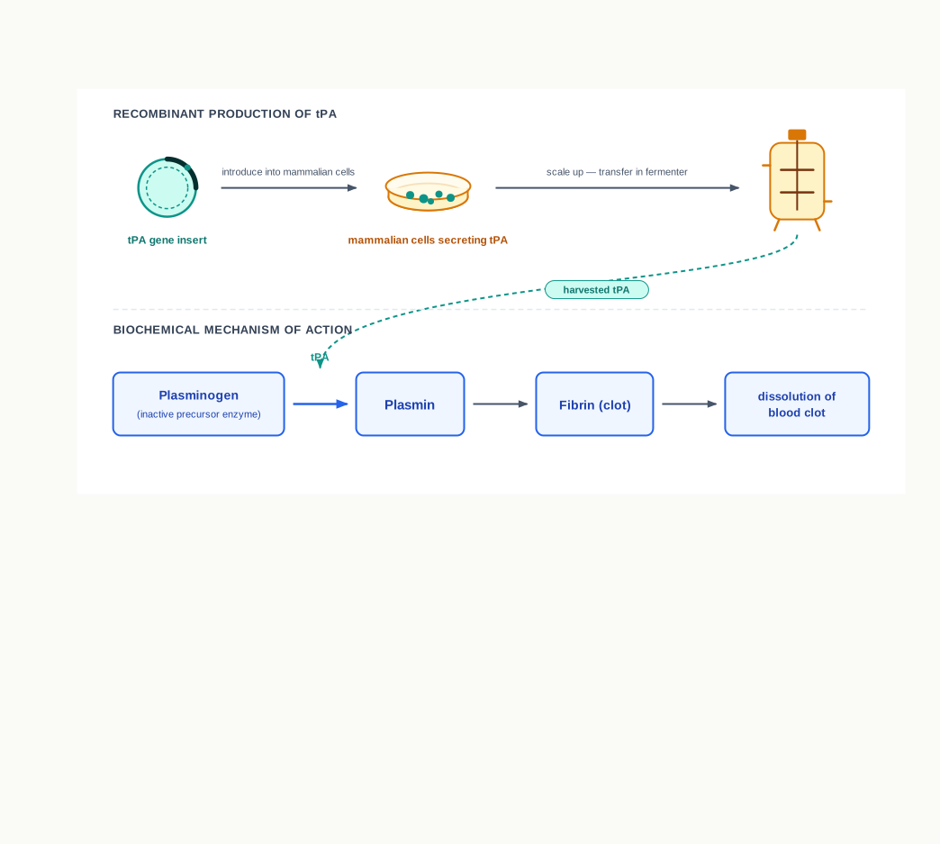
Fig. 8.21 – Production and mechanism of action of tPA.
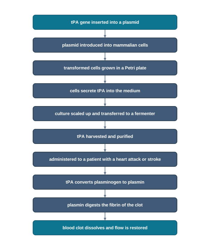
Fig. 8.22 – Making and using tPA.
Therapeutic MAB — OKT3
Muromonab-CD3, known as OKT-3, is a murine monoclonal antibody — that is, one derived from mouse cells — directed against the CD3 receptor. It is used as an immunosuppressant drug, given intravenously to reverse acute rejection of transplanted organs such as the kidney, the heart and the liver.
Its mechanism is precise. When OKT-3 is bound to CD3, the T-cell receptor (TCR) undergoes endocytosis — it is drawn into the cell — and the result is an inert T-cell. OKT-3 therefore acts by blocking the function of the T cells, which play a major role in acute graft rejection, as shown in Fig. 8.23. With the receptor withdrawn from the surface, subsequent recognition of the graft is prevented. The T cells are then eliminated by phagocytosis.
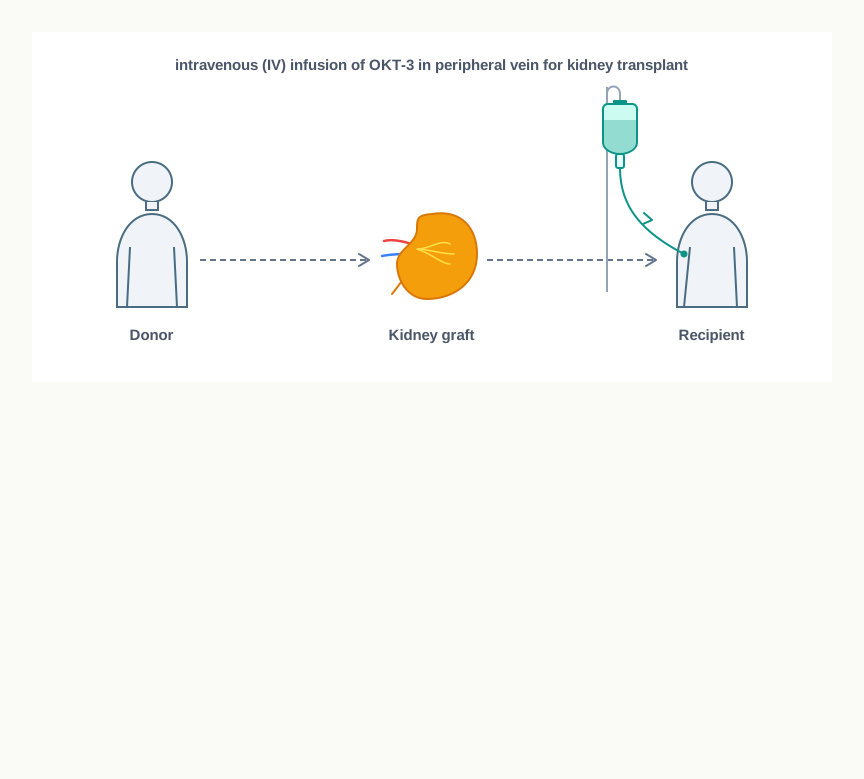
Fig. 8.23 – Use of OKT-3 during kidney transplantation.
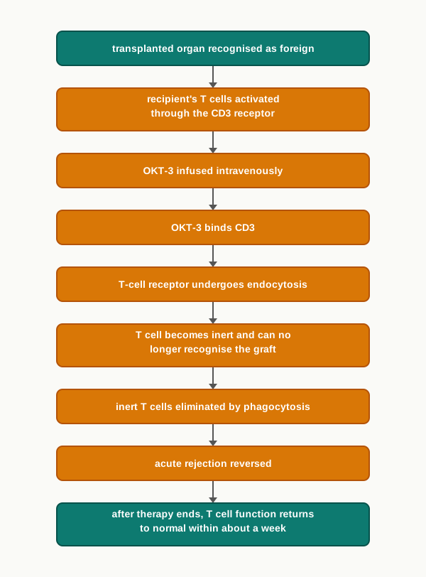
Fig. 8.24 – How OKT-3 prevents acute graft rejection.
The suppression is deliberately temporary. After OKT-3 therapy is over, T cell function usually returns to normal within a week, so the patient is not left permanently defenceless against infection. OKT-3 was the first monoclonal antibody to be approved for the treatment of acute rejection in clinics.
Trastuzumab
Trastuzumab, sold under the brand name Herceptin, is a monoclonal antibody approved for the therapy of early-stage breast cancer that is Human Epidermal growth factor Receptor 2-positive, written HER2⁺.
The distinction matters for treatment. Normal cells also express HER2, but cancer cells overexpress HER2⁺, along with a dysregulation of receptor activation. These cell surface receptors receive signals that stimulate the cells to proliferate, so a cell carrying too many of them is being told to divide far too often.
Trastuzumab works by attaching itself to the HER2 receptors and blocking them from receiving growth signals. The result is impaired growth of the breast cancer. Because the antibody recognises only that one receptor, the treatment is targeted at the cells that overexpress it — which is why the tumour must be tested for HER2 status before the drug is prescribed.
8.9 Summary
- Animal cell culture is the in vitro maintenance and proliferation of animal cells using an appropriate nutrient medium.
- The basic requirements for the optimal growth of animal cells are a controlled temperature, a correctly regulated pH, and an appropriate growth medium.
- Animal cell media fall into two major categories: natural media and artificial or synthetic media.
- Natural media consist of naturally occurring biological fluids such as plasma and tissue extract. They are suitable for culturing a wide range of animal cells.
- Artificial or synthetic media are made up of various nutrients, both organic and inorganic, together with vitamins, salts, O₂ and CO₂, serum, carbohydrates and cofactors. They can be modified according to the purpose of the culture, and are divided into four categories — serum-containing, serum-free, chemically defined and protein-free media.
- Cell culture can be classified as primary cell culture and secondary cell culture, the latter also being called a cell line.
- Primary cells can grow either as an adherent monolayer attached to the vessel surface, or freely in suspension.
- Secondary cell lines are derived from primary cells after sub-culturing.
- On the basis of the life span of the culture, cell lines are categorised as finite cell lines and continuous cell lines.
- Cell viability measurement — the determination of living or dead cells — is very significant in cell culture. It is done through dye exclusion viability assays and through metabolic viability assays.
- Cell culture technology has applications in many areas, including molecular genetics, immunological analyses, gene therapy, bioengineering and the pharmaceutical industry.
- Animal cell culture plays an important role in the research and development of drugs, and it helps to improve the health and quality of life of patients suffering from various diseases such as cancer and genetic disorders.
The overall sequence followed by the chapter, from a piece of tissue to a finished medicine, is condensed in Fig. 8.25.
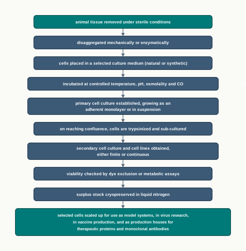
Fig. 8.25 – The complete cell culture sequence.
8.10 Key Terms
- Animal cell culture — The in vitro maintenance and proliferation of animal cells, which continue to grow outside the living organism when supplied with appropriate nutrients and growth conditions.
- Cell culture — The general process of growing cells under laboratory conditions.
- In vitro — Outside the living body, in laboratory glassware or plasticware.
- In vivo — Within the living organism.
- Clone — A homogenous population of cells derived from a single parental cell, all members of which are genetically identical.
- Cell line — A cell population that can be maintained and grown on in the laboratory through repeated transfers.
- HeLa — The first human cell line, established by George Gey from the cervical cancer of Henrietta Lacks.
- Growth factors — Proteins such as nerve growth factor, epidermal growth factor, insulin-like growth factor, FGF, platelet-derived growth factor and TGF, whose addition to the medium increases cellular proliferation.
- Biopharmaceuticals — Medicines produced by living cells rather than by chemical synthesis.
- Culture medium — The nutrient environment supplied to cultured cells, containing vitamins, amino acids, glucose, inorganic salts, serum and hormones, which also maintains the pH and osmolality of the culture.
- Natural media — Media consisting of natural biological substances such as plasma, serum and embryo extract, whose exact composition is not known.
- Synthetic or artificial media — Media composed of a basal medium together with supplements such as serum, growth factors and hormones.
- Basal medium — The defined nutrient base of a synthetic medium, to which supplements are added.
- Balanced salt solution (BSS) — A solution of inorganic salts that maintains the physiological pH and osmotic pressure of the culture, the ions also maintaining the membrane potential.
- Serum — The fluid remaining after blood has clotted, used as the most important medium supplement because it supplies proteins, growth factors, binding proteins and adhesion factors.
- Fetal bovine serum — The serum most commonly used to support the growth of cells in culture.
- Serum-containing media — Synthetic media in which human, bovine, equine or other serum is used as the supplement.
- Serum-free media — Media in which crude protein fractions such as bovine serum albumin or α- or β-globulin replace serum, allowing the medium to be made selective for a particular cell type.
- Xeno-free media — Media in which only human-source components such as human serum albumin are used as supplements, animal components being excluded.
- Protein-free media — Media supplemented with undefined components such as peptide fractions or protein hydrolysates.
- Chemically defined media — Media of fully known composition, comprising the basal media and the complex media such as EMEM and RPMI-1640.
- Mycoplasma — Very small bacteria that may contaminate serum and cultures without causing visible cloudiness of the medium.
- Osmolality — The concentration of osmotically active particles in the medium, formulated in commercial media to approximately 300 mOsmol.
- Osmometer — The instrument used to measure the osmolality of a medium.
- Natural buffering system — The pH-regulating system in which gaseous CO₂ balances with the CO₂/HCO₃⁻ content of the medium, requiring an atmosphere of 5–10 per cent CO₂.
- Chemical buffering system — The pH-regulating system using the zwitter ion HEPES, which buffers well over pH 7.2–7.4 without any controlled gaseous atmosphere.
- HEPES — 4-(2-hydroxyethyl)-1-piperazineethanesulfonic acid, the zwitter ion used as a chemical buffer in culture media.
- Phenol red — The pH indicator present in commercial media, which is yellow at acidic pH, pink at alkaline pH and bright red at the optimal pH of 7.4.
- HEPA filter — The High Efficiency Particulate Air filter that removes microbes and particulates from the air supplied to a laminar flow cabinet.
- Laminar flow hood — The cabinet supplying HEPA-filtered air in smooth parallel layers over the work area, in which all cell culture work is done aseptically.
- CO₂ incubator — The equipment that maintains chamber sterility, a constant temperature, a 5–10 per cent CO₂ atmosphere and about 95 per cent relative humidity for cultures.
- Inverted microscope — A microscope with its objectives below the stage and phase-contrast optics, used to examine living cells attached to the bottom of a culture vessel.
- Autoclave — The equipment that sterilises glassware, instruments and heat-stable solutions using saturated steam under pressure.
- Primary cell culture — The culture obtained directly from the host tissue, containing mostly heterogeneous cells similar to their parents.
- Explant — A fragment of tissue placed in a culture dish, from which cells grow out to establish a culture.
- Trypsin — A protease used to disaggregate tissue and to detach adherent cells from the growth surface.
- Collagenase — An enzyme used to disaggregate tissue by digesting the collagen of the extracellular matrix.
- Secondary cell culture — The culture obtained when a primary culture is sub-cultured or passaged into a new vessel with fresh growth medium.
- Senescence — The state in which cells, after several passages, lose their ability to proliferate.
- Finite cell line — A cell line with a limited number of cell divisions and a limited life span, derived from primary cultures of normal cells.
- Transformation — The spontaneous, chemically induced or virally induced change by which cells of a finite cell line regain the ability to divide indefinitely.
- Continuous cell line — A transformed cell line that can be sub-cultured and grown indefinitely, and which is immortal and tumorigenic.
- Adherent cells — Anchorage-dependent cells that require attachment to a solid or semi-solid substrate and proliferate as a monolayer.
- Monolayer culture — A culture in which the bottom of the vessel is covered by a continuous layer of cells one cell in thickness.
- Suspension cells — Anchorage-independent cells that are not attached to the vessel surface and float freely in the medium.
- Confluence — The stage at which the growth surface of the vessel is fully covered by the cells.
- Subculture or passaging — The process of detaching cells with an enzyme such as trypsin and transferring them to a fresh flask with fresh medium.
- Splitting — The common term for subculture, since the cells of one flask are equally divided into two flasks.
- Passage number — The number of times a cell line has been sub-cultured.
- Cryopreservation — The storage of cells at very low temperatures of –180 to –196°C in liquid nitrogen.
- Cryoprotective agent — A substance such as glycerol or DMSO added to the freezing medium to reduce the damage caused by ice crystal formation.
- Cell viability — The proportion of living cells in a culture, measured to assess the effect of pesticides, toxins and drugs.
- Dye exclusion viability assay — An assay in which live cells exclude a dye while dead cells, having lost membrane integrity, allow it to enter and stain them.
- Trypan blue — The dye most commonly used in the dye exclusion viability assay, which stains dead cells blue.
- Haemocytometer — The ruled counting chamber in which live and dead cells are counted under the microscope.
- Metabolic viability assay — An assay that estimates cell number from the ability of living cells to carry out a specific biochemical reaction, measured by absorbance, fluorescence or luminescence.
- MTT assay — A metabolic viability assay in which mitochondrial dehydrogenases of live cells reduce the yellow salt MTT to purple formazan crystals.
- Formazan — The purple, insoluble product of MTT reduction, dissolved in DMSO and measured, its quantity being proportional to the number of live cells.
- CHO cells — The Chinese Hamster Ovary cell line, used to manufacture most recombinant therapeutic proteins.
- Glycoprotein — A protein carrying attached sugar chains, a modification that animal cells can perform but bacteria cannot.
- Hybridoma — A cell formed by fusing an antibody-producing cell with a tumour cell, which secretes one specific antibody and divides indefinitely.
- Monoclonal antibody — A single uniform type of antibody produced by a hybridoma, all molecules of which recognise the same target site.
- Erythropoietin (EPO) — A glycoprotein hormone secreted by the kidney that stimulates the bone marrow to produce red cells, manufactured recombinantly in CHO cells.
- Factor VIII — The clotting factor absent in haemophilia A, a glycoprotein that may be produced in CHO cells.
- Factor IX — The clotting factor deficient in haemophilia B, or Christmas disease, produced recombinantly in CHO cells.
- Tissue plasminogen activator (tPA) — A serine protease that converts plasminogen to plasmin to dissolve blood clots, and the first drug produced through mammalian cell culture.
- OKT-3 (Muromonab-CD3) — A murine monoclonal antibody against the CD3 receptor, used to reverse acute rejection of transplanted organs.
- Trastuzumab — A monoclonal antibody that blocks HER2 receptors from receiving growth signals, used in the therapy of HER2-positive early-stage breast cancer.
8.11 Points to Ponder
- Animal cells take 18 to 24 hours to divide once, while a contaminating bacterium may divide in twenty minutes. Think about how this single difference in timing shapes almost every rule of practice in a cell culture laboratory — the hood, the filters, the antibiotics, the daily inspection. Which of these precautions would become unnecessary if animal cells divided as fast as bacteria?
- Serum is described as the most important component of culture media and, in the same chapter, as the source of four serious problems. Consider whether a substance can be genuinely indispensable and genuinely unsuitable at the same time, and what that tension tells you about the direction in which media design has moved.
- A medium of unknown composition once supported cells better than any defined mixture could. Reflect on what is gained, and what is quietly lost, when a natural biological fluid is replaced by a chemically defined recipe in which every ingredient is listed.
- The bicarbonate buffering system needs a gas cylinder, an incubator and a controlled atmosphere; HEPES needs none of these but costs more and can be toxic. Think about how a working laboratory decides between a cheap system with heavy requirements and an expensive one with few.
- Phenol red gives no number, no graph and no printout — only a colour. Consider why an indicator so crude has survived in commercial media alongside far more precise instruments, and what other measurements in science are valued for the same reason.
- Normal cells stop dividing after a limited number of divisions; transformed cells do not. Reflect on the fact that the property which makes a cell line useful and permanent in the laboratory is the same property that makes a cell dangerous in the body.
- Continuous cell lines are immortal and tumorigenic, and they are also the cells in which several life-saving medicines are manufactured. Think about what safeguards this situation demands during the purification of a product intended for a patient.
- The HeLa cell line has been used in laboratories worldwide for decades, and the woman whose cells they were never knew they had been taken. Consider what the scientific community owed her, whether that debt can be settled after the fact, and what rules would prevent the same situation arising today.
- Freezing must be done slowly and thawing quickly, though both pass through the same range of temperature. Reflect on why the correct rate of a physical process, and not merely its direction, can decide whether a cell lives or dies.
- A dye exclusion assay asks whether a cell’s membrane is intact; a metabolic assay asks whether its enzymes are working. Consider whether these two questions could ever give different answers for the same cell, and which answer you would trust when they disagree.
- A drug that leaves cultured human liver cells entirely unharmed may still injure a patient. Think about everything a flask of cells does not contain, and how a scientist should describe the limits of an in vitro result when reporting it.
- Bacteria grow faster and far more cheaply than animal cells, yet most recombinant therapeutic proteins are made in CHO cells. Reflect on how a single structural feature of a protein molecule can outweigh every economic argument in choosing a production system.
- Cell culture has reduced the number of animals used in experiments, and it began with tissue taken from animals and from people. Consider where, in the whole sequence from donor to medicine, the ethical questions actually arise, and who should be answerable for them.
8.12 NCERT Questions
- What is animal cell culture?
- Describe animal cell culture media and their types.
- Write the advantages and disadvantages of serum in the culture media.
- Describe any two chemically synthesised media.
- What is primary cell culture? Also discuss as to how it is developed.
- What is subculture or passaging of cells?
- Differentiate between finite and continuous cell lines.
- How is cell viability measurement performed?
- Write a detailed account of application of cell culture.
- The example of animal cell culture media is:
- DMEM
- MS media
- LB Media
- All of the above
- Name the type of culture that is prepared by inoculating directly from the tissue of an organism to culture media.
- Primary cell culture
- Secondary cell culture
- Cell lines
- Transformed cell culture
- Sodium bicarbonate is added to animal cell culture media to
- keep cells stuck to the plastic
- promote the uptake of CO₂ into animal cells
- maintain the correct pH when CO₂ is present
- keep iron soluble
- Which of the following is NOT present in growth medium for animal cell culture?
- Inorganic salts
- Bicarbonate
- Carbon source
- Starch
- Disaggregating of cells can be performed by:
- Physical disruption
- Enzymatic digestion
- Treating with chelating agents
- All of the above
- The approach in which genes are transferred into animals to obtain a large scale production of the proteins encoded by these genes in the milk, blood, etc., is called
- In situ culture
- Molecular pharming
- Gene therapy
- Hybridoma technology
- Which of the following is a protein-free animal cell culture media?
- RPMI-1640 Media
- MS media
- LB media
- None of the above
- MTT assay is used for
- Cell viability test
- Monitoring of variation in pH of culture media
- Transformation screening
- Cell dissociation from substratum
- Passaging of animal cells in animal cell culture is
- Sub-culturing of the cells
- Isolation of cells
- Passing the cells from culture tube to Petri dishes
- Counting of cells
- Which of the following is NOT the major function of serum?
- Enhance cell attachment
- Stimulate cell growth
- Promotion of tuber and bulb formation
- Provide transport proteins
- Assertion: Serum is the most important component of culture media. Reason: Serum is a good source of nutrients and also helps in cell proliferation and cell-matrix attachment.
- Both assertion and reason are true and the reason is the correct explanation of the assertion.
- Both assertion and reason are true but the reason is not the correct explanation of the assertion.
- Assertion is true but reason is false.
- Both assertion and reason are false.
- Assertion: Cell lines derived from the primary culture of normal cells are finite cell line. Reason: Some cells of the finite cell line undergoes transformation and retain the ability to divide indefinitely.
- Both assertion and reason are true and the reason is the correct explanation of the assertion.
- Both assertion and reason are true but the reason is not the correct explanation of the assertion.
- Assertion is true but reason is false.
- Both assertion and reason are false.
8.13 Objective Questions
- An animal cell in culture usually requires how long to complete one division?
- 20–30 minutes
- 2–4 hours
- 18–24 hours
- 5–7 days
- A homogenous population of cells derived from a single parental cell is called a
- clone
- explant
- hybridoma
- monolayer
- The first human cell line to be established was
- CHO
- HeLa
- L-cell
- Vero
- Which of the following is NOT a natural medium?
- Plasma clot
- Chicken embryo extract
- Amniotic fluid
- Ham’s F-12
- Which binding proteins supplied by serum carry other molecules into the cell?
- Albumin and transferrin
- Collagen and elastin
- Actin and tubulin
- Trypsin and collagenase
- Commercial culture media are generally formulated to a final osmolality of approximately
- 150 mOsmol
- 300 mOsmol
- 600 mOsmol
- 900 mOsmol
- The instrument used to measure the osmolality of a culture medium is the
- colorimeter
- haemocytometer
- osmometer
- osmoscope
- At the optimal pH of 7.4, medium containing phenol red appears
- yellow
- bright red
- pink
- colourless
- HEPES has a superior buffering capacity in the pH range
- 6.4–6.8
- 6.8–7.2
- 7.2–7.4
- 8.0–8.2
- A CO₂ incubator for mammalian cell culture maintains a relative humidity of about
- 45 per cent
- 65 per cent
- 75 per cent
- 95 per cent
- In a laminar flow hood, the air is first passed through
- the HEPA filter
- the pre-filter
- the fibre glass screen
- the exhaust valve
- The fragment of tissue from which cells grow out into a culture dish is called
- an explant
- a monolayer
- a passage
- a pellet
- Which enzyme disaggregates tissue by digesting the collagen of the extracellular matrix?
- Trypsin
- Collagenase
- Dehydrogenase
- Serine protease
- Cells that lose the ability to proliferate after several passages are said to have undergone
- transformation
- confluence
- senescence
- disaggregation
- Which of the following is an example of suspension cells?
- Fibroblasts
- Epithelial cells
- Haematopoietic stem cells
- Explant cells
- During subculture, the cell suspension is centrifuged at
- 30 × g for 5 min
- 300 × g for 5 min
- 3000 × g for 5 min
- 300 × g for 50 min
- Cells are stored in liquid nitrogen because the formation of ice crystals is reduced below
- –20°C
- –80°C
- –130°C
- –196°C
- A typical freezing medium used for cryopreservation contains
- 90 per cent serum and 10 per cent DMSO
- 10 per cent serum and 90 per cent DMSO
- 50 per cent serum and 50 per cent glycerol
- 100 per cent basal medium
- In the MTT assay, the yellow water-soluble salt is reduced to purple formazan crystals by
- mitochondrial dehydrogenase enzymes of live cells
- proteases released by dead cells
- the acidic pH of the medium
- trypsin present in the assay mixture
- The purple formazan crystals formed in an MTT assay are dissolved in
- water
- DMSO
- trypan blue
- phosphate buffered saline
- Which of the following is a dye exclusion viability assay stain?
- Phenol red
- MTT
- Propidium iodide
- Formazan
- Monoclonal antibodies listed in Table 8.3 are produced in
- CHO cells
- hybridoma cells
- fibroblasts
- haematopoietic stem cells
- Erythropoietin is secreted in the body by the
- liver
- kidney
- bone marrow
- spleen
- The first drug to be produced through mammalian cell culture was
- erythropoietin
- Factor VIII
- tissue plasminogen activator
- trastuzumab
- Muromonab-CD3 (OKT-3) is directed against
- the CD3 receptor
- the HER2 receptor
- fibrin
- plasminogen
- Assertion: Fetal bovine serum is commonly added to culture media. Reason: Serum supplies adhesion factors that help cells adhere to the substratum before they begin to divide.
- Assertion: Antibiotics such as penicillin and streptomycin are commonly added to culture media. Reason: Antibiotics are essential for the growth and proliferation of animal cells in culture.
- Assertion: A culture medium containing phenol red turns yellow when the cells inside are growing actively. Reason: Metabolising cells release CO₂ and acidic products, and phenol red turns yellow at acidic pH.
- Assertion: Media buffered with the natural bicarbonate system must be kept in an atmosphere of 5–10 per cent CO₂. Reason: The concentrations of CO₂, HCO₃⁻ and pH in such a medium are interrelated.
- Assertion: Vertical laminar flow hoods are preferred over horizontal ones for animal cell culture. Reason: Horizontal laminar flow hoods do not filter the air supplied to the work area.
- Assertion: Continuous cell lines can be grown up to a higher cell density than finite cell lines. Reason: Continuous cell lines are less adherent, are fast growing and are less demanding for nutrients.
- Assertion: During subculture, trypsin is removed after only 1–2 minutes. Reason: Prolonged exposure to trypsin digests the cells’ own surface proteins and damages them.
- Assertion: Cells are cooled slowly before storage but thawed as rapidly as possible. Reason: Slow cooling allows water to move out of the cells before they freeze, while rapid thawing minimises the growth of damaging ice crystals.
- Assertion: Recombinant erythropoietin is manufactured in CHO cells rather than in bacteria. Reason: Erythropoietin is a glycoprotein, and bacteria cannot carry out the glycosylation of proteins.
- Assertion: Primary cell cultures contain a heterogeneous population of cells. Reason: Repeated passaging causes the cells with the highest growth capacity to predominate.
8.14 Check Your Concepts
- Animal cells take 18 to 24 hours to divide. Explain how this single fact makes animal cell culture far more vulnerable to bacterial contamination than bacterial culture is.
- A student writes that “in vitro and in vivo mean the same thing, since the cells are alive in both”. Correct this statement and explain why the distinction matters when a result obtained in a flask is applied to a patient.
- Why must the osmolality of a culture medium be controlled so carefully? Describe what happens to an animal cell if the osmolality of the surrounding medium becomes (i) much lower and (ii) much higher than that inside the cell.
- Mammalian cells are cultured at 37°C. Give two separate reasons for the choice of this temperature, and predict what would happen to the culture if the incubator drifted to 45°C.
- A flask of bicarbonate-buffered medium is accidentally left on the bench overnight instead of in the CO₂ incubator. Predict the colour of the medium the next morning and explain the chemistry behind your prediction.
- Explain, using Le Chatelier’s principle, how the bicarbonate system resists a fall in the pH of the medium when the cells release acidic products.
- Pyruvate is sometimes added to a culture medium. Explain how its presence reduces the culture’s dependence on the external CO₂ supply.
- A researcher must film living cells on a microscope stage for three hours, outside any incubator. Which buffering system should the medium contain, and why would the alternative fail?
- Phenol red gives no numerical reading, yet it is present in almost every commercial medium. Justify its continued use, and state two different pH problems it can reveal.
- Serum is called the most important component of culture media and is also described as a source of serious problems. Set out both sides of this statement, and explain which of the disadvantages matters most when a therapeutic protein is being manufactured.
- Why does every new batch of serum need fresh testing before use, when a batch of a chemically defined basal medium does not?
- Explain why natural media show poor reproducibility and reduced uniformity, and state what synthetic media do differently to overcome this.
- The inorganic ions of a balanced salt solution are said to have more than one role. List these roles and explain why the concentration of NaCl in particular is the quantity generally adjusted.
- Antibiotics are described as “not essential” for the growth of cells, yet they are commonly added to culture media. Explain this apparent contradiction, and state one risk of relying on antibiotics instead of on good aseptic technique.
- In a laminar flow hood, air passes through a pre-filter before it reaches the HEPA filter. Explain why this order is used and what would happen if the pre-filter were removed.
- The laminar flow chamber is kept under constant positive pressure. Explain how this protects the culture, and state what would happen if the cabinet were held at negative pressure instead.
- Both horizontal and vertical laminar flow hoods supply HEPA-filtered air, yet only one is recommended for animal cell culture. Compare the two designs and justify the recommendation.
- Explain why the CO₂ cylinder is kept outside the laboratory and the gas piped in, rather than the cylinder being placed beside the incubator.
- A CO₂ incubator maintains about 95 per cent relative humidity. Explain what would go wrong in a culture flask if the chamber were dry, and relate your answer to the osmolality of the medium.
- Why is an inverted microscope, rather than an ordinary upright microscope, required in a cell culture laboratory? Explain also why phase-contrast optics are necessary for viewing living cells.
- Label the following parts on a diagram of an inverted microscope: ocular lens, objective lens, stage, condenser, light source, field aperture, and the coarse and fine adjustment. Beside the objective lens, write one line on why a 20x objective is normally sufficient.
- Draw and label a sectional diagram of a CO₂ incubator, marking the fan, perforated shelves, door gasket, water tray and control panel. Against each label, write the function it serves in maintaining the four essential conditions of the chamber.
- Arrange the following subculture steps in the correct order and explain the position you have given to the washing step: resuspend the pellet in fresh medium; add trypsin + EDTA; check the percentage of confluency; centrifuge at 300 × g for 5 min; remove the medium and wash the monolayer; reseed into fresh flasks; transfer the detached cells into a sterile tube.
- A student adds trypsin directly to a confluent monolayer without first removing the old medium and washing the cells. The cells do not detach. Identify the error and explain the chemistry behind it.
- Explain why EDTA is included along with trypsin in a dissociation solution, naming the ions it removes and the effect this has on cell adhesion.
- Why is a passage number recorded for every cell line? Explain why results obtained at a low passage number may differ from those obtained at a high one.
- Compare finite cell lines and continuous cell lines with respect to origin, life span, adherence, growth rate and tumorigenicity. State which type you would choose for a large-scale production process, and one precaution its use would demand.
- Explain how a cell belonging to a finite cell line can give rise to a continuous cell line. Name the ways in which this change can be brought about.
- Compare adherent cells and suspension cells. Explain why suspension cells are described as homogeneous in nature while adherent cells growing on a crowded surface are not.
- A culture is allowed to grow well past confluence and is not sub-cultured. Describe what happens to the cells and explain why overcrowding is fatal to a monolayer.
- Explain why cells are cooled slowly to –80°C before storage but thawed as rapidly as possible on revival. Your answer should refer to the movement of water and to the growth of ice crystals.
- A student prepares a freezing medium containing serum but forgets to add DMSO. Predict the viability on revival, and explain the role the cryoprotective agent would have played.
- Why are healthy cells in the log phase preferred for cryopreservation, and why is the medium replaced 24 hours before freezing?
- Explain the principle of the dye exclusion assay. Why does the entry of the dye into a cell indicate that the cell is dead?
- A student mixes cells with trypan blue and returns to count them an hour later, obtaining a viability of 40 per cent. A colleague repeating the count immediately obtains 92 per cent for the same suspension. Explain the discrepancy and state which value should be reported.
- Explain why the amount of formazan produced in an MTT assay is proportional to the number of living cells. What two facts about dehydrogenase enzymes make this proportionality possible?
- Compare dye exclusion assays and metabolic viability assays in terms of what each actually measures. Suggest a situation in which the two could give different answers for the same population of cells.
- In a multi-well MTT plate, one well in the middle of a row of replicates shows almost no purple colour while its neighbours are deeply coloured. Give one biological and one technical explanation, and state how you would distinguish between them.
- Match the following and write the matched pairs:
| Column A | Column B |
|---|---|
| a. Trypan blue | i. pH indicator in commercial media |
| b. Phenol red | ii. Zwitter ion used as a chemical buffer |
| c. HEPES | iii. Stains dead cells in a viability assay |
| d. MTT | iv. Cryoprotective agent |
| e. DMSO | v. Reduced to purple formazan by live cells |
- Match the following and write the matched pairs:
| Column A | Column B |
|---|---|
| a. Collagenase | i. Number of times a cell line has been sub-cultured |
| b. Explant | ii. Digests the collagen of the extracellular matrix |
| c. Senescence | iii. Fragment of tissue from which cells grow out |
| d. Passage number | iv. Cell line that divides indefinitely |
| e. Continuous cell line | v. Loss of the ability to proliferate after several passages |
- Cell culture is said to require no ethical clearance, and yet the chapter raises ethical questions about it. Explain where in the sequence — from donor tissue to finished medicine — the ethical questions actually arise.
- A compound shows no toxicity at all towards cultured human liver cells. Explain, giving three distinct reasons, why this cannot be accepted as evidence that the compound is safe for patients.
- Bacteria grow faster and far more cheaply than animal cells, yet seven of the eight products in Table 8.3 are made in CHO cells. Explain the structural reason behind this choice, and state what would be wrong with the product if bacteria were used.
- Explain how a hybridoma combines two abilities that no single cell possesses, and why this makes it suitable for producing monoclonal antibodies.
- Recombinant human EPO is described as advantageous over blood transfusion for the treatment of anaemia. Give two reasons, and explain why a patient in chronic renal failure needs this hormone at all.
- Plasminogen circulates continuously in the blood but does not dissolve clots on its own. Explain how tPA changes this, and why the body’s arrangement of an inactive precursor is a sensible design.
- Complete and explain the following sequence for the production of tPA: tPA gene inserted into a plasmid → ? → cells grown in a Petri plate → ? → transfer to fermenter → ? Explain what is achieved at each of the missing steps.
- Explain the mechanism by which OKT-3 reverses acute graft rejection. Why does T cell function return to normal within about a week after the therapy is stopped?
- Normal cells also express HER2, yet trastuzumab is prescribed only after a tumour has been tested. Explain the reason for this test and how trastuzumab impairs the growth of the cancer.
- A laboratory reports that its cell line has begun to grow much faster, needs less serum and no longer stops growing when the surface is covered. Suggest what may have happened to the cells, and state what this means for any experiment already in progress with that line.
8.15 Case-Based Questions
A trainee sets up a fresh culture on Monday afternoon. On Tuesday morning the medium in the flask is yellow and slightly cloudy, and under the inverted microscope the spaces between the cells appear to shimmer with fine moving particles. The trainee argues that the yellow colour proves the cells have grown well and that the flask should simply be given fresh medium.
- State whether you agree with the trainee’s interpretation, and give your reasons.
- Explain how the appearance of the medium and the microscopic picture together point to a different conclusion.
- Explain why this problem develops so much faster than the growth of the animal cells themselves.
- List three practices that should have prevented it.
A biopsy of connective tissue arrives in a laboratory that has been asked to establish a culture from it. One worker proposes chopping the tissue into small fragments and attaching them to a dish. Another proposes treating the tissue with an enzyme solution to release single cells before plating them.
- Name the two routes being proposed and the term used for the tissue fragment in the first route.
- Name two enzymes suitable for the second route, and state what each one acts upon.
- State one advantage of each route.
- The culture that is finally obtained is described as heterogeneous. Explain what this means and why it is expected at this stage.
A cell line is maintained by a research group over three years. In the first year the cells stopped dividing after a fixed number of passages and the stock had to be replaced from frozen vials. In the third year, one flask continues to divide indefinitely, grows faster, tolerates a lower serum concentration and no longer stops growing when the surface is covered.
- Identify the type of cell line described in the first year and the type described in the third year.
- Name the process by which the change occurred and state three ways in which it can be brought about.
- Explain why the changed cells are described as immortal and tumorigenic.
- State one advantage and one serious caution associated with using the changed cells for manufacturing a medicine.
Over a long weekend, the CO₂ cylinder supplying an incubator runs empty. On Monday the flasks inside are found to have medium of a deep pink colour. The cells are still attached but appear rounded and granular, and a viability check gives a value of 55 per cent.
- Explain why the medium turned pink rather than yellow.
- Write the equilibrium involved and state the direction in which it shifted.
- Explain what the rounded, granular appearance of the cells indicates.
- Suggest one change to the buffering of the medium that would have limited the damage, and state one disadvantage of that change.
A student prepares vials of a valuable cell line for long-term storage. Cells from an overgrown flask are suspended in medium containing 90 per cent serum and 10 per cent DMSO and the vials are placed directly into liquid nitrogen. Two weeks later a vial is revived by leaving it to warm slowly on the bench, and almost no cells survive.
- Identify three separate errors in this procedure.
- For each error, explain the damage it would have caused inside the cells.
- State the correct temperature range for long-term storage and the reason for the value chosen.
- Write the correct sequence of steps the student should have followed.
A company manufactures a recombinant therapeutic glycoprotein. The cells are grown in a serum-containing medium and the protein is secreted into that medium. During purification the team finds it very difficult to separate the product from other proteins present in the harvest, and one batch is rejected after tests detect mycoplasma.
- Explain why the presence of serum makes the purification difficult.
- Explain how mycoplasma is likely to have entered the process and why it was not noticed by simple inspection of the flasks.
- Recommend a change of medium, naming the type you would use, and give two reasons for your recommendation.
- Explain why the company uses animal cells rather than bacteria for this product, even though bacteria would be cheaper.
A patient who received a kidney transplant three weeks ago develops signs of acute rejection. The transplant team administers an intravenous monoclonal antibody preparation. The rejection is reversed, and about a week after the therapy ends the patient’s immune function is found to have returned to normal.
- Name the monoclonal antibody used and the receptor it is directed against.
- Describe, step by step, how binding of the antibody makes the T cell inert.
- Explain why the suppression of immunity is temporary rather than permanent.
- State one reason why the patient must be monitored for infection during the course of therapy.
A hospital preserves surgically removed tumour tissue and, from one sample, establishes a cell line that grows indefinitely. The line is later distributed to laboratories worldwide and used in the development of a commercially successful medicine. The patient was never told that a cell line had been established from the tissue.
- Identify the ethical issues raised by this situation.
- State the arguments that could be advanced on each side of the question of consent.
- Name the safeguards that a hospital should have in place before human tissue is cultured or commercialised.
- Explain why the scientific value of the cell line does not, by itself, settle the ethical question.
8.16 Practice With Data
- In a trypan blue exclusion assay, a worker counts 276 clear cells and 24 blue-stained cells in the fields examined. Calculate the percentage viability of the culture. If the laboratory rule is that cells must show at least 95 per cent viability before they may be used to set up an experiment, state whether this culture qualifies and by what margin it passes or fails.
- A cell suspension is diluted by mixing 0.1 mL of the suspension with 0.9 mL of trypan blue solution, and the mixture is loaded into a haemocytometer. The average count over the large squares is 85 cells per square. Using the relation
\[\\text{Cells per mL} = (\\text{average count per large square}) \\times (\\text{dilution factor}) \\times 10^4\]
calculate the cell density of the original suspension, and then calculate the total number of cells in the 5 mL of suspension available.
- A flask is seeded with \(2 \\times 10^5\) animal cells that divide once every 24 hours. Using
\[N = N\_0 \\times 2^n\]
where \(N\_0\) is the initial number of cells, \(n\) is the number of divisions and \(N\) is the final number, calculate the number of cells present after 4 days. Calculate also how long it would take for the population to first exceed \(1 \\times 10^7\) cells.
A flask containing \(1 \\times 10^6\) animal cells, which divide once every 24 hours, is accidentally contaminated with 50 bacteria, which divide once every 20 minutes. Using \(N = N\_0 \\times 2^n\) for both populations, calculate the number of each after 12 hours. Comment on what your two answers explain about the importance of aseptic technique in animal cell culture.
A confluent flask at passage number 15 contains \(1.2 \\times 10^7\) cells. The cells are trypsinized and split equally into six fresh flasks of the same size.
- Calculate the number of cells seeded into each new flask.
- State the passage number of the new flasks.
- Taking the doubling time as 24 hours, calculate how many days each new flask will take to return to \(1.2 \\times 10^7\) cells.
A technician must prepare 250 mL of complete growth medium containing 10 per cent (v/v) fetal bovine serum and 1 per cent (v/v) antibiotic solution, with basal medium making up the remainder. Calculate the volume of each of the three components required. If the serum is supplied in 50 mL bottles, calculate how many complete 250 mL batches can be prepared from one bottle.
A freezing medium is to be prepared with the standard composition of 90 per cent serum and 10 per cent DMSO.
- Calculate the volume of serum and of DMSO needed to prepare 30 mL of this freezing medium.
- If each cryovial is filled with 1.5 mL, calculate the number of vials that can be prepared.
- If each vial is to contain \(2 \\times 10^6\) cells, calculate the total number of cells required.
A medium contains NaCl at a concentration of 0.11 mol L⁻¹ and glucose at 5.5 mmol L⁻¹. Taking NaCl as contributing two osmotically active particles per formula unit and glucose as contributing one, calculate the osmolality contributed by each solute in mOsmol L⁻¹ and their combined total. Compare your total with the value of approximately 300 mOsmol to which commercial media are formulated, and state what adjustment would be made to bring the medium to that value.
Cells are cooled from 25°C to –80°C at a controlled rate of 1°C per minute before being transferred to liquid nitrogen at –196°C.
- Calculate the time taken for the controlled cooling stage, in hours and minutes.
- Calculate the further fall in temperature that occurs on transfer to liquid nitrogen.
- Explain, with reference to the value –130°C given in the chapter, why the second stage need not be slow.
- In an MTT assay, known numbers of cells were placed in wells and the absorbance of the dissolved formazan was measured.
| Cells per well | Absorbance |
|---|---|
| 0 | 0.04 |
| 5 000 | 0.22 |
| 10 000 | 0.41 |
| 20 000 | 0.80 |
| 40 000 | 1.58 |
- Plot absorbance against the number of cells per well.
- Comment on the relationship shown by your graph and explain its biological basis.
- A test well gives an absorbance of 0.60. Use your graph to estimate the number of live cells in that well.
- Suggest why the well containing no cells still gives a small absorbance reading.
- Cultured tumour cells were treated with increasing concentrations of an anticancer drug and assayed by the MTT method. The absorbance of the untreated control wells was 1.20.
| Drug concentration (µmol L⁻¹) | Absorbance |
|---|---|
| 0 | 1.20 |
| 1 | 1.08 |
| 5 | 0.84 |
| 10 | 0.60 |
| 25 | 0.30 |
| 50 | 0.12 |
- Calculate the percentage viability at each concentration, taking the untreated control as 100 per cent.
- Plot percentage viability against drug concentration.
- Estimate from your graph the concentration of the drug that reduces viability to 50 per cent.
- State what the reading at 50 µmol L⁻¹ suggests about the drug, and one further test you would carry out before calling it a promising anticancer agent.
- A normal human cell line is known to divide about 50 times before entering senescence. The line is maintained by splitting each confluent flask 1:4 at every passage, and the cells are at passage 6 when received.
- Calculate the number of divisions each cell undergoes between one 1:4 split and the next return to confluence.
- Estimate the number of further passages the line can support before senescence.
- Explain why a laboratory would freeze a large stock of vials as soon as such a line is received.
- Four flasks were inspected on the same morning and the following observations recorded.
| Flask | Colour of medium | Cell appearance | Viability (%) |
|---|---|---|---|
| A | Bright red | Spread, confluent 70% | 96 |
| B | Yellow | Spread, confluent 100% | 91 |
| C | Deep pink | Rounded, granular | 58 |
| D | Yellow, turbid | Indistinct, particles moving | 34 |
- State the approximate pH indicated by the colour of the medium in flasks A, B and C.
- For each flask, state the most likely explanation of the observations and the action you would take.
- Explain why flask B and flask D show the same colour but require completely different responses.