Chapter 2: Cellular Organelles
In the early 1950s, a Belgian scientist named Christian de Duve was not looking for a new cell organelle at all. He was studying how insulin acts on liver cells, and to do that he had to break liver tissue open and measure the activity of several enzymes in the broken-up mixture.
One enzyme behaved oddly. Acid phosphatase, an enzyme that works best in acidic surroundings, showed very little activity when a freshly prepared sample was tested. But when the same sample was left standing for a few days, or shaken roughly, its activity rose several times over. The enzyme seemed to be appearing out of nowhere.
Most people would have blamed a faulty measurement. de Duve asked a different question: what if the enzyme had been present all along, but locked away? He reasoned that it must be sealed inside tiny membrane-bound bags, and that his test could reach it only after those bags were torn open.
He was right. The bags were later named lysosomes — the cell’s digestive compartments. de Duve had discovered an organelle without ever seeing one, purely by reasoning from the behaviour of an enzyme. The electron microscope confirmed it soon afterwards, and in 1974 he shared the Nobel Prize for work on how a cell is organised inside.
That story carries the central idea of this chapter. A cell is not a bag of chemicals. It is a set of separated workshops, each walled off, each doing a job that would be dangerous or impossible anywhere else.
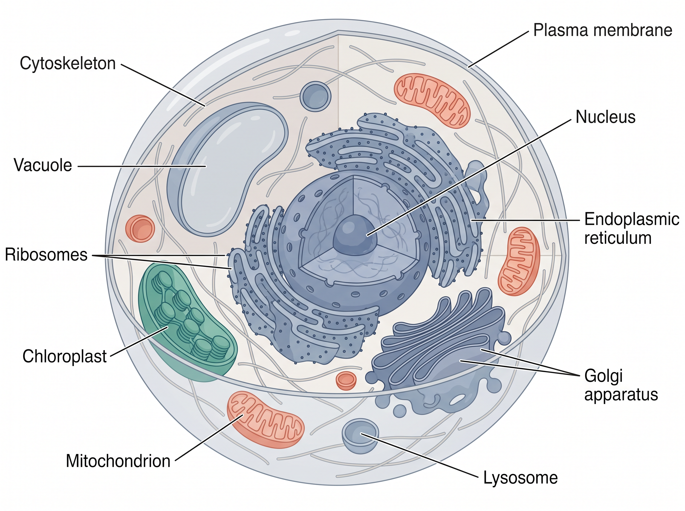
Fig. 2.0 – Eukaryotic cell structure and major compartments.
Keep these five questions in mind as you read. By the end of this chapter, you will be able to answer every one of them.
2.1 Plasma Membrane
Why a Cell Needs Compartments
At this very moment your body is doing a great many jobs at once. Food is being digested, electrical messages are racing along nerves, the heart is pumping blood, nutrients are circulating, proteins are being built and the kidneys are filtering urine. All of this is possible because of cells, which are considered to be the basic unit of life.
Each cell is fitted with different machineries known as organelles, and each organelle is responsible for a different function. Keeping these jobs in separate compartments matters, because reactions that would interfere with one another can then run side by side without clashing.
You already know that cells — whether they make up a unicellular organism or a multicellular one — are broadly placed in two categories on the basis of nuclear organisation and the presence of membrane-bound organelles: prokaryote and eukaryote.
Some components are common to both types. These are the plasma membrane, cytoplasm, ribosomes, DNA and so on. Prokaryotic cells are without an organised nucleus and contain numerous ribosomes and mesosomes (folds in the plasma membrane), and some of them also carry locomotory structures such as flagella.
A eukaryotic cell, in contrast, has a well-organised nucleus, a cell membrane, and membrane-bound cell organelles such as endoplasmic reticulum, Golgi apparatus, mitochondria, plastids, vacuole, lysosomes, peroxisomes and many more. Advancement in microscopic techniques played a very crucial role in exploring the detailed structure of the cell, because most of these structures are far below the resolving power of an ordinary light microscope.
With that picture in mind, let us look at an individual cell to understand its structure and functioning, and the role each part plays in establishing cell function and life itself. The natural place to begin is at the boundary.
The Boundary of the Cell
The plasma membrane forms the boundary of the cytoplasm, and from the outside it is guarded by the extracellular matrix. It is the membrane that is responsible for the relationship of a cell with its surroundings — everything the cell takes in, everything it sends out, and every signal it receives has to deal with this layer first.
The plasma membrane is semipermeable in nature. It does not treat all substances alike; it lets some pass and holds others back. That single property is what allows a cell to keep an internal composition different from the fluid around it.
For a long time nobody could say what this boundary was actually made of. The major breakthrough in understanding the detailed structure of the cell membrane was realised only after two things came together: an understanding of its chemical composition, which is mainly lipid and protein, and the invention of the electron microscope.
The Fluid Mosaic Model
A widely accepted model for the organisation of the plasma membrane was proposed by Seymour Jonathan Singer and Garth L. Nicolson (1972) as ‘The Fluid Mosaic Model’, shown in Fig. 2.1. The model suggests the plasma membrane to be a lipid bilayer surrounding the cell, with a mosaic of globular proteins set into it.
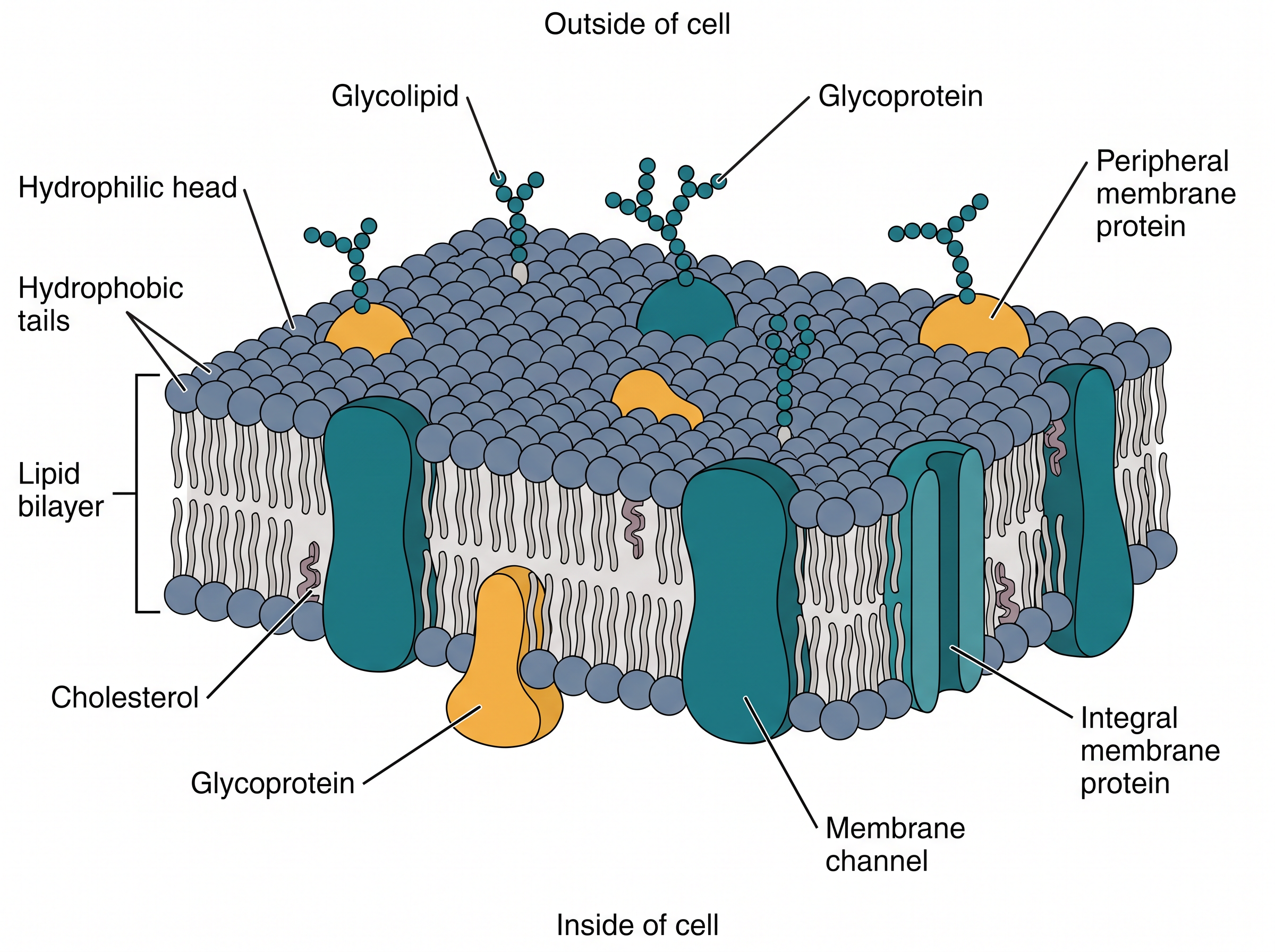
Fig. 2.1 – Fluid mosaic model of the plasma membrane.
The word ‘mosaic’ is doing real work in that name. In a mosaic, separate tiles of different kinds are set into a common background; here the proteins are the tiles and the lipid bilayer is the background. The word ‘fluid’ matters just as much, and we shall come to it in a moment.
The composition of lipid and protein varies in different cells. For example, the human erythrocyte membrane contains approximately 52 per cent protein and 40 per cent lipids. A membrane that has to carry out a great deal of transport or signalling work needs more protein; a membrane whose main job is to act as a barrier can manage with relatively more lipid.
The lipid bilayer makes the cell boundary a quasifluid structure, and it is dynamic in nature. Because of this fluid nature, lipids and proteins can freely diffuse laterally across the membrane — that is, they can slide sideways within the plane of the membrane, rather than being locked in fixed positions like bricks in a wall.
Why the Bilayer Forms at All
Phospholipids are the major membrane lipid. A phospholipid is composed of a hydrophilic (water-loving) head facing the exterior, and a long hydrophobic (water-fearing) tail of hydrocarbon chains occupying the interior of the lipid bilayer.
This double nature explains the whole arrangement. In a watery environment the heads are comfortable facing the water, while the tails are not; so the tails turn away from water and face each other, and two sheets of phospholipid come together tail-to-tail. The bilayer is not held together by strong covalent bonds — it assembles itself because that is the arrangement in which the hydrophobic tails are best hidden from water.
Two Kinds of Membrane Proteins
Two different types of proteins have been identified in the plasma membrane, classified on the basis of their location and association: peripheral and integral membrane proteins.
Peripheral membrane proteins are mainly involved in cell signalling, and these are superficially attached to the lipid bilayer. They sit on the surface rather than passing into it, which is exactly what a signalling protein needs — a position from which it can receive a message at the surface and pass it on.
Integral membrane proteins are partially or fully buried in the plasma membrane. Transmembrane proteins, which run right through from one face to the other, are the most abundant type of integral membrane protein. Only a protein that spans the full thickness of the bilayer can form a continuous passage from outside to inside, which is why transporters and channels belong to this class.
Structurally, the prokaryotic cell membrane is similar to that of eukaryotes. The same bilayer plan, with the same two classes of proteins, works in a bacterium and in a human cell alike.
A special membranous structure is formed by extension of the plasma membrane into the cell. This structure is called the mesosome, and it takes the form of vesicles, tubules and lamellae. Mesosomes increase the surface of the plasma membrane. Since a prokaryote has no internal membrane-bound organelles, folding its own boundary inwards is one way of gaining extra membrane surface on which work can be done.
What the Fluidity Is For
The quasifluid nature of the membrane is useful for different cellular functions such as cell division, cell growth, communication at intercellular junctions, cell secretion and endocytosis.
Each of these depends on the membrane being able to change shape without breaking. A dividing cell must pinch its boundary into two; a growing cell must add new material into an existing sheet; a secreting cell must let a vesicle fuse with the boundary and open outwards. A rigid membrane could do none of these things.
Transport Across the Membrane
The plasma membrane, being selectively permeable, restricts molecular movement and thereby maintains cell composition. Deciding what crosses is not a side job of the membrane — it is the main one.
Some molecules move passively across the membrane along the concentration gradient, without any expenditure of energy. This is called passive transport. Passive movement of molecules occurs by the process of diffusion and osmosis.
However, a few molecules cannot cross the plasma membrane by simple diffusion. These include charged particles, for example ions and amino acids, and also certain uncharged ones, for example glucose. The reason lies in the structure we have just described: the interior of the bilayer is hydrophobic, and a charged or strongly polar molecule cannot pass through an oily layer on its own.
Movement of such molecules is facilitated by carrier proteins, for example the glucose transporter, and by channel proteins [Fig. 2.2 (a, b)]. Such molecular movement is known as facilitated movement.
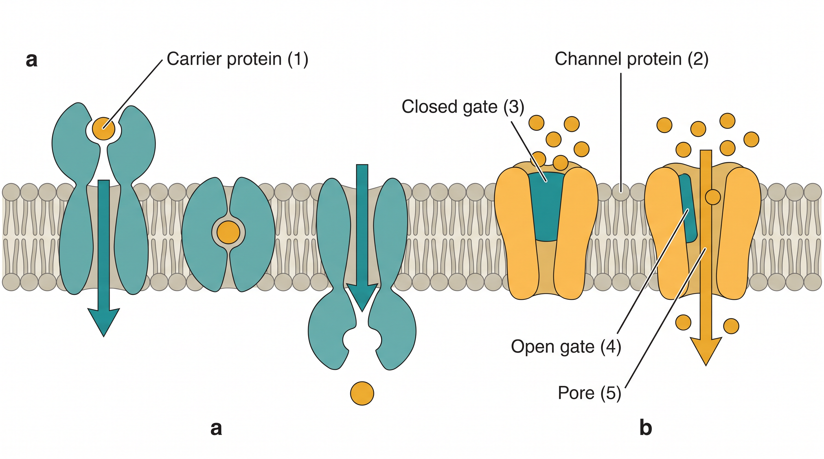
Fig. 2.2 – Membrane transport mechanisms: (a) carrier protein and (b) channel protein.
It is worth being clear about the difference between the two proteins in Fig. 2.2. A carrier protein binds the molecule and changes its own shape to release it on the other side, as in Fig. 2.2(a). A channel protein forms a water-filled pore through which the particle passes, and the gate shown in Fig. 2.2(b) decides whether that pore is open or closed at a given moment.
Aquaporins are one of the critical channel proteins for the transport of water in plant and animal cells across the plasma membrane. Some of the well-studied channel proteins in the membrane of muscle and nerve cells are ion channels [Fig. 2.2(b)].
Moving Uphill: Active Transport
Molecules which are transported against the concentration gradient — that is, from lower concentration to higher concentration — require utilisation of energy from ATP molecules. An example is the Na⁺-K⁺ pump (Fig. 2.3). This is referred to as active transport.
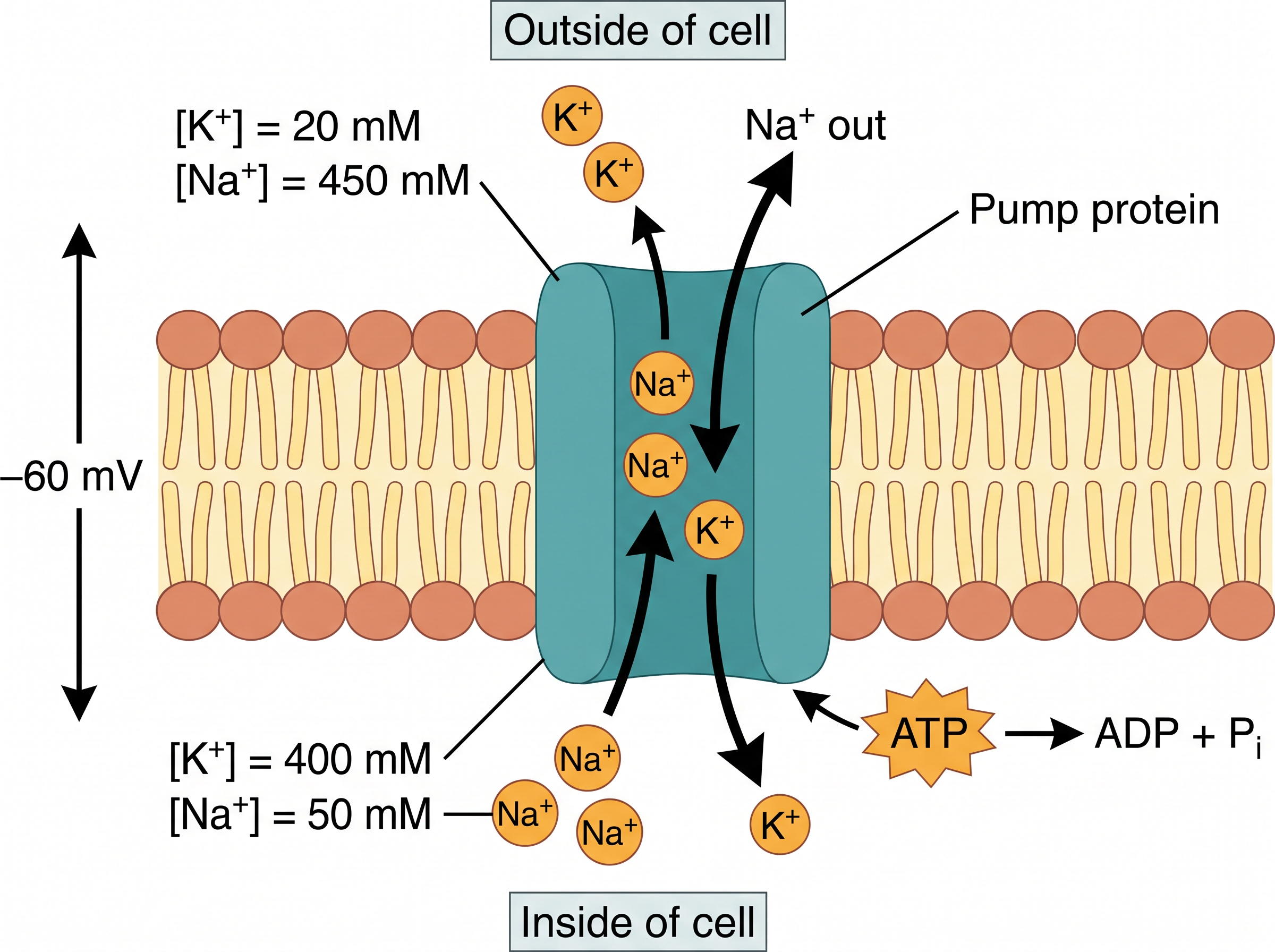
Fig. 2.3 – Active transport by the Na⁺-K⁺ pump.
Read the numbers in Fig. 2.3 carefully, because they show why a pump is needed at all. Outside the cell, \([\text{K}^+] = 20\ \text{mM}\) and \([\text{Na}^+] = 450\ \text{mM}\). Inside the cell, \([\text{K}^+] = 400\ \text{mM}\) and \([\text{Na}^+] = 50\ \text{mM}\). Here \([\ ]\) stands for concentration and mM is the millimolar, a unit of concentration.
So potassium is far more concentrated inside than outside, and sodium is far more concentrated outside than inside — in each case the exact opposite of what simple diffusion would produce. Left to itself, diffusion would even the two sides out. The pump keeps pushing both ions the “wrong” way, and that constant uphill work is what costs ATP. The figure also shows a membrane potential of \(-60\ \text{mV}\), an electrical difference across the membrane that exists because the distribution of charged ions on the two sides is unequal.
Active Transport Without ATP
However, some active transports are ATP-independent. In these, molecules are transported against the concentration gradient with no energy utilisation from ATP hydrolysis. Instead, the transport of such a molecule is coupled with a second molecule that is transported along the concentration gradient. Examples are the active transport of ions, sugars and amino acids using energy derived from the Na⁺ gradient.
This is a neat piece of cellular economy, and it is worth seeing the link. The Na⁺-K⁺ pump has already spent ATP to build a steep sodium gradient. When sodium is then allowed to rush back into the cell down that gradient, the energy released is used to drag another molecule uphill. The ATP was spent earlier, at the pump; the second transporter simply cashes in on the gradient the pump created.
Naming Transport by Direction
In coupled transport, if two molecules are transported in the same direction — for example, the uptake of glucose and Na⁺ together — it is called symport. If active transport involves transport of two molecules in the opposite direction — for example, the transport of Na⁺ and Ca²⁺ by the Na⁺-Ca²⁺ antiporter — it is called antiport. While facilitated diffusion transports only a single molecule, for example glucose, it is known as uniport.
The whole classification of membrane transport is summarised in Fig. 2.4, and it is worth fixing this branching in your mind, because questions often ask you to place a named example in the correct branch.
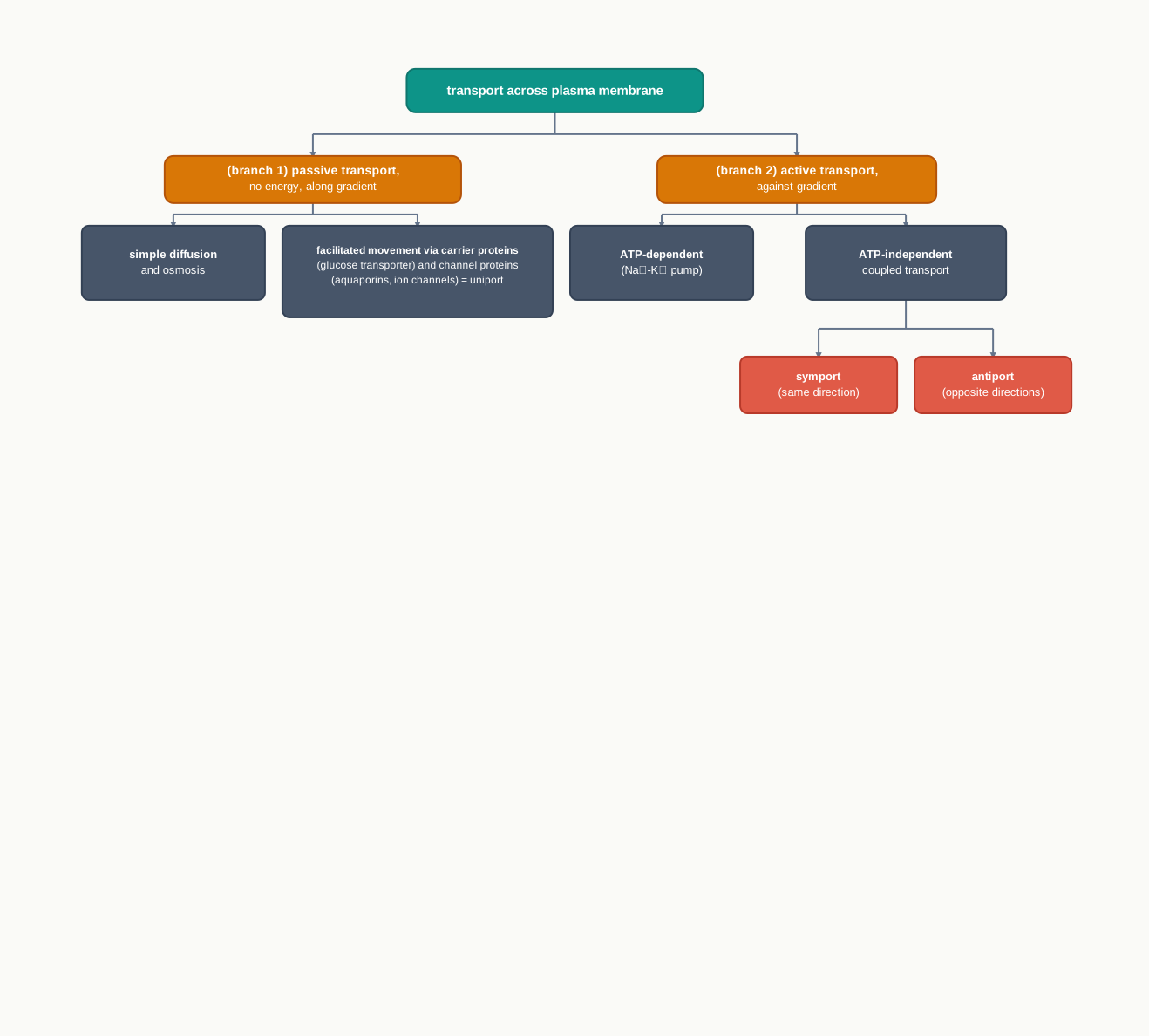
Fig. 2.4 – Classification of plasma membrane transport.
2.2 Cell Wall
Cells of bacteria, algae, fungi and higher plants are surrounded by a rigid cell wall in addition to the plasma membrane. It is not found in animal cells. The wall lies outside the membrane, so anything entering or leaving such a cell has to cross the wall first and the membrane afterwards.
The cell wall differs structurally in bacteria and in eukaryotes. In bacteria, it is composed of polysaccharide cross-linked by small peptides, which provides rigidity, shape and protection from osmotic pressure. In eukaryotes — that is, in plants and fungi — it is primarily made up of polysaccharides.
What the Wall Does
The cell wall not only determines the cell shape, but also prevents cell bursting caused due to osmotic pressure. It also helps in cell–cell interaction, and provides mechanical strength and protection from infection.
The point about bursting deserves a moment’s thought. You have already seen that water crosses the plasma membrane by osmosis. If a cell sits in a dilute solution, water keeps entering, and a bare membrane would stretch and rupture. A rigid wall stops the swelling early: as the cell presses outward, the wall pushes back, and the inward flow of water comes to a halt before any damage is done.
The Bacterial Cell Wall
Gram-positive bacteria have a thick cell wall with a single plasma membrane [Fig. 2.5(a)]. In contrast, Gram-negative bacteria have a thin cell wall surrounded by a dual plasma membrane [Fig. 2.5(b)].
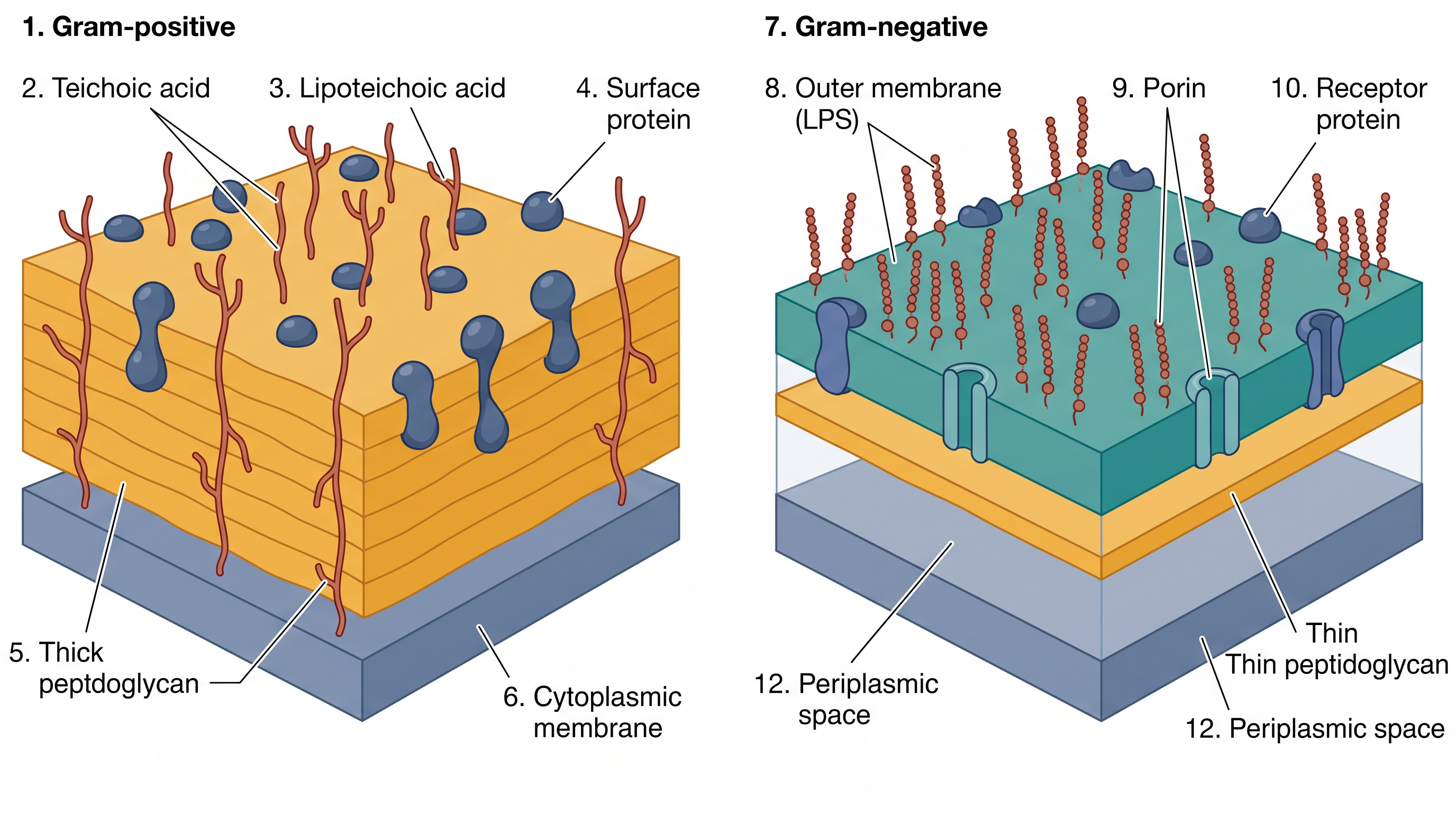
Fig. 2.5 – Prokaryotic cell wall: (a) Gram-positive and (b) Gram-negative bacteria.
Compare the two halves of Fig. 2.5 carefully. In (a) the peptidoglycan layer is thick and lies directly above one membrane. In (b) the peptidoglycan layer is thin and is sandwiched in a periplasmic space between two membranes, with the outer membrane carrying lipopolysaccharide (LPS) on its surface and porins forming passages through it.
The cell wall continuously grows and changes its shape as the bacterium grows and divides. It is not a finished, permanent shell — it is being rebuilt all the time, and that fact turns out to matter enormously in medicine.
Structurally, the bacterial cell wall is a sturdy covalent shell of linear peptidoglycan chains cross-linked by tetrapeptides. Think of the long peptidoglycan chains as parallel threads and the tetrapeptide cross-links as stitches tying neighbouring threads together. Threads alone would slide apart; it is the stitching that turns them into a single strong fabric enclosing the whole cell.
Commonly used antibiotics are known to inhibit this cross-linking of peptidoglycan strands, thereby interfering with bacterial growth. Because the bacterium is constantly making new wall as it grows, blocking the cross-linking means the new wall is laid down unstitched and weak, and the cell cannot withstand its own internal pressure.
Glycocalyx, Slime Layer and Capsule
In prokaryotic cells, especially in bacteria, the cell wall is further covered with a heavily glycosylated protein known as glycocalyx. It acts as a barrier to invading pathogens and protects the cell from mechanical and ionic stresses. Glycocalyx is also involved in cell–cell interactions.
In some cases the glycocalyx is present as a loose sheath called a slime layer, and in others it is thick and tough and is called a capsule. The difference is one of how firmly the layer is attached and how well organised it is, not of what it is made from.
The Eukaryotic Cell Wall
Among eukaryotes, the cell wall is mainly composed of polysaccharide (Fig. 2.6). It may be of cellulose, a linear polymer of glucose residues, as in most higher plants, or of chitin, a linear polymer of N-acetylglucosamine, as in fungi.

Fig. 2.6 – Eukaryotic cell wall polysaccharides: (a) cellulose and (b) chitin.
Notice in Fig. 2.6 that both polymers use the same kind of joint, the \(\beta(1\rightarrow4)\) glycosidic linkage, in which carbon 1 of one sugar ring is joined to carbon 4 of the next. This particular linkage produces long, straight, unbranched chains that can lie side by side and hydrogen-bond into tough fibres — which is exactly the property a wall needs. The two polymers differ only in the sugar being repeated: β-D-glucose in cellulose, and N-acetylglucosamine in chitin.
Layers of the Plant Cell Wall
In plants, a growing cell is surrounded by a comparatively thin primary cell wall, which leaves scope for cell expansion. A thin, extensible wall is essential at this stage — a cell that is still enlarging cannot be locked inside a rigid box.
As the cell ceases growing, a new layer called the secondary cell wall is formed between the primary cell wall and the plasma membrane. Note the position carefully: the secondary wall is laid down on the inner side, so the primary wall remains outermost. A secondary cell wall is very rigid and thick compared to the primary cell wall, due to the deposition of lignin.
A layer of calcium pectate, known as the middle lamella, holds together neighbouring cells. Their cytoplasm is connected through a structure called plasmodesmata. These fine channels running from one cell into the next allow a walled plant tissue to behave as a continuous, communicating whole rather than as a set of sealed boxes.
The whole classification of cell walls, from bacteria through to plants, is brought together in Fig. 2.7.
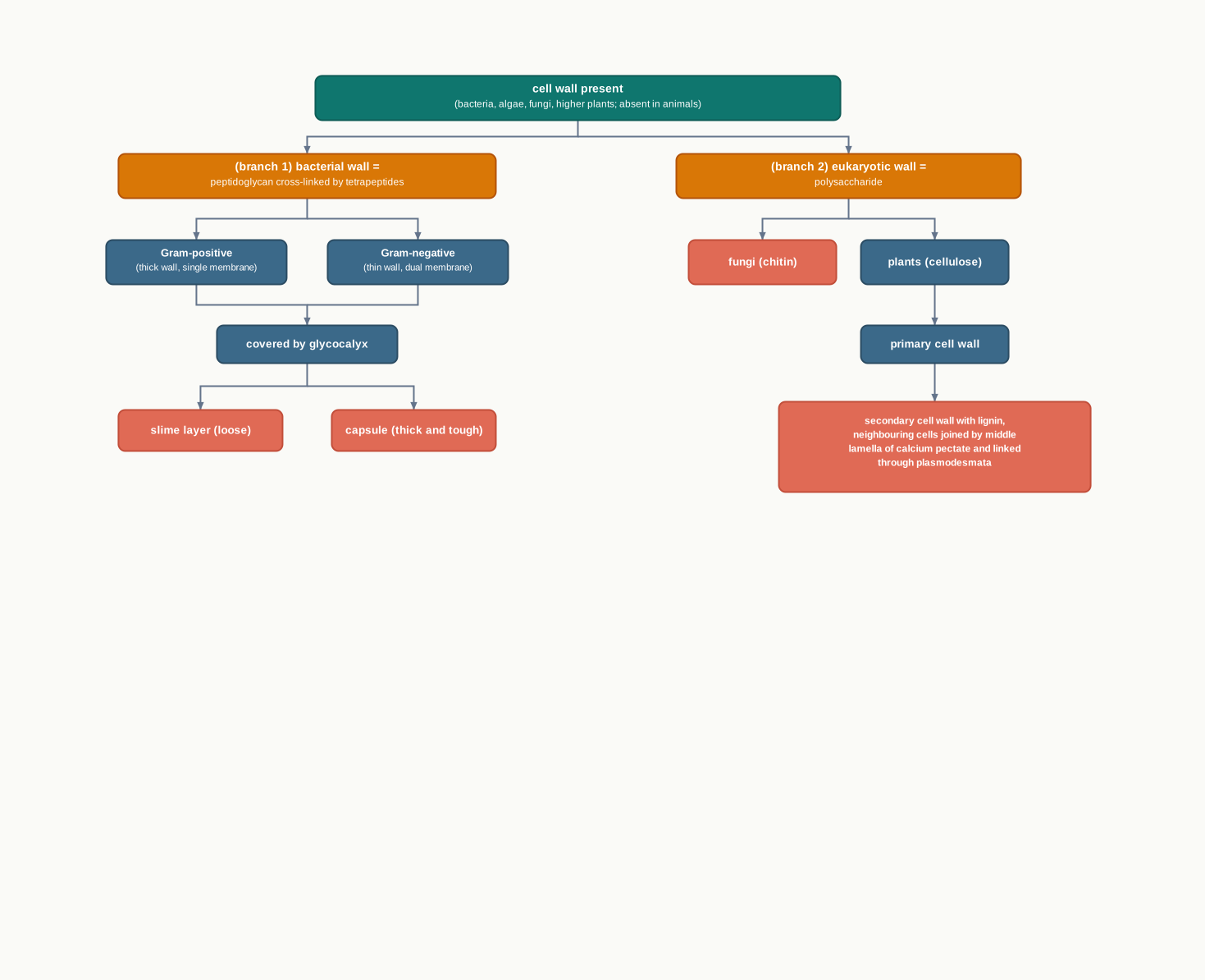
Fig. 2.7 – Types of cell wall.
2.3 Endomembrane System
Among eukaryotes there are many cell organelles which are bound by a membrane similar to that of the cell membrane, and these are distinct from one another in terms of structure and function. Yet some of these membrane-bound organelles work together in a system known as the endomembrane system, because their functions are co-ordinated with each other. The prefix endo- means ‘within’.
It comprises a group of membrane-bound organelles that work together in protein and lipid synthesis, and in the processing, packaging and transport of these products to their respective locations inside a cell. The endomembrane system includes the endoplasmic reticulum, the Golgi complex, lysosomes and vacuoles.
An Assembly Line, Not a Collection
It is worth being clear about why these four are grouped together while mitochondria and plastids are not. The organelles of the endomembrane system pass material to one another in a definite order, using small membrane sacs called vesicles that bud off from one compartment and fuse with the next.
Because a vesicle carries its cargo enclosed in membrane, the cargo never has to be released into the cytosol on the way. A protein made for export can therefore travel the whole distance to the cell surface without once mixing with the general contents of the cell.
That pathway is the backbone of this whole section, and it is set out in Fig. 2.8.

Fig. 2.8 – Pathway of a secretory protein.
2.3.1 Endoplasmic Reticulum
The endoplasmic reticulum (ER) is an extensive network of membrane-enclosed tubules and cisternae located near the nucleus and the Golgi apparatus. It is exclusively present in a eukaryotic cell.
The ER is a large and dynamic structure, continuously involved in protein synthesis, calcium storage and lipid metabolism. Its enormous folded surface is the point of it: by extending membrane throughout the cytoplasm, the cell creates a very large working area and, at the same time, a separate internal space in which newly made proteins can be handled.
Based on the presence or absence of ribosomes, the endoplasmic reticulum may either be rough ER or smooth ER (Fig. 2.9).
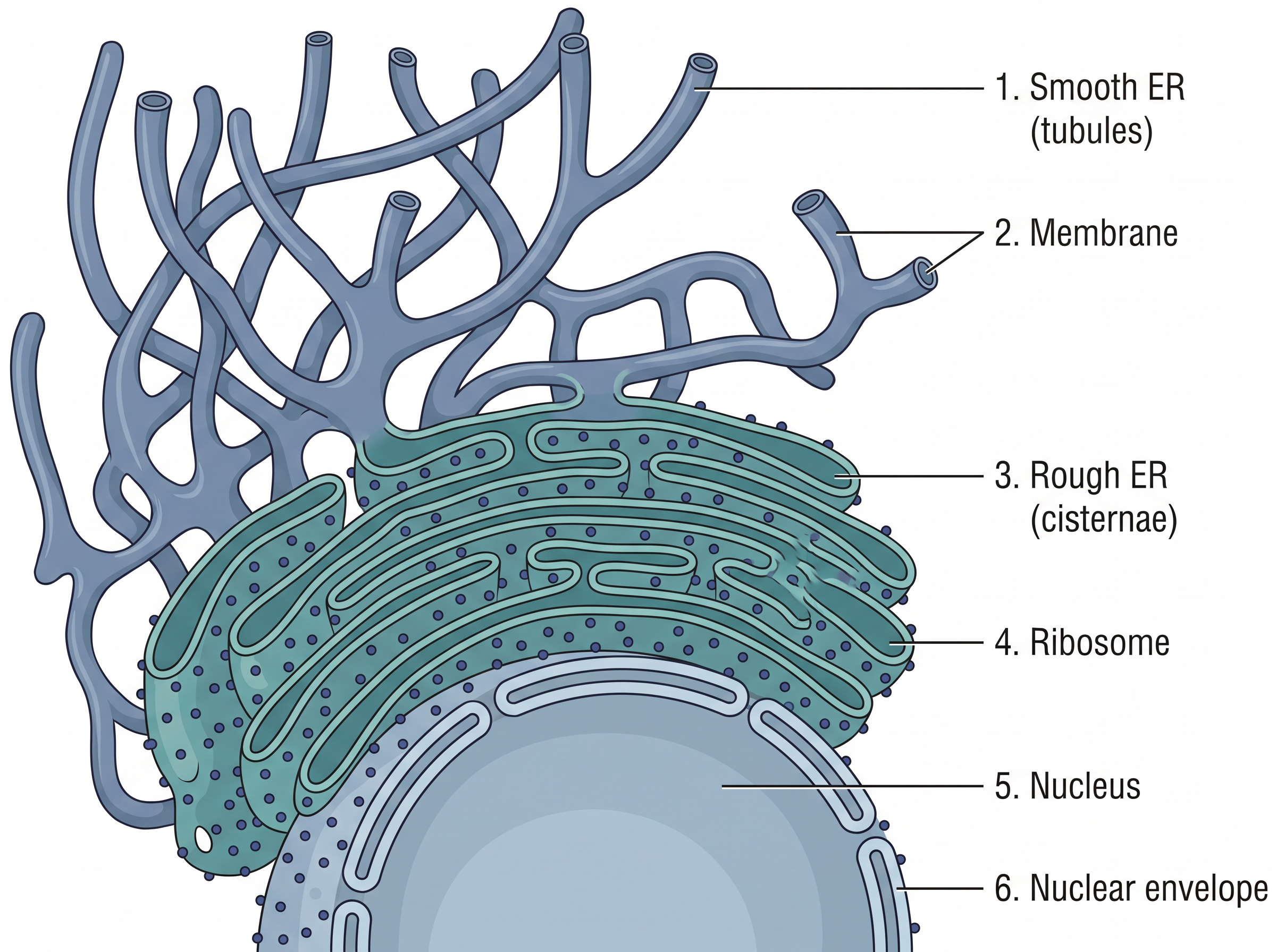
Fig. 2.9 – Endoplasmic reticulum: (a) smooth ER and (b) rough ER.
Notice in Fig. 2.9 that the two forms are not two separate organelles. They are two regions of one continuous membrane network, differing in whether ribosomes are attached to the outer surface — and, as a result, differing in what work goes on there.
Rough Endoplasmic Reticulum (RER)
Rough ER can be distinguished from smooth ER by the presence of ribosomes on its cytosolic surface. Ribosomes are the protein synthesising factory of a cell; their own structure is taken up in Section 2.6.
Proteins synthesised on free ribosomes are released into the cytoplasm, and they are directly transported to the nucleus, mitochondria, chloroplast and peroxisomes to be used within the cell [Fig. 2.10(a)].
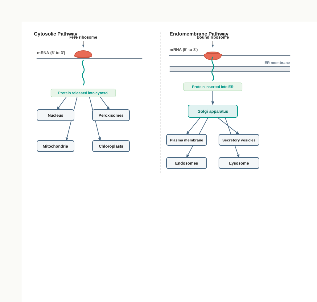
Fig. 2.10 – Protein sorting in higher eukaryotes: (a) free and (b) bound ribosomes.
In the case of bound ribosomes, after initiation of protein synthesis the ribosome–protein complex is transferred to a receptor on the ER in eukaryotes. There the nascent protein — that is, the protein still under synthesis by the ribosome — is inserted into the ER.
These proteins may either be retained in the ER, or transported to their destinations via the Golgi complex through the secretory pathway [Fig. 2.10(b)]. The ER plays a significant role in the trafficking of secretory proteins to the Golgi apparatus, lysosomes and the plasma membrane within eukaryotic cells.
Compare the two halves of Fig. 2.10 as a single decision. Every protein begins on a ribosome; what differs is whether that ribosome stays free in the cytosol or docks on the ER. A protein finished in the cytosol goes to compartments the cell keeps for itself. A protein pushed into the ER enters the vesicle system and can be sent to the cell surface or out of the cell altogether.
Smooth Endoplasmic Reticulum (SER)
As mentioned earlier, ribosomes are not present on the surface of smooth ER [Fig. 2.9(a)]. It is mainly involved in lipid metabolism.
As lipids are hydrophobic, they cannot be synthesised in the cytosol, which is watery. A lipid being built needs a hydrophobic environment to sit in, and the interior of a membrane provides exactly that. Most of the lipids are therefore synthesised in the SER and transported to their respective destinations as transport vesicles.
Phospholipids are one of the crucial components of a membrane, and are derived from glycerol. Their synthesis takes place on the outer side, that is the cytosolic side, of the SER membrane. The SER is also an essential site for cholesterol synthesis.
2.3.2 Golgi Apparatus
The Golgi apparatus was first observed by Camillo Golgi, an Italian biologist, in 1898, as a dark stained reticular structure located near the cell nucleus. This was later found to be present in other cell types as well, and was named the Golgi apparatus or the Golgi complex.
It is a membrane-bound cell organelle consisting of a series of flattened membranous sacs that look like stacked pouches, called cisternae. A varied number of cisternae are present in a stack. The membrane of each cisterna in a stack separates its internal space from the cytosol, so each sac remains a closed compartment with its own contents.
The Golgi cisternae are concentrically arranged near the nucleus, with a distinct cis face — the cisternae nearest the endoplasmic reticulum, also called the forming face — and a trans face — the cisternae away from the endoplasmic reticulum, also called the maturing face (Fig. 2.11).
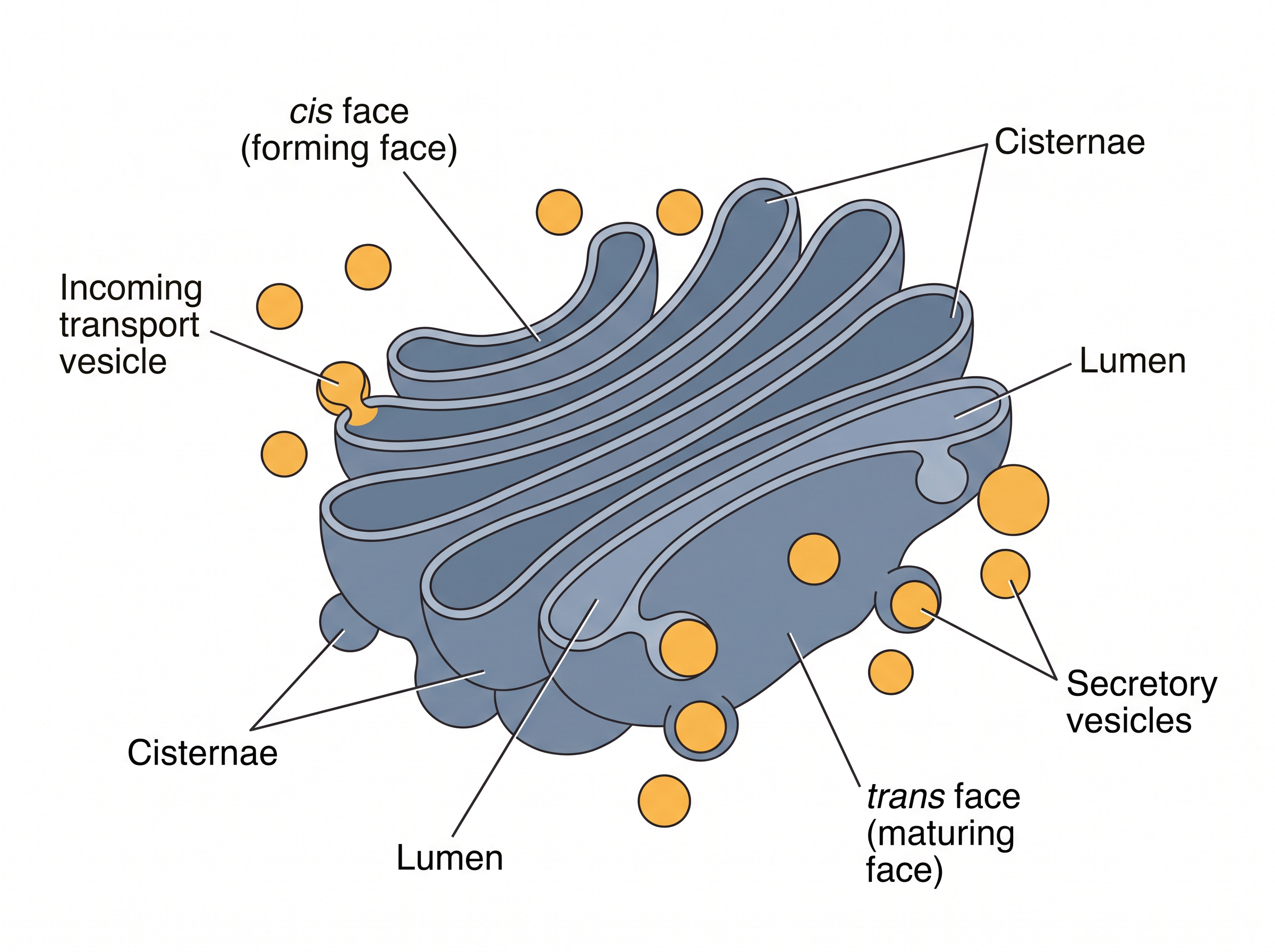
Fig. 2.11 – Structure of the Golgi apparatus.
The two names given to each face tell you what happens there. The cis or forming face is where new material arrives and a new cisterna begins; the trans or maturing face is where fully processed material leaves. The stack therefore has a direction built into it, and cargo moves through it one way.
The Golgi apparatus principally performs the function of packaging materials and preparing them for secretion — it is, in effect, the receiving and shipping department of the cell. Transport vesicles move material from the ER to the Golgi complex.
The material to be secreted moves from the ER to the Golgi apparatus, during which vesicles are budded off from the ER. These vesicles travel to the Golgi apparatus and fuse with the cis face. The trans face gives rise to vesicles that pinch off and travel to other sites. These vesicles can fuse with the plasma membrane, releasing their contents outside the cell. Some vesicles deliver their contents to other organelles instead.
Proteins synthesised by ribosomes of the RER are modified in the cisternae of the Golgi apparatus before they are released from the trans face. For example, specific types of sugars are attached to some proteins before they are released from the cell.
The Golgi apparatus is thus a central membrane organelle for trafficking and post-translational modification of protein and lipid in the cell. ‘Post-translational’ simply means after translation — after the protein has already been built by the ribosome. It is also an important site of formation for glycoproteins and glycolipids, that is, proteins and lipids carrying attached sugar chains.
2.3.3 Lysosomes
Present in the cytoplasm, lysosomes are small spherical vesicles of approximately 0.2 – 0.5 micron in diameter, which are bound by a single membrane containing hydrolytic enzymes capable of breaking down macromolecules.
These special vesicles of animal cells and some other eukaryotes are formed either from the Golgi apparatus or directly from the endoplasmic reticulum — which is precisely why lysosomes belong to the endomembrane system.
Lysosomal enzymes show optimal activity at an acidic pH, and thus they are acid hydrolases. They are used in the dissolution and digestion of redundant structures or damaged macromolecules from within or outside the cell.
For example, when an animal cell ingests food into a food vacuole, lysosomes fuse with the vacuole and break down the contents — carbohydrates, proteins, fats and other components — enzymatically. Lysosomes carry out intracellular digestion in a variety of circumstances.
They also use their hydrolytic enzymes to recycle the cell’s own organic material through a process called autophagy, and in this way the cell continually renews itself. Worn-out organelles and damaged molecules are broken down to their building blocks, which the cell then uses again.
What happens when this digestive machinery fails can be seen in disease. In Tay-Sachs disease, the brain becomes impaired due to accumulation of lipids in the cells, because of a lack of, or the inactivation of, the lipid-digesting enzymes in them. The lipids that should have been broken down simply build up inside the cells, and nerve cells are damaged as a result — a reminder that recycling is not an optional extra for a cell but a continuous necessity.
2.3.4 Vacuoles
Vacuoles are membrane-bound intracellular organelles found in the cytoplasm of most plants, fungi and also some animal cells. The term ‘vacuole’, meaning ‘empty’, comes from its transparent morphology and lack of cytoplasmic material — a name given from appearance rather than from function. Its major functions are storage, structural support and recycling.
The number and size of vacuoles varies per plant cell. Young cells have a large number of small vacuoles. As the plant cell matures, the vacuoles amalgamate to form a large central vacuole that occupies almost 90 per cent of the cytoplasmic volume.
The central vacuole contains water, cell sap, solid inclusions and other metabolites. Other types of vacuoles found in a plant cell are lytic vacuoles, protein storage vacuoles (PSV) and other storage vacuoles. The vacuoles are covered by a membrane called the tonoplast.
The major function of vacuoles in plant cells is storage, maintaining cell turgor, and also protecting cells during biotic stresses.
Fungal vacuoles, however, are comparatively complex organelles performing a variety of functions apart from storage, such as helping in degradative processes, playing a role in osmoregulation, and maintaining intracellular pH.
Food vacuoles help in engulfing food particles, and contractile vacuoles help in excretion. In the case of Amoeba, the contractile vacuole plays a critical role in excretion and osmoregulation. In many protists, food vacuoles are formed by engulfing food particles.
2.4 Mitochondria
Mitochondria (singular: mitochondrion) are found in nearly all eukaryotic cells. Some cells may have a single large mitochondrion, but more often a cell has hundreds or even thousands of mitochondria, placed at variable locations within the cell depending upon the cell’s function.
That variation is itself informative. A cell doing a great deal of energy-demanding work needs more of these organelles, and it tends to keep them close to wherever the energy is being spent, rather than scattered at random.
Mitochondria possess recognisable morphological characteristics despite considerable variability in appearance. A typical mitochondrion is sausage shaped. In electron micrographs, they appear mostly as rod-shaped or cylindrical. They vary in size within the range 3.0 to 10.0 μm long and 0.5 – 1.5 μm wide.
A Cell Within a Cell
Each mitochondrion is a double membrane bound structure, that is, it has an outer and an inner membrane, each consisting of a phospholipid bilayer with proteins (Fig. 2.12). You have already met this bilayer-with-proteins arrangement at the plasma membrane; the same basic design is reused here, twice over.
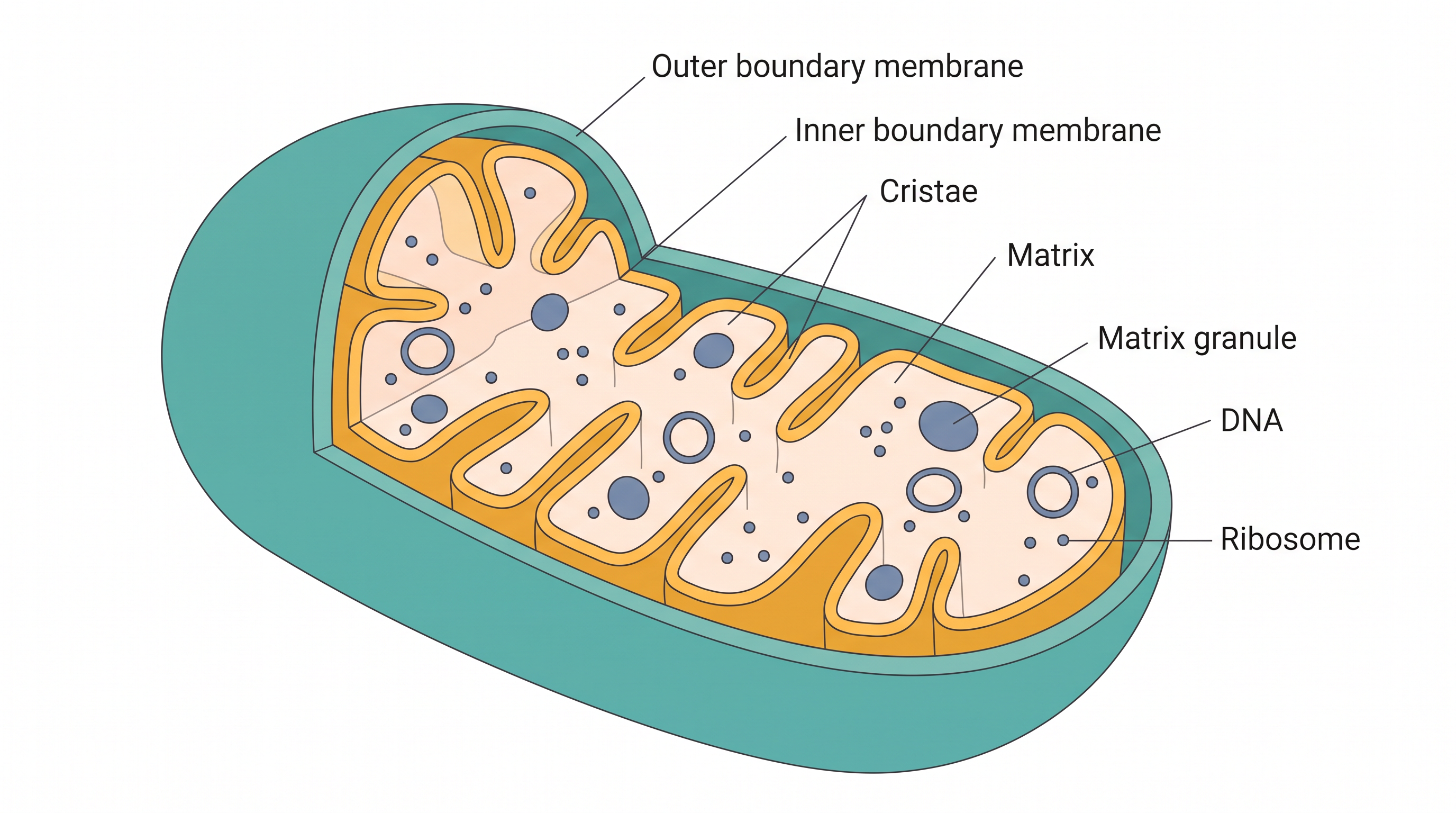
Fig. 2.12 – Internal structure of a mitochondrion.
The outer membrane is smooth, but the inner membrane has infoldings called cristae (singular: crista) that provide it with a comparatively larger surface area.
This is a structure–function link worth holding on to. The reactions of respiration are carried out by enzymes and proteins seated in and near the inner membrane, so the amount of work a mitochondrion can do depends directly on how much inner membrane it has. Folding that membrane into cristae packs far more surface into the same small volume, without making the organelle any bigger.
The inner membrane divides the mitochondrion into two internal compartments. The narrow region between the inner and the outer membrane is the peri-mitochondrial space, and the innermost compartment lined by the inner membrane is called the mitochondrial matrix.
Getting these compartments in the right order is a common requirement in diagram-labelling questions, so trace them from the outside inwards as shown in Fig. 2.13.

Fig. 2.13 – Compartments of a mitochondrion.
Where the Energy Work Happens
The inner membrane and the matrix contain all the enzymes and proteins involved in the process of the tricarboxylic acid (TCA) cycle and cellular respiration, for the purpose of ATP synthesis. The details of these processes are given in Chapter 5.
You have already seen ATP at work in Section 2.1, driving the Na⁺-K⁺ pump against a concentration gradient. This is where that ATP comes from. Placing the whole set of respiratory enzymes inside a sealed double membrane also means the cell can maintain conditions inside the mitochondrion that differ from those in the cytoplasm — which is exactly what these reactions require.
The Mitochondrion’s Own Genetic System
Mitochondria also contain DNA molecules, ribosomes (70S) and a few RNA molecules. Some of the mitochondrial proteins are synthesised by genes present on the mitochondrial DNA.
Therefore, the mitochondrial matrix is the site of organellar DNA replication, transcription, protein synthesis and other enzymatic processes. In other words, this organelle carries a small working copy of the machinery that the rest of the cell keeps in its nucleus and cytoplasm. Keep the detail about 70S ribosomes in mind — we shall return to what it suggests when ribosomes are taken up in Section 2.6.
Not everything the organelle needs is made this way, however. We know that mitochondria consist of well over 1000 proteins, which vary within and between species in response to the needs of the organisms. Only a small share of these is encoded by mitochondrial DNA; the rest are made elsewhere in the cell and imported.
Mitochondrial DNA has become useful far outside cell biology. Because a cell contains many mitochondria and each carries its own DNA, this DNA is present in many copies, and it can often still be recovered from old, burnt or badly degraded material in which the nuclear DNA has broken down beyond use. It is also passed on through the mother, so all maternal relatives share the same mitochondrial sequence. Forensic scientists use both facts when identifying human remains from disasters or from very old skeletal material, where a maternal relative can be matched to the sample.
2.5 Plastids
Plastids are usually present in the cytoplasm of plant cells. The term plastid is derived from the Greek word Plastikas, meaning formed or moulded. They are easily observed under the microscope, as they are large.
Plastids bear some specific pigments, and thus impart specific colours to plants. Three different types of plastids are recognised on the basis of their pigments or colouration.
Chromoplasts — These are coloured plastids containing variously coloured pigments such as the yellow, red, pink and violet colours of flowers, fruits, leaves and so on. In chromoplasts, fat soluble pigments like carotene, xanthophylls and others are present.
Leucoplasts — These are colourless plastids. They are usually involved in the storage of various kinds of reserve food materials, and are named accordingly: amyloplasts (storage of starch), aleuroplasts (storage of protein) and elaioplasts or lipoplasts (storage of oil).
Chloroplasts — These are the green plastids, universally found in all the green parts of the plant, specially the green leaves. A chloroplast contains large quantities of green pigments, called chlorophyll. Chlorophyll is a collection of four pigments: chlorophyll a, chlorophyll b, and the yellow pigments — carotenoids and xanthophylls.
The three types are set out in Fig. 2.14, which is worth memorising in this branched form rather than as a flat list, because the storage plastids are further named by what they store.
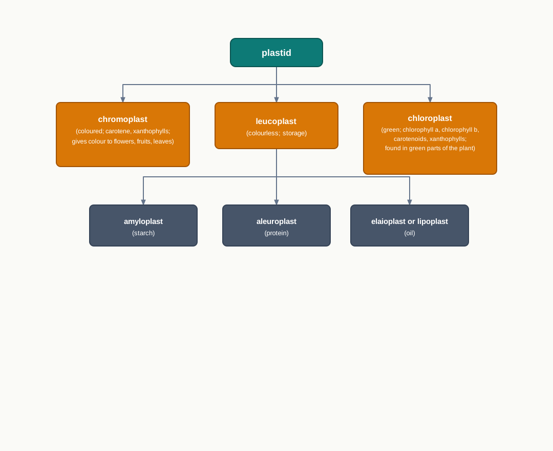
Fig. 2.14 – Classification of plastids.
These categories are easier to remember once you connect them to plants you already know. The white flesh of a potato tuber is packed with amyloplasts, which is why it is a starch crop. The colour of a ripening tomato or a marigold petal comes from chromoplasts. And the green of every leaf comes from chloroplasts.
Carotene, one of the pigments named above, is also the substance our bodies convert into vitamin A. Rice grain does not accumulate it in the edible part, and vitamin A deficiency remains a serious health problem in populations that depend heavily on rice. This is what led Ingo Potrykus and Peter Beyer to engineer a rice variety in which the grain accumulates carotene, giving it a golden colour — Golden Rice. It is a direct example of biotechnology working on the pigment chemistry of plastids to address a nutritional problem.
The Structure of a Chloroplast
Chloroplasts are located predominantly in the mesophyll cells of the leaves. The chloroplasts of higher plants are generally lens-shaped — oval, spherical, discoid, flat ellipsoids, or even ribbon-like organelles — and on average 2 to 4 μm wide and 5 to 10 μm long. They are the largest organelles in the plant cell.
The chloroplast is bounded by a double membrane, separated by a very narrow intermediate space. So far this resembles the mitochondrion, but the chloroplast then does something the mitochondrion does not.
It also contains a third inner membrane, which is organised into flattened membranous sacs called thylakoids. Thylakoids are arranged in orderly stacks called grana (singular: granum), which resemble stacks of coins.
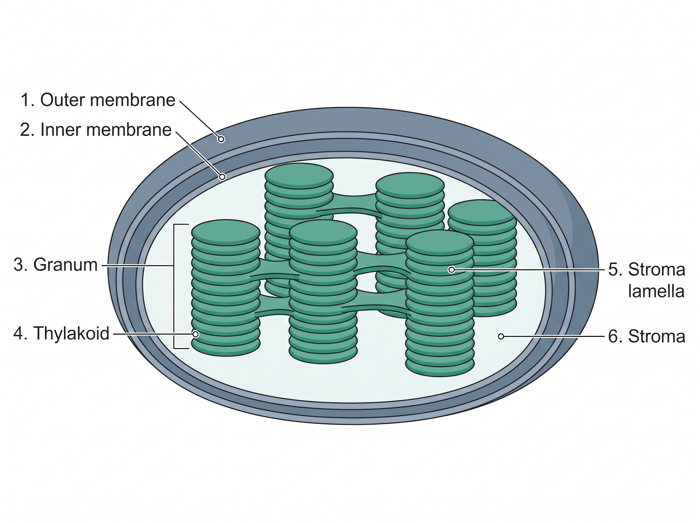
Fig. 2.15 – Sectional view of a chloroplast.
The space, or fluid, outside the thylakoids and within the chloroplast envelope is the stroma, which contains the chloroplast DNA and ribosomes as well as many enzymes. In addition, there are flat membranous tubules called the stroma lamellae, connecting the thylakoids of the different grana. The membrane of the thylakoids encloses a space called a lumen.
Trace this in Fig. 2.15 before going further. The membranes of the chloroplast divide the chloroplast space into three compartments: the intermediate space, the stroma, and the thylakoid space. Note that this is one more compartment than a mitochondrion has, and the extra one exists because of the third membrane system.
Where Photosynthesis Begins
Chlorophyll pigments are present in the thylakoids. The thylakoid membrane contains light-harvesting proteins, reaction centres, electron-transport chains and ATP synthase, which carry out the primary events of photosynthesis. The details of photosynthesis are given in Chapter 5.
Each of those four components has a clear job in sequence: the light-harvesting proteins collect light energy, the reaction centres convert it into a flow of electrons, the electron-transport chains pass those electrons along, and ATP synthase uses the result to make ATP. All four are held in the thylakoid membrane, which is why the amount of thylakoid membrane matters so much to a leaf.
The ribosomes of the chloroplasts are smaller (70S) than the cytoplasmic ribosomes (80S). You have already met 70S ribosomes once, inside the mitochondrion. The chloroplast, too, has its own DNA and its own ribosomes of this smaller type — and the significance of that pattern will become clear in Section 2.6.
2.6 Ribosomes
Ribosomes are the ‘protein synthesising factories’ scattered throughout the cytoplasm in both prokaryotic and eukaryotic cells. Each ribosome is a membraneless cell organelle, which was first observed by George Palade in 1955 under the electron microscope.
Note the two things that make ribosomes unusual among the structures in this chapter. They are found in prokaryotes as well as eukaryotes, unlike most organelles you have met so far; and they have no membrane of their own. Everything else in the endomembrane system is defined by its membrane — a ribosome is defined by what it is built from.
The number of ribosomes per cell is quite variable. A rapidly growing mammalian cell has approximately 10 million ribosomes. A cell that is growing fast must build protein fast, and since every protein in the cell is made on a ribosome, the count rises with demand.
How Ribosomes Are Named
Ribosomes have been classified on the basis of their sedimentation rate in a centrifuge machine — prokaryotic ribosomes as 70S and eukaryotic ribosomes as 80S.
Because S is a rate and not a mass, S values do not simply add up. This is worth fixing firmly in mind before the next paragraph, since the numbers look like arithmetic and are not.
The Two Subunits
Ribosomes are mainly composed of rRNA and proteins. Each ribosome has a small and a large subunit (Fig. 2.16).
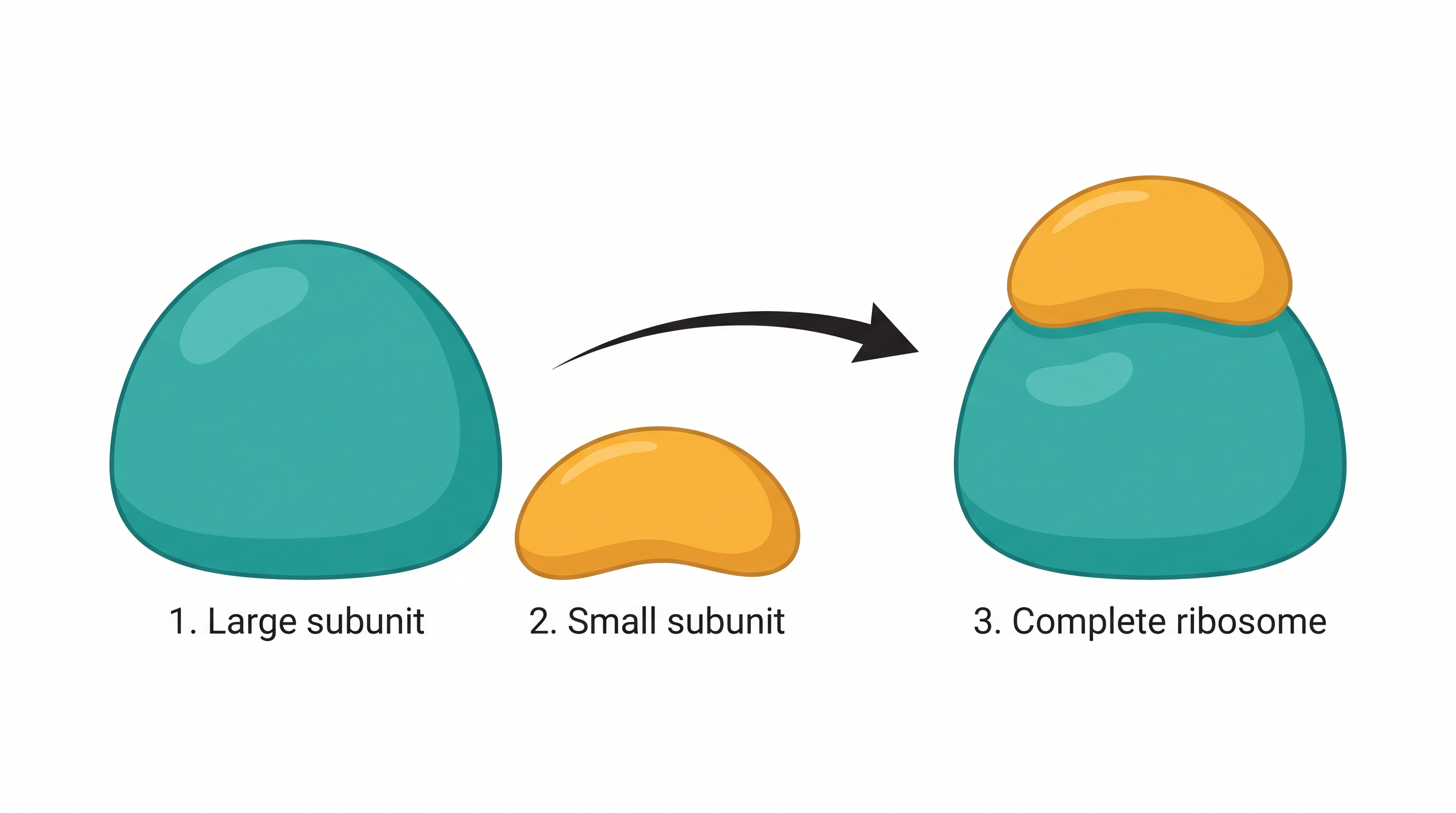
Fig. 2.16 – Subunits of a ribosome.
The 70S prokaryotic ribosome has a larger 50S and a smaller 30S subunit. On the other hand, 80S eukaryotic ribosomes also contain two subunits, that is, a larger subunit of 60S and a smaller subunit of 40S.
Look carefully at those figures. A 50S and a 30S subunit combine to give a 70S ribosome, not an 80S one; a 60S and a 40S subunit give 80S, not 100S. Nothing has gone wrong. Sedimentation rate depends on shape as well as on mass, and the joined particle is more compact than the two separate pieces, so it does not sediment at the sum of their rates.
In both prokaryotes and eukaryotes, the subunits of ribosomes remain dissociated in the cytoplasm when the ribosome is not involved in protein synthesis. They come together only when there is work to do — which is why Fig. 2.16 shows both the separated and the joined state.
What the rRNA Does
In a ribosomal subunit, rRNAs assume a characteristic secondary structure by utilising complementary base pairing, and form a distinct three-dimensional structure by associating with ribosomal proteins.
That is worth unpacking. A single rRNA strand folds back on itself wherever its bases can pair with one another, and this folding gives the molecule a definite shape. The ribosomal proteins then bind to that folded rRNA, and the finished subunit takes on its precise three-dimensional form. The shape is not incidental — a ribosome must hold messenger RNA and transfer RNA in exactly the right positions, and only a precisely shaped particle can do so.
Some rRNAs present in the large subunit have catalytic activities and are called ribozymes.
This is a striking finding, because catalysis had long been regarded as the business of proteins alone. In the ribosome, it is an RNA molecule that performs a catalytic role.
Why the 70S Ribosome Turns Up Inside Eukaryotes
It is peculiar to note that 70S ribosomes are also present in the mitochondria and chloroplasts of a eukaryotic cell, suggesting their relatedness to the prokaryotic cell.
Put the evidence from the last three sections side by side. Mitochondria and chloroplasts are each bounded by a double membrane; each carries its own DNA; each carries out its own replication, transcription and protein synthesis; and each contains ribosomes of the 70S type — the prokaryotic type — rather than the 80S ribosomes found in the surrounding cytoplasm of the very same cell.
This difference between 70S and 80S is not merely a matter of classification. Several groups of antibiotics work by binding to the 70S ribosome and blocking bacterial protein synthesis, while leaving the 80S ribosomes of our own cytoplasm largely untouched. That is selective toxicity of the same kind you met with cell-wall antibiotics in Section 2.2, but acting at a completely different target — a reminder that a drug is useful only when the pathogen has something the patient does not.
2.7 Microbodies
These are small, single-membrane bound cell organelles, present only in eukaryotic cells. They are usually present near the endoplasmic reticulum. Based on their functional properties, microbodies are classified into two types: peroxisomes and glyoxysomes.
Notice the word single. Mitochondria and chloroplasts are bounded by two membranes; microbodies by one. That single point separates them at once in a classification question.
2.7.1 Peroxisomes
Peroxisomes are small, membrane-bound organelles. They are involved in energy metabolism in the cell, and thus serve as a site for enzymes involved in metabolic reactions.
They are derived from the ER and replicate by fission — that is, an existing peroxisome divides into two, rather than a new one being built from nothing. Unlike mitochondria and chloroplasts, the peroxisome lacks its own genome. Every protein it contains must therefore be made elsewhere in the cell and brought in.
In animal cells, peroxisomes perform two major functions: oxidation and lipid biosynthesis. They contain oxidases that help in peroxide production, and catalase that neutralises the harmful products of the oxidation reaction.
This pairing of enzymes is the whole logic of the organelle. Oxidation reactions generate hydrogen peroxide, which would damage the cell if it were released freely into the cytoplasm. So the cell keeps both the enzyme that produces it and the enzyme that destroys it inside the same sealed compartment. Catalase removes the peroxide as fast as it forms:
\[2\text{H}_2\text{O}_2 \rightarrow 2\text{H}_2\text{O} + \text{O}_2\]
Here \(\text{H}_2\text{O}_2\) is hydrogen peroxide, \(\text{H}_2\text{O}\) is water and \(\text{O}_2\) is oxygen. The dangerous product is converted into two entirely harmless ones.
In liver cells, peroxisomes also help in alcohol detoxification. This is a familiar, everyday role for an organelle most students never hear about: a considerable share of the alcohol that reaches the liver is broken down through oxidation reactions of this kind.
In plant cells, peroxisomes have two major roles to play — the conversion of fatty acids into carbohydrates in seeds, and photorespiration in leaves.
2.7.2 Glyoxysomes
Glyoxysomes are specialised peroxisomes found in fungi and other higher plants, especially in the fat storage tissues in germinating seeds. When oil-filled seeds germinate, the number and activity of glyoxysomes also increase.
Glyoxysomes contain all the enzymes necessary for fatty acid oxidation, the glyoxylate cycle and gluconeogenesis. The seedling uses these sugars synthesised from fats until it is mature enough to produce them by photosynthesis.
The conversion of lipids into glucose requires the coordinated function of glyoxysomes, mitochondria and plastids. No single organelle carries out the whole route; the pathway is handed on from one compartment to the next, as summarised in Fig. 2.17.
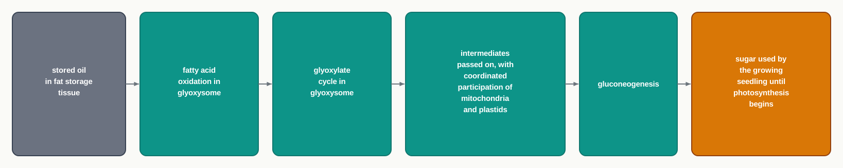
Fig. 2.17 – Conversion of stored fat into sugar.
2.8 Cytoskeleton
The cytoskeleton is a multi-component system made of fibrous proteins. It maintains cell organisation and shape. It is a vital component of the cell that provides mechanical support, and plays a very crucial role especially during cell division, cell movement and intracellular transport.
The name can be misleading, so it is worth correcting straight away. A skeleton suggests something rigid and permanent, but the cytoskeleton is neither. Its filaments are built up from protein subunits and can be taken apart again, which is exactly what lets a cell change shape, divide in two, or move from one place to another.
The cytoskeleton is composed of three major filaments, varying in their protein composition and diameter: microtubules (25 nm) made of tubulin protein, actin filaments (6 nm) made of actin protein, and intermediate filaments (10 nm) made of a combination of different subunits of protein.
The unit used here, the nanometre (nm), equals \(10^{-9}\ \text{m}\). These filaments are therefore far below the resolving power of a light microscope, which is why the cytoskeleton was among the later features of the cell to be described. Note also that the three are named by diameter in a way that is easy to muddle — the intermediate filament sits between the other two in thickness, which is where its name comes from.
The Three Filaments
1. Microtubules — These are composed of globular proteins called tubulin, which is a dimer having α and β subunits. Tubulin proteins undergo polymerisation to form a protofilament. Microtubules are hollow rod-like structures, and may contain 10–15 protofilaments (Fig. 2.18).
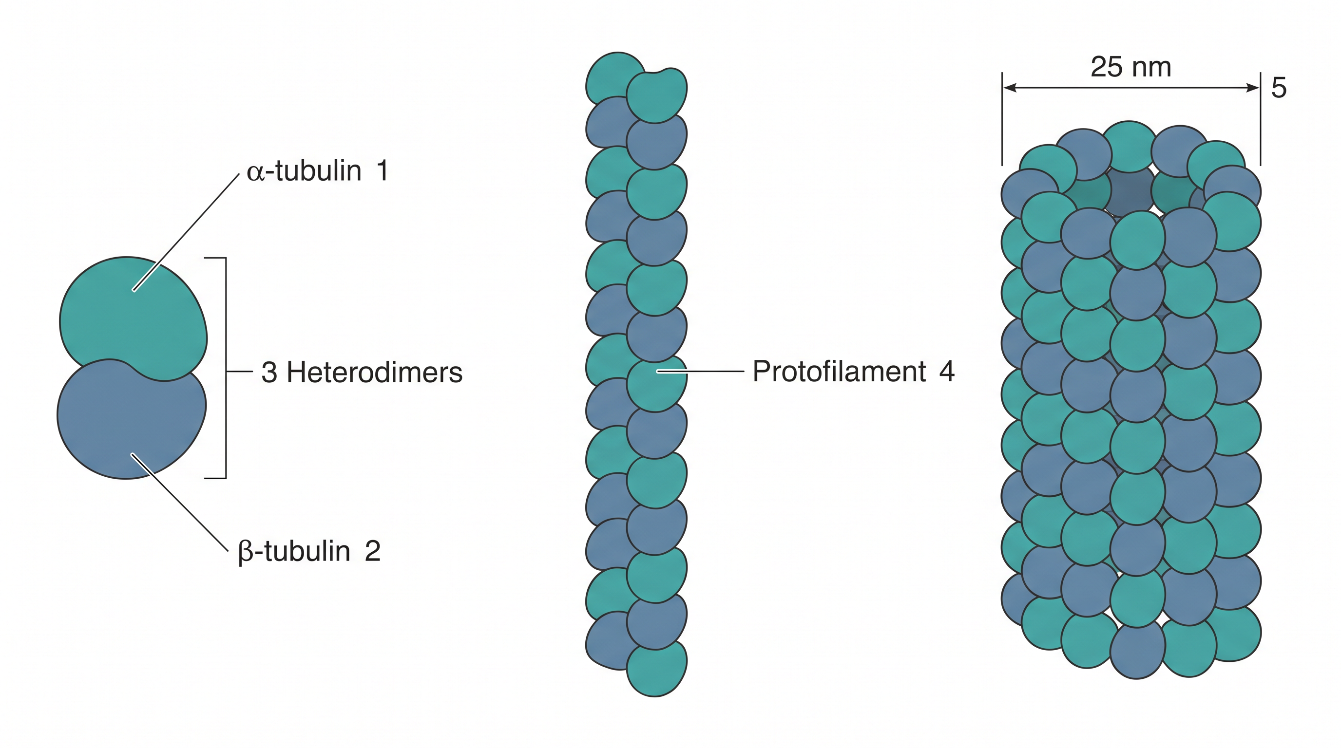
Fig. 2.18 – Structure of a microtubule.
Follow the building sequence in Fig. 2.18, because the terms are easy to confuse. An α and a β subunit together form one dimer; dimers joined end to end form a protofilament; and 10–15 protofilaments arranged side by side form the hollow tube that is the microtubule. Being hollow rather than solid gives the structure considerable stiffness for the amount of protein used, in the same way that a hollow steel pipe resists bending better than a solid rod of equal weight.
In addition to its other functions, the microtubule is also responsible for the rhythmic movement of cilia and flagella — structures taken up in Section 2.9 — and for the spindle apparatus of cell division, which is dealt with in Section 2.10.
2. Actin filament — This is observed in skeletal muscle and plays a very important role in muscle contraction. It is richly found in the cytoplasm closer to the plasma membrane. Its main function is to provide strength to the cell, facilitating cytokinesis and cell movement.
Its position just beneath the plasma membrane fits its job. Cytokinesis — the division of the cytoplasm at the end of cell division — requires the cell surface to be drawn inwards, and a cell that crawls must push its surface forward. Both are surface events, so the filaments that carry them out are concentrated where the surface is.
3. Intermediate filaments — These are strong filaments resembling a rope, primarily involved in providing mechanical strength to the cell.
The comparison with a rope is exact and useful. A rope is made of many thin strands twisted together, which is why it bears a heavy pull without snapping even though no single strand could. Intermediate filaments are built the same way, and this is why they are the cell’s main defence against being torn by mechanical stress.
A Target for Medicine
Because microtubules must assemble and disassemble for a cell to divide, they are a natural target for drugs meant to stop rapidly dividing cells. Vinblastine and vincristine, obtained from the Madagascar periwinkle Catharanthus roseus, act in exactly this way: they interfere with the assembly of tubulin into microtubules, so the spindle cannot form and division stalls. Both are used in cancer chemotherapy.
This is a good example of why structural detail is worth learning. Knowing that a microtubule is a polymer of tubulin dimers is not merely descriptive — it identifies the precise step at which a drug can intervene.
2.9 Cilia and Flagella
Cilia (singular: cilium) and flagella (singular: flagellum) are hair-like, microscopic, filamentous protoplasmic structures. Both cilia and flagella are involved in cell motility.
Although cilia and flagella are morphologically and physiologically identical, they can be differentiated on the basis of their size, number and function (Table 2.1). That is an important sentence to read carefully: they are built the same way, and they differ in how long they are, how many there are, and how they move — not in what they are made of.
Cilia are smaller in size and present in large number in a ciliated cell, whereas flagella are longer in size and typically vary from one or two in number.
Where They Are Found
The body of a Paramecium, a unicellular protozoan, is fully covered by a few thousands of cilia. Cells present in the upper respiratory tract of mammals bear cilia to expel particulate matter present in inhaled air.
This second example is worth pausing on, because it is happening inside you as you read. Dust, smoke particles and microorganisms are trapped in the mucus lining your airways, and the cilia of the epithelial cells beat in a coordinated rhythm that sweeps that mucus upwards and out. Anything that damages or paralyses these cilia — tobacco smoke being the best-known example — leaves the particles where they land, which is one reason smokers cough persistently and suffer more chest infections.
A mammalian spermatozoan contains a single flagellum, while a unicellular Chlamydomonas, a green alga, has two flagella. Notice how the number relates to the job: the sperm cell must propel itself as a whole, and one long flagellum does that, whereas a lining of thousands of short cilia is what a sheet of stationary cells needs in order to move something across its surface.
The Structure Inside
Cilia and flagella are fibrillar and made up of microtubules. You met microtubules in Section 2.8, where it was noted that they are responsible for the rhythmic movement of cilia and flagella; this section shows how they are arranged to achieve it.
Their fundamental structure is the same. Both arise from a centriole-like structure known as the basal body, which is taken up further in Section 2.10.
The electron microscopic view shows that they are bounded by a unit membrane of thickness 90 Å, which is continuous with the plasma membrane (Fig. 2.19). The unit used here, the angstrom, equals \(1\ \text{Å} = 10^{-10}\ \text{m}\), so 90 Å is \(9 \times 10^{-9}\ \text{m}\), or 9 nm.
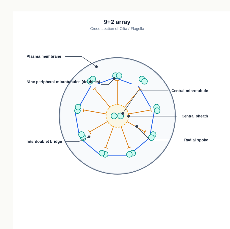
Fig. 2.19 – Eukaryotic cilium showing the 9+2 array.
The fact that this membrane is continuous with the plasma membrane matters. A cilium is not a separate object stuck onto the cell — it is an outgrowth of the cell itself, wrapped in the same boundary membrane you studied in Section 2.1.
They have a core known as the axoneme in the matrix, containing nine peripheral and two central microtubules. Such an arrangement is referred to as the 9+2 array. The central fibrils are enclosed by a sheath.
Read Fig. 2.19 as a cross-section, as though the cilium had been cut across and you were looking down the cut end. The nine doublets lie in a ring near the membrane, the two central microtubules lie at the centre within their sheath, and the radial spokes and interdoublet bridges link these parts into one mechanical unit. Without those connections the microtubules could slide past one another freely; because they are tied together, sliding is converted into bending, and the cilium beats.
Prokaryotes also contain cilia/flagella, but these differ structurally from the eukaryotic ones in terms of the 9+0 array.
2.10 Centrosome and Centrioles
The centrosome is present in the animal cell cytoplasm near the nucleus. It consists of two cylindrical centrioles placed perpendicular to each other, embedded in amorphous pericentriolar materials.
The word amorphous means without a definite shape, so the pericentriolar material is a shapeless mass surrounding the two centrioles rather than a structure with a form of its own. The centrioles sit within it at right angles to each other, which is the arrangement shown in Fig. 2.20.
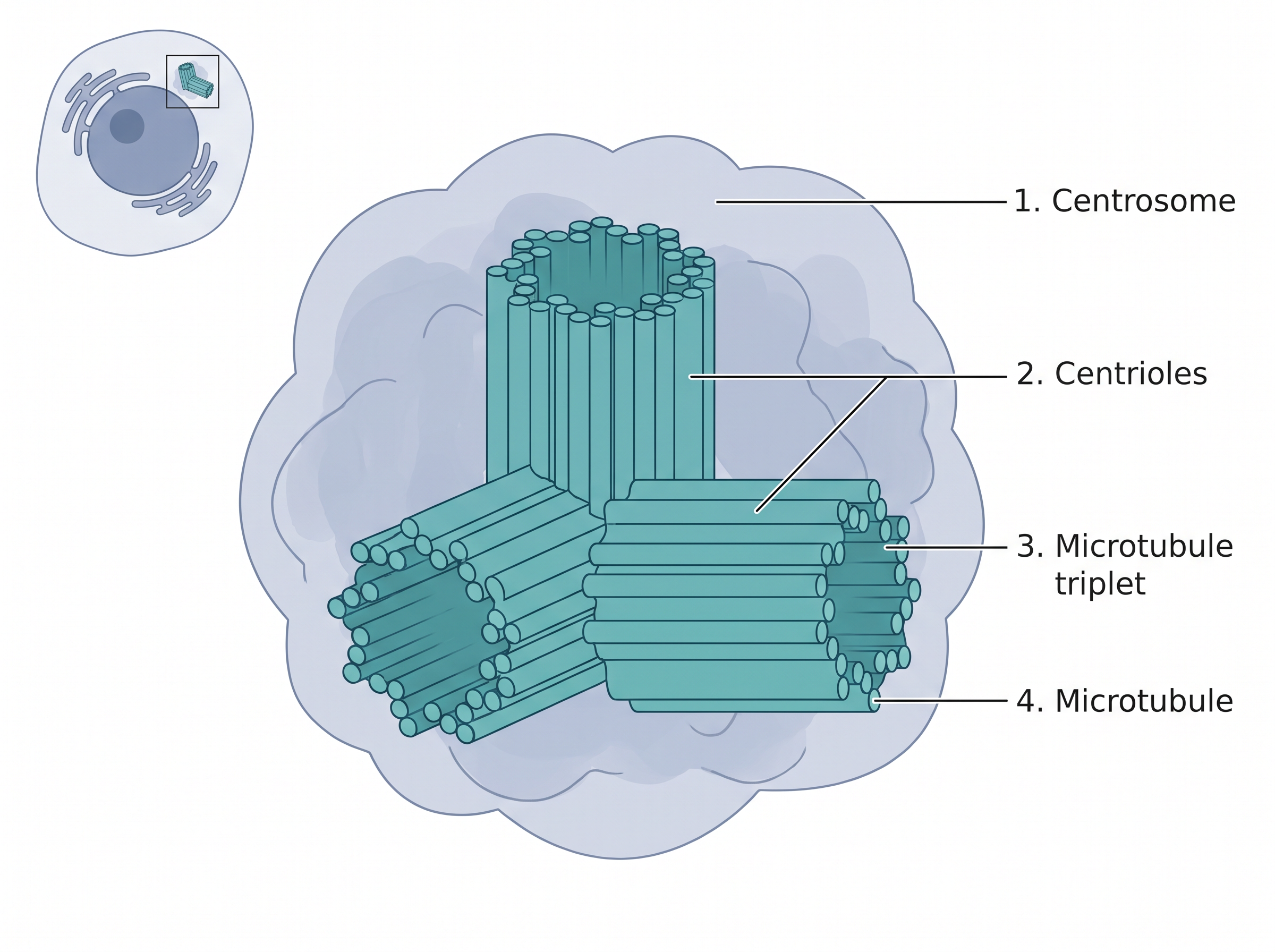
Fig. 2.20 – Centrosome and centrioles.
Behaviour During Cell Division
During cell division, the centrosome duplicates in S phase and separates to opposite directions during mitosis, the M-phase.
Two terms there need unpacking. The S phase is the stage of the cell cycle in which DNA is synthesised, that is, copied. The M phase is the stage in which the cell actually divides. So the cell makes its second centrosome well before it needs it, at the same time as it is copying its DNA — and only later does the pair move apart to opposite ends of the cell.
Structure and Role of Centrioles
Centrioles are two cylindrical structures composed of nine triplets of microtubules of tubulin, arranged around a central cavity (Fig. 2.20). They act as a centre of mitotic spindle assembly during cell division.
The mitotic spindle is the framework of microtubules that attaches to the chromosomes and draws them apart, so that each daughter cell receives a complete set. The centrioles, having moved to opposite ends of the cell, mark the two poles from which that framework is organised. Since microtubules are themselves made of tubulin, as you saw in Section 2.8, a structure built of tubulin triplets is well suited to serve as the organising point for them.
Centrosome and centrioles also form the basal body of the cilia and flagella, which facilitate locomotion. This is the centriole-like structure referred to in Section 2.9, and it explains why the two topics belong together: the same kind of microtubule-based assembly organises the spindle inside the cell and anchors the beating structures on its surface.
These two arrangements are a favourite pairing in objective questions, precisely because both involve the number nine. Fix the difference by the grouping: triplets in the centriole, doublets plus a central pair in the cilium.
2.11 Nucleus
As compared to prokaryotes, eukaryotes have a well-defined nucleus (Fig. 2.21) — a master controller of cell activities and a vast repository of genomic information. It contains the genetic material and regulates gene expression by various mechanisms that are exclusive to eukaryotes.
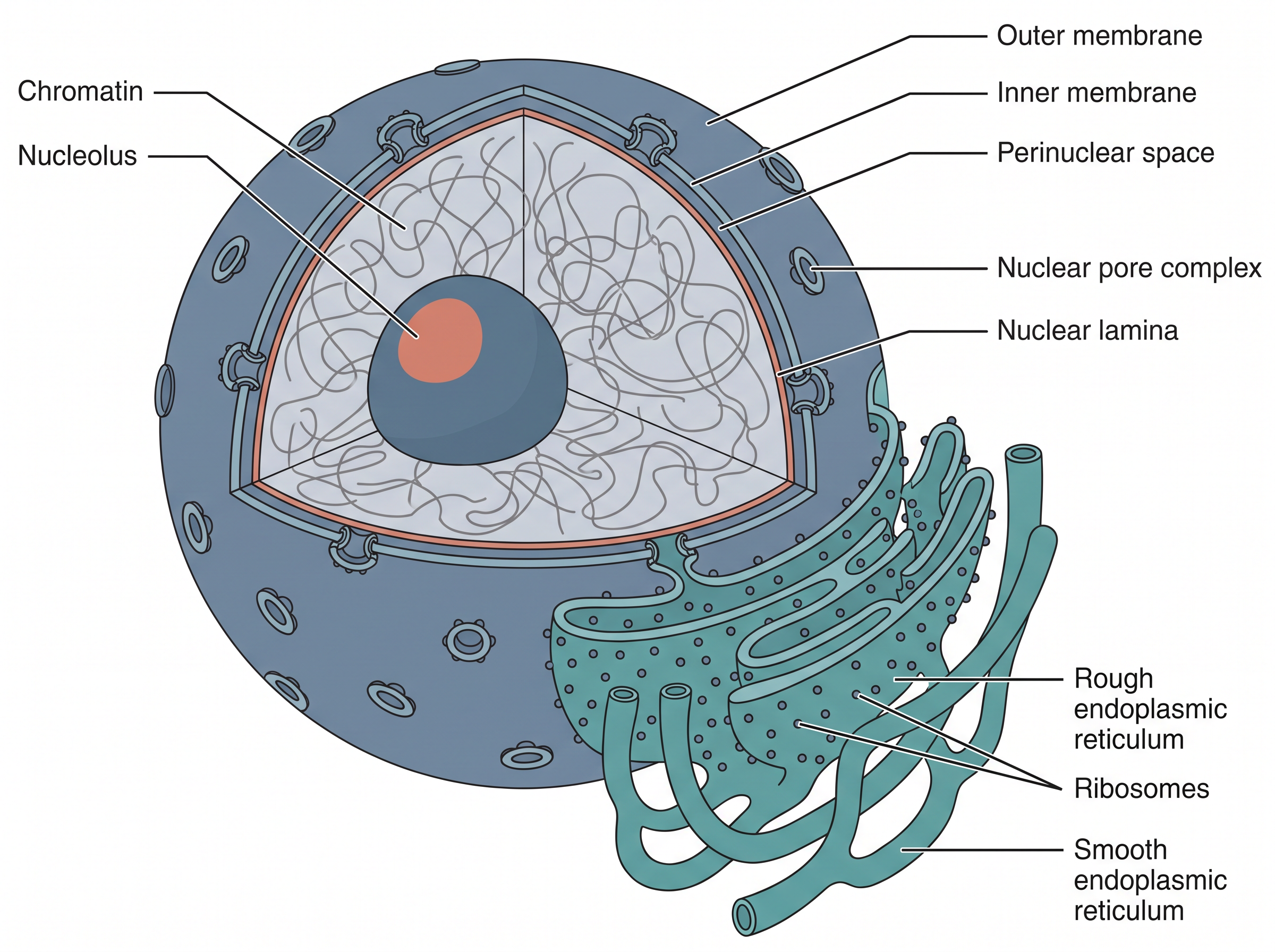
Fig. 2.21 – Structure of the eukaryotic nucleus.
The phrase ‘exclusive to eukaryotes’ is the key to this whole section. A prokaryote has its genetic material lying in the cytoplasm, so the moment a message is transcribed it is available for use. A eukaryote keeps its genetic material behind a membrane, and that separation creates room for an extra layer of control over which genes are expressed and when.
2.11.1 The Nuclear Envelope
The nuclear envelope, or nuclear membrane, is a double lipid bilayer similar to the plasma membrane. It surrounds the nucleus and acts as a barrier to keep molecules from freely moving between the nucleus and the cytoplasm.
A complete barrier, however, would be useless. Messages have to leave the nucleus and the proteins that act on the genetic material have to enter it. So the nuclear envelope is porous, having pores called nuclear pores.
At the nuclear pores, the outer and inner nuclear membranes are continuous, and each pore is fitted with a proteinous nuclear pore complex. Each nuclear pore complex is composed of structural protein subunits called nucleoporins, arranged in a ring-like manner around a central channel (Fig. 2.22). It allows trafficking of RNA and proteins.
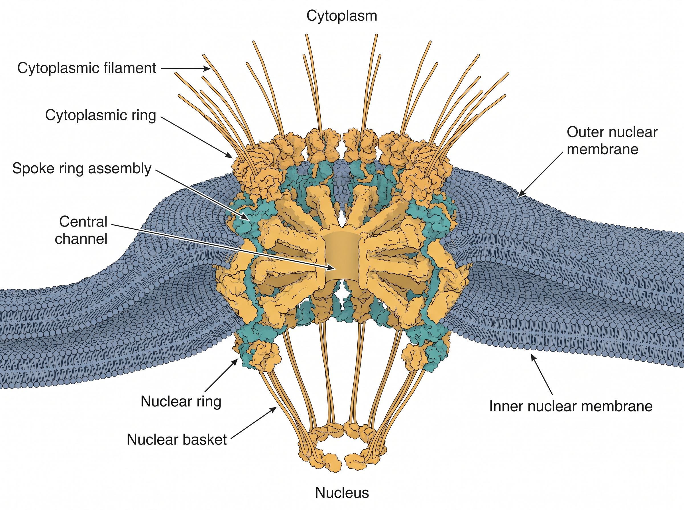
Fig. 2.22 – Nuclear pore complex.
Notice in Fig. 2.22 that the two membranes turn and join each other at the rim of the pore, so the pore is not a hole punched through two separate sheets — it is a continuous opening where the envelope folds back on itself. The rings, spokes and filaments of the complex then fill that opening, so that passage through it is controlled rather than free.
The outer nuclear membrane is continuous with the endoplasmic reticulum. This connection explains something you met in Section 2.3: the rough ER was described as lying near the nucleus, studded with ribosomes on its cytosolic surface. The two are, in fact, parts of one continuous membrane system — which is why a protein can begin its journey at the nuclear surface and end up in a secretory vesicle.
A fibrous network of lamin proteins is present below the inner membrane, called the nuclear lamina, which strengthens the structural framework of the nucleus. It plays much the same supporting role for the nucleus that the cytoskeleton of Section 2.8 plays for the cell as a whole.
Nuclear pore complexes are exclusively designed to transport proteins and RNA, along with small polar and charged molecules.
2.11.2 Nucleoplasm
The nuclear envelope encloses a clear fluid called nucleoplasm, also known as karyolymph or nuclear sap, which contains protein fibrils called the nuclear matrix. It helps to maintain the shape of the nucleus.
The enzymes associated with DNA replication and transcription are located in the nuclear matrix. This is worth noting carefully. The enzymes that copy DNA and that transcribe it into RNA are not simply floating loose in the nuclear fluid; they are held on a protein framework, which keeps them organised in place while they work.
The nucleolus and the chromatin are suspended in the nucleoplasm.
2.11.3 Nucleolus
The nucleus comprises a distinct set of organelles referred to as nuclear bodies, but these lack a well-defined membrane. They help in compartmentalising nuclear processes.
This is compartmentalisation of a different kind from what you have seen so far in this chapter. Every compartment up to now has been defined by a membrane. Inside the nucleus, distinct working regions exist without any membrane around them at all — the materials simply gather together in one region of the nucleoplasm.
The nucleolus (Fig. 2.21) is one of the most distinct nuclear bodies, involved in the synthesis of rRNA and ribosomes.
Here the chapter closes a loop. In Section 2.6 you saw that ribosomes are made of rRNA and proteins, and that their subunits stay dissociated in the cytoplasm until needed. The nucleolus is where that rRNA is made and where the subunits are put together, before they pass out through the nuclear pore complexes into the cytoplasm. The whole route is set out in Fig. 2.23.

Fig. 2.23 – Formation of a working ribosome.
Besides the nucleolus, various other structures present in the nucleus are involved in many different activities, which include transcriptional regulation, gene silencing, DNA repair, rRNA transcription and processing, and many others.
The chromosomal region occupied by the nucleolus comprises a large number of rRNA synthesising genes; therefore, it was named the nucleolar organising region.
That name makes the causal order clear, and it is worth stating plainly: the nucleolus is not a permanent structure that happens to contain some genes. It forms around those rRNA genes because that is where rRNA synthesis is taking place — the activity organises the structure, not the other way round.
2.12 Chromosome
A chromosome is a thread-like microscopic structure formed by the coiling of DNA packaged with protein, containing all the genetic material of an organism.
Chromosomes can be categorised into two types: autosomes, the body chromosomes, and allosomes, the sex chromosomes. Certain hereditary traits are linked to a person’s sex and are passed on through the sex chromosomes. Autosomes contain the rest of the genetic information.
Cells in a human being have 23 pairs of chromosomes — 22 pairs of autosomes and one pair of sex chromosomes — with a total of 46 per cell.
Chromosome Number Is Fixed for a Species
Table 2.2 shows the number of chromosomes in some plants and animals.
Table 2.2: Number of chromosomes in different eukaryotic organisms
| Organism | No. of Chromosomes |
|---|---|
| Arabidopsis thaliana (diploid) | 10 |
| Maize (diploid) | 20 |
| Wheat (hexaploid) | 42 |
| Common fruit fly (diploid) | 8 |
| Earthworm (diploid) | 36 |
| Mouse (diploid) | 40 |
| Human (diploid) | 46 |
| Elephant (diploid) | 56 |
| Donkey (diploid) | 62 |
| Dog (diploid) | 78 |
| Gold Fish (diploid) | 100 |
| Tobacco (tetraploid) | 18 |
| Oat (hexaploid) | 12 |
Read this table for the pattern rather than for the individual figures. The number is fixed for a species but does not rise with the complexity of the organism — a dog has 78 chromosomes and a human 46, while a fruit fly manages with 8. What a chromosome number tells you is how an organism’s genetic material is parcelled up, not how much of it there is or how elaborate the organism is.
That error stood for more than thirty years, and it is worth asking why. Chromosomes clump together on a slide and are extremely difficult to count reliably; once a figure was published and accepted, later workers tended to see what they expected to see. Improved preparation methods, which spread the chromosomes apart cleanly, were what finally made an accurate count possible.
Chromosomes in Prokaryotes
Prokaryotes like bacteria or blue-green algae usually contain a single circular chromosome, called the nucleoid, in the cytoplasm, as they do not contain a well-defined nucleus or other membrane-bound organelles. However, in some prokaryotes there may be more than one chromosome, for example Vibrio cholerae.
How DNA Is Packed Inside the Nucleus
In eukaryotes, the chromosomes are present inside a well-defined nucleus. During the interphase of the cell cycle, chromosomes are present in the form of long threads called chromatin fibres.
The chromatin fibre is composed of nucleosomes. Nucleosomes consist of DNA wrapped around histone proteins. Chromatin enables the long strands of DNA molecules to fit into the cell nucleus.
If all of the DNA molecules in a single human cell are unwound from the histones and placed end-to-end, they would stretch to 6 feet — roughly 1.8 m of thread to be fitted inside a structure a few micrometres across.
During cell division, chromatin fibres condense further to form microscopically visible, long and slender chromosomes. The structure of chromosomes therefore varies through the different phases of the cell cycle, and this variation is not accidental: DNA that is being read must be accessible, while DNA that is being moved must be compact and tidy.
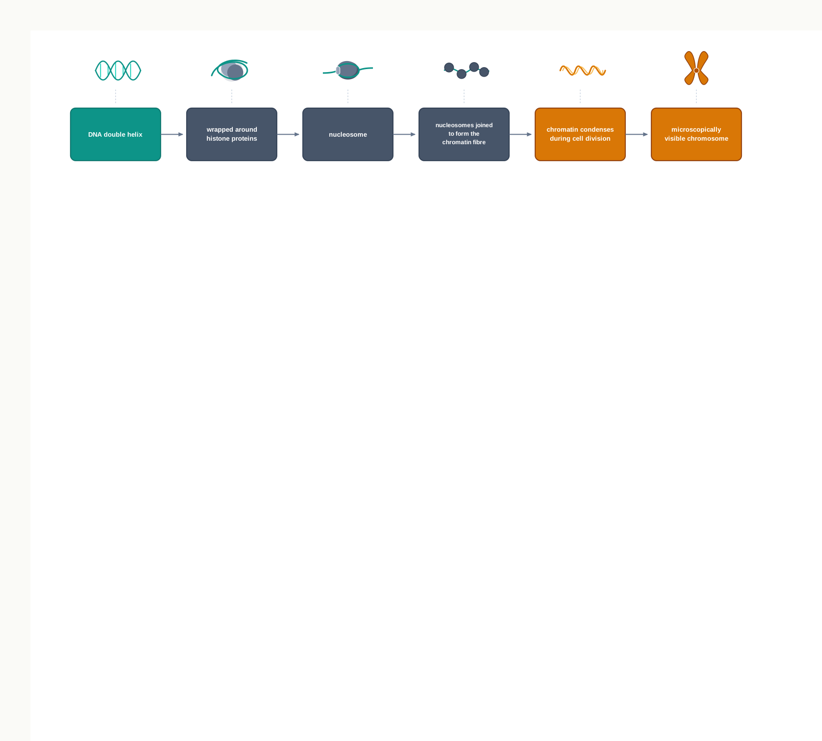
Fig. 2.24 – DNA packaging in a eukaryotic cell.
When Things Go Wrong
During cellular division, chromosomes are replicated along with their DNA, divided, and passed successfully to their daughter cells. Sometimes, errors do occur, leading to changes in the number or structure of chromosomes in the new cells, which may lead to serious problems.
A familiar example is Down syndrome, in which an extra copy of chromosome 21 is present, so the cells carry 47 chromosomes instead of 46. It is the sort of change that karyotyping is designed to detect, and it shows why the accuracy of chromosome distribution during division matters so much — a single extra chromosome alters the dosage of every gene it carries.
2.13 Summary
The millions of tasks performed by our body are possible because of the cell, which is the basic unit of life. Cells are broadly categorised into two types: prokaryotic cells, which have no membrane-bound organelles and carry a nucleoid, and eukaryotic cells, which have membrane-bound organelles and a well-defined nucleus.
Both prokaryotic and eukaryotic cells are surrounded by a plasma membrane. The plasma membrane is mainly composed of phospholipids. It is selectively permeable and facilitates the transport of molecules into and out of the cell.
Besides the plasma membrane, the cells of bacteria, algae, fungi and some higher plants are surrounded by a rigid cell wall.
A eukaryotic cell has two major compartments: the nucleus and the cytoplasm.
The nucleus is enclosed by a nuclear envelope, which has nuclear pores. The nuclear envelope encloses the nucleoplasm, the nucleolus and the genetic material in the form of chromatin. The nucleolus helps in rRNA synthesis.
A pair of centrioles forms the spindle apparatus during cell division in animal cells. The centrosome and centrioles also form the basal body of the cilia and flagella, which facilitate locomotion.
The endomembrane system includes the endoplasmic reticulum, the Golgi apparatus, lysosomes and vacuoles. The ER is constituted by tubules called cisternae, and is of two types — rough, with ribosomes, and smooth, without ribosomes.
Ribosomes are non-membranous structures involved in protein synthesis. They may be present freely in the cytoplasm or in a bound state on the rough ER.
The function of the ER is to help in the synthesis and transport of proteins, lipoproteins and glycogen.
The Golgi apparatus is a membranous organelle composed of flattened sacs. It performs the packaging of secretory substances and their transport from the cell.
Lysosomes are single-membrane structures containing enzymes for the digestion of all types of macromolecules.
Vacuoles are membrane-bound organelles that function in storage, structural support and recycling in the cell.
Peroxisomes and microbodies take part in oxidation reactions within the cell. Glyoxysomes are peroxisomes involved in fat metabolism.
Mitochondria are bound by two membranes. The inner membrane has infoldings called cristae. Mitochondria help in oxidative phosphorylation and the generation of ATP.
In plant cells, the pigment-containing granules are called plastids. The plastids containing the green pigment chlorophyll are known as chloroplasts, and chloroplasts are essential for photosynthesis.
The cytoskeleton is a network of fibrous proteins — microtubules, actin filaments and intermediate filaments — that maintains cell shape and organisation and acts in cell division, cell movement and intracellular transport.
A chromosome is formed by the coiling of DNA packaged with protein. Chromatin, made of nucleosomes, allows very long DNA molecules to fit inside the nucleus, and condenses further into visible chromosomes during cell division.
The chapter as a whole moves inwards from the cell boundary to the genetic material, and that route is worth carrying in your head as a single sequence.
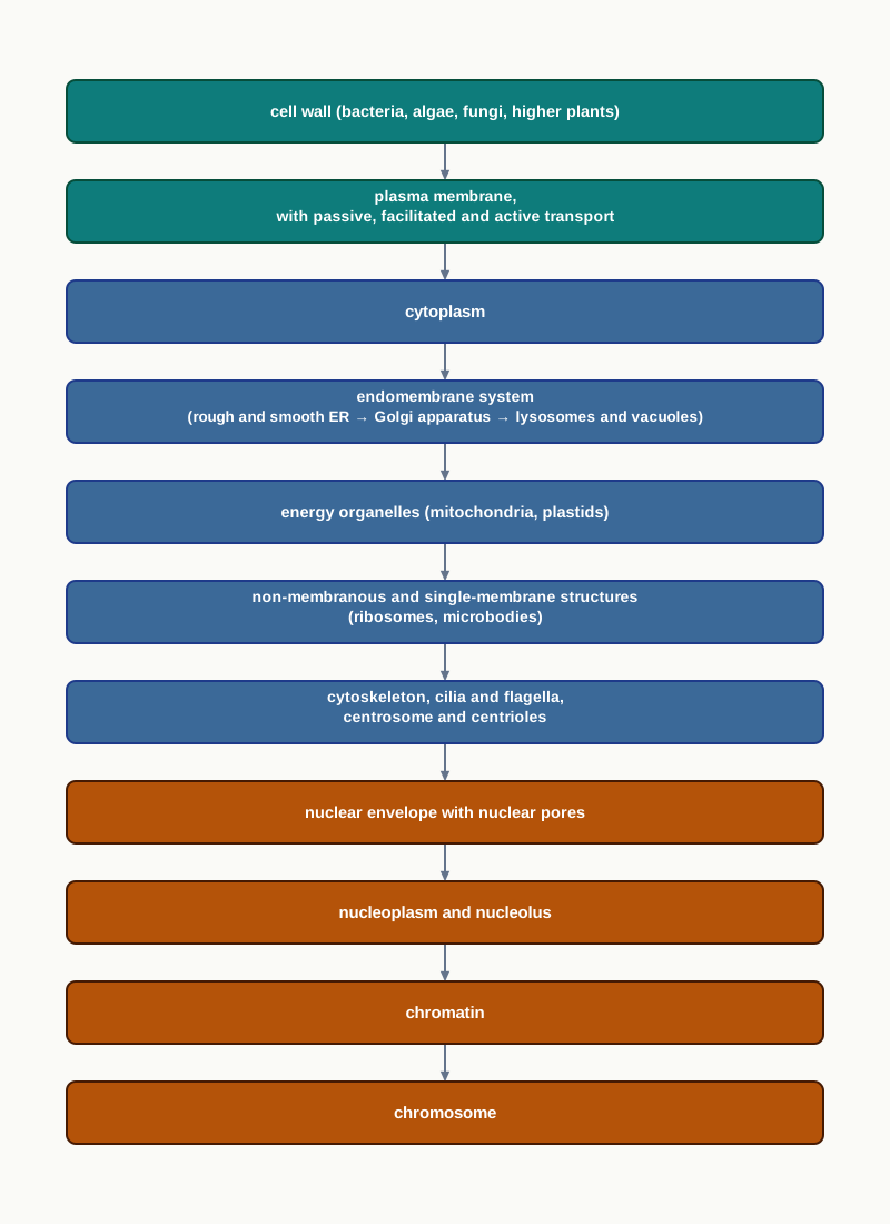
Fig. 2.25 – Route of the chapter: outside to inside.
2.14 Key Terms
Organelles — The different machineries inside a cell, each responsible for a different function.
Prokaryote — A cell without an organised nucleus and without membrane-bound organelles, carrying its genetic material as a nucleoid.
Eukaryote — A cell with a well-organised nucleus and membrane-bound cell organelles.
Plasma membrane — The semipermeable boundary of the cytoplasm that controls the relationship of a cell with its surroundings.
Fluid Mosaic Model — The accepted model of membrane organisation, proposed by Singer and Nicolson in 1972, describing a quasifluid lipid bilayer with globular proteins set into it like a mosaic.
Lipid bilayer — The double sheet of lipid molecules that forms the basic structure of a membrane.
Phospholipid — The major membrane lipid, with a hydrophilic head facing the exterior and a long hydrophobic hydrocarbon tail occupying the interior of the bilayer.
Peripheral membrane proteins — Membrane proteins superficially attached to the lipid bilayer, mainly involved in cell signalling.
Integral membrane proteins — Membrane proteins partially or fully buried in the plasma membrane.
Transmembrane proteins — The most abundant type of integral membrane protein, spanning the full thickness of the membrane.
Mesosome — A membranous structure formed by inward extension of the plasma membrane, in the form of vesicles, tubules and lamellae, which increases the surface of the plasma membrane.
Passive transport — Movement of molecules across the membrane along the concentration gradient, by diffusion and osmosis, without expenditure of energy.
Carrier proteins — Membrane proteins, such as the glucose transporter, that bind a molecule and carry it across the membrane.
Channel proteins — Membrane proteins that form a pore through which suitable particles pass.
Facilitated movement — Movement of molecules across the membrane through carrier proteins or channel proteins, still along the concentration gradient.
Aquaporins — Channel proteins critical for the transport of water across the plasma membrane in plant and animal cells.
Ion channels — Well-studied channel proteins of muscle and nerve cell membranes that allow the passage of ions.
Active transport — Movement of molecules against the concentration gradient, from lower to higher concentration, using energy.
Na⁺-K⁺ pump — An ATP-driven pump that moves Na⁺ and K⁺ against their concentration gradients across the plasma membrane.
Symport — Coupled transport in which two molecules are carried in the same direction.
Antiport — Transport in which two molecules are carried in opposite directions.
Uniport — Transport of a single molecule, as in facilitated diffusion of glucose.
Cell wall — The rigid layer outside the plasma membrane in bacteria, algae, fungi and higher plants, which gives shape, prevents osmotic bursting and provides mechanical strength.
Peptidoglycan — The linear polysaccharide chain of the bacterial cell wall, cross-linked by tetrapeptides.
Gram-positive bacteria — Bacteria with a thick cell wall and a single plasma membrane.
Gram-negative bacteria — Bacteria with a thin cell wall surrounded by a dual plasma membrane.
Glycocalyx — A heavily glycosylated protein layer covering the bacterial cell wall, acting as a barrier to pathogens and protecting against mechanical and ionic stress.
Slime layer — A loose sheath form of the glycocalyx.
Capsule — A thick and tough form of the glycocalyx.
Cellulose — A linear polymer of glucose residues forming the cell wall of most higher plants.
Chitin — A linear polymer of N-acetylglucosamine forming the cell wall of fungi.
Primary cell wall — The comparatively thin wall of a growing plant cell, which allows for cell expansion.
Secondary cell wall — The rigid, thick wall formed between the primary cell wall and the plasma membrane after the cell stops growing.
Lignin — The substance deposited in the secondary cell wall that makes it rigid and thick.
Middle lamella — A layer of calcium pectate that holds neighbouring plant cells together.
Plasmodesmata — Structures connecting the cytoplasm of neighbouring plant cells.
Endomembrane system — The co-ordinated group of membrane-bound organelles — ER, Golgi complex, lysosomes and vacuoles — that work together in the synthesis, processing, packaging and transport of proteins and lipids.
Endoplasmic reticulum (ER) — An extensive network of membrane-enclosed tubules and cisternae, present exclusively in eukaryotic cells, involved in protein synthesis, calcium storage and lipid metabolism.
Rough ER (RER) — The region of ER bearing ribosomes on its cytosolic surface, where proteins of the secretory pathway are inserted.
Smooth ER (SER) — The region of ER without ribosomes, mainly involved in lipid metabolism and cholesterol synthesis.
Cisternae — The flattened membranous sacs that make up the ER and the stacks of the Golgi apparatus.
Golgi apparatus — A membrane-bound organelle of stacked cisternae that packages materials for secretion and carries out post-translational modification of proteins and lipids.
cis face — The forming face of the Golgi stack, nearest the endoplasmic reticulum, where transport vesicles arrive.
trans face — The maturing face of the Golgi stack, away from the ER, from which vesicles pinch off.
Lysosomes — Small single-membrane vesicles, about 0.2 – 0.5 micron across, containing hydrolytic enzymes that digest macromolecules.
Acid hydrolases — The lysosomal enzymes, which show optimal activity at acidic pH.
Autophagy — The process by which a cell uses lysosomal enzymes to digest and recycle its own organic material.
Vacuoles — Membrane-bound organelles of plants, fungi and some animal cells, functioning in storage, structural support and recycling.
Tonoplast — The membrane covering a vacuole.
Contractile vacuole — A vacuole that plays a critical role in excretion and osmoregulation, as in Amoeba.
Mitochondria — Double-membrane-bound organelles of eukaryotic cells that house the enzymes of the TCA cycle and cellular respiration for ATP synthesis.
Cristae — The infoldings of the inner mitochondrial membrane, which increase its surface area.
Peri-mitochondrial space — The narrow region between the outer and inner mitochondrial membranes.
Mitochondrial matrix — The innermost compartment of a mitochondrion, lined by the inner membrane, and the site of organellar DNA replication, transcription and protein synthesis.
Plastids — Large, usually pigment-bearing organelles of plant cell cytoplasm.
Chromoplasts — Coloured plastids containing fat soluble pigments such as carotene and xanthophylls.
Leucoplasts — Colourless plastids involved in the storage of reserve food materials.
Amyloplast — A leucoplast that stores starch.
Aleuroplast — A leucoplast that stores protein.
Elaioplast (lipoplast) — A leucoplast that stores oil.
Chloroplasts — Green plastids of the mesophyll cells, and the largest organelles of the plant cell, in which photosynthesis takes place.
Chlorophyll — The green pigment of chloroplasts, a collection of four pigments: chlorophyll a, chlorophyll b, carotenoids and xanthophylls.
Thylakoids — Flattened membranous sacs formed by the third inner membrane of the chloroplast, containing the chlorophyll pigments.
Grana — Orderly stacks of thylakoids resembling stacks of coins.
Stroma — The fluid outside the thylakoids and within the chloroplast envelope, containing chloroplast DNA, ribosomes and many enzymes.
Stroma lamellae — Flat membranous tubules connecting the thylakoids of different grana.
Ribosomes — Membraneless organelles of rRNA and protein, present in prokaryotic and eukaryotic cells, which are the protein synthesising factories of the cell.
Svedberg unit (S) — The unit of sedimentation rate, named after Theodor Svedberg, used to classify ribosomes.
70S ribosome — The prokaryotic ribosome, with a 50S large subunit and a 30S small subunit, also found in mitochondria and chloroplasts.
80S ribosome — The eukaryotic cytoplasmic ribosome, with a 60S large subunit and a 40S small subunit.
Ribozymes — rRNA molecules of the large ribosomal subunit that possess catalytic activity.
Microbodies — Small, single-membrane-bound organelles of eukaryotic cells found near the ER, comprising peroxisomes and glyoxysomes.
Peroxisomes — Microbodies containing oxidases and catalase, involved in oxidation, lipid biosynthesis, alcohol detoxification in liver cells, and photorespiration in leaves.
Catalase — The peroxisomal enzyme that neutralises the harmful products of oxidation reactions.
Glyoxysomes — Specialised peroxisomes of fungi and higher plants, containing the enzymes of fatty acid oxidation, the glyoxylate cycle and gluconeogenesis.
Cytoskeleton — A multi-component network of fibrous proteins that maintains cell shape and organisation and functions in cell division, cell movement and intracellular transport.
Microtubules — Hollow rod-like cytoskeletal filaments, 25 nm in diameter, made of tubulin and responsible for the rhythmic movement of cilia and flagella.
Tubulin — The globular protein of microtubules, a dimer of α and β subunits.
Protofilament — The chain formed by polymerisation of tubulin, 10–15 of which make up a microtubule.
Actin filament — A cytoskeletal filament 6 nm in diameter, made of actin, important in muscle contraction, cytokinesis and cell movement.
Intermediate filaments — Strong, rope-like cytoskeletal filaments 10 nm in diameter, providing mechanical strength to the cell.
Cilia — Short, numerous hair-like structures occurring over the cell surface, moving in a co-ordinated sweeping rhythm.
Flagella — Long hair-like structures, one or two per cell, occurring at one end and showing undulatory or whiplash movement.
Basal body — The centriole-like structure from which cilia and flagella arise.
Axoneme — The microtubule core of a cilium or flagellum, with nine peripheral and two central microtubules.
9+2 array — The arrangement of nine peripheral and two central microtubules in a eukaryotic cilium or flagellum.
Centrosome — A structure near the nucleus of an animal cell, consisting of two perpendicular centrioles embedded in amorphous pericentriolar material.
Centrioles — Two cylindrical structures of nine microtubule triplets arranged around a central cavity, acting as the centre of mitotic spindle assembly.
Nucleus — The well-defined master controller of cell activities in a eukaryote, containing the genetic material and regulating gene expression.
Nuclear envelope — The double lipid bilayer surrounding the nucleus, acting as a barrier between nucleus and cytoplasm.
Nuclear pores — The pores of the nuclear envelope, at which the inner and outer membranes are continuous.
Nuclear pore complex — The proteinous assembly fitted into each nuclear pore, which allows trafficking of RNA and proteins.
Nucleoporins — The structural protein subunits of the nuclear pore complex, arranged in a ring around a central channel.
Nuclear lamina — The fibrous network of lamin proteins beneath the inner nuclear membrane that strengthens the structural framework of the nucleus.
Nucleoplasm — The clear fluid enclosed by the nuclear envelope, also called karyolymph or nuclear sap.
Nuclear matrix — The protein fibrils of the nucleoplasm that maintain nuclear shape and hold the enzymes of DNA replication and transcription.
Nuclear bodies — Distinct structures within the nucleus that lack a well-defined membrane and help compartmentalise nuclear processes.
Nucleolus — The most distinct nuclear body, involved in the synthesis of rRNA and ribosomes.
Nucleolar organising region — The chromosomal region occupied by the nucleolus, containing a large number of rRNA synthesising genes.
Chromosome — A thread-like microscopic structure formed by coiling of DNA packaged with protein, containing the genetic material of an organism.
Autosomes — The body chromosomes, 22 pairs in a human cell, carrying the general genetic information.
Allosomes — The sex chromosomes, one pair in a human cell, carrying traits linked to sex.
Nucleoid — The single circular chromosome of a prokaryote, lying free in the cytoplasm.
Chromatin fibre — The long thread form in which chromosomes exist during interphase, composed of nucleosomes.
Nucleosome — The repeating unit of chromatin, consisting of DNA wrapped around histone proteins.
2.15 Points to Ponder
A cell is often described as a bag of chemicals. After studying this chapter, consider how far that description holds. What would a living cell lose if all its internal membranes were removed, leaving the same chemicals in the same amounts?
The lipid bilayer blocks charged and polar molecules, and membrane proteins then let selected ones through. Think about what would happen to a cell that had the proteins but no bilayer, and to one that had the bilayer but no proteins.
The Na⁺-K⁺ pump spends ATP continuously to maintain gradients that diffusion is constantly trying to undo. Reflect on why a cell should be willing to spend energy on a task that is never finished.
A cell wall protects a bacterium from bursting, yet it is also the feature that certain antibiotics exploit. Consider why a structure that provides protection can also become the point of greatest vulnerability.
Animal cells have no cell wall at all. Think about what they gain from this absence, and what they must do instead to stay safe.
Both mitochondria and chloroplasts increase their working surface by folding membranes — cristae in one, grana in the other. Reflect on why folding, rather than simply becoming larger, is the solution that both organelles have arrived at.
Mitochondria and chloroplasts carry their own DNA and 70S ribosomes, yet the great majority of their proteins are made outside them. Consider what kind of relationship this suggests between the organelle and the rest of the cell.
Lysosomal enzymes are kept behind a membrane and work best at acidic pH. Think about which of these two safeguards you would regard as the more important, and what your reasoning depends on.
The nucleolus has no membrane around it, and yet it is a distinct region where a particular job gets done. Reflect on what this suggests about the different ways a cell can keep processes separate.
The number of chromosomes is fixed for a species but does not increase with the complexity of the organism. Consider what a chromosome number does and does not tell you about an organism.
For over thirty years, the human chromosome number was believed to be 48 before it was corrected to 46. Think about how an error can persist for so long in a science that depends on direct observation, and what this suggests about the role of improved methods in scientific work.
The cytoskeleton is described as a skeleton, yet its filaments are assembled and disassembled continually. Reflect on how well the name fits, and what alternative name you might propose.
Every compartment studied in this chapter separates something from something else. Try to identify, for each organelle, exactly what is being kept apart from what, and what would go wrong if that separation failed.
2.16 NCERT Questions
The Fluid Mosaic Model has been proposed by
- Robert Brown
- Schleiden and Schwann
- Robert Virchow
- Singer and Nicolson
Ribosomes are composed of
- only rRNA
- rRNA and proteins
- rRNA, proteins and DNA
- lipids, proteins and DNA
Tonoplast is
- a membrane covering the cell wall in plant cells
- the inner membrane of the mitochondria
- a membrane covering the vacuoles
- a membrane covering the plastids
Describe the various mechanisms of transport across the plasma membrane with the help of labelled diagrams.
Match the following
| Column I | Column II |
|---|---|
| a. Nucleolus | i. Alcohol detoxification |
| b. Mesosome | ii. Infoldings of inner mitochondrial membrane |
| c. Vacuoles | iii. Protein synthesis |
| d. Cristae | iv. Disc shaped sacs in Golgi |
| e. Ribosomes | v. rRNA synthesis |
| f. Thylakoid | vi. Membranous extensions of plasma membrane |
| g. Peroxisomes | vii. Storage and structural support |
| h. Cisternae | viii. Membranous sacs in chloroplast |
What is the significance of the ratio of protein and lipids in membranes? How does varying the concentration of lipids in a membrane affect its function?
State the importance of the cell wall in prokaryotic cells.
A eukaryotic cell contains organelles which may be bound by a single membrane, bound by a double membrane, or non-membrane bound. Classify the various eukaryotic organelles into these three types.
Mention the different types of vacuoles.
Peroxisomes share similarities as well as differences with mitochondria and chloroplast. Comment.
What are glyoxysomes? Where are these present? Mention their functions.
Cell is the structural and functional unit of life. Justify the statement.
Distinguish between
- cilia and flagella
- primary and secondary cell wall
- lysosomes and vacuoles
- microtubules and actin filaments
- active and passive transport
2.17 Objective Questions
Peripheral membrane proteins are mainly involved in
- ATP synthesis
- cell signalling
- forming pores for ion movement
- synthesis of phospholipids
The human erythrocyte membrane contains approximately
- 40 per cent protein and 52 per cent lipids
- 52 per cent protein and 40 per cent lipids
- 52 per cent protein and 52 per cent lipids
- 40 per cent protein and 40 per cent lipids
Aquaporins are
- carrier proteins for glucose
- channel proteins for the transport of water
- peripheral proteins involved in signalling
- pumps that use ATP directly
The transport of Na⁺ and Ca²⁺ in opposite directions by the Na⁺-Ca²⁺ antiporter is an example of
- uniport
- symport
- antiport
- simple diffusion
The rigidity and thickness of the secondary cell wall is due to the deposition of
- cellulose
- chitin
- lignin
- calcium pectate
The middle lamella that holds neighbouring plant cells together is a layer of
- peptidoglycan
- calcium pectate
- lignin
- N-acetylglucosamine
Chitin is a linear polymer of
- β-D-glucose
- N-acetylglucosamine
- glycerol
- tubulin
The cis face of the Golgi apparatus is
- the maturing face, away from the ER
- the forming face, nearest the ER
- the face from which secretory vesicles pinch off
- the face that fuses with the plasma membrane
Lysosomal enzymes are described as acid hydrolases because they
- are made only in acidic cells
- show optimal activity at an acidic pH
- produce acid inside the lysosome
- digest only acidic macromolecules
Unlike mitochondria and chloroplasts, a peroxisome
- is bound by a double membrane
- contains 70S ribosomes
- lacks its own genome
- arises only from pre-existing peroxisomes of the nucleus
Glyoxysomes contain all the enzymes necessary for
- photosynthesis and photorespiration
- fatty acid oxidation, the glyoxylate cycle and gluconeogenesis
- the TCA cycle and oxidative phosphorylation
- rRNA synthesis and ribosome assembly
A microtubule may contain
- 2–4 protofilaments
- 9 protofilaments
- 10–15 protofilaments
- 25 protofilaments
The diameters of microtubules, actin filaments and intermediate filaments are respectively
- 6 nm, 10 nm and 25 nm
- 25 nm, 6 nm and 10 nm
- 10 nm, 25 nm and 6 nm
- 25 nm, 10 nm and 6 nm
The 70S prokaryotic ribosome consists of subunits of
- 60S and 40S
- 50S and 30S
- 50S and 20S
- 40S and 30S
Ribozymes are
- ribosomal proteins with catalytic activity
- rRNAs of the large subunit with catalytic activity
- enzymes that assemble ribosomal subunits
- enzymes that digest rRNA
Nucleoporins are the structural protein subunits of the
- nuclear lamina
- nuclear matrix
- nuclear pore complex
- nucleolar organising region
Stroma lamellae are flat membranous tubules that connect
- the outer and inner chloroplast membranes
- the thylakoids of different grana
- the chloroplast to the endoplasmic reticulum
- the stroma to the intermediate space
In eukaryotic cilia and flagella, the axoneme shows an arrangement of
- nine triplets around a central cavity
- nine peripheral doublets and two central microtubules
- nine peripheral microtubules with no central microtubule
- ten to fifteen protofilaments
Assertion (A): Lysosomal enzymes do not digest the cytoplasm of the cell that contains them. Reason (R): Lysosomal enzymes are acid hydrolases, which show optimal activity at an acidic pH.
Assertion (A): Cristae allow a mitochondrion to accommodate more of the machinery of cellular respiration. Reason (R): Cristae are infoldings of the outer mitochondrial membrane.
Assertion (A): 70S ribosomes are present in the mitochondria and chloroplasts of eukaryotic cells. Reason (R): These organelles show relatedness to prokaryotic cells.
Assertion (A): A 50S and a 30S subunit together form a 70S ribosome and not an 80S one. Reason (R): The Svedberg unit measures sedimentation rate, which depends on the shape of a particle as well as on its mass.
Assertion (A): The outer nuclear membrane is continuous with the endoplasmic reticulum. Reason (R): The nuclear envelope is a double lipid bilayer perforated by nuclear pores.
Assertion (A): Antibiotics that block the cross-linking of peptidoglycan strands cause growing bacteria to burst. Reason (R): The bacterial cell wall prevents cell bursting caused due to osmotic pressure.
Assertion (A): A growing plant cell is surrounded by a comparatively thin primary cell wall. Reason (R): The primary cell wall leaves scope for cell expansion.
Assertion (A): Facilitated movement is classified as a form of passive transport. Reason (R): Facilitated movement takes place through carrier proteins and channel proteins.
2.18 Check Your Concepts
Why can a phospholipid bilayer reseal itself after being torn, while a sheet of paper cannot? Base your answer on the arrangement of the hydrophilic heads and hydrophobic tails.
A membrane is described as being in a “quasifluid” state. Name three cellular processes that would fail if the membrane were completely rigid, and explain why each one requires fluidity.
Glucose is an uncharged molecule, and yet it cannot cross the plasma membrane by simple diffusion. Explain why, and name the type of protein that makes its entry possible.
Facilitated movement requires a membrane protein but is still classified as passive transport, whereas the Na⁺-K⁺ pump also requires a membrane protein and is classified as active transport. What exactly is the basis of this difference?
A cell is treated with a substance that stops all ATP production. Predict what will happen, over time, to the concentrations of Na⁺ and K⁺ on the two sides of its plasma membrane, and give the reason for your prediction.
Distinguish between symport, antiport and uniport, giving one example of each from the chapter.
Why does an antibiotic that blocks the cross-linking of peptidoglycan affect a growing bacterial culture much more than a culture that has stopped growing?
A student performs a Gram staining procedure but forgets to add iodine after the crystal violet. Identify what will go wrong with the result, and explain why that step cannot be skipped.
Both cellulose and chitin are built from repeating sugar units joined by β(1→4) glycosidic linkages. Explain how these two polymers differ, and why this type of linkage suits a structural role.
The secondary cell wall is laid down between the primary cell wall and the plasma membrane, and not on the outside. Explain why this position is necessary.
Arrange the following in the correct sequence for a protein that is destined to be secreted from the cell: fusion with the plasma membrane; insertion into the ER; budding of a vesicle from the ER; modification in the Golgi cisternae; pinching off from the trans face; fusion with the cis face.
Two proteins are being made in the same cell at the same time. One ends up inside a peroxisome; the other is secreted from the cell. At what point did their routes separate, and what determined the difference?
Explain why lipids cannot be synthesised in the cytosol, and relate your answer to the function of the smooth endoplasmic reticulum.
The Golgi apparatus is described as the “receiving and shipping department” of the cell. Justify this description by referring to the cis face, the trans face and the vesicles involved.
A cell’s lysosomal membranes are damaged so that their contents leak into the cytoplasm. Explain why the damage to the cell may still be limited, and state the condition under which it would become severe.
In Tay-Sachs disease, lipids accumulate inside brain cells. Working backwards from this observation, explain what has gone wrong at the level of the organelle, and why the material builds up instead of being removed.
Both cristae and grana are folded membrane systems. Compare them in terms of the organelle they belong to, the membrane that is folded, and the function they support.
The chloroplast has three compartments while the mitochondrion has two. Account for this difference in terms of the membranes present in each organelle.
List the evidence given in this chapter which suggests that mitochondria and chloroplasts are related to prokaryotic cells. State clearly which of your points concern structure and which concern the genetic system.
Explain why an antibiotic that acts on the 70S ribosome can be used to treat a bacterial infection in a human patient, and state one reason why such drugs must still be used with care.
A peroxisome contains both an enzyme that produces hydrogen peroxide and an enzyme that destroys it. Explain the logic of keeping the two together inside one membrane-bound compartment.
Arrange the following in the correct sequence for a germinating oilseed: gluconeogenesis; stored oil in fat storage tissue; sugar used by the seedling; the glyoxylate cycle; fatty acid oxidation in the glyoxysome. Then state why this pathway becomes unnecessary once the shoot reaches light.
Redraw the transverse section of a cilium as shown in Fig. 2.19 and label the following: plasma membrane, peripheral microtubule doublets, central microtubules, central sheath, radial spoke and interdoublet bridge. State the array shown by this arrangement.
Both the centriole and the axoneme of a cilium are built around the number nine. Distinguish between the two arrangements, and state the function associated with each.
The nuclear envelope must act as a barrier and yet allow RNA and proteins to pass. Explain how the structure of the nuclear pore complex resolves these two opposing requirements.
The nucleolus has no membrane of its own, and yet it is a distinct region of the nucleus. Explain how such a compartment can exist without a membrane, and what determines where it forms.
Arrange the following in the correct order of increasing packing: chromatin fibre; DNA double helix; condensed chromosome visible during cell division; nucleosome. Then explain why the cell does not keep its DNA in the most condensed form at all times.
In a karyotyping procedure, a student adds colchicine to the cell culture. Explain the purpose of this step by referring to the cytoskeleton, and predict what the slide would look like if the step were left out.
Match the following.
| Column I | Column II |
|---|---|
| a. Nucleoporins | i. Storage of starch |
| b. Tonoplast | ii. Ring around the central channel of a nuclear pore |
| c. Amyloplast | iii. Loose sheath form of the glycocalyx |
| d. Ribozyme | iv. Membrane covering a vacuole |
| e. Slime layer | v. Nine triplets of microtubules |
| f. Centriole | vi. rRNA with catalytic activity |
Classify each of the following as bounded by a single membrane, bounded by a double membrane, or non-membranous: ribosome, lysosome, mitochondrion, peroxisome, nucleus, chloroplast, vacuole, centriole.
For any five organelles studied in this chapter, state in one line each what is being kept separated from what, and what would go wrong in the cell if that separation failed.
2.19 Case-Based Questions
A sample from a patient with a wound infection is brought to a hospital laboratory. A smear is prepared and stained; under the microscope, almost all the bacterial cells appear violet. The physician prescribes an antibiotic that acts by preventing the cross-linking of peptidoglycan strands. Within two days the patient improves.
- What does the violet colour of the cells indicate about the structure of their cell wall?
- Explain, step by step, how an antibiotic that only prevents cross-linking is able to kill the bacterium completely.
- Why does this antibiotic not damage the patient’s own cells in the same way?
- Would you expect the same drug to be equally effective against a bacterium whose cells appear pink after staining? Give a reason.
A student breaks open cultured animal cells to make a homogenate and spins it in an ultracentrifuge. Two fractions are collected. On analysis, Fraction A contains ribosomes that sediment at 80S, while Fraction B contains an organelle bounded by two membranes, whose interior yields DNA and ribosomes that sediment at 70S.
- Identify the likely origin of the ribosomes in Fraction A and the organelle in Fraction B.
- On what physical property does the separation in an ultracentrifuge depend?
- What conclusion about the evolutionary origin of the organelle in Fraction B is suggested by these findings?
- The student is surprised that the 70S ribosomes in Fraction B, when broken apart, give subunits of 50S and 30S rather than of 35S each. Explain why the numbers behave in this way.
A batch of oil-rich sunflower seeds is germinated in complete darkness. Over the first few days the seedlings grow and their stored oil content falls sharply, while their sugar content rises. Microscopic examination of the fat storage tissue shows a marked increase in the number of glyoxysomes. After a week in darkness, growth stops.
- Account for the fall in oil content and the simultaneous rise in sugar content.
- Name the three processes for which the glyoxysome supplies enzymes in this conversion.
- Which two other organelles must function in coordination with the glyoxysome for this conversion to be completed?
- Explain why growth stops after a week in darkness, and what would have been different if the seedlings had been exposed to light.
A cell sample is cultured, treated with colchicine, swollen in a hypotonic solution, fixed on a slide and stained. The chromosomes are photographed and arranged in pairs. The count comes to 47 chromosomes, with three copies of one chromosome instead of two.
- Name the technique described, and state why the cells must be arrested at metaphase for it to work.
- Explain how colchicine achieves this arrest, referring to the cytoskeleton.
- At what stage of the cell’s life would an error producing an extra chromosome have occurred?
- Explain why a single extra chromosome can have such wide-ranging effects on an individual.
A student places a few drops of human blood in a beaker of distilled water and, in a second beaker, a piece of onion peel in distilled water. After some time, the blood sample appears clear and the red cells can no longer be found, while the onion cells remain intact and appear firm and swollen under the microscope.
- Explain what has happened to the red blood cells, and why.
- Explain why the onion cells survived the same treatment.
- State the structure responsible for the difference in outcome, and describe how it prevents further entry of water.
- Suggest one advantage that animal cells gain from lacking this structure.
A biotechnology company grows cultured eukaryotic cells that have been engineered to make a therapeutic protein. The plan is to collect the protein from the culture medium. On testing, however, almost none of the protein is found in the medium; instead, it is recovered from the broken-open cells, in the fraction containing the cytosol.
- Trace the route that a protein must take in order to be released from a eukaryotic cell into the surrounding medium.
- On the basis of where the protein was found, at which point in this route did the process fail?
- What difference in the behaviour of the ribosome making this protein would explain the observation?
- Explain why a protein that travels the correct route never mixes with the general contents of the cell on its way out.
A long-term smoker complains of a persistent cough and repeated chest infections. Examination shows damage to the epithelium lining the upper respiratory tract. In a healthy person, this epithelium bears large numbers of short, hair-like structures that beat in a coordinated rhythm.
- Name these structures and state their normal function in the respiratory tract.
- Describe the internal arrangement of microtubules found in each of them.
- Explain how damage to these structures leads to both the persistent cough and the repeated infections.
- How would you distinguish these structures from flagella in terms of size, number and type of movement?
2.20 Practice With Data
- The concentrations shown in Fig. 2.3 for a cell maintained by the Na⁺-K⁺ pump are: outside the cell, \([\text{K}^+] = 20\ \text{mM}\) and \([\text{Na}^+] = 450\ \text{mM}\); inside the cell, \([\text{K}^+] = 400\ \text{mM}\) and \([\text{Na}^+] = 50\ \text{mM}\).
- Calculate the ratio of \([\text{K}^+]\) inside to \([\text{K}^+]\) outside.
- Calculate the ratio of \([\text{Na}^+]\) outside to \([\text{Na}^+]\) inside.
- State which ion is being held against the steeper concentration difference.
- Predict, with reasons, the direction in which each ion would move if the pump stopped working, and state what the two ratios would eventually become.
- The human erythrocyte membrane contains approximately 52 per cent protein and 40 per cent lipids by mass.
- Calculate the percentage of the membrane mass accounted for by components other than protein and lipid.
- For a membrane sample of mass 2.5 mg, calculate the mass of protein and the mass of lipid present.
- A different membrane is found to contain 75 per cent lipid and 20 per cent protein. Suggest, with a reason, whether this membrane is likely to be carrying out a great deal of transport work.
- In an experiment of the kind that led to the bilayer model, the lipids extracted from a known number of red blood cells were spread out as a single layer on a water surface. The total surface area of the cells used was \(1.0\ \text{m}^2\), and the lipid layer obtained covered \(2.0\ \text{m}^2\).
- Calculate the ratio of the area covered by the extracted lipid to the surface area of the cells.
- State what this ratio shows about the number of lipid layers in the cell membrane.
- What result would have been expected if the membrane had been a monolayer?
- A cilium is bounded by a unit membrane of thickness 90 Å, where \(1\ \text{Å} = 10^{-10}\ \text{m}\). The cytoskeletal filaments of the cell have diameters of 25 nm, 10 nm and 6 nm.
- Express the membrane thickness of 90 Å in nanometres and in metres, giving your answer in the form \(a \times 10^{n}\).
- Calculate how many times thicker a microtubule is than this membrane.
- Arrange the three cytoskeletal filaments in increasing order of diameter and name each one.
- As a plant cell matures, its small vacuoles amalgamate into a central vacuole that occupies almost 90 per cent of the cytoplasmic volume. Consider a mature cell whose cytoplasmic volume is \(12\,000\ \mu\text{m}^3\).
- Calculate the volume occupied by the central vacuole.
- Calculate the volume remaining for the rest of the cytoplasm.
- A young cell of the same species has a cytoplasmic volume of \(2000\ \mu\text{m}^3\) with only 10 per cent occupied by small vacuoles. Calculate how many times the non-vacuolar cytoplasm has increased as the cell matured, and comment on what this suggests about how a plant cell enlarges.
- A rapidly growing mammalian cell contains approximately 10 million ribosomes, and each ribosome consists of one large and one small subunit.
- Express the number of ribosomes in the form \(N = a \times 10^{n}\).
- Calculate the total number of ribosomal subunits the cell must assemble.
- If one-fifth of these ribosomes are bound to the rough ER at a given moment, calculate the number of free ribosomes present in the cytoplasm.
- State the difference in the destination of the proteins made by these two populations of ribosomes.
- Use the chromosome numbers given in Table 2.2 to answer the following.
- Arrange the following organisms in increasing order of chromosome number: mouse, common fruit fly, human, dog, maize, gold fish.
- Calculate the number of chromosomes expected in a gamete of a human, a mouse and a common fruit fly, all of which are diploid.
- Wheat is hexaploid with 42 chromosomes. Calculate the number of chromosomes in one basic set.
- A student concludes from this table that a more complex organism must have more chromosomes. Using two specific entries from the table, explain why this conclusion is not supported by the data.
- A laboratory needs to prepare salt solutions matching the outside concentrations shown in Fig. 2.3. The dilution of a stock solution follows the relation \[C_1V_1 = C_2V_2\] where \(C_1\) and \(C_2\) are the concentrations of the stock and the final solution, and \(V_1\) and \(V_2\) are the corresponding volumes.
- Calculate the volume of a 1.0 M KCl stock needed to prepare 250 mL of a 20 mM KCl solution.
- Calculate the volume of a 2.0 M NaCl stock needed to prepare 500 mL of a 450 mM NaCl solution.
- State what each symbol in the relation represents, and give the SI-based unit in which each is measured here.
- If all the DNA molecules in a single human cell are unwound from the histones and placed end to end, they would stretch to about 6 feet, which is approximately 1.8 m. Take the diameter of a typical nucleus to be \(5\ \mu\text{m}\).
- Express 1.8 m in micrometres, in the form \(a \times 10^{n}\).
- Calculate how many times longer the extended DNA is than the diameter of the nucleus.
- Name the level of packaging that makes this possible, and state the two components of its repeating unit.
- Explain why this figure would be different if measured on a cell in the middle of cell division.
- A mitochondrion measures 3.0 to 10.0 μm in length and 0.5 – 1.5 μm in width. A chloroplast measures 5 to 10 μm in length and 2 to 4 μm in width. Cilia are 5–10 µm long and flagella up to 150 µm long.
- Calculate the length-to-width ratio of the longest mitochondrion and of the longest chloroplast, and state which organelle is the more elongated.
- Calculate how many times longer the longest flagellum is than the longest cilium.
- A single mammalian cell contains a flagellum of maximum length. Calculate how many chloroplasts of maximum length could be laid end to end along it.
- Comment on why the chloroplast is described as the largest organelle in the plant cell, using the figures above to support your answer.