Chapter 3: Biomolecules
In the years just after the Second World War, a quiet biochemist in Cambridge sat down with a problem that most scientists of the time thought was unsolvable. Frederick Sanger wanted to know the exact order of the amino acids in a protein. Many chemists then believed that proteins were shapeless, variable mixtures — that asking for their exact sequence was like asking for the exact arrangement of grains in a heap of sand.
Sanger chose insulin, the hormone that controls blood sugar. He broke the molecule into small fragments, separated them, identified the amino acid sitting at the end of each fragment, and then fitted the overlapping pieces together like the pieces of a jigsaw. It took him years. By the mid-1950s he had the complete answer: insulin is not a heap of sand at all. It is two chains of amino acids, held together by sulphur bridges, in one fixed and exact order — the same order in every insulin molecule. The work earned him the Nobel Prize in Chemistry in 1958.
That result changed biology. If a protein has a definite sequence, then that sequence must be written down somewhere in the cell, and it must be readable. The molecules that store, carry and execute this information are what this chapter is about.
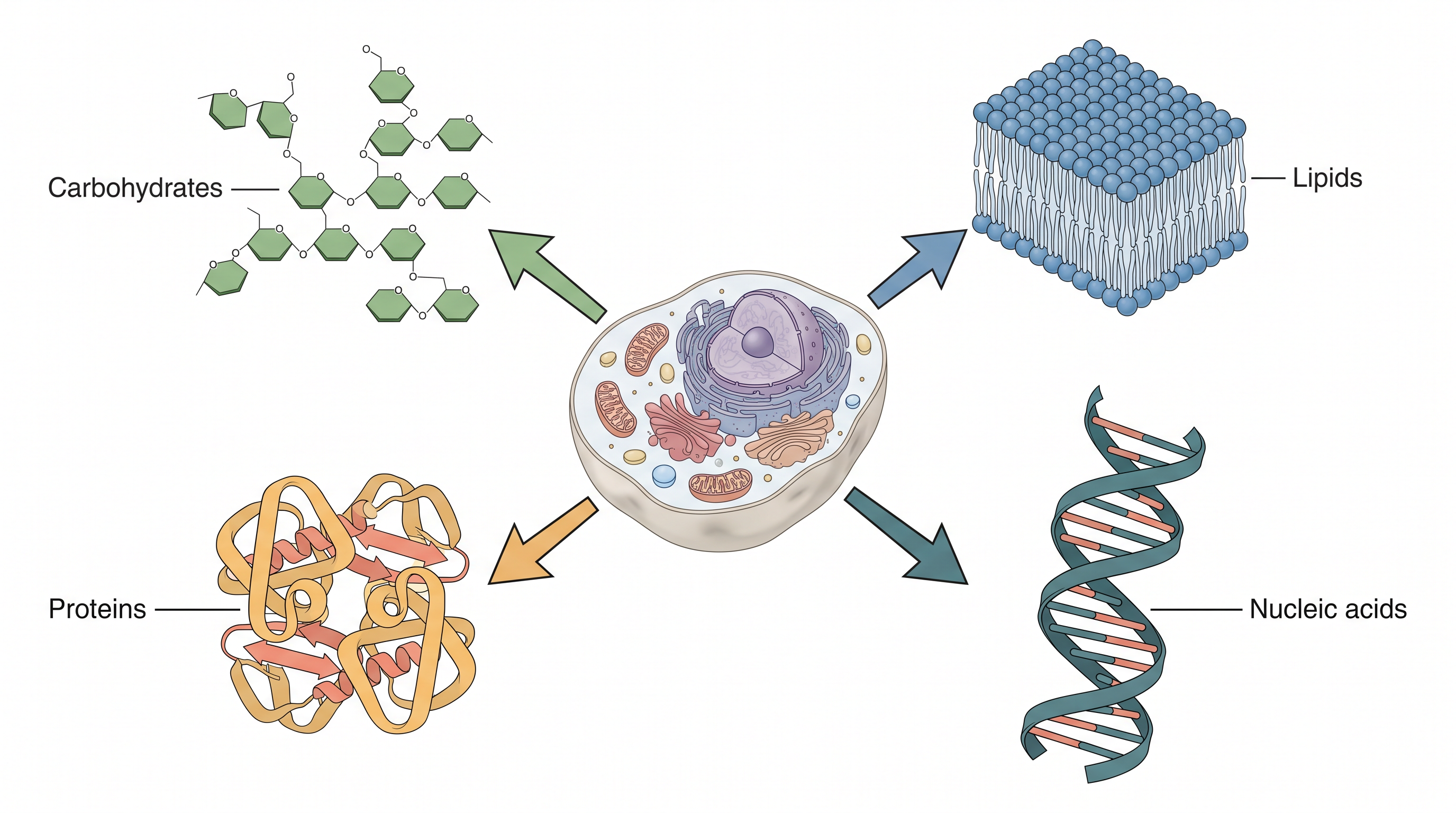
Fig. 3.0 – The four major classes of biomolecules.
Keep these five questions in mind as you read. By the end of this chapter you will be able to answer every one of them.
3.1 Carbohydrates
Carbohydrates are one of the most abundant classes of biomolecules in nature, and they are found widely distributed in all life forms. From the sugar dissolved in your blood to the wood of a tree and the shell of a crab, the same family of molecules turns up again and again.
Chemically, carbohydrates are aldehyde and ketone derivatives of the polyhydric alcohols. That single sentence packs in three ideas worth unpacking. A polyhydric alcohol is a molecule carrying many hydroxyl (–OH) groups. An aldehyde group (–CHO) sits at the end of a carbon chain, while a ketone group (>C=O) sits somewhere in the middle. So a carbohydrate is essentially a short carbon chain wearing many –OH groups, with one carbon carrying a C=O group either at the end or in the middle.
The major role of carbohydrates in living organisms is to function as a primary source of energy. This is why glucose is the first fuel a cell reaches for, and why a patient who cannot eat is given a glucose drip rather than a protein or fat drip.
But energy supply is only one of their jobs. These molecules also serve as energy stores, as metabolic intermediates, and as one of the major components of bacterial and plant cell wall. The cell walls of bacteria and plants are made up of polymers of carbohydrates. Carbohydrates are also part of DNA and RNA, which you will study later in this chapter.
Carbohydrates also act as informational materials. They are linked to the surfaces of proteins and lipids, where they act in cell–cell interaction, and in the interaction between cells with other elements in the cellular environment. In other words, the sugar coat on a cell surface works like a name tag: it tells a neighbouring cell, or a passing antibody, what kind of cell it is looking at. The A, B and O blood groups are decided in exactly this way — by small differences in the sugar chains attached to the surface of your red blood cells.
Classification of Carbohydrates
Carbohydrates are found in various forms ranging from simple sugars to complex polymers of more than one unit, and accordingly these have been classified. They are commonly classified into three categories, namely monosaccharides, oligosaccharides, and polysaccharides.
The basis of this classification is simply the number of sugar units joined together. The overall scheme is summarised in Fig. 3.1.
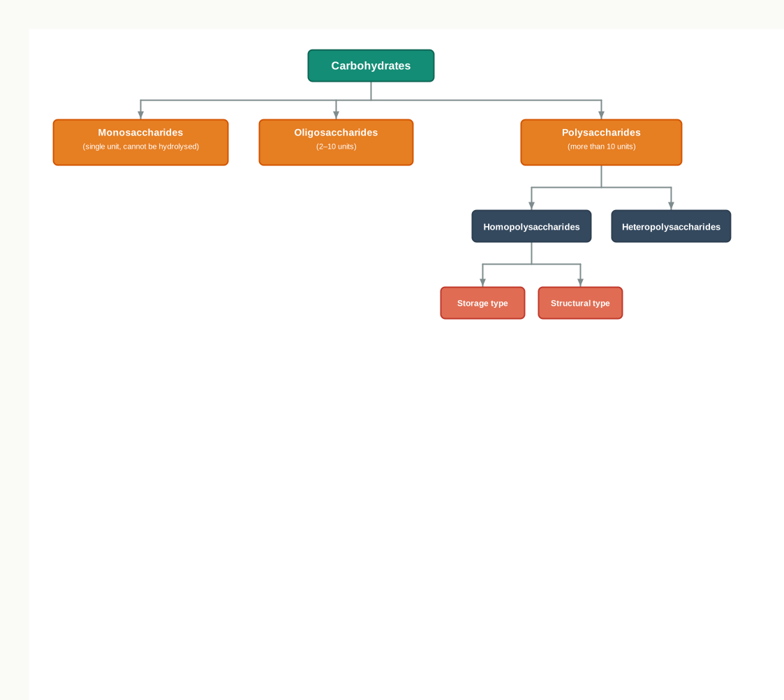
Fig. 3.1 – Classification of carbohydrates.
Monosaccharides
Monosaccharides are simple sugars which cannot be further hydrolysed into simpler forms. Hydrolysis means splitting a molecule by adding water across a bond; if you try this on a monosaccharide, you do not get two smaller sugars, because there is nothing left to split.
These monosaccharides contain free aldehyde (–CHO) and ketone (>C=O) groups, with two or more hydroxyl (–OH) groups, and have a general formula of
\[C\_n(H\_2O)\_n\]
where \(n\) is the number of carbon atoms in the molecule.
Based on the number of carbon atoms and functional groups, monosaccharides are classified as given in Table 3.1. Read the table in two directions at once: moving down the rows tells you how long the carbon chain is, while moving across the columns tells you whether the C=O group is at the end (an aldose) or in the middle (a ketose).
Table 3.1: Classification of monosaccharides
| S. No. | Class of monosaccharides based on number of carbon atoms | Aldoses | Ketoses |
|---|---|---|---|
| 1. | Trioses (\(C\_3H\_6O\_3\)) | Glyceraldehyde (an aldotriose) | Dihydroxyacetone (a ketotriose) |
| 2. | Tetroses (\(C\_4H\_8O\_4\)) | Erythrose | Erythrulose |
| 3. | Pentoses (\(C\_5H\_{10}O\_5\)) | Ribose | Ribulose |
| 4. | Hexoses (\(C\_6H\_{12}O\_6\)) | Glucose | Fructose |
Notice that glucose and fructose appear in the same row. Both are hexoses with the formula \(C\_6H\_{12}O\_6\), yet glucose is an aldose and fructose is a ketose. Two molecules with an identical formula but a different arrangement of atoms behave very differently in the body — this is your first hint that in biology, shape matters more than composition.
Oligosaccharides
Conventionally, oligosaccharides are carbohydrates having two to ten units of monosaccharides joined together by glycosidic bond. A glycosidic bond is the covalent link that joins one sugar unit to the next; you will see exactly how it is formed a little later in this section.
Some commonly occurring oligosaccharides are maltose, lactose, sucrose, etc., which are disaccharides (having two monosaccharides).
Polysaccharides
Polysaccharides are polymers of more than ten monosaccharide units joined together by glycosidic linkages. A polymer is a long molecule built by repeating a small unit, called the monomer, many times over — here the monomer is a monosaccharide.
These are classified in a number of ways depending upon the type of repeating monosaccharide unit (homo- and hetero-polysaccharides); in the degree of branching; and in the type of glycosidic linkage between the monomeric units. A homopolysaccharide repeats a single kind of sugar unit, while a heteropolysaccharide uses more than one kind.
Examples of some common polysaccharides are starch, glycogen, cellulose, and chitin.
Glycoconjugates
Carbohydrates can be conjugated to proteins and lipids to form glycoconjugates. There are three types of glycoconjugates: glycoproteins, proteoglycans and glycolipids.
If the protein component is predominant in the association of carbohydrate and protein, it is called glycoprotein. If the association contains major amount of carbohydrate than protein, then it is called proteoglycan. When the carbohydrate conjugates with lipids, it is called glycolipid.
Structure and Properties of Carbohydrates
Having classified carbohydrates, we now look at how they are actually built — because every property discussed above, from sweetness to digestibility to rigidity, follows from structure.
Structure of Monosaccharides
Structures of some common monosaccharides are given in Fig. 3.2. Compare the chains in that figure carefully: the only differences between several of these sugars lie in whether a particular –H and –OH pair is drawn to the left or to the right.
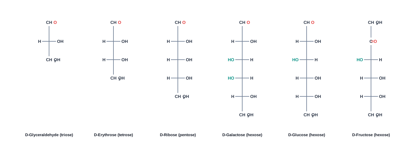
Fig. 3.2 – Open-chain structures of common monosaccharides.
A monosaccharide such as glucose exists both as straight chain structure and cyclic structure, as shown in Fig. 3.3. In water, the cyclic form is the one that dominates. Cyclic structures are the result of hemiacetal formation by intramolecular reaction between carbonyl group and a hydroxyl group. “Intramolecular” simply means that the two reacting groups belong to the same molecule — the chain bends around and one of its own –OH groups attacks its own C=O group, closing the chain into a ring.
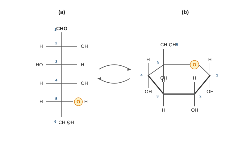
Fig. 3.3 – Straight-chain and cyclic structures of glucose.
Chiral Centres and Stereoisomers
All monosaccharides except dihydroxyacetone contain one or more asymmetric (chiral) carbon. A chiral carbon is a carbon atom bound by four different groups. Such a carbon has no plane of symmetry, so the molecule and its mirror image cannot be superimposed on each other — just as your left hand cannot be laid exactly over your right.
Because of this, these sugars are optically active isomers (enantiomers). “Optically active” means a solution of the sugar rotates the plane of polarised light passing through it.
A molecule with \(n\) chiral centres can have \(2^n\) stereoisomers, where \(n\) is the number of chiral carbon atoms in the molecule. Thus, glyceraldehyde with one chiral centre has \(2^1 = 2\) stereoisomers, and glucose with four chiral centres has \(2^4 = 16\) stereoisomers.
D and L Sugars
The orientation of the –OH group that is most distant from the carbonyl carbon determines whether the sugar belongs to D or L sugars. When this –OH group is on the right side of the carbon atom bearing it, then the sugar is D-isomer, and when it is on the left, the sugar is L isomer, as shown in Fig. 3.4.
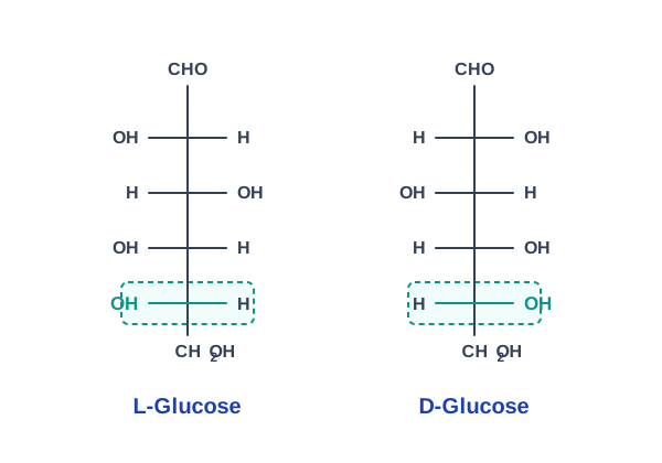
Fig. 3.4 – L and D forms of glucose.
Most of the sugars present in the biological system are D sugars. This is not a trivial detail. Enzymes are themselves three-dimensional, so an enzyme built to grip a D sugar cannot grip its mirror image, exactly as a right-handed glove cannot fit a left hand. L-glucose tastes sweet but the body cannot metabolise it at all.
Anomers and Mutarotation
When the open chain closes into a ring, the carbonyl carbon becomes a new chiral centre, and this creates a fresh kind of isomerism.
Isomeric forms of monosaccharide that differ only in their configuration about the hemiacetal or hemiketal carbon atom are called anomers. A hemiacetal is formed due to reaction between alcoholic and aldehyde groups of a monosaccharide, and a hemiketal is formed due to reaction between alcoholic and keto groups of a monosaccharide.
The carbonyl carbon atom is called the anomeric carbon. In the α-anomer, the –OH group of the anomeric carbon is on the opposite side of the sugar ring from the CH₂OH group at the chiral centre that designates the D and L configuration (C-5 in case of glucose). The other anomer is known as β-anomer.
The interconversion of α and β anomers in aqueous solution is called mutarotation, in which one ring form opens briefly into the linear form, then closes again to produce β anomers, as shown in Fig. 3.5. So in a glass of glucose solution, the two ring forms are constantly converting into each other through the open-chain form.
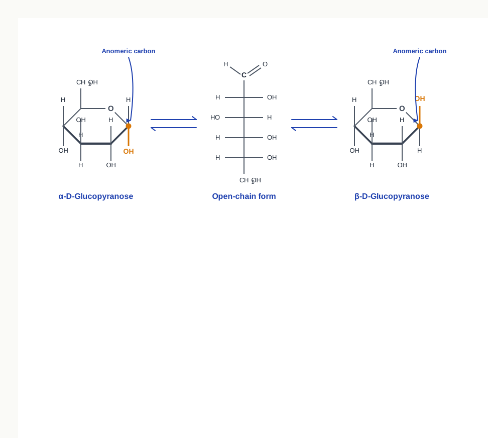
Fig. 3.5 – Cyclic anomers of D-glucose and mutarotation.
Epimers
Isomers having different configuration of –OH only at one carbon atom are known as epimers. The most important epimers of glucose are mannose (epimers at C-2) and galactose (epimers at C-4), as shown in Fig. 3.6.
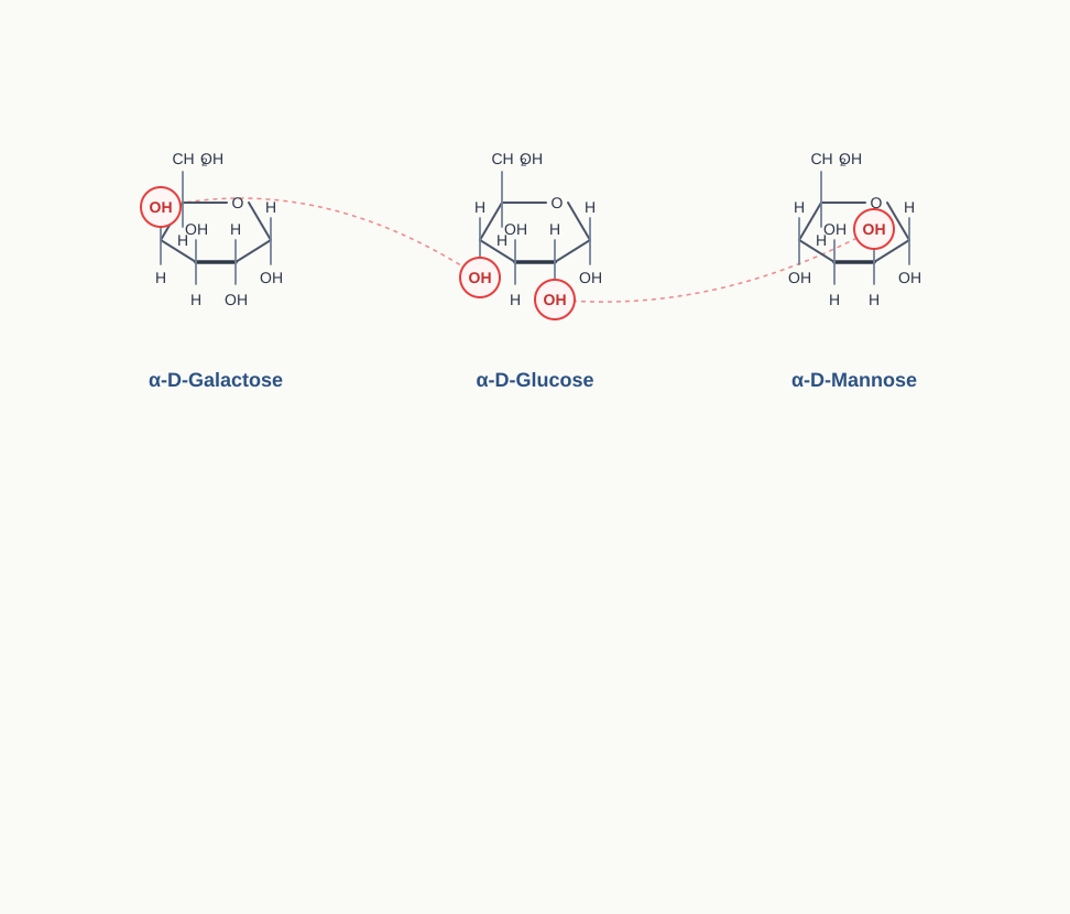
Fig. 3.6 – Epimers of glucose: galactose and mannose.
Students very often mix up anomers with epimers, and examiners know it. The safe way to remember the difference is that anomers differ only at the anomeric carbon created during ring closure, whereas epimers differ at any one carbon other than that.
Structure of Oligosaccharides
Oligosaccharides consist of two to ten monosaccharides joined by glycosidic linkage.
For example, the disaccharide maltose contains two D-glucose residues joined by a glycosidic linkage, which is a covalent bond formed by joining of –OH group of one monosaccharide with the anomeric carbon of the other sugar unit. Its formation from two molecules of α-D-glucose is shown in Fig. 3.7. Note that a molecule of water is released as the bond forms, which is why this is a condensation reaction, and why adding water back (hydrolysis) reverses it.
\[2\\ \\text{α-D-glucose} \\rightleftharpoons \\text{Maltose} + \\mathrm{H\_2O}\]
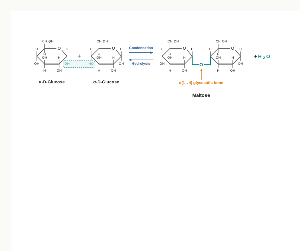
Fig. 3.7 – Formation of maltose by condensation.
Lactose is made up of D-galactose and D-glucose residues, joined by a β(1→4) glycosidic bond, as shown in Fig. 3.8.
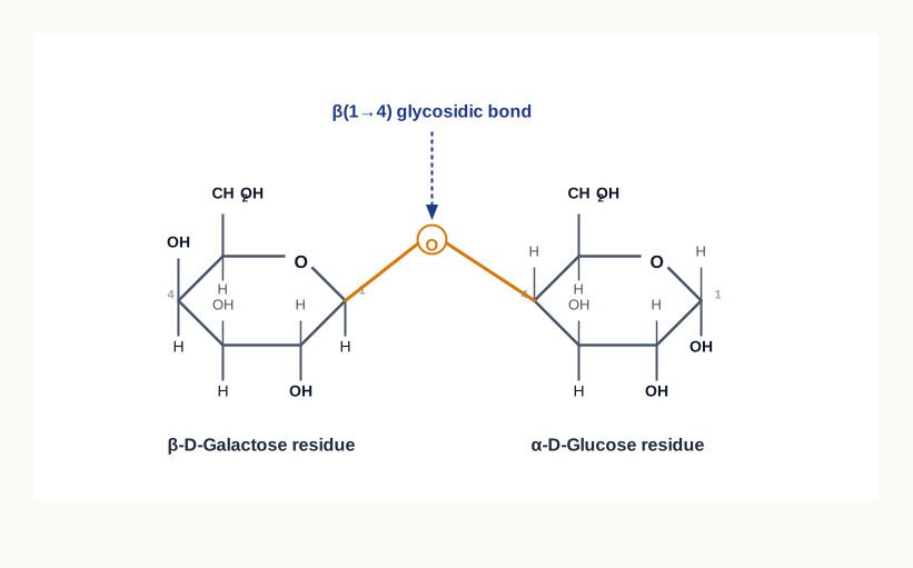
Fig. 3.8 – Structure of lactose.
Lactose is the sugar of milk, and it explains a familiar everyday observation. Infants make plenty of the enzyme lactase, which hydrolyses the β(1→4) bond of lactose. In many adults the production of this enzyme falls, the undigested lactose passes into the large intestine, and gut bacteria ferment it — producing the gas and discomfort of lactose intolerance. The lactose-free milk sold in shops is ordinary milk in which the bond has already been broken by adding lactase.
Disaccharides can be hydrolysed to yield their constituent monosaccharides by boiling with dilute acid. For example, hydrolysis of sucrose yields a mixture of glucose and fructose.
\[\\text{Sucrose} + \\mathrm{H\_2O} \\rightarrow \\text{Glucose} + \\text{Fructose}\]
Other oligosaccharides are trisaccharides (for example, raffinose, which is made up of glucose, galactose, and fructose units), tetra-, penta- and hexasaccharides.
Structure of Polysaccharides
Depending on the functional roles played, homopolysaccharides are divided into storage polysaccharides and structural polysaccharides.
Storage polysaccharides serve as storage form of monosaccharide that is used as fuel. Starch is an example of storage polysaccharide in plants, and glycogen is the storage polysaccharide in animals.
Why store glucose as a polymer at all, instead of keeping it free? Because thousands of free glucose molecules inside a cell would raise its osmotic concentration enormously and draw in water until the cell burst. Locking them into one large, insoluble polymer stores the same fuel at almost no osmotic cost.
Structural polysaccharides such as cellulose and chitin serve as structural elements in plant cell wall and animal exoskeleton, respectively.
Heteropolysaccharides, unlike homopolysaccharides, provide extracellular support for organisms. In extracellular space of animal tissues, these form a matrix that holds individual cells together and provides shape, support and protection to the cells and tissues.
The names and properties of some homopolysaccharides are given in Table 3.2.
Table 3.2: List of some common homopolysaccharides
| Name | Constituent monosaccharide | Biological significance |
|---|---|---|
| Starch | α-D-glucose | Storage of energy in plants |
| Glycogen | α-D-glucose | Storage of energy in bacteria and animals |
| Cellulose | β-D-glucose | It plays structural role and provides rigidity and strength to the cell wall. |
| Chitin | β-N-acetyl-D-glucosamine | It plays structural role and provides rigidity to exoskeleton of insects. |
| Inulin | β-D-fructose | Storage of energy in plants. |
| Pectin | α-D-galacturonic acid | It has structural role: holds cellulose fibrils together in plant cell walls. |
| Dextran | α-D-glucose | It plays structural role as an extracellular adhesive in bacteria. |
| Xylan | β-D-xylose | It has storage and supporting roles in plants. |
Look down the second column of Table 3.2 and one pattern stands out: starch, glycogen, cellulose and dextran are all built from glucose, yet their jobs could hardly be more different. The difference lies entirely in whether the linkage is α or β, and in how much the chain branches.
Examples of Some Common Polysaccharides
(a) Starch
Starch occurs in plants as reserve carbohydrate in tubers, seeds, fruits, and roots. It is composed of two homopolysaccharides, amylose (15–20%) and amylopectin (80–85%).
Amylose is a linear polymer of α-D-glucose monomers and linked by α(1→4) bonds, as shown in Fig. 3.9.
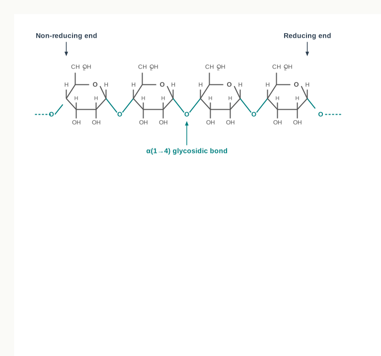
Fig. 3.9 – Structure of amylose.
Amylopectin, on the other hand, consists of glucose units linked by α(1→4) glycosidic linkage, like amylose. However, unlike amylose, it is highly branched. Branch points occur every 24 to 30 glucose residues, and linkage at the branch points is α(1→6) glycosidic, as shown in Fig. 3.10.
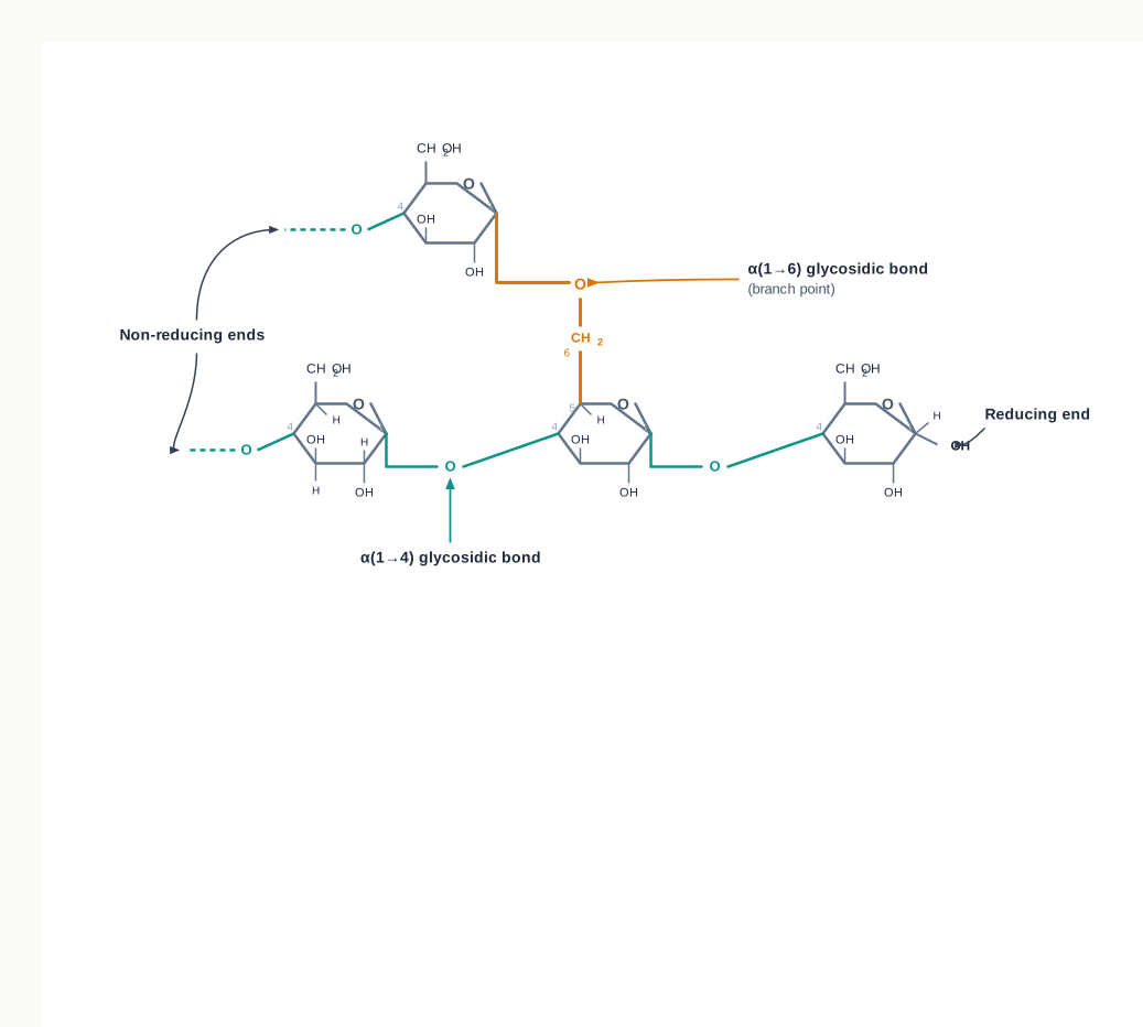
Fig. 3.10 – Structure of amylopectin.
Characteristic blue colour of the starch with iodine is due to amylose. In contrast, amylopectin gives only dull reddish brown colour with iodine. The reason is that the long, unbranched amylose chain coils into a helix that traps iodine inside it; the heavily branched amylopectin cannot form long helices, so the colour is much weaker. This is the basis of the iodine test you may have performed on a potato slice in the laboratory.
The enzymes present in saliva (salivary amylase) and pancreatic juice (pancreatic amylase) hydrolyse α(1→4) glycosidic linkages of starch, therefore digesting it into monomeric glucose residues. This is why a piece of plain bread begins to taste faintly sweet if you keep chewing it — salivary amylase has started releasing glucose in your mouth. The complete route is shown in Fig. 3.11.

Fig. 3.11 – Digestion of starch in the human body.
(b) Glycogen
Glycogen is an extensively branched storage homopolysaccharide found in animals. Similar to amylopectin, it consists of glucose units joined together by α(1→4) glycosidic linkage and having α(1→6) linkage at branching points.
Branching is not decoration; it is design. Every branch creates one more free end at which enzymes can begin clipping off glucose, so a heavily branched molecule can release fuel far faster than a linear one — exactly what a muscle needs at the start of a sprint.
Muscle cells contain glycogen at 1–2 per cent of their dry weight, and liver cells contain up to 10 per cent of their dry weight as glycogen. The division of labour is worth noting: muscle glycogen fuels the muscle itself, while liver glycogen is broken down to keep the glucose level of the whole blood stream steady between meals.
(c) Cellulose
Cellulose is the most abundant extracellular structural polysaccharide found in plants. It is also the most abundant of all biomolecules in the biosphere. It is the primary structural component of plant cell wall.
Structurally, cellulose is a linear polymer of upto 15000 D-glucose units linked by β(1→4) glycosidic bonds, as shown in Fig. 3.12. The β linkage makes each successive glucose unit flip over relative to its neighbour, so the chain stays straight and flat instead of coiling. Straight chains can pack side by side and hydrogen bond into tough fibres — which is precisely why cotton, paper and wood are strong.
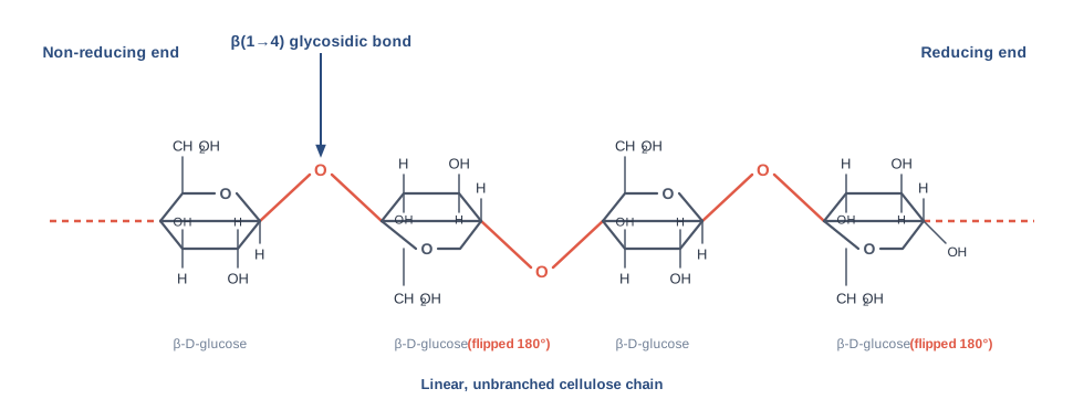
Fig. 3.12 – Structure of cellulose.
Unlike starch, cellulose cannot be digested by humans, as the human gut lacks the β(1→4) glycosidic bond hydrolysing enzyme known as cellulase. However, cattle and termites can digest cellulose, as their gut harbours symbiotic microorganisms that secrete cellulase, which hydrolyses and digests cellulose.
(d) Chitin
Chitin is a linear polysaccharide of β(1→4) linked N-acetyl-D-glucosamine residues. It is the main structural component of the exoskeleton of invertebrates (crustaceans, insects and spiders), and main component of cell walls of most fungi.
Chitin and cellulose have similar structures except that the OH group at second carbon position of cellulose is replaced by an acetamido group in chitin. Extensive hydrogen bonding of N-acetyl side chains makes chitin a tough and insoluble polymer.
That toughness has been put to industrial use. Chitin recovered from prawn and crab shells — otherwise a waste product of the seafood industry — is converted into chitosan, which is used in wound dressings and in water treatment.
(e) Peptidoglycan
Peptidoglycan constitutes the rigid component of bacterial cell wall. It is a heteropolysaccharide of alternating β(1→4) linked N-acetyl muramic acid (NAM) and N-acetyl-D-glucosamine (NAG) residues.
The linear polysaccharide chains are cross linked by short peptides attached to N-acetyl muramic acid. Cross linking by peptide welds the polysaccharide chains into a strong sheath that envelops the entire cell and prevents osmotic rupture of the cell. A bacterium usually sits in surroundings more dilute than its own cytoplasm, so water keeps entering it; without this sheath the cell would swell and burst.
Because the sheath is essential and because human cells have nothing like it, peptidoglycan is an ideal drug target. Penicillin acts by blocking the cross-linking step, so a growing bacterium builds a wall that cannot be stitched together, and bursts — while the patient’s own cells are untouched.
Lysozyme, which is an enzyme present in human tears, kills bacteria by hydrolysing the β(1→4) glycosidic linkage of peptidoglycan. Tears, then, are not only for crying; they are a continuous antibacterial wash for the surface of the eye.
Agar is worth pausing on, because a structural polysaccharide made by seaweed turns out to be one of the most useful materials in the whole of biotechnology. Its value lies in what it does not do: it does not react with the molecules placed in it, and it stays solid at the temperatures at which bacteria and plant cells grow.
3.5 Nucleic Acids
The cell organelles, namely nucleus, mitochondria and chloroplast, contain nucleic acid within them. Within nucleus, nucleic acids are associated with histone proteins to form chromatin. **Histones** are small, positively charged proteins; because the nucleic acid itself carries negative charges, the two bind tightly, and the enormously long molecule is packed into the small space of a nucleus.
Nucleic acids are polymers of nucleotides linked through phosphodiester linkages. Two types of nucleic acids are present in cells, namely deoxyribonucleic acid (DNA) and ribonucleic acid (RNA).
DNA acts as a genetic material and inherits the information from one generation to next. However, RNA serves as genetic material in some viruses. Every organism you have studied so far — bacteria, plants, animals — stores its information in DNA; RNA takes over that role only in certain viruses.
The Three Parts of a Nucleotide
Nucleotides of DNA and RNA are made up of nitrogenous base, sugar, and phosphate. Every nucleotide is built from exactly these three parts, so a nucleic acid is far simpler in composition than a protein.
The Sugar
The sugar that is present in nucleic acids is pentose sugar, which is of two types: the one present in DNA is **2’-deoxy-D-ribose**, and the other present in RNA is **D-ribose**. Both the pentoses are present as closed five membered rings, as shown in Fig. 3.13.
The difference between the two is a single oxygen atom. “Deoxy” means one oxygen fewer — the 2’ carbon of the DNA sugar carries only an –H where the RNA sugar carries an –OH. That missing –OH is what makes DNA the more chemically stable of the two, and therefore the better molecule in which to keep information for a lifetime.
For numbering pentoses of nucleotides, the carbon numbers are given a prime (′) designation to differentiate them from the numbered atoms of the nitrogenous bases. So the carbons of the sugar are 1′, 2′, 3′, 4′ and 5′, while the atoms of the base are numbered 1, 2, 3 and so on without primes. Keeping the two systems apart is essential — 5′ refers to the sugar, never to the base.
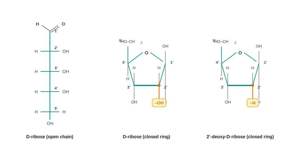
Fig. 3.13 – Structure of pentose sugars present in nucleic acids.
The Nitrogenous Bases
Nitrogenous bases are of two types: **purines** and **pyrimidines**. Purines are built from two fused rings and pyrimidines from a single ring, so purines are the larger of the two.
The two purine bases of DNA and RNA are **adenine (A)** and **guanine (G)**. Among pyrimidines, **cytosine (C)** is present in both DNA and RNA, **thymine (T)** is present in DNA only, and **uracil (U)** is present in RNA only. The structure of the five major bases is shown in Fig. 3.14.
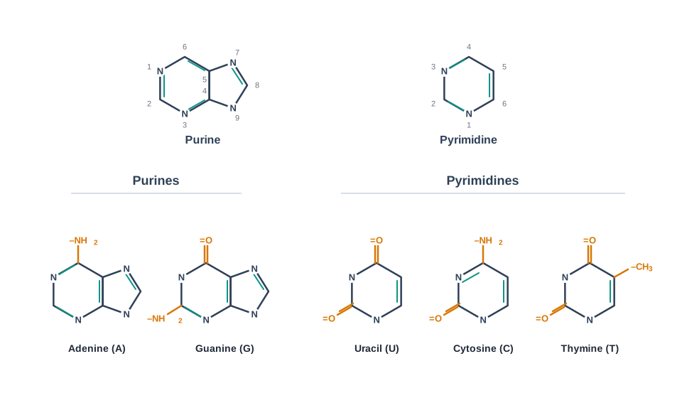
Fig. 3.14 – Structure of nitrogenous bases present in nucleic acids.
The purine and pyrimidine bases contain aromatic ring structures which absorb light at a wavelength near 260 nm. This is not a stray physical fact — it is the property on which the routine measurement of DNA in every molecular biology laboratory depends.
Nucleosides and Nucleotides
A base (either purine or pyrimidine), a pentose sugar unit and a phosphate group are linked together to form a **nucleotide**. On the other hand, a base linked only to a pentose sugar unit without a phosphate group is called a **nucleoside**.
The distinction is simply the phosphate: nucleoside plus phosphate gives nucleotide. Students lose marks on this every year, so fix it now — the shorter word, nucleoside, is the smaller molecule.
The four nucleosides of DNA are named as deoxyadenosine, deoxyguanosine, deoxycytidine, and deoxythymidine.
The nucleotides in DNA are of four types, namely deoxyadenylate (deoxyadenosine-5’-monophosphate; dAMP), deoxyguanylate (deoxyguanosine-5’-monophosphate; dGMP), deoxycytidylate (deoxycytidine-5’-monophosphate; dCMP) and deoxythymidylate (deoxythymidine-5’-monophosphate; dTMP), as shown in Fig. 3.15. Nucleotides in DNA are indicated by prefix ‘d’, as they contain deoxyribose rather than ribose sugar.
The four nucleosides of RNA are called adenosine, guanosine, cytidine, and uridine, which, when bound to a phosphate group, make ribonucleotides or ribonucleoside-5’-monophosphate. There are four types of ribonucleotides, namely adenylate (adenosine-5’-monophosphate; AMP), guanylate (guanosine-5’-monophosphate; GMP), cytidylate (cytidine-5’-monophosphate; CMP), and uridylate (uridine-5’-monophosphate; UMP), also shown in Fig. 3.15.
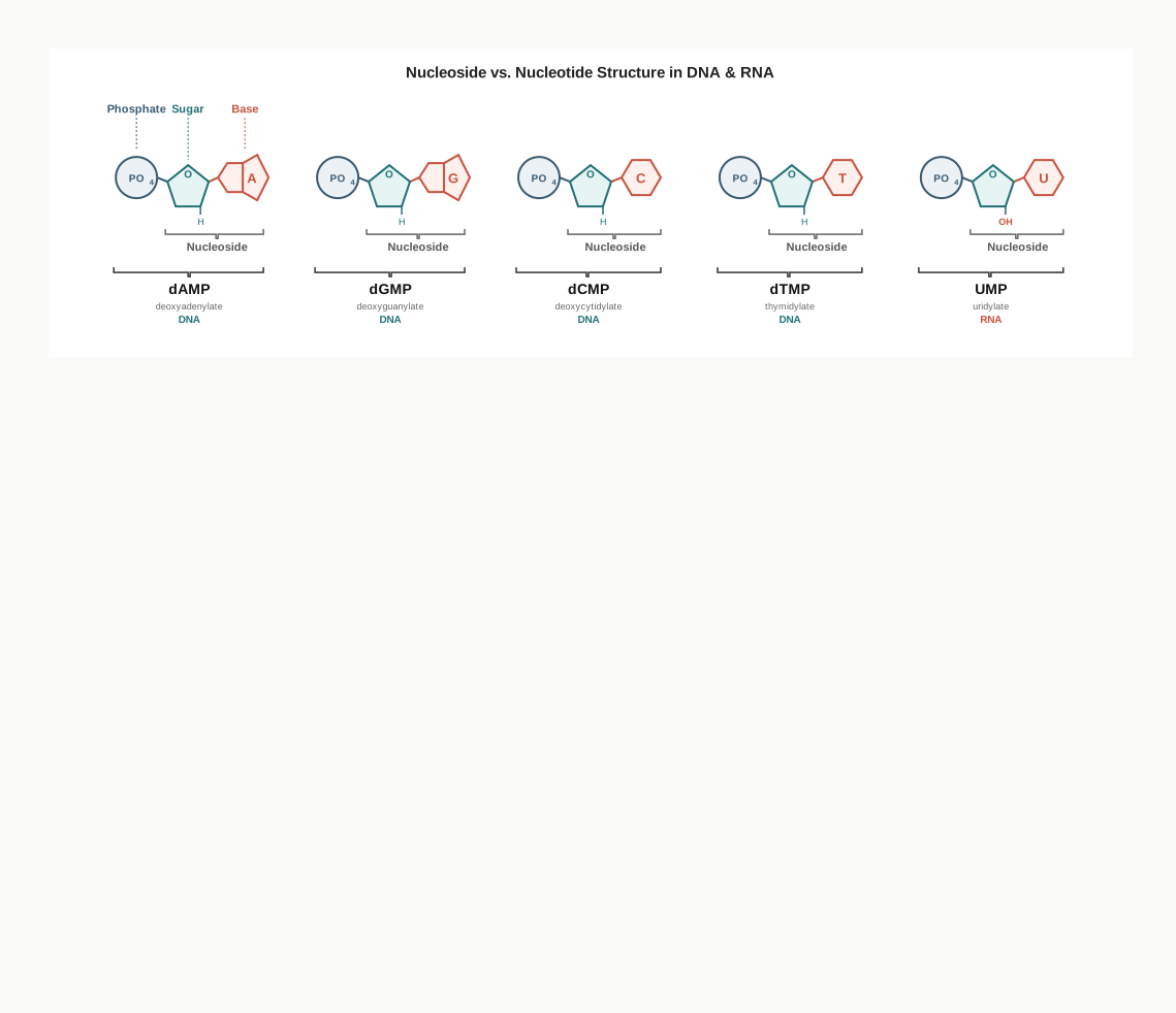
Fig. 3.15 – Nucleosides and nucleotides present in DNA and RNA.
The naming looks intimidating but follows one rule. Take the base, turn it into a nucleoside name, add “5’-monophosphate” for the phosphate, and add “deoxy-” or the prefix ‘d’ if the sugar is deoxyribose. Once you see the pattern, all eight names can be worked out rather than memorised.
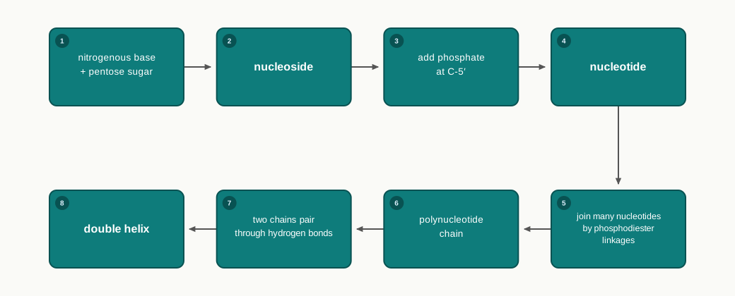
Fig. 3.16 – Building a nucleic acid.
3.5.1 Polynucleotide chain
Nucleotides of both DNA and RNA are covalently linked, in which the 3’ hydroxyl (–OH) group of the sugar of one nucleotide unit is esterified to the –OH group of phosphate attached to 5’ carbon atom of the sugar of the next nucleotide, forming a **phosphodiester linkage**, as shown in Fig. 3.17.
The name explains the structure. One phosphate group forms two ester bonds — one to the 3’ carbon of the sugar behind it and one to the 5’ carbon of the sugar ahead of it — hence phospho-di-ester. The result is an unvarying backbone of sugar–phosphate–sugar–phosphate, with the bases branching off it.
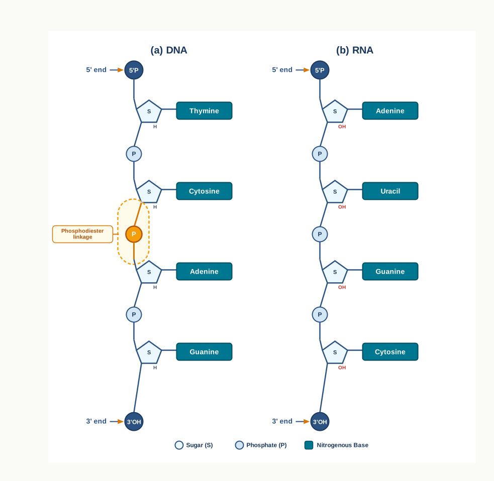
Fig. 3.17 – Primary structure of (a) DNA and (b) RNA.
Like a polypeptide chain, a polynucleotide chain has a specific polarity, i.e., distinct 5’ and 3’ ends. The 5’ end has a phosphate group at C-5’ of sugar, while the 3’ end has a free –OH group at C-3’ of ribose.
The base sequence in a polynucleotide chain is written in the 5’→3’ direction. This is a strict convention, not a preference. The sequence 5'-ATGC-3' and the sequence 5'-CGTA-3' contain the same four bases, but they are different molecules — so a sequence written without its polarity marked is incomplete information.
The Watson–Crick Double Helix
James Watson and Francis Crick in 1953 proposed the double helical (three dimensional) structure of DNA, shown in Fig. 3.18 (b). It consists of two polynucleotide chains of DNA wound around the same axis to form a right handed double helix.
The two strands are oriented antiparallel, i.e., their 3’ and 5’ phosphodiester bonds run in opposite directions. If one strand runs 5’→3’ from top to bottom, its partner runs 3’→5’ in the same direction — rather like two lanes of a road carrying traffic in opposite directions.
The sugar and phosphate form the backbones of the double helix and remain exposed to the polar environment. The bases of both the strands are stacked inside the core of the double helix and make it hydrophobic. This arrangement follows the same logic you met with proteins in Section 3.4: the water-loving parts face the water, and the water-avoiding parts are buried inside.
Within the helix, each nucleotide base of one strand makes hydrogen bonds in the same plane with the base of the other strand. A of one strand forms two hydrogen bonds with T of the other strand (A=T) and *vice versa*, and G on one strand forms three hydrogen bonds with C on the other strand (G≡C) and *vice versa*, as shown in Fig. 3.18 (a).

Fig. 3.18 – Base pairing in DNA and the Watson–Crick model.
Why must A pair only with T, and G only with C? Because a purine is large and a pyrimidine is small. Pairing a purine with a pyrimidine always spans the same width, so the helix keeps a constant diameter along its whole length. Two purines would bulge and two pyrimidines would pinch. The hydrogen bonding groups then decide which purine goes with which pyrimidine.
This pairing rule has a consequence that made DNA convincing as the genetic material: if you know the sequence of one strand, you know the sequence of the other. 5'-GAATTC-3' on one strand demands 3'-CTTAAG-5' on the other. A molecule that carries its own backup copy is exactly what heredity requires.
Forms of DNA
The Watson–Crick structure of DNA is also known as **B-DNA**. The B-DNA is the most stable form of DNA, and it is the form found in the cell under normal conditions.
The other two structural variants are A-DNA and Z-DNA. The A-DNA is a right handed double helix. It is wider and contains 11 base pairs per helix. However, Z-DNA is a left handed double helix with 12 base pairs per helix. The name Z-DNA comes from the zig-zag path its backbone follows.
Denaturation and Annealing
The bases of two strands of DNA double helix are joined by hydrogen bonds, as seen in Fig. 3.18 (a). A hydrogen bond is weak on its own, and this weakness is deliberate — the two strands must be separable, or the information in them could never be read or copied.
Heating disrupts hydrogen bonds between the base pairs, and thereby separation of the two strands of DNA takes place, called **denaturation** or **melting**.
The temperature at which half of the DNA is denatured is called **melting temperature (T_m)**. Apart from heating, melting is also caused by acid or alkali, both of which interfere with hydrogen bonding.
The two strands of nucleic acids that are separated by melting can spontaneously reassociate to form a double helix if temperature is decreased below \(T\_m\). This process of reassociation or renaturation is called **annealing**. The complete cycle is shown in Fig. 3.19.
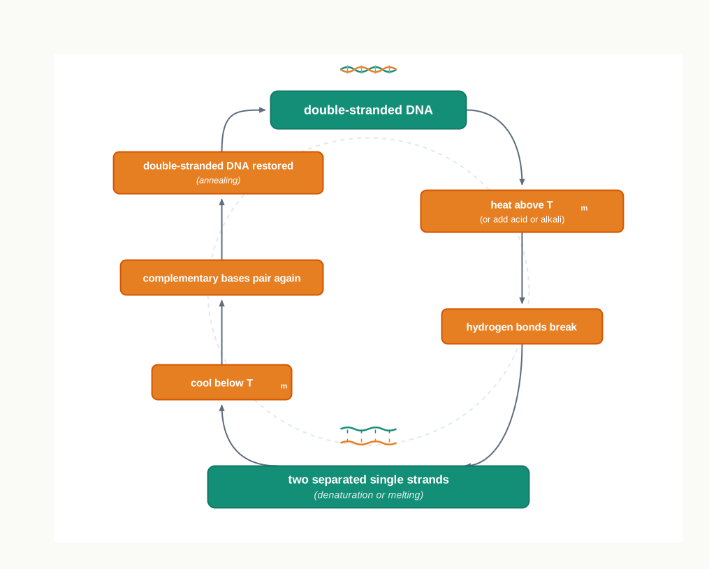
Fig. 3.19 – Cycle of DNA denaturation and annealing.
Notice that \(T\_m\) is not the same for every DNA sample. A G≡C pair is held by three hydrogen bonds while an A=T pair is held by only two, so a DNA rich in G and C needs more heat to come apart and has a higher melting temperature.
This pair of processes is the basis of a great deal of modern biotechnology. Any method that needs to open a double helix and then let a short piece of DNA bind to a chosen sequence — including the amplification of DNA in the laboratory — is built on controlled cycles of melting and annealing.
3.5.2 Types of RNA
Three kinds of RNA work together to convert the information held in DNA into protein.
Messenger RNA (mRNA)
It is a single strand linear polyribonucleotide chain that carries genetic information from DNA to ribosome. The name is exact: mRNA is a message, a working copy sent out from the DNA archive so that the original need never leave the nucleus.
At the 5’ end is UTR (untranslated region), containing no genetic information for polypeptide synthesis. It is followed by initiation codon, coding region, and stop codon. At the 3’ end is present another UTR, as shown in Fig. 3.20. The untranslated regions are not wasted sequence — they are the parts the ribosome uses to locate and hold the message before translation begins.
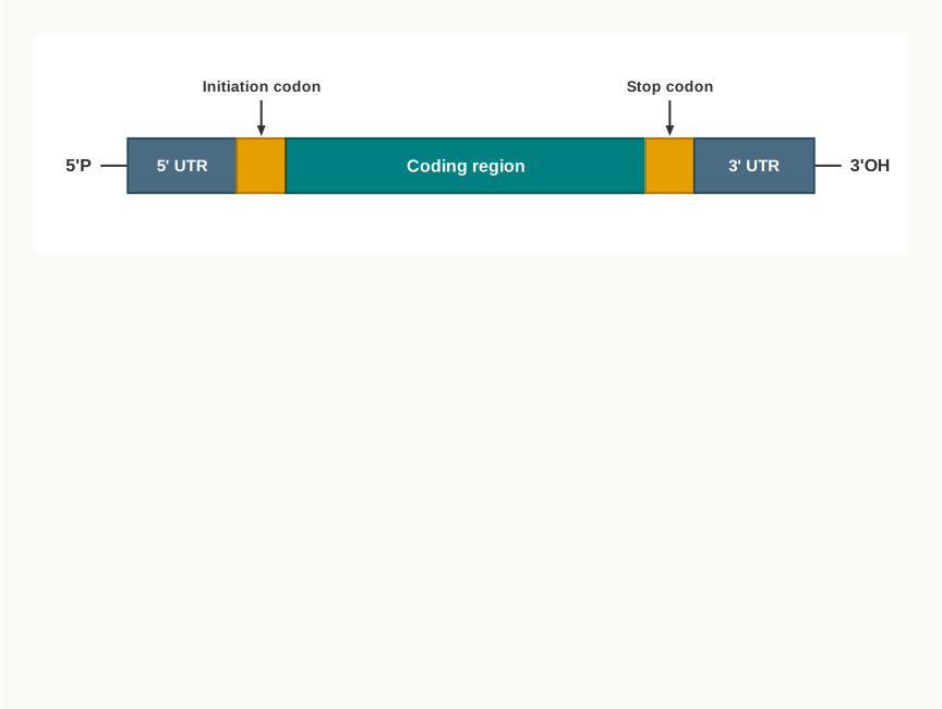
Fig. 3.20 – Structure of a typical prokaryotic mRNA.
In eukaryotic mRNA, 5’ end contains guanylate which is methylated at its N-7. This process is called **capping**. The 3’ end of mRNA also undergoes **polyadenylation** (addition of several adenylate residues).
Both modifications act as protection. A eukaryotic message has to travel out of the nucleus and survive in the cytoplasm, and the cap at one end and the run of adenylate residues at the other keep it from being destroyed on the way.
Ribosomal RNA (rRNA)
It forms the structural components of ribosomes. So rRNA is not a message at all — it is part of the machine that reads the message.
70S prokaryotic ribosomes have 16S rRNA in the smaller subunit (30S), and 23S and 5S rRNAs in the larger subunit (50S).
The eukaryotic ribosome (80S) has 18S rRNA in the smaller subunit (40S) and 28S, 5.8S and 5S rRNAs in the larger subunit (60S).
The S in these figures stands for the Svedberg unit, a measure of how fast a particle settles when spun in a centrifuge. Larger, denser particles settle faster and so carry a higher S value. Because the value depends on shape as well as mass, these numbers do not add up in the ordinary way — which is why a 30S and a 50S subunit together make a 70S ribosome, not an 80S one.
Transfer RNA (tRNA)
These are small RNA molecules that transfer amino acids to ribosome during protein synthesis. tRNA is therefore the adaptor that connects the two languages of the cell — the four-base language of nucleic acids and the twenty-amino-acid language of proteins.
It is a single polyribonucleotide chain folded to form four arms, having a clover-leaf secondary structure, as shown in Fig. 3.21. The folding comes from the chain pairing with itself wherever short stretches of its own sequence are complementary.
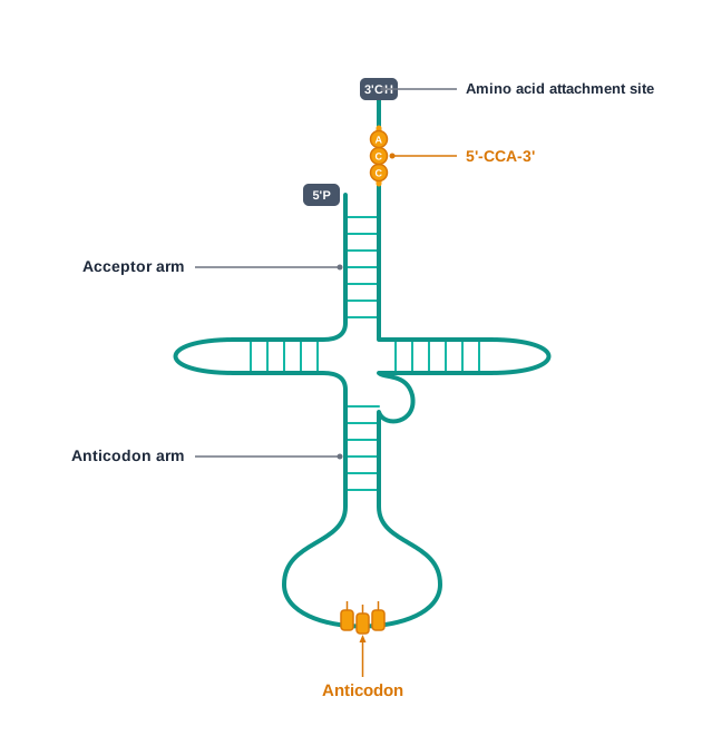
Fig. 3.21 – Clover-leaf secondary structure of tRNA.
The acceptor arm has a 5'-CCA-3' sequence at the 3’OH end, which is the amino acid binding site.
The anticodon arm has the anticodon, a set of three bases which recognises a specific codon of mRNA during translation. The recognition works by the same base pairing rule as in DNA, which is why one tRNA can read one codon and no other.
3.6 Summary
- Carbohydrates are made up of carbon, hydrogen, and oxygen atoms, and are widely distributed in animal and plant tissues.
- Carbohydrates are categorised into four major classes: monosaccharides (for example, glucose and ribose), disaccharides (for example, sucrose and lactose), oligosaccharides (for example, raffinose), and polysaccharides (for example, starch and glycogen). Disaccharides are simply the smallest oligosaccharides, carrying two units.
- Monosaccharides can be classified as trioses, tetroses, pentoses, hexoses, and so on, depending on the number of carbon atoms in the molecule. They are also classified by functional group as aldoses and ketoses.
- A pentose sugar such as ribose is an important component of nucleic acids and coenzymes.
- Two monosaccharides linked by a glycosidic bond make a disaccharide, for example sucrose.
- Polysaccharides perform various roles, both as the storage form of energy and as structural components. The type of glycosidic linkage decides which role a polysaccharide plays.
- Lipids are organic compounds found in living organisms. These are made up of hydrophobic fatty acid chains linked to glycerol via ester bond, and they dissolve in organic solvents rather than in water.
- Fatty acids are long chain hydrocarbons containing a carboxylic acid group. Fatty acids may be saturated (having no double bonds) or unsaturated (having one or more double bonds). Nomenclature of fatty acids is based on total number of carbons, total number of double bonds, and position of double bonds.
- Lipids are broadly classified into two classes — simple and compound lipids.
- Simple lipids include triacylglycerol and waxes, which are esters of glycerol with fatty acids, and esters of high molecular weight alcohol with fatty acids, respectively.
- Compound lipids include membrane lipids, which are of two types: glycerophospholipids and sphingolipids. Compound lipids are amphipathic molecules, carrying a hydrophilic phosphate head together with hydrophobic fatty acid tails, and they arrange themselves in water as micelles.
- Glycerophospholipid contains a glycerol backbone, and sphingolipids contain a sphingoid base backbone.
- Steroids are another class of compound lipids, made up of a four-fused ring structure. Cholesterol is the most common steroid found in animals. It is the precursor of all steroid hormones and vitamin D.
- The amino acids are organic compounds containing amine (–NH₂) and carboxyl (–COOH) functional groups, along with a side chain (R group) specific to each amino acid. At physiological pH they exist as zwitterions.
- There are 20 standard amino acids, some non-standard amino acids (for example, 4-hydroxyproline and 5-hydroxylysine), and some non-protein amino acids (for example, L-ornithine and L-citrulline).
- In a polypeptide chain, the amino acids are linked covalently via peptide bond in a linear fashion to make a protein. Only L isomers of amino acids are used.
- There are four levels of protein structures, namely primary, secondary, tertiary, and quaternary.
- Primary structure of proteins is the linear chain of amino acid sequences linked through peptide bonds, running from the N-terminal end to the C-terminal end.
- Secondary structure of protein describes the regular folding taken up by stretches of the polypeptide chain, held in place by hydrogen bonds between the NH and CO groups of the main chain.
- The two major types of secondary structures are α-helix and β-sheets.
- Tertiary structure is the three dimensional arrangement of protein, held together by disulfide, hydrogen, hydrophobic and ionic bonds between side chains, with polar R groups on the outside and non-polar R groups buried inside.
- Quaternary structure is the complex arrangement of folded protein subunits, stabilised through hydrogen bonds, electrostatic interactions, ionic bonds and disulfide bridges.
- Nucleic acids are polymers of nucleotides, which contain nitrogenous bases (adenine, guanine, cytosine, thymine and uracil), sugar and phosphate, joined by phosphodiester linkages.
- Deoxyribonucleic acid (DNA) and ribonucleic acid (RNA) are the two types of nucleic acids.
- DNA is the genetic material of almost all the organisms, except some viruses where RNA is the genetic material.
- DNA contains deoxyribose sugar and thymine, while RNA contains ribose sugar and uracil in place of thymine.
- In DNA, adenine forms 2 hydrogen bonds with thymine (A=T) and cytosine forms 3 hydrogen bonds with guanine (C≡G). Breaking these bonds by heat separates the strands (denaturation), and cooling below \(T\_m\) lets them re-pair (annealing).
- J. Watson and F. Crick (1953) described the three dimensional double helical structure of DNA, in which two antiparallel strands wind around the same axis. B-DNA is the most stable form; A-DNA and Z-DNA are the two structural variants.
- The mRNA, tRNA, rRNA are the major types of RNA — the message, the adaptor that carries amino acids, and the structural component of ribosomes, respectively.
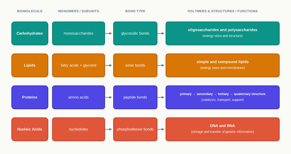
Fig. 3.22 – The four classes of biomolecules.
3.7 Key Terms
- **Biomolecules** — The macromolecules that build cellular organelles and carry out cellular processes, namely carbohydrates, proteins, lipids and nucleic acids.
- **Carbohydrate** — An aldehyde or ketone derivative of a polyhydric alcohol, serving as the primary energy source, an energy store, a structural material and an informational molecule on cell surfaces.
- **Polyhydric alcohol** — A molecule carrying many hydroxyl (–OH) groups.
- **Monosaccharide** — A simple sugar that cannot be hydrolysed into any simpler sugar, with the general formula \(C\_n(H\_2O)\_n\).
- **Hydrolysis** — The splitting of a molecule by the addition of water across one of its bonds.
- **Aldose** — A monosaccharide carrying its C=O group at the end of the chain as an aldehyde (–CHO) group.
- **Ketose** — A monosaccharide carrying its C=O group in the middle of the chain as a ketone (>C=O) group.
- **Oligosaccharide** — A carbohydrate made of two to ten monosaccharide units joined by glycosidic bonds.
- **Glycosidic bond** — The covalent bond formed by joining the –OH group of one monosaccharide with the anomeric carbon of another sugar unit.
- **Polysaccharide** — A polymer of more than ten monosaccharide units joined by glycosidic linkages.
- **Polymer** — A large molecule built by repeating a small unit, the monomer, many times over.
- **Homopolysaccharide** — A polysaccharide built from only one kind of repeating monosaccharide unit.
- **Heteropolysaccharide** — A polysaccharide built from more than one kind of monosaccharide unit, which provides extracellular support in organisms.
- **Glycoconjugate** — A carbohydrate covalently joined to a protein or a lipid.
- **Glycoprotein** — A glycoconjugate in which the protein component predominates.
- **Proteoglycan** — A glycoconjugate in which the carbohydrate component predominates.
- **Glycolipid** — A glycoconjugate formed when a carbohydrate conjugates with a lipid.
- **Chiral (asymmetric) carbon** — A carbon atom bonded to four different groups, so that the molecule and its mirror image cannot be superimposed.
- **Enantiomers** — The optically active mirror-image isomers that arise from the presence of chiral carbon atoms.
- **D and L sugars** — Isomers distinguished by the orientation of the –OH group most distant from the carbonyl carbon; on the right gives the D-isomer and on the left the L-isomer.
- **Hemiacetal** — The group formed by reaction between the alcoholic and aldehyde groups of a monosaccharide, which closes the sugar into a ring.
- **Hemiketal** — The group formed by reaction between the alcoholic and keto groups of a monosaccharide.
- **Anomeric carbon** — The carbonyl carbon of a sugar, which becomes a new chiral centre when the ring closes.
- **Anomers** — Isomeric forms of a monosaccharide differing only in their configuration about the hemiacetal or hemiketal carbon atom.
- **Mutarotation** — The interconversion of α and β anomers in aqueous solution, in which the ring briefly opens into the linear form and closes again.
- **Epimers** — Isomers having a different configuration of –OH at only one carbon atom, such as mannose and galactose relative to glucose.
- **Starch** — The storage polysaccharide of plants, composed of linear amylose and branched amylopectin.
- **Amylose** — The linear component of starch, a polymer of α-D-glucose joined by α(1→4) bonds, responsible for the blue colour with iodine.
- **Amylopectin** — The branched component of starch, with α(1→4) linkages along the chain and α(1→6) linkages at branch points every 24 to 30 glucose residues.
- **Glycogen** — The extensively branched storage homopolysaccharide of animals, with α(1→4) linkages and α(1→6) linkages at branch points.
- **Cellulose** — The most abundant biomolecule in the biosphere, a linear polymer of up to 15000 D-glucose units joined by β(1→4) bonds, forming the plant cell wall.
- **Cellulase** — The enzyme that hydrolyses the β(1→4) glycosidic bond of cellulose, absent in humans but secreted by symbiotic microorganisms in cattle and termites.
- **Chitin** — A linear polysaccharide of β(1→4) linked N-acetyl-D-glucosamine residues, forming invertebrate exoskeletons and most fungal cell walls.
- **Peptidoglycan** — The rigid heteropolysaccharide of the bacterial cell wall, made of alternating β(1→4) linked NAM and NAG residues cross linked by short peptides.
- **Lysozyme** — The enzyme of human tears that kills bacteria by hydrolysing the β(1→4) glycosidic linkage of peptidoglycan.
- **Agar** — A gelatinous heteropolysaccharide from the cell wall of marine red algae, used to form a surface for growing bacterial and plant tissue cultures.
- **Agarose** — The component of agar with very few charged groups, used as an inert support for the electrophoretic separation of nucleic acids.
- **Lipid** — A hydrophobic, non-polar organic compound, soluble in organic solvents, made largely of hydrocarbon chains linked to glycerol by ester linkage.
- **Ester linkage** — The bond formed when the –COOH group of an acid reacts with the –OH group of an alcohol, with the loss of water.
- **Fatty acid** — A long hydrocarbon chain carrying a carboxylic acid group, obtained by hydrolysis of fats and usually containing an even number of carbon atoms.
- **Saturated fatty acid** — A fatty acid whose hydrocarbon chain contains no double bonds.
- **Unsaturated fatty acid** — A fatty acid whose chain contains one or more double bonds; monounsaturated with one, polyunsaturated with more than one.
- **Simple lipid** — A fatty acid ester of an alcohol with no other group attached, such as a fat or a wax.
- **Triacylglycerol** — A simple lipid formed by esterifying glycerol with three fatty acids, the main vehicle of energy storage in adipose tissue.
- **Wax** — A simple lipid formed by esterification of a fatty acid with a high molecular weight monohydric alcohol, serving as a protective surface coating.
- **Compound lipid** — A fatty acid ester of an alcohol that also carries an additional group, such as a phospholipid or a steroid.
- **Amphipathic molecule** — A molecule carrying both a hydrophilic region and a hydrophobic region within the same structure.
- **Phospholipid** — An amphipathic compound lipid with two hydrophobic fatty acid tails and a hydrophilic phosphate head, found mainly in cell membranes.
- **Glycerophospholipid** — A phospholipid built on a glycerol backbone, with a modified phosphate group on the third carbon of glycerol.
- **Sphingophospholipid** — A phospholipid built on a sphingoid base backbone, with the fatty acid attached by an amide linkage instead of an ester linkage.
- **Ceramide** — A sphingolipid whose head group contains only a hydrogen atom.
- **Sphingomyelin** — A sphingolipid whose head group is phosphocholine.
- **Micelle** — The sphere-shaped structure adopted by phospholipids in water, with tails turned inward and heads facing outward.
- **Steroid** — A hydrophobic lipid with a characteristic four-fused ring structure, acting in cells as a receptor ligand.
- **Cholesterol** — The most common steroid, made chiefly in the liver, precursor of all steroid hormones and a source of rigidity in eukaryotic plasma membranes.
- **Ergosterol** — A fungal steroid that is an important precursor of vitamin D.
- **Amino acid** — An organic molecule whose α-carbon carries a carboxylic (–COOH) group, an amino (–NH₂) group, a hydrogen atom and a variable side chain.
- **α-carbon** — The carbon atom to which the carboxylic group is attached, bearing all four groups of an amino acid.
- **Side chain (R group)** — The only variable part of an amino acid, which decides its polarity, charge and behaviour in a folded protein.
- **Zwitterion** — The dipolar form of an amino acid at physiological pH, carrying –COO⁻ and –⁺NH₃ within one overall neutral molecule.
- **Non-standard amino acid** — An amino acid formed in cells after protein synthesis by modification of a standard residue, such as 4-hydroxyproline.
- **Non-protein amino acid** — An amino acid that takes no part in proteins, such as L-ornithine, L-citrulline, β-alanine, creatine and γ-aminobutyrate.
- **Protein** — A macromolecule built from 20 naturally occurring amino acids linked covalently into a linear sequence and folded into a working shape.
- **Peptide bond** — The covalent amide linkage between the α-carboxyl group of one amino acid and the α-amino group of the next, formed with loss of water.
- **Residue** — Each amino acid unit within a polypeptide, so called because a water molecule is lost when it joins the chain.
- **N-terminal and C-terminal ends** — The two different ends of a polypeptide, bearing a free amino group and a free carboxy group respectively, which give the chain its polarity.
- **Dalton (Da)** — The unit of molecular mass; the mean molecular weight of an amino acid residue is about 110 Da.
- **Primary structure** — The linear sequence of amino acids in a polypeptide, linked through peptide bonds.
- **Dihedral (torsion) angles φ and ψ** — The angles of rotation about the Cα–N and Cα–C bonds, which together determine the path of the polypeptide chain.
- **Ramachandran plot** — A two-dimensional plot of the permissible values of φ and ψ, identified by G.N. Ramachandran in 1963.
- **Secondary structure** — The regular local folding of the polypeptide chain, stabilised by hydrogen bonds between main chain NH and CO groups.
- **α helix** — A rod-like secondary structure in which the CO group of each amino acid hydrogen bonds with the NH group four residues ahead.
- **β pleated sheet** — A secondary structure in which strands distant in the sequence lie side by side, hydrogen bonded either in parallel or in anti-parallel orientation.
- **Tertiary structure** — The overall three-dimensional arrangement of all residues in a protein, held by disulfide, hydrogen, hydrophobic and ionic bonds.
- **Disulfide bond** — A covalent bond formed by oxidation of a pair of cysteine residues.
- **Quaternary structure** — The spatial arrangement of two or more folded polypeptide subunits, which may be identical (homodimer) or different (heterodimer).
- **Nucleic acid** — A polymer of nucleotides linked through phosphodiester linkages; DNA and RNA are its two types.
- **Chromatin** — The complex formed within the nucleus when nucleic acids associate with histone proteins.
- **Nucleoside** — A nitrogenous base linked to a pentose sugar, without a phosphate group.
- **Nucleotide** — A nitrogenous base, a pentose sugar and a phosphate group linked together.
- **Purine** — A double-ringed nitrogenous base; adenine and guanine are the purines of DNA and RNA.
- **Pyrimidine** — A single-ringed nitrogenous base; cytosine occurs in both DNA and RNA, thymine in DNA only, and uracil in RNA only.
- **Phosphodiester linkage** — The bond joining the 3’ –OH of one nucleotide’s sugar to the phosphate on the 5’ carbon of the next.
- **5’ and 3’ ends** — The two different ends of a polynucleotide chain, bearing a phosphate at C-5’ and a free –OH at C-3’ respectively, which is why sequences are written 5’→3’.
- **Antiparallel** — The arrangement of the two DNA strands in which their 3’ and 5’ phosphodiester bonds run in opposite directions.
- **B-DNA** — The Watson–Crick right handed double helix, the most stable form of DNA.
- **A-DNA** — A wider right handed double helical variant of DNA containing 11 base pairs per helix.
- **Z-DNA** — A left handed double helical variant of DNA containing 12 base pairs per helix.
- **Denaturation (melting)** — The separation of the two DNA strands when heat, acid or alkali disrupts the hydrogen bonds between base pairs.
- **Melting temperature (\(T\_m\))** — The temperature at which half of the DNA in a sample has been denatured.
- **Annealing** — The spontaneous reassociation of separated strands into a double helix when the temperature falls below \(T\_m\).
- **Messenger RNA (mRNA)** — A single stranded linear polyribonucleotide chain that carries genetic information from DNA to the ribosome.
- **Untranslated region (UTR)** — The stretch at each end of an mRNA that carries no information for polypeptide synthesis.
- **Capping** — The methylation at N-7 of the guanylate present at the 5’ end of eukaryotic mRNA.
- **Polyadenylation** — The addition of several adenylate residues at the 3’ end of mRNA.
- **Ribosomal RNA (rRNA)** — The RNA that forms the structural components of ribosomes, 70S in prokaryotes and 80S in eukaryotes.
- **Transfer RNA (tRNA)** — A small clover-leaf shaped RNA that transfers amino acids to the ribosome during protein synthesis.
- **Anticodon** — The set of three bases on the anticodon arm of tRNA that recognises a specific codon of mRNA during translation.
3.8 Points to Ponder
- Starch, glycogen and cellulose are all built from glucose, yet one feeds a plant, one feeds an animal and one holds a tree upright. If the monomer is the same in all three, where exactly does the difference in function come from — and what does this tell you about the importance of bond type and branching in biology?
- A cell could store its glucose as free molecules instead of locking it into a polymer. Think about what would happen to the water balance of such a cell, and why evolution has settled on polymers for storage in every group of organisms you have studied.
- Only D sugars and only L amino acids are used by living systems, even though the mirror-image forms are chemically identical in composition. What might this uniformity suggest about the origin of the enzymes that handle these molecules?
- Peptidoglycan protects a bacterium from bursting, and human cells have nothing like it. Consider why this single fact has made bacterial cell wall synthesis one of the most successful targets in the history of medicine, and what happens when bacteria find ways around it.
- A double bond puts a permanent bend in a fatty acid chain. Trace this one structural feature all the way through to the physical state of an oil, the fluidity of a membrane, and the texture of the food on your plate.
- Phospholipids form a micelle in water without any enzyme, any energy input or any instruction. Reflect on what it means that a biological structure can assemble itself simply because of the shape and polarity of its molecules.
- In sickle cell anaemia, one residue out of 146 is changed and the consequences are lifelong. What does this suggest about how much of a protein’s sequence is “important”, and how would you decide which positions in a protein matter most?
- The Ramachandran plot is mostly empty space. Consider what it means that of all the shapes a polypeptide chain could theoretically take, only a small number are physically allowed — and whether this makes protein folding easier or harder to predict.
- Boiling an egg destroys the protein’s shape but leaves every peptide bond intact. Think about why the loss of shape alone is enough to destroy function, and what this implies about the relationship between structure and activity in enzymes.
- DNA holds its two strands together with hydrogen bonds, which are among the weakest bonds in chemistry. Consider why a molecule entrusted with the information of a whole organism should be designed to come apart so easily, and what would go wrong if the strands were joined covalently instead.
- G≡C pairs are held by three hydrogen bonds and A=T pairs by two. Think about how the base composition of a DNA sample would affect the temperature needed to separate its strands, and where in the laboratory such a difference would matter.
- RNA serves as the genetic material in some viruses but not in cellular organisms. Using what you know about the 2’ position of the two pentose sugars, reflect on why DNA might have become the preferred long-term store of information.
- Bacterial ribosomes are 70S and human cytoplasmic ribosomes are 80S. Consider how such a difference in a piece of cellular machinery becomes a difference between a medicine and a poison.
- Four classes of biomolecules — carbohydrates, lipids, proteins and nucleic acids — each use a different kind of bond to join their units. Look back through the chapter and ask why each class needed its own kind of linkage, rather than all of life settling on one.
3.9 NCERT Questions
- Describe the classification of carbohydrates.
- Differentiate between D- and L-forms of glucose.
- Draw the structure of a disaccharide made up of two monosaccharides glucose and fructose.
- Draw the partial structure of starch and glycogen.
- Write the major functions of carbohydrates.
- Describe isomerisation in monosaccharides.
- Differentiate between sphingolipids and glycerolipids.
- Why are membrane lipids called amphipathic?
- Differentiate between saturated and unsaturated fatty acids.
- Describe the various categories of amino acids.
- What is zwitterion? Draw the structure of zwitterion.
- What are non-standard and non-protein amino acids?
- How the peptide bonds are formed?
- Draw the structure of Lys-Glu-Lys.
- Describe the various secondary structures of protein.
- Differentiate between tertiary and quaternary structure of proteins.
- Differentiate between nucleosides and nucleotides.
- Explain the primary structure of DNA.
- Draw the structure of A-T-C-G oligonucleotide.
- Explain Watson and Crick model of DNA.
- Describe the various forms of DNA.
- Describe the clover leaf model of tRNA.
- In carbohydrates which of these functional groups are present
- Alcohol and carboxyl groups
- Aldehyde and ketone groups
- Hydroxyl and hydrogen groups
- Ether and ester groups
- Which of the following is a non-reducing disaccharide?
- Maltose
- Lactose
- Sucrose
- Cellobiose
- The repeating units of proteins are
- Glucose units
- Amino acids
- Fatty acids
- Nucleotides
- Which of the following is the most common secondary structure of proteins
- α-helix
- β-pleated sheet
- Both (a) and (b)
- None of the above
- A nucleotide contains
- Nitrogenous base, sugar and phosphate
- Sugar and phosphate
- Nitrogenous base and sugar
- None of the above
- The two strands in DNA double helix are joined by
- Covalent bond
- Hydrogen bond
- Glycosidic bond
- Phosphodiester bond
- Which is an example of storage lipid?
- Fatty acids
- Triacylglycerol
- Sphingolipids
- Eicosanoids
- In glycerolipids fatty acids are joined to glycerol through which bond?
- Phosphodiester bond
- Glycosidic bond
- Peptide bond
- Ester bond
3.10 Objective Questions
- The general formula of a monosaccharide is
- \(C\_n(H\_2O)\_{n-1}\)
- \(C\_n(H\_2O)\_n\)
- \(C\_{2n}(H\_2O)\_n\)
- \(C\_n(H\_2O)\_{2n}\)
- Which one of the following is a ketotriose?
- Glyceraldehyde
- Erythrulose
- Dihydroxyacetone
- Ribulose
- Glucose contains four chiral centres. The number of stereoisomers possible for glucose is
- 4
- 8
- 16
- 32
- Mannose and galactose are related to glucose as
- Anomers at C-1 and C-4 respectively
- Epimers at C-2 and C-4 respectively
- Enantiomers
- Ketose forms of glucose
- The interconversion of α and β anomers of a sugar in aqueous solution is called
- Isomerisation
- Mutarotation
- Epimerisation
- Hydrolysis
- Branch points in amylopectin occur every
- 4 to 6 glucose residues
- 10 to 12 glucose residues
- 24 to 30 glucose residues
- 100 to 120 glucose residues
- The characteristic blue colour of starch with iodine is due to
- Amylopectin
- Amylose
- Both amylose and amylopectin equally
- The α(1→6) branch points
- Peptidoglycan of the bacterial cell wall is a heteropolysaccharide made of alternating
- NAM and NAG residues
- D-galactose and D-glucose residues
- α-D-glucose and β-D-glucose residues
- N-acetyl-D-glucosamine residues only
- Agarose gels are used in the laboratory as
- A source of energy for bacterial cultures
- An inert support for the electrophoretic separation of nucleic acids
- A reducing agent for disulfide bonds
- A solvent for extracting lipids
- A fatty acid denoted as 18:2 (\(\\Delta^{9,12}\)) has
- 18 double bonds and 2 carbon atoms
- 18 carbon atoms and 2 double bonds, beginning at C-9 and C-12
- 18 carbon atoms and 12 double bonds
- 2 carbon atoms and double bonds at positions 9 and 18
- Naturally occurring fatty acids usually contain an even number of carbon atoms because
- Odd-numbered chains cannot form ester bonds
- They are synthesised from two carbon units
- They always contain a double bond
- Glycerol contains three carbon atoms
- In sphingolipids, the fatty acid chain is attached to the backbone through
- An ester linkage
- A glycosidic linkage
- An amide linkage
- A phosphodiester linkage
- Which of the following lipids does NOT contain a fatty acid chain?
- Triacylglycerol
- Wax
- Phosphatidylcholine
- Cholesterol
- Which one of the following amino acids has a hydrogen atom as its R group?
- Alanine
- Glycine
- Serine
- Valine
- The single letter symbols of tryptophan, glutamine and lysine are, respectively
- T, G and L
- W, Q and K
- W, G and K
- Y, Q and L
- 4-hydroxyproline is best described as
- A standard amino acid
- A non-protein amino acid
- A non-standard amino acid formed by modification after protein synthesis
- A D-isomer of proline
- In an α helix, the CO group of each amino acid forms a hydrogen bond with the NH group of the amino acid situated
- Immediately next to it
- Two residues ahead
- Four residues ahead
- On a neighbouring polypeptide chain
- Taking the mean molecular weight of an amino acid residue as 110 Da, a polypeptide of 400 residues would have an approximate molecular weight of
- 4,400 Da
- 11,000 Da
- 44,000 Da
- 4,40,000 Da
- Disulfide bonds of the tertiary structure are formed by the oxidation of a pair of
- Serine residues
- Methionine residues
- Cysteine residues
- Histidine residues
- A protein made of two identical subunits and two different subunits shows
- Only primary structure
- Only secondary structure
- Tertiary structure but no quaternary structure
- Quaternary structure
- The prime (′) designation is used for the carbon atoms of
- The nitrogenous base
- The pentose sugar
- The phosphate group
- The peptide backbone
- Purine and pyrimidine bases absorb light strongly at a wavelength near
- 260 nm
- 280 nm
- 340 nm
- 560 nm
- In a phosphodiester linkage, the phosphate group joins
- The 3’ –OH of one sugar to the 5’ carbon of the next sugar
- The 2’ –OH of one sugar to the 5’ carbon of the next sugar
- Two nitrogenous bases of opposite strands
- The base to the sugar within one nucleotide
- Z-DNA differs from B-DNA in that it is
- Right handed with 11 base pairs per helix
- Left handed with 12 base pairs per helix
- Left handed with 11 base pairs per helix
- Right handed and wider
- The temperature at which half of the DNA in a sample is denatured is called
- The annealing temperature
- The renaturation temperature
- The melting temperature
- The optimum temperature
- Capping in eukaryotic mRNA involves
- Addition of several adenylate residues at the 3’ end
- Methylation at N-7 of the guanylate present at the 5’ end
- Removal of the untranslated regions
- Attachment of the anticodon arm
- The smaller subunit of the prokaryotic 70S ribosome is
- 40S, containing 18S rRNA
- 30S, containing 16S rRNA
- 50S, containing 23S and 5S rRNA
- 60S, containing 28S, 5.8S and 5S rRNA
- The amino acid binding site of a tRNA molecule is
- The anticodon arm
- The
5'-CCA-3'sequence at the 3’OH end of the acceptor arm - The 5’P end of the chain
- The loop opposite the acceptor arm
- **Assertion (A):** Humans cannot digest cellulose although they digest starch readily. **Reason (R):** The human gut lacks cellulase, the enzyme that hydrolyses the β(1→4) glycosidic bond.
- **Assertion (A):** Storing glucose as glycogen rather than as free glucose protects a cell from taking in excess water. **Reason (R):** A large number of free glucose molecules would raise the osmotic concentration of the cell, whereas one large polymer contributes far less.
- **Assertion (A):** Oleic acid is a liquid at room temperature while stearic acid, of the same chain length, is a solid. **Reason (R):** A double bond puts a permanent bend in the hydrocarbon chain, so the chains cannot pack closely against one another.
- **Assertion (A):** Phospholipids do not spontaneously mix with water and instead form micelles. **Reason (R):** Phospholipids are amphipathic, carrying a hydrophilic phosphate head and two hydrophobic fatty acid tails.
- **Assertion (A):** Glycine does not exist in L and D forms. **Reason (R):** The R group of glycine is a hydrogen atom, so its α-carbon is not attached to four different groups.
- **Assertion (A):** An amino acid exists as a zwitterion at physiological pH. **Reason (R):** A proton migrates from the carboxyl group to the amino group, giving –COO⁻ and –⁺NH₃ within the same molecule.
- **Assertion (A):** The peptide bond cannot rotate freely. **Reason (R):** Resonance interactions give the peptide bond about 40 per cent double bond character, making the six atoms around it planar.
- **Assertion (A):** In the tertiary structure of a globular protein, non-polar R groups are found in the interior. **Reason (R):** Non-polar side chains are hydrophobic and are therefore buried away from the surrounding water.
- **Assertion (A):** DNA rich in guanine and cytosine has a higher melting temperature than DNA rich in adenine and thymine. **Reason (R):** A G≡C pair is held by three hydrogen bonds while an A=T pair is held by only two.
- **Assertion (A):** Separated DNA strands can reassociate into a double helix when the temperature falls below \(T\_m\). **Reason (R):** Melting breaks only the hydrogen bonds between base pairs, leaving the phosphodiester backbone and the base sequence of each strand intact.
3.11 Check Your Concepts
- Glucose and fructose have the same molecular formula, \(C\_6H\_{12}O\_6\), and both are hexoses. Explain what makes them different molecules, and name the class to which each belongs.
- Why can a monosaccharide not be hydrolysed into anything simpler, while a disaccharide can? Support your answer by describing what happens when sucrose is boiled with dilute acid.
- A student writes that “an anomer and an epimer are the same thing, since both differ at one carbon.” Identify the error and rewrite the statement correctly, giving one example of each.
- Dihydroxyacetone is the only monosaccharide that is not optically active. Explain why, using the idea of a chiral carbon.
- What would happen to a bacterial cell placed in a dilute medium if its peptidoglycan cross-links were prevented from forming? Explain your answer in terms of the function of the peptidoglycan sheath.
- Amylose gives a deep blue colour with iodine while amylopectin gives only a dull reddish brown colour. Relate this difference to the branching pattern of the two molecules.
- Cattle can live on grass but human beings cannot. Explain this difference at the level of a single chemical bond, naming the enzyme involved and where it comes from in cattle.
- Compare and contrast starch and glycogen under the following headings: occurrence, monomer, type of linkage, degree of branching, and biological role.
- Chitin and cellulose have very similar structures. State the single structural difference between them, and explain how this difference makes chitin a tough and insoluble polymer.
- Match the polysaccharide in Column I with its constituent monosaccharide in Column II.
| Column I | Column II |
|---|---|
| a. Cellulose | i. β-D-fructose |
| b. Inulin | ii. β-N-acetyl-D-glucosamine |
| c. Pectin | iii. β-D-xylose |
| d. Chitin | iv. β-D-glucose |
| e. Xylan | v. α-D-galacturonic acid |
- Why is agar, rather than starch or cellulose, used to prepare the solid surface on which bacterial and plant tissue cultures are grown? Give two properties of agar that make it suitable.
- A fatty acid is described as 20:4 (\(\\Delta^{5,8,11,14}\)). Write down everything this symbol tells you about the molecule, and state whether it is monounsaturated or polyunsaturated.
- Explain why animal fats are usually solid at room temperature while plant oils are usually liquid, using the structure of their fatty acid chains.
- Distinguish between a simple triglyceride and a mixed triglyceride, and state where in the body triglycerides are chiefly stored.
- Both a wax and a triacylglycerol are simple lipids, yet only one of them contains three ester linkages. Explain why, referring to the alcohol used in each.
- Why are membrane lipids described as amphipathic, and how does this property explain the sphere-shaped arrangement they take up in water?
- Compare glycerophospholipids and sphingophospholipids under the headings: backbone, linkage of the fatty acid chain, and one named example.
- Steroids contain no long fatty acid chain, and yet they are classified as lipids. Justify this classification.
- The α-carbon of an amino acid is described as chiral or asymmetric. Explain what this means, and state which isomeric form is found in proteins.
- Only the side chain R varies among the twenty standard amino acids. Explain how such a small point of variation can produce proteins of completely different properties.
- Draw a labelled structure of the zwitterion of glycine. Mark the group that carries the negative charge and the group that carries the positive charge, and explain why the molecule is nevertheless neutral overall.
- Classify the following amino acids into their R group categories: tyrosine, aspartate, valine, arginine, and threonine.
- Distinguish between non-standard amino acids and non-protein amino acids, giving two examples of each and stating how each type arises.
- Why is each amino acid unit in a polypeptide called a residue and not simply an amino acid? Explain with reference to what happens during peptide bond formation.
- The pentapeptides
Tyr-Ala-Gly-Ser-LeuandLeu-Ser-Gly-Ala-Tyrcontain exactly the same amino acids. Explain why they are nevertheless two different molecules, and identify the N-terminal and C-terminal residue in each. - Arrange the following in the correct order of increasing structural complexity, and write one sentence describing each: quaternary structure, primary structure, tertiary structure, secondary structure.
- The backbone of a protein is described as a linked sequence of rigid planar peptide bonds, and yet proteins can fold in a great many ways. Explain how both statements can be true, naming the two angles involved.
- Left-handed α helices lie in a sterically allowed region of the Ramachandran plot, and yet they are not found in proteins. Explain why.
- Compare the α helix and the β pleated sheet under the headings: shape, which residues hydrogen bond with which, and one named protein in which it occurs.
- Explain why parallel β sheets are less twisted than anti-parallel β sheets, and state how the two types are distinguished.
- In a globular protein, polar R groups are found on the outside and non-polar R groups in the interior. Explain the reason for this arrangement, and predict what would happen to the folding if such a protein were placed in a non-polar solvent instead of water.
- Boiling an egg makes the egg white turn solid and white, but no peptide bonds are broken. Explain which level or levels of protein structure have been destroyed, and why the protein no longer works.
- Differentiate between a homodimer and a heterodimer, and state which types of bonds hold the subunits of a quaternary structure together.
- A nucleoside and a nucleotide differ by a single component. Name it, and write the nucleoside and nucleotide names for adenine in both DNA and RNA.
- Why are the carbons of the pentose sugar given a prime (′) designation? Explain what confusion this convention prevents.
- Explain why a purine always pairs with a pyrimidine in the DNA double helix, and never a purine with a purine.
- A student writes a DNA sequence simply as
ATGCCAwithout any further marking. Explain why this is incomplete, and rewrite it correctly along with its complementary strand. - Label the following on a diagram of the DNA double helix: the sugar–phosphate backbone, the stacked bases, an A=T pair, a G≡C pair, the 5’ end and the 3’ end of each strand. State which parts face the polar environment and which form the hydrophobic core.
- Compare B-DNA, A-DNA and Z-DNA under the headings: handedness of the helix, base pairs per helix where stated, and relative stability.
- Arrange the following steps in the correct sequence and name the two processes involved: strands reassociate into a double helix; hydrogen bonds between base pairs break; temperature is lowered below \(T\_m\); two single strands are formed; the sample is heated above \(T\_m\).
- A student prepares to separate the two strands of a DNA sample and reports that “boiling the DNA breaks it into individual nucleotides.” Identify what is wrong with this statement and correct it, naming the bonds that are actually broken and the bonds that survive.
- Explain why the untranslated regions of an mRNA are not wasted sequence, and describe the two modifications made to eukaryotic mRNA together with their purpose.
- Draw and label the clover-leaf structure of tRNA, marking the acceptor arm, the
5'-CCA-3'sequence, the amino acid attachment site and the anticodon arm. Explain how the anticodon selects the correct place on an mRNA. - A 30S subunit and a 50S subunit combine to form a 70S ribosome rather than an 80S one. Explain why these numbers do not add up in the ordinary way, and state what the S value actually measures.
- Compare mRNA, rRNA and tRNA under the headings: structure, function, and one distinguishing feature of each.
- Four different kinds of bond join the units of the four classes of biomolecules — glycosidic, ester, peptide and phosphodiester. Name the class of biomolecule associated with each, and state which two of these bonds are broken by the addition of water during digestion in the human alimentary canal.
3.12 Case-Based Questions
- A ten-year-old boy drinks a glass of milk and, within an hour, develops stomach cramps and bloating. He has no such trouble with curd or with lactose-free milk. A doctor explains that his body has stopped producing enough of a particular enzyme, so the milk sugar passes undigested into his large intestine, where gut bacteria act on it.
- Name the disaccharide present in milk and the two monosaccharides that make it up.
- Name the type of glycosidic bond joining these two units, and the enzyme the boy is short of.
- Explain why lactose-free milk causes him no discomfort, given that it is prepared from ordinary milk.
- The boy’s father suggests he should avoid all carbohydrates. Comment on whether this advice is scientifically justified.
- In a school laboratory, a student places a drop of dilute iodine solution on four samples: a slice of raw potato, a piece of filter paper, a spoon of boiled rice water, and a small piece of prawn shell. The potato and the rice water turn blue-black. The filter paper and the prawn shell show no such colour.
- Name the polysaccharide responsible for the blue-black colour and the specific component of it that produces the colour.
- Name the polysaccharide present in filter paper and in prawn shell, and state the type of glycosidic linkage in each.
- All four samples contain carbohydrate polymers. Explain why only two of them respond to the test.
- The student chews a piece of plain bread for two minutes and finds that it begins to taste sweet. Explain this observation, naming the enzyme and the bond it acts upon.
- A patient with a bacterial infection is given an antibiotic that prevents the short peptide cross-links of the bacterial cell wall from forming. The bacteria continue to grow, but within a few hours large numbers of them burst. The patient’s own cells are unaffected by the drug.
- Name the heteropolysaccharide of the bacterial cell wall and the two alternating residues from which it is built.
- Explain why the bacteria burst once cross-linking is blocked.
- Explain why the same drug does not damage the patient’s own cells.
- Human tears contain an enzyme that also destroys bacteria. Name it and state the bond it hydrolyses.
- A food company markets two cooking media. Product A is a mustard oil that pours freely at room temperature. Product B is a solid fat that holds its shape. Laboratory analysis shows that Product A is rich in 18:1 (\(\\Delta^{9}\)) and 18:2 (\(\\Delta^{9,12}\)) chains, while Product B is rich in 16:0 and 18:0 chains.
- Classify the fatty acids in each product as saturated, monounsaturated or polyunsaturated, giving your reason from the symbols.
- Explain, in terms of chain shape and packing, why Product A is liquid and Product B is solid at the same temperature.
- Both products are triacylglycerols. Explain what this means and name the alcohol common to both.
- The label on Product B claims it is “cholesterol free”. Explain whether the absence of cholesterol tells a consumer anything about the fatty acids present.
- A pharmaceutical team wants to deliver a drug that will not dissolve in blood. They enclose the drug inside tiny hollow spheres built from phospholipids, and inject the preparation. The spheres travel through the bloodstream and release the drug at the target tissue.
- Explain the property of phospholipids that allows them to form such spheres in a watery medium without any energy being supplied.
- Draw a simple labelled sketch of the arrangement of phospholipid molecules in the sphere, marking the heads, the tails, and the position of the water.
- State which part of the phospholipid gives it its identity and name two common modifiers of the phosphate group.
- Suggest why a naturally occurring lipid is a suitable material for carrying a drug through the body.
- A young patient tires easily and suffers periodic episodes of severe pain. A blood report shows red blood cells that are rigid and curved rather than round. Sequencing shows that in the β-chain of her haemoglobin, a single glutamate residue has been replaced by valine. The remaining 145 residues are unchanged.
- State which level of protein structure has been altered directly by this substitution.
- Using Table 3.4, state the R group category of glutamate and of valine, and explain why the exchange matters so much.
- Explain how a change in one residue can go on to affect the folding and the behaviour of the whole molecule.
- Haemoglobin is built of four subunits. Name the level of protein structure this represents, and state two types of interaction that hold such subunits together.
- A student purifies an enzyme and finds it works well at 30 °C. She then heats one sample to 90 °C for five minutes and cools it back to 30 °C. The solution turns cloudy and the enzyme no longer works. A second sample, kept at 30 °C throughout, works normally. Chemical analysis shows that the number of peptide bonds in the heated sample is unchanged.
- Explain which levels of protein structure have been affected by heating and which level has survived.
- Name the four types of interaction that stabilise the tertiary structure, and state which of them is covalent.
- Explain why an unfolded protein loses its activity even though its amino acid sequence is intact.
- The student suggests repeating the experiment at pH 2 instead of high temperature. Predict what she would observe, and justify your prediction.
- A technician isolates DNA from two bacterial species, X and Y. She measures the absorbance of each solution at 260 nm to check the yield. She then heats each solution slowly and records the temperature at which half of the DNA has separated into single strands. Species X gives a value of 72 °C and species Y a value of 85 °C. Both solutions are then cooled slowly back to room temperature.
- Explain why 260 nm is the wavelength chosen for the concentration measurement.
- Name the process taking place on heating and the term for the temperature being recorded.
- Which of the two species has DNA richer in guanine and cytosine? Justify your answer in terms of hydrogen bonds.
- State what happens to the separated strands as the solutions cool, name this process, and explain why the original double helix — and not a random pairing — is restored.
3.13 Practice With Data
- A molecule with \(n\) chiral centres can have \(2^n\) stereoisomers. Calculate the number of stereoisomers for (i) glyceraldehyde, which has one chiral centre, (ii) glucose, which has four, and (iii) a hypothetical sugar with six chiral centres. State by what factor the number of stereoisomers rises for each additional chiral carbon.
- A sample of starch weighs 250 g. Taking amylose as 20 per cent and amylopectin as 80 per cent of starch by mass, calculate the mass of each component present. If a second sample of the same mass were found to contain 15 per cent amylose, state which of the two samples would give the deeper blue colour with iodine and why.
- In amylopectin, branch points occur every 24 to 30 glucose residues. For a chain of 3,000 glucose residues, calculate the maximum and the minimum number of branch points expected. State the type of glycosidic linkage found at these branch points and the type found along the rest of the chain.
- Muscle cells contain glycogen at 1–2 per cent of their dry weight, and liver cells contain up to 10 per cent of their dry weight as glycogen. A person has 28 kg of muscle tissue and 1.5 kg of liver tissue, and in both, the dry weight is one-third of the fresh weight. Calculate the maximum mass of glycogen stored in each tissue, and comment on which store is larger in total even though the liver is far richer per gram.
- Cellulose is a linear polymer of up to 15000 D-glucose units. Given that a single glucose residue in the chain adds about 0.5 nm to its length, estimate the maximum length of one cellulose molecule in nanometres and in micrometres. Comment on how this length compares with a typical plant cell, which is about 50 µm across.
- Study the fatty acid data below and answer the questions that follow.
| Fatty acid | Symbol | No. of Carbons |
|---|---|---|
| P | 16:0 | 16 |
| Q | 18:1 (\(\\Delta^{9}\)) | 18 |
| R | 18:2 (\(\\Delta^{9,\\,12}\)) | 18 |
| S | 20:4 (\(\\Delta^{5,\\,8,\\,11,\\,14}\)) | 20 |
a. Calculate the total number of double bonds present in one molecule of each. b. State how many hydrogen atoms fewer than a fully saturated chain of the same length each of Q, R and S carries. c. Arrange P, Q, R and S in the expected order of increasing melting point, and justify the order you have chosen. d. Name the fatty acid in the table that matches the description “monounsaturated”.
- One molecule of triacylglycerol is formed by esterifying one molecule of glycerol with three molecules of fatty acid. Calculate the number of water molecules released when 0.5 mol of triacylglycerol is formed. If a mixed triglyceride is made using two molecules of palmitic acid (16:0) and one of oleic acid (18:1 \(\\Delta^{9}\)), calculate the total number of carbon atoms contributed by the three fatty acid chains.
- In a paper chromatography experiment on a mixture of amino acids, the solvent front travelled 15.0 cm from the starting line. The spots moved as follows: serine 3.6 cm, alanine 7.5 cm, leucine 11.4 cm. Using \(R\_f = \\dfrac{\\text{distance travelled by the amino acid}}{\\text{distance travelled by the solvent front}}\), calculate the \(R\_f\) value of each amino acid, and arrange the three in order of increasing polarity of their side chains.
- Any of the 20 standard amino acids may occupy any position in a short peptide. Calculate the number of different sequences possible for (i) a dipeptide, (ii) a tripeptide, and (iii) a pentapeptide. Hence state, as a power of 20, the number of possible sequences for a chain of 50 residues.
- The mean molecular weight of an amino acid residue is about 110 Da. (i) Estimate the molecular weight of a polypeptide containing 220 residues. (ii) Estimate the number of residues in a protein of molecular weight 1,32,000 Da. (iii) Natural polypeptides usually contain 50 to 2,000 residues; calculate the molecular weight range this corresponds to.
- Ferritin has 75 per cent of its amino acid residues in the α-helical form. For a ferritin polypeptide of 180 residues, calculate the number of residues present in α helices and the number not in α helices. In an α helix, the CO group of each amino acid hydrogen bonds with the NH group four residues ahead; for an unbroken helical stretch of 40 residues, calculate how many such hydrogen bonds can form.
- An α helix has a pitch of 5.4 Å, that is, the helix advances 5.4 Å along its axis for every complete turn. Calculate the length of a helical segment containing 12 complete turns, in Å and in nm (\(1\\ \\text{nm} = 10\\ \\text{Å}\)). If a second helix measures 43.2 Å in length, calculate the number of complete turns it contains.
- For double-stranded DNA, an absorbance of 1.0 at 260 nm in a 1 cm path length corresponds to a concentration of about 50 µg mL⁻¹. A student dilutes her DNA preparation 50 times and records an absorbance of 0.44. (i) Calculate the concentration of DNA in the diluted sample. (ii) Calculate the concentration in the original preparation. (iii) Calculate the total mass of DNA present if the original preparation had a volume of 2.5 mL.
- A double-stranded DNA molecule is 1,200 base pairs long. Analysis shows that adenine accounts for 30 per cent of its total bases. Using the base pairing rules of the double helix, calculate (i) the percentage of thymine, guanine and cytosine, (ii) the number of A=T pairs and the number of G≡C pairs, and (iii) the total number of hydrogen bonds holding the two strands together.
- A stretch of DNA is 264 base pairs long. Calculate the number of complete helical turns it would contain if it existed (i) as A-DNA, which has 11 base pairs per helix, and (ii) as Z-DNA, which has 12 base pairs per helix. State which of the two forms is left handed.
- Four DNA samples were heated slowly and their melting temperatures recorded.
| Sample | G + C content (%) | \(T\_m\) (°C) |
|---|---|---|
| W | 30 | 69 |
| X | 45 | 78 |
| Y | 55 | 84 |
| Z | 70 | ? |
a. Describe the trend shown by the data, and explain it in terms of the number of hydrogen bonds in each type of base pair. b. Estimate the \(T\\\_m\) of sample Z, showing how you arrived at your estimate. c. State what is happening to the DNA molecules at the temperature recorded as \(T\\\_m\). d. Predict what would be observed if sample Y were cooled slowly from 95 °C back to 25 °C, and name the process.
- A prokaryotic ribosome is described as 70S, made of a 30S and a 50S subunit, while a eukaryotic ribosome is 80S, made of a 40S and a 60S subunit. (i) Show that in both cases the subunit values do not add up arithmetically to the value of the whole ribosome. (ii) Explain why this is so, stating what the S value actually measures. (iii) List the rRNA molecules present in each subunit of both ribosome types.