Chapter 4: Enzymes and Bioenergetics
For a long time, chemists could not agree on what a “ferment” actually was. Living cells clearly carried out reactions that no chemist could copy in a flask, and something inside them was doing the work. But what was that something? Many leading scientists of the time believed it was a mysterious force that could never be separated from the living cell, and certainly never bottled.
In 1926, an American biochemist named James B. Sumner set out to settle the question in the most direct way possible: by holding the substance in his hand. Working with jack bean extract, he managed to obtain tiny crystals of urease, the enzyme that breaks down urea. When he tested the dissolved crystals, they still split urea exactly as the living extract had. Then he tested the crystals chemically and found that they were protein.
Sumner had done two things at once. He had crystallised an enzyme for the first time, and he had shown that an enzyme is an ordinary chemical substance, not a life force. Many colleagues doubted him for years. He was proved right, and in 1946 he received the Nobel Prize in Chemistry for that work.
Everything in this chapter follows from Sumner’s crystals: enzymes are molecules, they obey the rules of chemistry, and their behaviour can be measured, graphed and predicted.
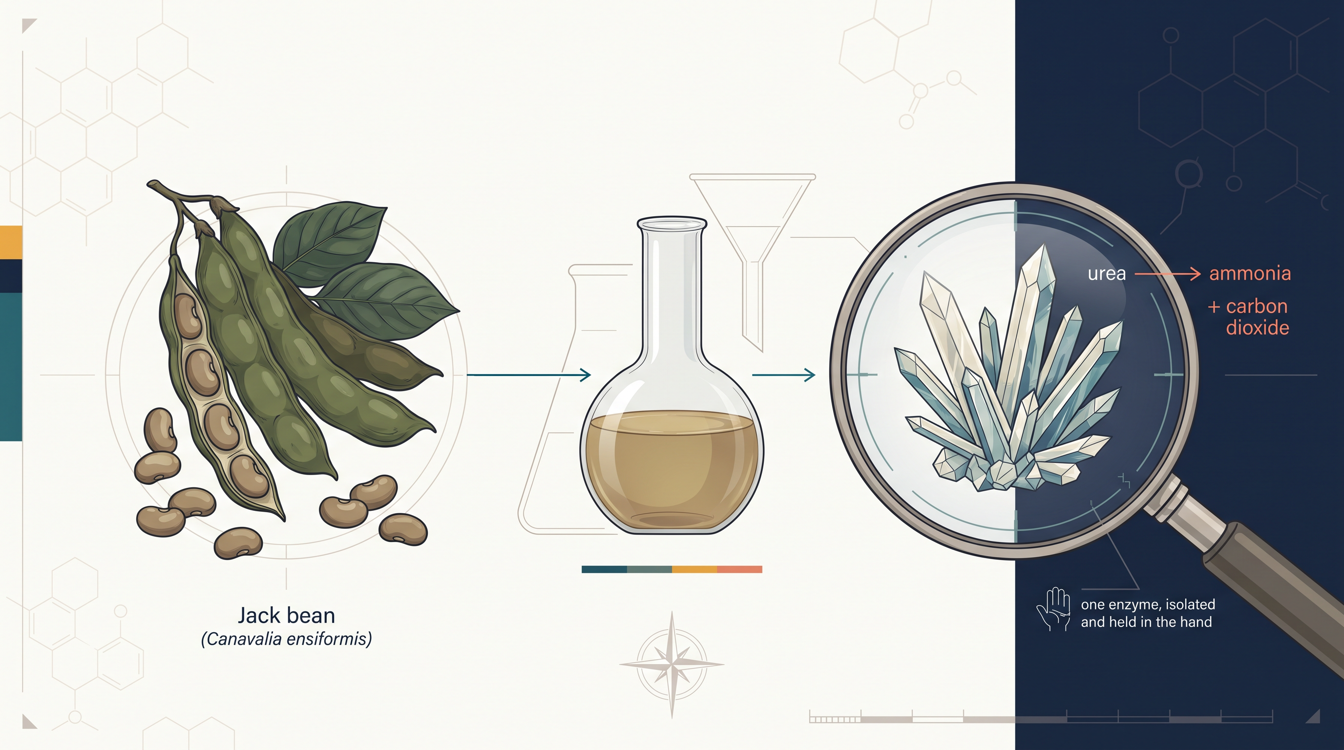
Fig. 4.0 – Jack bean extract, urease crystals and urea breakdown.
Keep these five questions in mind as you read. Each one is answered somewhere in this chapter, and you will meet the answers exactly where the science that explains them is built up.
4.1 Enzymes: Classification and Mode of Action
Every second, thousands of chemical reactions run inside a living cell — sugars are broken down, proteins are built, DNA is copied, wastes are neutralised. Left to themselves in a test tube at body temperature, most of these reactions would take hours, days or even years. Inside the cell they finish in fractions of a second. The molecules responsible for this enormous speeding up are the enzymes.
Enzymes are biocatalysts: they catalyse biochemical reactions both in vivo (inside the living body) and in vitro (outside the body, in a test tube or reaction vessel). This second property is what makes them so valuable to biotechnology. An enzyme purified out of a cell does not stop working simply because it has left the cell — as long as it is given the right temperature, pH and substrate, it continues to do its job in a laboratory tube or an industrial vessel.
Two features make enzymes remarkable. First, they are highly specific to substrates — each enzyme normally acts only on one substance or one narrow group of related substances. Second, they have great catalytic power — they enhance the rate of a reaction tremendously without themselves being changed. An enzyme molecule that has just converted one substrate molecule into product emerges intact and immediately picks up the next substrate molecule. This is why a very small amount of enzyme can transform a very large amount of substrate.
What Enzymes Are Made Of
All enzymes are proteins, with the exception of a small group of catalytic RNA molecules called ribozymes. This exception matters: it tells us that catalytic power depends on a molecule having a precisely folded three-dimensional shape, and not on it being a protein as such. RNA, which can also fold into complex shapes, can therefore also catalyse reactions.
Like other proteins, enzymes cover a huge range of sizes. Their molecular weight ranges from about 2000 to more than one million Dalton. The enzymatic activity of proteinaceous enzymes may be affected depending on the conformational structure of the protein as well as on its denaturation. Conformation means the exact three-dimensional folding of the polypeptide chain; denaturation means the loss of that folding. Because the catalytic site is built out of this folding, anything that disturbs the shape — heat, extreme pH, certain chemicals — reduces or destroys activity, even though the chain of amino acids itself is still present.
Cofactors: The Helpers Many Enzymes Need
There are many enzymes which require cofactors for their catalytic activity. A cofactor is a non-protein partner without which the protein part simply cannot work. The cofactor may be a complex organic molecule called a coenzyme (Table 4.1), or it may be a metal ion such as \(\text{Fe}^{2+}\), \(\text{Mn}^{2+}\), \(\text{Zn}^{2+}\) or \(\text{Mg}^{2+}\) (Table 4.2).
Why should a protein need such a partner at all? The twenty amino acids of a protein can supply acidic, basic, polar and non-polar groups, but they cannot easily carry electrons, transfer a whole methyl group, or hold a positive charge exactly where it is needed. Coenzymes and metal ions fill those chemical gaps.
An enzyme plus its cofactor is called a holoenzyme — this is the complete, catalytically active form. In such cases, the protein component of a cofactor-requiring enzyme is called an apoenzyme. The apoenzyme on its own is inactive. The relationship is shown in Fig. 4.1.
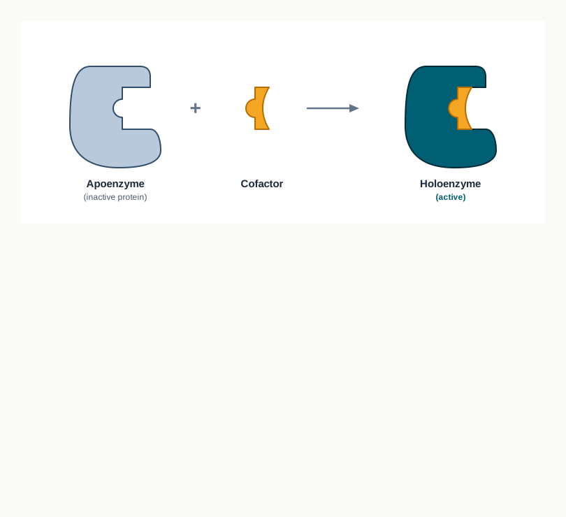
Fig. 4.1 – Formation of a holoenzyme.
Table 4.1: Some coenzymes and their precursor vitamins and their role
| Coenzyme | Precursor vitamin | Role in the catalytic reaction |
|---|---|---|
| Biocytin | Biotin (vitamin B7) | Transfer of \(\text{CO}_2\) |
| Coenzyme B12 (5’-adenosylcobalamin) | Vitamin B12 | Transfer of an alkyl group |
| Flavin adenine dinucleotide (FAD) | Riboflavin (vitamin B2) | Transfer of electrons |
| Coenzyme A | Pantothenic acid (vitamin B5) | Transfer of acyl and alkyl group |
| Nicotinamide adenine dinucleotide (NAD) | Niacin (vitamin B3) | Transfer of hydride (\(:\text{H}^-\)) |
| Pyridoxal phosphate | Pyridoxine (vitamin B6) | Transfer of amino group |
| Thiamine pyrophosphate | Thiamine (vitamin B1) | Transfer of aldehydes |
| Tetrahydrofolate | Folic acid (vitamin B9) | Transfer of one carbon group |
Coenzymes take part in catalysis transiently and are carriers of specific functional groups. “Transiently” is the key word: a coenzyme picks up a chemical group at one enzyme, carries it away, and hands it over at another enzyme, so it is not permanently attached to any single protein. Most of the coenzymes are derived from vitamins (organic nutrients required in small amounts in the diet).
This single sentence explains a great deal of nutrition. We need vitamins in milligram quantities, not gram quantities, precisely because they are not fuel — they are raw material for coenzymes, which are used over and over again. It also explains why a deficiency of one vitamin can produce such widespread symptoms: if niacin is short, every enzyme that depends on NAD works below par at once.
Table 4.2: Metal ions that serve as cofactors for enzymes
| Metal Ions | Enzyme name |
|---|---|
| \(\text{Fe}^{2+}\) or \(\text{Fe}^{3+}\) | Catalase, peroxidase, cytochrome oxidase |
| \(\text{Cu}^{2+}\) | Cytochrome oxidase |
| \(\text{Mg}^{2+}\) | DNA polymerase |
| \(\text{Mn}^{2+}\) | Arginase |
| \(\text{K}^+\) | Pyruvate kinase |
| \(\text{Mo}^{2+}\) | Nitrogenase, nitrate reductase |
| \(\text{Zn}^{2+}\) | Carbonic anhydrase, alcohol dehydrogenase |
| \(\text{Ni}^{2+}\) | Urease |
Notice the entry for DNA polymerase. This enzyme needs \(\text{Mg}^{2+}\), which is exactly why magnesium chloride is added to every DNA-copying reaction set up in a biotechnology laboratory. Leave the magnesium out and the reaction fails completely, however perfect the rest of the mixture may be. A cofactor is not an optional extra in a recipe; it is part of the enzyme.
When a coenzyme or metal ion is tightly bound through a covalent bond with the enzyme protein, it is called a prosthetic group. The difference between a prosthetic group and an ordinary coenzyme is therefore one of attachment, not of chemical type — the same kind of molecule may be loosely and briefly associated in one enzyme and permanently bolted on in another.
4.1.1 Classification of enzymes
Thousands of enzymes are already known and new enzymes may also be discovered. Without an agreed system, the same enzyme could easily be given different names in different laboratories, and two quite different enzymes could end up sharing a name. In order to have a systematic study and to avoid ambiguities, the International Union of Biochemistry (I.U.B.) in 1964 adopted a classification of enzymes depending on the type of reactions they catalyze.
The logic of this scheme is worth noticing. Enzymes are grouped not by the organism they come from, not by their size, and not by their shape, but purely by the kind of chemical change they bring about. According to this commission, all enzymes are classified into 6 major classes, but recently, another class of enzymes namely translocase has been added (Table 4.3).
Table 4.3: Classification of enzymes adopted by I.U.B.
| Class No. | Class name | Type of reaction catalyze |
|---|---|---|
| 1. | Oxidoreductases | Oxidation-reduction reactions (transfer of electrons) |
| 2. | Transferases | Transfer of groups |
| 3. | Hydrolases | Hydrolytic reactions (transfer of functional groups to water) |
| 4. | Lyases | Addition or removal of groups to form double bonds |
| 5. | Isomerases | Transfer of groups within molecules to yield isomeric forms |
| 6. | Ligases | Condensation of two molecules coupled through ATP hydrolysis |
| 7. | Translocase | Transfer of ion/molecules across the membrane |
Two of these classes will reappear constantly in later chapters of biotechnology. Hydrolases include the enzymes that cut bonds using water — the DNA-cutting enzymes belong here. Ligases join two molecules together, and because joining costs energy, this class is defined as condensation coupled through ATP hydrolysis. The enzyme that seals two DNA fragments together is a ligase for exactly this reason.
Isozymes
Many enzymes are present in multiple forms (more than one molecular form) in the same species, tissue or even in the same cell. These enzymes are called isoenzymes or isozymes.
Isoenzymes catalyse the same reaction but have different amino acid composition, and hence possess different physicochemical properties. They may differ in the temperature or pH at which they work best, in how strongly they hold their substrate, and in their electrical charge — which is why they can be separated from one another in the laboratory even though they do the same chemical job.
For example, a glycolytic enzyme, hexokinase, exists in four isozyme forms in various tissues. Similarly, lactate dehydrogenase (LDH), involved in anaerobic glucose metabolism, has five isozyme forms in human.
Why would a body bother maintaining several versions of one enzyme? Because different tissues face different demands. A form tuned for a muscle working without enough oxygen is not the form best suited to heart tissue, which works aerobically almost all the time. Isozymes let the same reaction be regulated differently in different places.
Enzyme active site
The catalytic reaction performed by enzymes occurs at a particular site on the enzyme. This site is called the active site, and represents only a small part of the total size of the enzyme.
The active site is a clearly defined pocket or cleft in the enzyme molecule where the whole or a portion of the substrate can fit. Notice the consequence of this: most of the enzyme’s bulk is not directly involved in chemistry at all. Its job is structural — to hold the few crucial amino acid residues in exactly the right positions in space, and to shield the reacting groups from the surrounding water.
The active site has a three-dimensional structure since it consists of portions of a polypeptide chain. The residues that line the pocket may be far apart along the chain and are brought together only when the protein folds. This is precisely why denaturation destroys activity: unfolding scatters the residues of the active site, and the pocket ceases to exist.
Various non-covalent bonds involved in enzyme–substrate binding are electrostatic interactions, hydrogen bonds, Van der Waals forces and hydrophobic interactions. Every one of these is a weak interaction. That weakness is essential rather than accidental — the substrate must be held firmly enough for the reaction to occur, but loosely enough for the product to be released quickly so that the enzyme can start again. A covalently trapped substrate would end catalysis instead of assisting it.
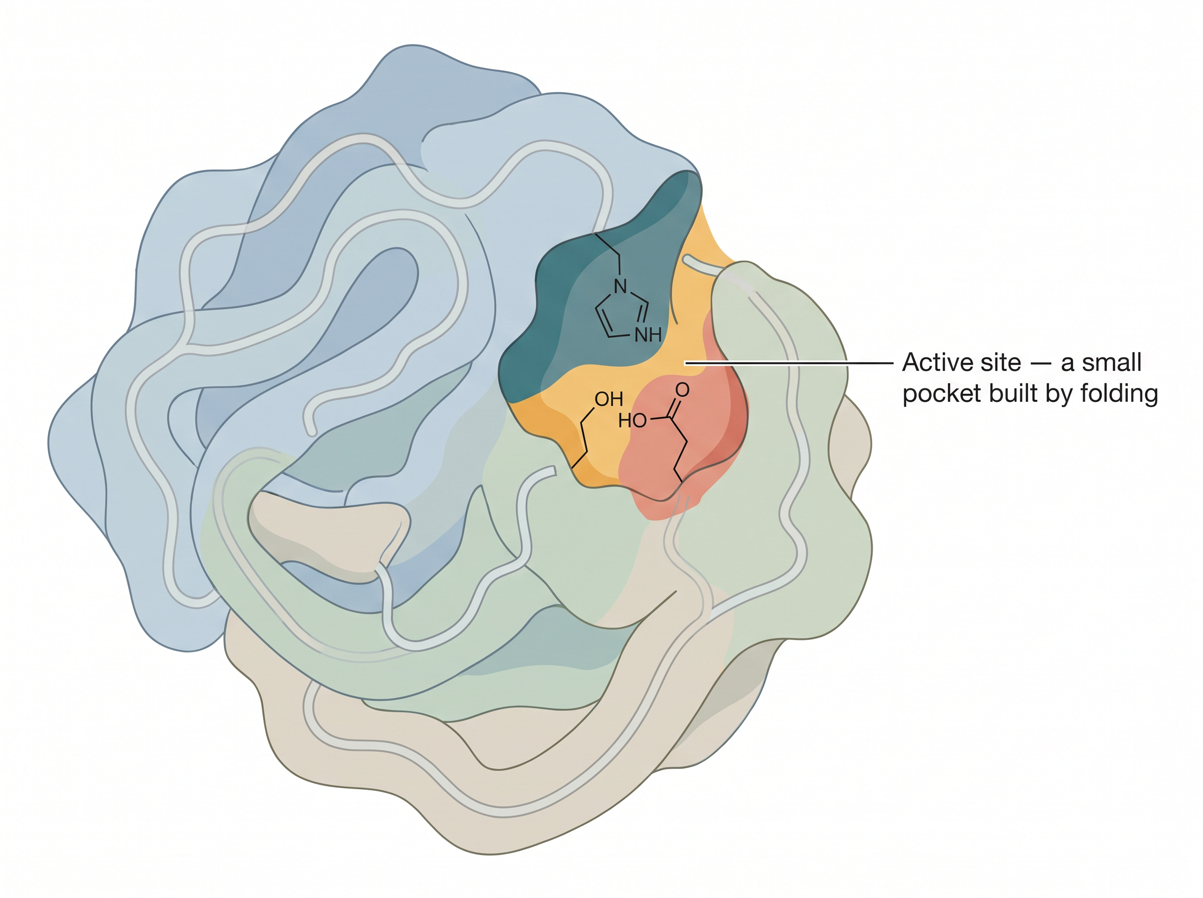
Fig. 4.2 – The enzyme active site.
Fischer’s Lock and Key Model
How exactly does a substrate meet its active site? The first serious answer came in 1894, when the Lock and Key Model for the substrate and enzyme interaction was proposed by Emil Fischer.
According to this model, complementary structural features are present between enzyme and substrate, and the active site is pre-shaped to fit the substrate. The substrate can fit into its complementary site on the enzyme as a key fits into a lock. This results in the formation of an enzyme–substrate complex, as shown in Fig. 4.3.
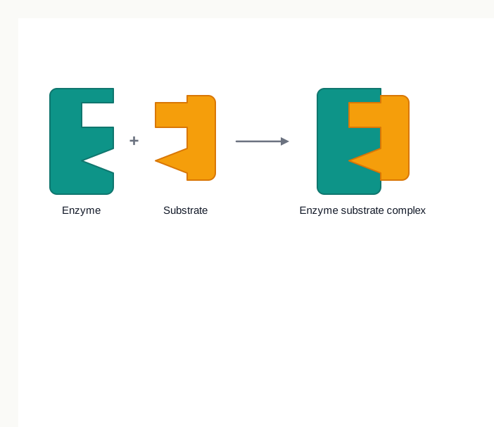
Fig. 4.3 – The lock and key model.
The model explains specificity beautifully. A key of the wrong shape will not enter the lock at all, and in the same way a molecule of the wrong shape cannot occupy the active site. What the model does not explain is flexibility — a real protein is not a rigid metal lock.
Koshland’s Induced Fit Model
Daniel Koshland in 1958 proposed the Induced Fit Hypothesis. He suggested that the structure of a substrate may be complementary to that of the active site in the enzyme–substrate complex but not in the free enzyme.
In other words, the perfect fit is not there in advance; it is created. The interaction between the substrate and the enzyme induces conformational changes in the enzyme which align the amino acid residues or other groups for substrate binding, catalysis, or both. This is shown in Fig. 4.4.
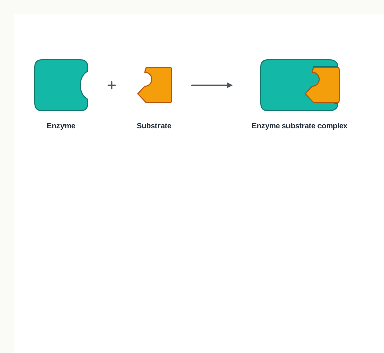
Fig. 4.4 – The induced fit model.
The relationship between a substrate and an active site resembles a hand and a glove. During interaction, the structure of one component, i.e., the substrate or the hand, remains rigid, and the shape of the second component, i.e., the active site or the glove, is flexible enough to become complementary to that of the first.
This small change of picture has a large consequence. Because the enzyme must be moulded by the substrate before catalysis begins, a molecule that merely looks similar to the substrate may bind but fail to trigger the correct conformational change — and therefore fail to react. Induced fit thus explains specificity even better than the lock and key model does.
Enzyme specificity
The enzymes are highly specific in action. In fact, the properties that make enzymes such a strong catalyst are their specificity of substrate binding and their ideal arrangement of catalytic groups. The two work together: binding brings the right molecule to the right place, and the arrangement of catalytic groups then acts on it.
Various types of enzyme specificity are: group specificity, absolute specificity, stereospecificity, and geometrical specificity.
When enzymes act on several different closely related substrates then it is called group specificity. Such an enzyme recognises a chemical feature shared by a family of molecules rather than one molecule alone.
When enzymes act only on one particular substrate, it is called absolute specificity. This is the strictest form — one enzyme, one substrate.
Stereochemical or optical specificity occurs when a substrate exists in two stereochemical forms (chemically identical but with a different arrangement of atoms in three-dimensional space); then only one of the isomers will undergo reaction by a particular enzyme. For example, D-amino acid oxidase catalyses oxidation of the D-amino acids to keto acids. The mirror-image L-form is untouched, even though it contains exactly the same atoms joined in exactly the same order.
In geometrical specificity, enzymes are specific towards cis and trans forms. For example, fumarase catalyses the interconversion of fumarate and malate.
The ability of an enzyme to tell two mirror images apart is not a curiosity — it is the basis of an entire industry. Many drugs are active in one stereochemical form and useless or harmful in the other, so enzymes are used industrially to produce a single desired isomer rather than a mixture.
4.1.2 Factors affecting enzyme activity
The rate of enzyme catalysed reactions is influenced by changing the environmental conditions. The important factors that influence the velocity of enzyme catalysed reactions are temperature, pH, substrate concentration, and modulators.
Since all four factors act on the same underlying structure — a folded protein holding a substrate in a pocket — they can be understood together. Anything that changes the folding changes activity; anything that changes how often substrate reaches the pocket changes activity as well.
Temperature
The rate of an enzyme catalysed reaction increases with the increase in temperature up to a maximum and then falls. When a graph is plotted between temperature versus enzyme activity, a bell-shaped curve is obtained, as shown in Fig. 4.5.
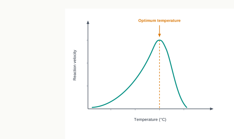
Fig. 4.5 – Effect of temperature on enzyme activity.
Read the curve in two halves, because two different things are happening on either side of the peak. On the rising side, heat is simply making molecules move faster, so enzyme and substrate collide more often and more energetically. On the falling side, heat has begun to unfold the protein; active sites are being destroyed faster than the extra collisions can compensate. The peak is where these two effects balance.
The temperature at which the maximum rate of reaction occurs is called the enzyme’s optimum temperature. The optimum temperature is different for different enzymes; but for most of the enzymes it is between 40-45°C.
Majority of enzymes in the human body have an optimum temperature of around 37°C (98.6°F) and are denatured or degraded at extreme temperatures. This is one reason a high fever is dangerous — the body’s enzymes are being pushed towards the descending limb of their curve.
However, a few enzymes like Taq DNA polymerase present in the thermophilic bacterium Thermus aquaticus, venom phosphokinase and muscle adenylate kinase are active even at 100°C. Their polypeptide chains are folded and bonded in ways that resist unfolding, so the descending limb of their curve begins far to the right of ours.
Hydrogen Ion Concentration (pH)
Enzyme activity is also affected by pH. A plot of enzyme activity against pH results in a bell shaped curve, as shown in Fig. 4.6.
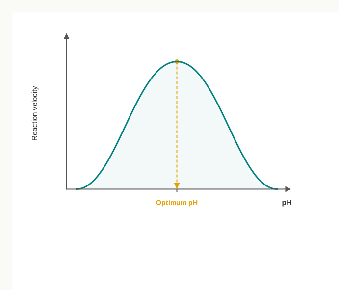
Fig. 4.6 – Effect of pH on enzyme activity.
Each enzyme has its unique optimum pH at which the rate of reaction is greatest. The optimum pH is the pH at which the activity of a particular enzyme is at maximum.
The reason lies in charge. Several amino acid side chains in the active site are acidic or basic, and whether they carry a charge depends on the surrounding pH. At the optimum pH, the right groups carry the right charges to bind the substrate and to perform catalysis. Shift the pH, and those charges change.
Many enzymes of higher organisms show optimum reaction rate around neutral pH (pH 6-8). However, there are several exceptions such as pepsin (pH 1-2), acid phosphatases (pH 4-5) and alkaline phosphatases (pH 10-11).
These exceptions make sense once you look at where the enzyme works. Pepsin operates in the strongly acidic stomach, so an optimum of pH 1-2 is exactly what its surroundings demand. An enzyme is adapted to its compartment.
Below and above the optimum pH, the enzyme activity is much lowered, and at extreme pH the enzyme becomes totally inactive. At extremes the protein is not merely mis-charged but denatured, and this loss is generally not recovered by simply restoring the correct pH.
Substrate concentration
The substrate concentration also influences enzyme activity. As the substrate concentration increases the rate of reaction also increases. This is because the more substrate molecules will interact with enzyme molecules, the more products will be formed.
However, after a certain concentration, further increase in substrate concentration will have no effect on the rate of reaction, since the substrate concentration will no longer be the limiting factor. This flattening is shown in Fig. 4.7.
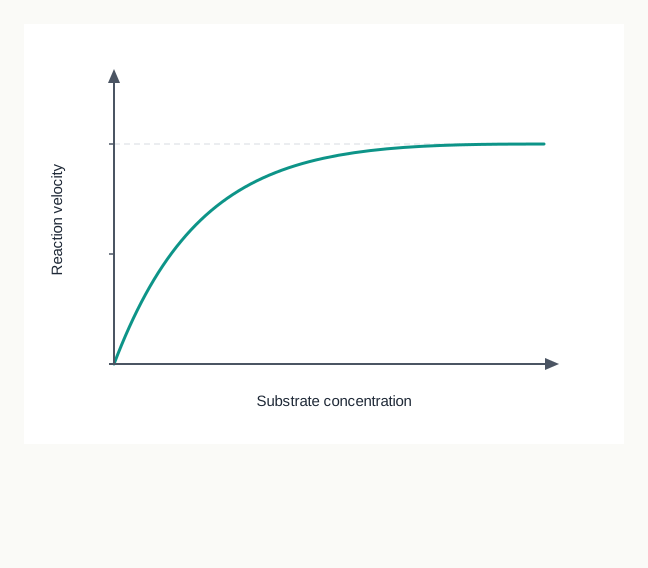
Fig. 4.7 – Effect of substrate concentration on reaction velocity.
At this stage, enzyme molecules become saturated and work at their maximum possible rate. A useful picture is a set of billing counters in a shop. When customers are few, adding more customers means more bills per minute. Once every counter has a queue, adding more customers only lengthens the queues; the billing rate cannot rise further because the counters, not the customers, are now the limit.
Learning to read this plateau matters, because the same curve returns in a quantitative form later in this section, where it is used to define two constants that describe an enzyme numerically.
Modulators
The fourth factor is the presence of modulators — substances that bind to an enzyme and change its activity. Some modulators reduce activity and are called inhibitors; others increase it and are called activators. Because they are important enough to need their own treatment, inhibitors are dealt with in Section 4.1.6, and enzymes whose activity is deliberately tuned by regulatory molecules are dealt with in Section 4.1.7.
4.1.3 Unit of enzyme activity
You cannot buy an enzyme by weight and know what you are getting, because a gram of partly denatured enzyme does far less work than a gram of intact enzyme. What matters is not how much protein is present, but how much reaction it can drive. Enzymes are therefore measured by activity.
The enzyme unit (U) is the amount of enzyme that catalyses the conversion of 1 micromole of substrate into product per minute under standard conditions. The phrase “under standard conditions” is essential — the same enzyme sample would give a different figure at a different temperature, pH or substrate concentration, so those conditions must be fixed before any comparison is meaningful.
The International Union of Biochemistry (I.U.B.) adopted the enzyme unit as the unit of enzyme activity in 1964. But it was discouraged in favour of the katal, since the minute is not an SI unit.
One katal (kat) is the amount of enzyme that catalyses the conversion of 1 mole of substrate per second into product (\(1\ \text{kat} = 6 \times 10^{7}\ \text{IU}\)).
4.1.4 Specific activity
Activity alone tells you how much work a preparation can do, but not how clean it is. A crude cell extract may show high total activity simply because it is a large volume of soup containing a great many proteins, only a few of which are the enzyme you want.
Specific activity is the unit of enzyme activity per milligram of enzyme protein. Specific activity represents a measurement of enzyme purity in the mixture.
The logic is worth spelling out. During purification, unwanted proteins are progressively removed while the enzyme itself is retained. Total activity therefore stays roughly constant or falls slightly, but the total protein falls sharply — so the ratio of the two rises. A rising specific activity is the standard evidence that a purification step has worked.
4.1.5 Mechanism of enzyme action
For understanding the enzyme mechanism, you should consider two thermodynamic properties of a reaction. These are the free energy difference (\(\Delta G\)) between products and reactants, and the energy needed to initiate the conversion of reactant into product.
These two quantities answer two different questions, and confusing them is the commonest error in this topic. The former energy, i.e., \(\Delta G\), determines whether the reaction is spontaneous — that is, whether it can happen at all. The latter determines the rate of reaction — that is, how fast it happens.
Enzymes affect the energy which determines the rate of reaction. The enzymes cannot change the laws of thermodynamics and therefore cannot alter the equilibrium of a biochemical reaction. They speed up the attainment of equilibrium.
Put plainly, an enzyme changes the speed of a journey, not the destination. A reaction that would eventually settle at a particular balance of reactants and products will settle at exactly the same balance in the presence of an enzyme — it will simply get there far sooner.
The rate of reaction rather depends on the free energy of activation (\(\Delta G^A\)), which is not related to \(\Delta G\). The substrate S of a reaction is converted into product P via formation of a transition state which has higher free energy than either S or P. The transition state is the strained, half-way arrangement of atoms that must be reached before old bonds can break and new ones form, and reaching it is the bottleneck of the whole reaction.
The difference between the free energy of the transition state and the substrate is called the Gibbs free energy of activation or simply activation energy (\(\Delta G^A\)). The enzymes enhance reaction rate without altering \(\Delta G\) of the reaction; rather, they lower the activation energy, \(\Delta G^A\). This is shown clearly in Fig. 4.8.
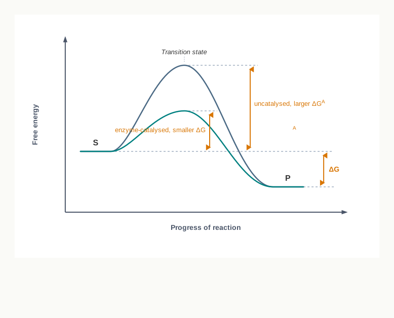
Fig. 4.8 – Free energy profile of a reaction.
The way an enzyme lowers this barrier follows directly from the induced fit picture. By binding the substrate in a strained orientation and surrounding it with precisely placed catalytic groups, the enzyme makes the transition state easier to reach, so a far larger fraction of molecules can cross the barrier at body temperature.
Kinetics of enzyme-catalysed reaction
During catalysis, substrate S binds at the active site of the enzyme E and results in formation of an enzyme substrate complex ES, which is finally converted into product P. The reaction can be represented as:
\[\text{E} + \text{S} \rightleftharpoons [\text{ES}] \longrightarrow \text{E} + \text{P}\]
Here, E forms a weakly bonded complex ES with the substrate S. The ES complex decomposes to yield product P and the free enzyme E. Note that the first step is shown as reversible — the substrate may leave again without reacting — while the second step yields product and regenerates the enzyme, which is then free to begin another cycle. The complete cycle is summarised in Fig. 4.9.
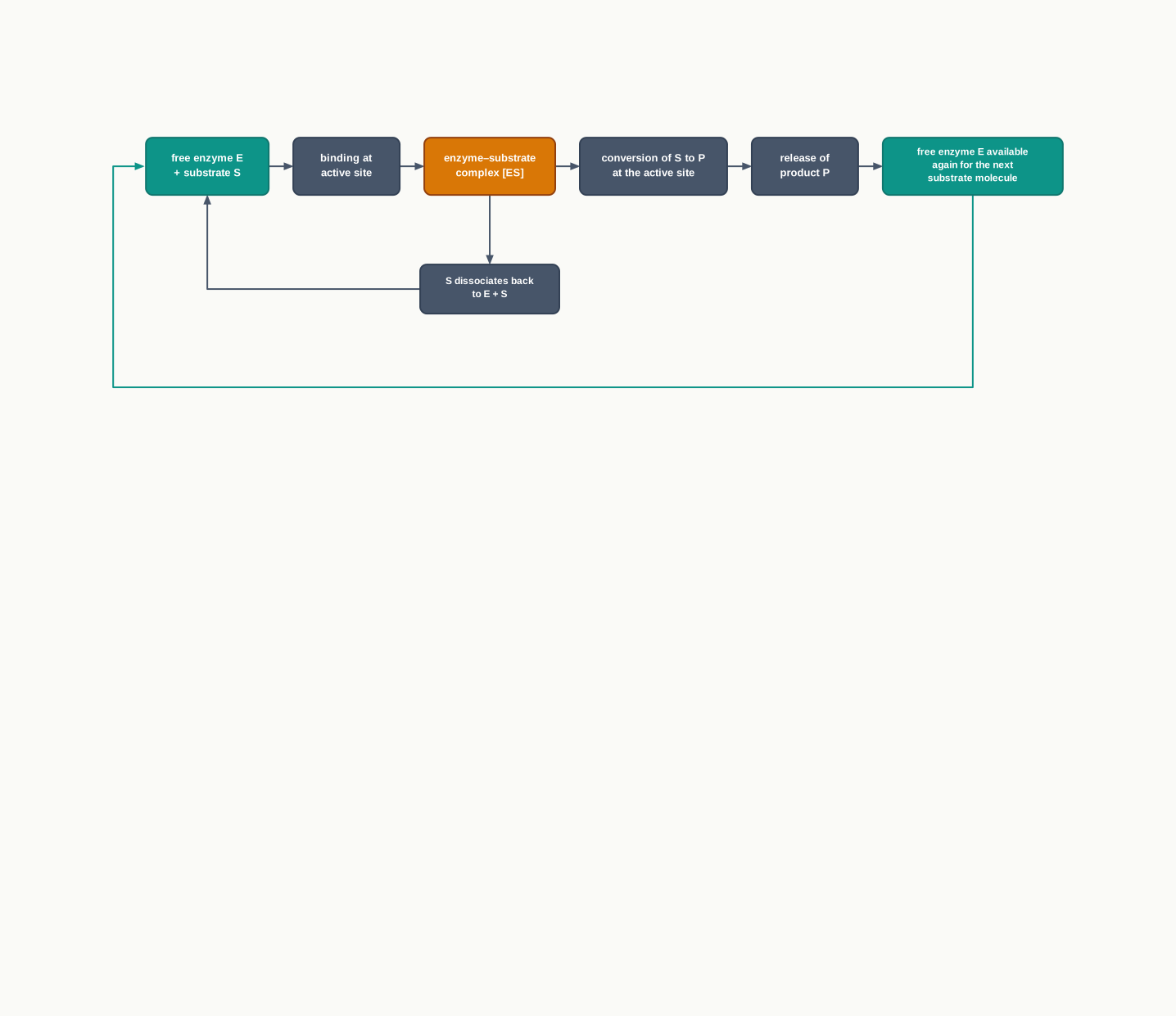
Fig. 4.9 – Catalytic cycle of an enzyme.
The kinetics of enzyme catalysed reactions was explained by Leonor Michaelis and Maud Menten in 1913. The most remarkable feature of this kinetics is that a specific ES complex is an intermediate during catalysis. Michaelis-Menten theory of enzyme kinetics is the simplest one that accounts for the kinetic properties of many enzymes.
Further simplifying the reaction, Michaelis and Menten derived the following equation for a one substrate reaction:
\[v_0 = \frac{V_{max}\,[\text{S}]}{K_m + [\text{S}]}\]
The equation is called the Michaelis-Menten equation. Here, \(K_m\) is called the Michaelis constant, \(v_0\) is the initial velocity, \(V_{max}\) is the maximum velocity of reaction, and \([\text{S}]\) is the substrate concentration. Velocities are expressed as amount of product formed per unit time, and both \(K_m\) and \([\text{S}]\) are expressed in units of concentration, such as mol L\(^{-1}\).
A graph of \(v_0\) against \([\text{S}]\) results in a rectangular hyperbola, as shown in Fig. 4.10. \(V_{max}\) is the maximum velocity at a particular enzyme concentration. \(V_{max}\) and \(K_m\) can be determined from the graph as shown in Fig. 4.10.
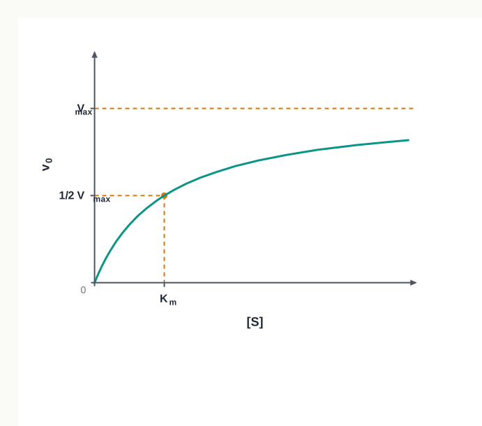
Fig. 4.10 – Michaelis-Menten curve for a single-substrate enzyme.
Reading this graph is a standard examination skill, so work through it slowly. The curve rises steeply at first and then flattens. In the graph, we can see that at very low substrate concentration (when \([\text{S}] \ll K_m\)), the reaction rate is directly proportional to substrate concentration, so that \(v_0 \propto [\text{S}]\) — doubling the substrate doubles the velocity, because most enzyme molecules are still free and waiting.
At high substrate concentration (when \([\text{S}] \gg K_m\)), \(v_0 = V_{max}\), i.e., the reaction rate is maximum and independent of substrate concentration. Every active site is occupied, and adding more substrate changes nothing. This is the same saturation you met in Fig. 4.7, now expressed quantitatively.
When \([\text{S}] = K_m\), then \(v_0 = V_{max}/2\). Thus, \(K_m\) is the substrate concentration at which half of the maximum reaction rate is obtained.
Because of this, \(K_m\) carries a practical meaning. A low \(K_m\) means the enzyme reaches half its maximum speed at a very small substrate concentration — that is, it works efficiently even when substrate is scarce. A high \(K_m\) means the enzyme needs plenty of substrate before it gets going.
4.1.6 Enzyme inhibition
Substances which decrease the rate of an enzyme catalysed reaction are called enzyme inhibitors, and the process is known as enzyme inhibition. Inhibition is not always something to be avoided. Many medicines work precisely by inhibiting a chosen enzyme, and cells themselves use inhibition to switch pathways off when a product is no longer needed.
Enzyme inhibition can be classified as reversible inhibition and irreversible inhibition.
In irreversible inhibition, the inhibitor binds very tightly to the enzyme and does not dissociate from it. The affected enzyme molecule is permanently out of action; activity can return only when the cell makes fresh enzyme.
For example, the antibiotic penicillin acts as an inhibitor and binds with the enzyme transpeptidase, which is responsible for the synthesis of the bacterial cell wall. Hence, binding of this drug to the enzyme prevents cell wall synthesis, thus killing the bacteria. Notice the elegance of the target: human cells have no cell wall and no transpeptidase, so the drug damages the bacterium and not the patient. Selectivity of this kind is the central goal of drug design.
In the same way, the drug aspirin inhibits the enzyme cyclooxygenase, thus reducing inflammation.
In reversible inhibition, the inhibitor rapidly dissociates from the enzyme–inhibitor complex. The block is temporary, and removing the inhibitor restores activity. There are three types of reversible inhibitions: competitive, non-competitive and uncompetitive inhibition.
(i) Competitive inhibition
In competitive inhibition, there is close resemblance in the structure of the inhibitor I and the substrate S; therefore, they both compete for the same active site on the enzyme. Because the inhibitor mimics the substrate, the active site accepts it — but no product results, and the site is occupied meanwhile.
The enzyme can form the enzyme-substrate ES complex or it can form the enzyme-inhibitor EI complex, as shown in Fig. 4.11, but not ESI. This last point is the logical heart of competitive inhibition: since both molecules need the same pocket, they cannot both be bound at once.

Fig. 4.11 – Competitive inhibition.
Competitive inhibitors decrease the rate of reaction by reducing the amount of active enzyme molecules bound to a substrate. At very high substrate concentration, the chances of binding of the inhibitor molecule to the enzyme will be reduced, so \(V_{max}\) for the reaction will not be changed. Flood the system with substrate and the substrate wins the competition by sheer numbers, so the enzyme can still reach its full speed.
However, the \(K_m\) is increased in the presence of the inhibitor and is denoted by the symbol \(K'_m\), as shown in Fig. 4.12. A raised \(K_m\) says exactly what you would expect: more substrate than before is now needed to reach half the maximum velocity.
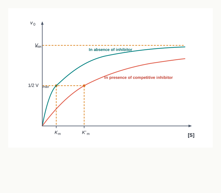
Fig. 4.12 – Michaelis-Menten plot for competitive inhibition.
(ii) Non-Competitive inhibition
In this type of inhibition, the inhibitor has no structural similarity with the substrate and binds with the enzyme at a different site other than the active site. Therefore, the inhibitor can bind with E as well as with the ES complex, and formation of ES, EI and ESI takes place, as shown in Fig. 4.13.
The inhibitor I and substrate S can bind simultaneously to the same enzyme molecule as their binding sites are different and hence do not overlap.
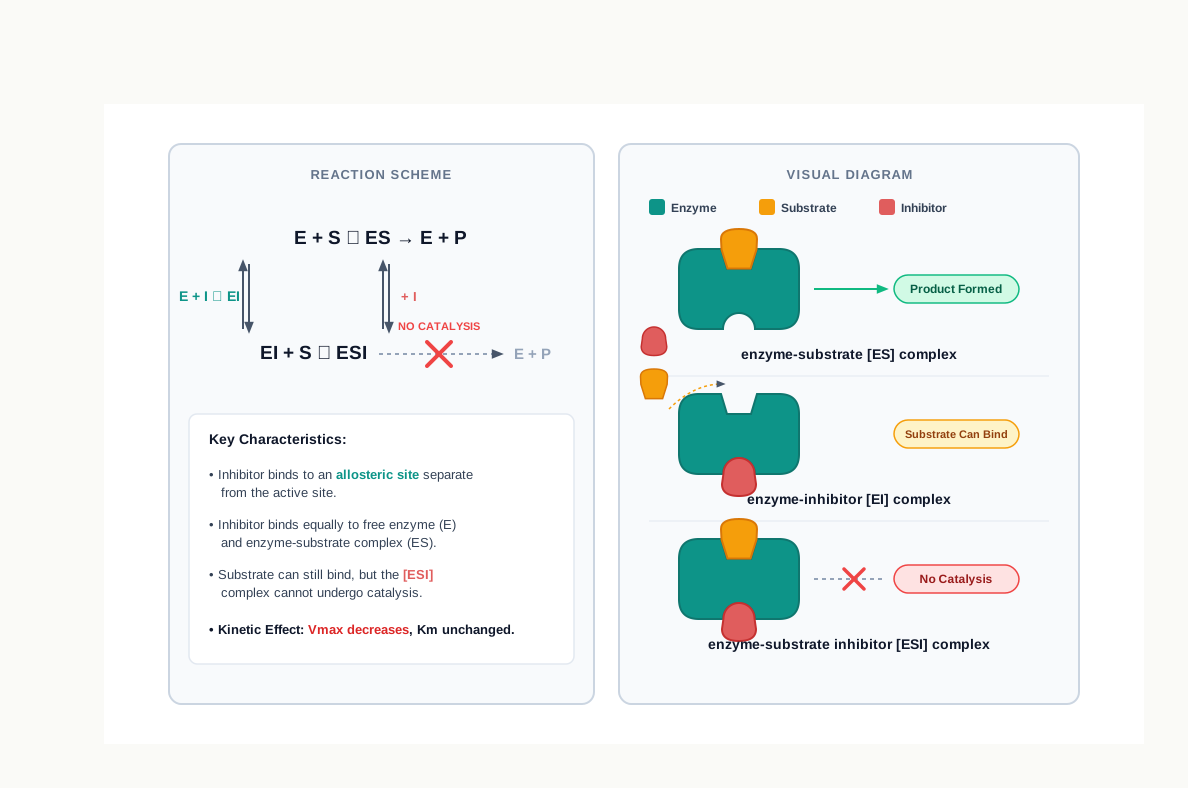
Fig. 4.13 – Non-competitive inhibition.
A non-competitive inhibitor lowers the \(V_{max}\) rather than decreasing the proportion of enzyme molecules that are bound to the S. Thus, non-competitive inhibition, in contrast to competitive inhibition, cannot be overcome by increasing substrate concentration — adding substrate does not help, because the substrate and the inhibitor were never competing for the same place.
The substrate can still bind to the EI complex. However, the ESI does not form product. The inhibitor effectively lowers the concentration of active enzyme and hence lowers the \(V_{max}\). There is no effect on \(K_m\) as the inhibitor decreases the amount of functional enzyme, as shown in Fig. 4.14.
It helps to think of it this way: the enzyme molecules that remain unbound by inhibitor behave completely normally, so the substrate concentration needed to half-saturate them is unchanged. There are simply fewer of them, so the ceiling is lower.
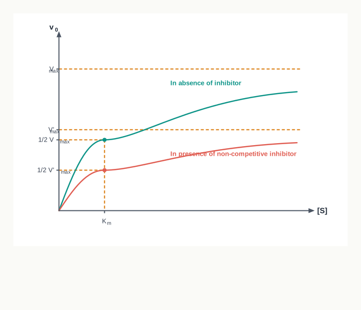
Fig. 4.14 – Michaelis-Menten plot for non-competitive inhibition.
(iii) Uncompetitive inhibition
In this type of inhibition, the inhibitor does not bind to the free enzyme. It binds only to the enzyme substrate (ES) complex, as shown in Fig. 4.15.
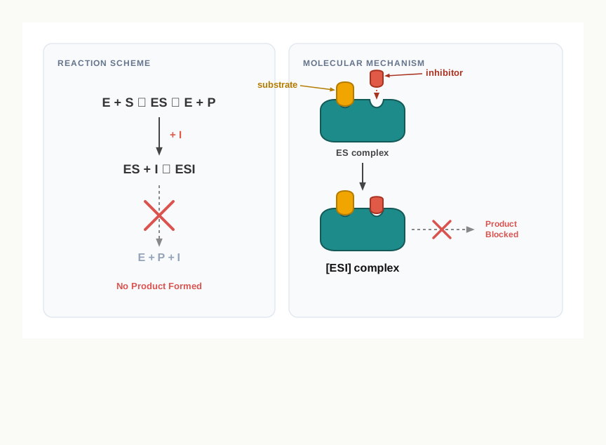
Fig. 4.15 – Uncompetitive inhibition.
The inhibitor does not compete with the substrate for the same binding site. Therefore, the inhibition cannot be overcome by increasing substrate concentration. In fact, more substrate means more ES complex, which is precisely what this inhibitor needs in order to bind.
Both \(K_m\) and \(V_{max}\) values are altered.
The three types of reversible inhibition can therefore be told apart purely from what happens to the two constants, which is exactly how such questions are set in examinations. The reasoning route is set out in Fig. 4.16.
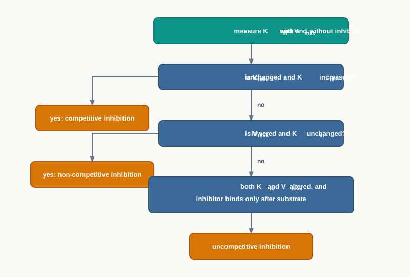
Fig. 4.16 – Identifying the type of reversible inhibition.
4.1.7 Allosteric enzymes
Allosteric enzymes do not obey Michaelis-Menten kinetics. These enzymes result in a sigmoidal graph instead of a rectangular hyperbola when \(v_0\) is plotted against substrate concentration \([\text{S}]\), as shown in Fig. 4.17.
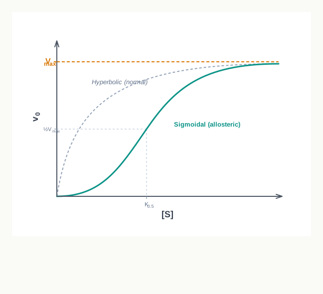
Fig. 4.17 – Kinetics of an allosteric enzyme.
The shape of this curve is the point. At low substrate concentration the curve rises only gently, then suddenly steepens, then levels off. This means the enzyme is relatively unresponsive until substrate reaches a certain level, and then switches on sharply over a narrow range — behaviour that suits a control point far better than a smooth hyperbola would.
Each subunit of an allosteric enzyme also contains a regulatory site along with an active site. Regulatory molecules may reversibly bind to the regulatory site and alter the affinity of the enzyme for substrate binding. Since the binding is reversible, the control can be applied and released as the cell’s needs change.
Whereas most of the enzymes obeying Michaelis-Menten kinetics are the normal enzymes, allosteric enzymes are the key regulators of metabolic pathways in the cell. They are typically placed at the first or a branching step of a pathway, where switching the enzyme off stops the whole sequence and prevents the cell from wasting materials and energy on a product it already has enough of.
4.2 Brief Introduction to Bioenergetics
Biological energetics, or bioenergetics, deals with the transformation and use of energy by living cells. A cell is never at rest chemically. It builds molecules, pumps ions, moves, divides and repairs itself, and every one of these activities has to be paid for in energy.
In biological reactions, the energy is released as the reactions move from a higher to a lower energy level. Energy liberated in this way is utilised to perform work. The energetics of these processes are governed by the laws of thermodynamics, as described below.
Notice how this section connects with the previous one. Section 4.1 dealt with how fast reactions go and how enzymes control that speed. Bioenergetics asks a different question: which reactions can go at all, in which direction, and what it costs to make the unfavourable ones happen anyway.
4.2.1 The laws of thermodynamics
Thermodynamics is the branch of physical chemistry that deals with energy changes. Energy is the capacity to do work, and it exists in a variety of forms such as electrical, mechanical, chemical, heat and light. These forms of energy are interconvertible.
That last property is what living systems exploit constantly. A leaf converts light energy into chemical energy; a muscle converts chemical energy into mechanical energy; the electric organ of certain fishes converts chemical energy into electrical energy. In every case the accounting is the same, which is why one set of laws covers all of them.
Thus, bioenergetics is concerned with the changes in energy during biochemical processes. It does not deal with the mechanism or the speed of the process. This is an important limitation to keep in mind: thermodynamics can tell you that a reaction is possible, but not how long you will have to wait for it. The waiting time is decided by activation energy and by enzymes, which you met in Section 4.1.
There are two fundamental laws of thermodynamics which explain the interconversions of various forms of energy. The two laws also help to understand the following:
- The direction of a reaction, whether forward or reverse.
- The accomplishment of work, whether useful or non-useful.
- Whether the energy for executing a reaction must be delivered from an external source.
The first law of thermodynamics
The first law of thermodynamics states that ‘energy can neither be created nor destroyed, but can be converted into other forms of energy’. It means the total amount of energy in the universe (system plus surroundings) remains constant.
According to this law, energy exchange in any process takes place between the ‘system’ and the ‘surroundings’. A system is matter within a defined region, and surroundings constitute matter in the rest of the universe. Thus, the system and surroundings make the universe, which includes the entire earth and the outer space.
In biology the chosen system may be a single mitochondrion, a whole cell, an organ, or an entire organism — the boundary is drawn by the investigator to suit the question being asked. Everything outside that boundary is then, by definition, the surroundings. This is illustrated in Fig. 4.18.
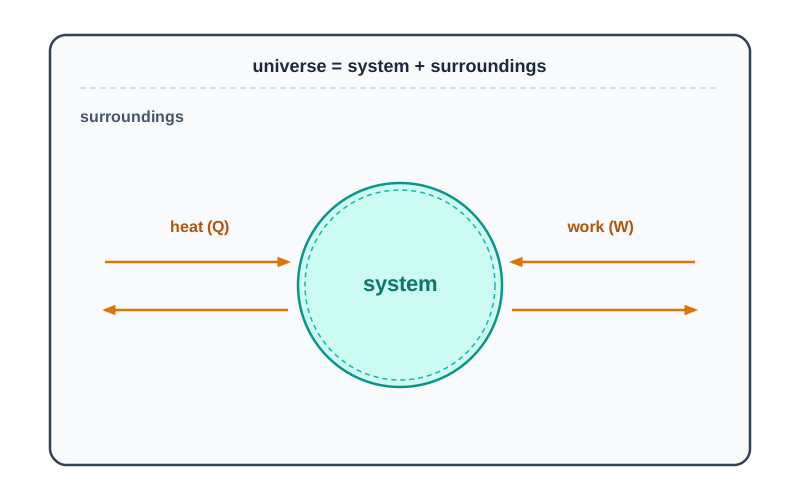
Fig. 4.18 – System, surroundings and universe.
Thus, the first law of thermodynamics can be expressed in the form of the following equation:
\[\Delta E = E_B - E_A = Q - W\]
Where,
- \(\Delta E\) = change in internal energy
- \(E_A\) = energy of a system at the start of a process
- \(E_B\) = energy of a system at the end of a process
- \(Q\) = heat absorbed by the system
- \(W\) = work done by the system
Read the equation as a simple account of income and expenditure. Heat absorbed by the system is energy coming in; work done by the system is energy going out; the difference is what stays behind as a change in the system’s internal energy. Energy in SI units is measured in joules (J), although biochemical data are often quoted in calories or kilocalories.
According to this equation, the change in the energy of a system depends only on the initial and the final stages, and not on the path of the transformation.
This path independence has a very useful biological consequence. Glucose oxidised rapidly in a flame and glucose oxidised slowly through the long, enzyme-catalysed pathways of a cell release exactly the same total energy, because the starting materials and end products are the same. The cell’s advantage is not that it extracts more energy, but that it releases it in small, controlled instalments that can be captured rather than lost as a burst of heat.
The second law of thermodynamics
The first law of thermodynamics cannot be used to predict the spontaneity or non-spontaneity of a reaction. It tells us that energy will be conserved whichever way the reaction goes, but not which way it will actually go.
On the other hand, the second law introduces a term ‘entropy’, which is denoted by the symbol ‘S’. Entropy is the degree of randomness or disorder of a system, and it explains whether a reaction takes place spontaneously. The entropy of a system increases when it becomes more disordered. Entropy reaches a maximum in a system as it nears equilibrium.
Biological systems, however, do not follow the second law, as life is a state of higher organisation or lower entropy rather than increased entropy. A cell assembles ordered proteins from scattered amino acids and ordered membranes from dispersed lipids, which is a decrease in entropy — the opposite of what the law appears to demand.
The apparent conflict disappears once the surroundings are included in the accounting. An organism is not a sealed box; it continuously takes in ordered energy and matter and discards heat and waste into its surroundings. The order built up inside is more than paid for by the disorder created outside, so the sum still increases.
This highly ordered form of life is maintained for a while by the consumption of chemical energy (food), and in the case of photosynthetic organisms through light energy. This energy is either converted to a less organised form of energy (heat) or utilised to perform work. Ultimately, thermodynamic equilibrium is certain, and entropy increases after the death and subsequent decomposition of every organism.
The second law of thermodynamics states that the entropy or disorder of the universe is always increasing. According to this law, a process can occur spontaneously only if the sum of the entropies of the system and its surroundings increases. This can be represented as:
\[(S_{system} + S_{surroundings}) > 0 \quad \text{(for a spontaneous process)}\]
Thus, for a process to occur spontaneously, the total entropy of a system must increase. However, the entropy of a system can decrease even during a spontaneous process, provided the entropy of the surroundings increases to an extent that their sum becomes positive.
The difference between the first and the second law is that the first law is concerned with the transformation of various kinds of energy involved in a given process, whereas the second law is concerned with the availability of the energy of a given system for doing work.
Combining the two laws
Entropy is not used as a criterion to know whether a biochemical reaction is spontaneous or not. There are two practical reasons for this. First, changes in entropy of a reaction cannot be measured. Second, changes in entropy of the surroundings and of the system would both have to be known before spontaneity could be judged — and a biochemist working with a reaction in a tube has no way of measuring what the rest of the universe is doing.
Therefore, a different thermodynamic function, ‘free energy’, is used to overcome this problem. Free energy is denoted by the symbol \(\Delta G\). Its great advantage is that it refers only to the system, yet still predicts spontaneity correctly.
In 1878, Gibbs created the free energy function by combining the first and second laws of thermodynamics. The following equation was obtained:
\[\Delta G = \Delta H - T\Delta S\]
Where,
- \(\Delta G\) = change in free energy of a reacting system
- \(\Delta H\) = change in heat content or enthalpy of this system
- \(T\) = absolute temperature at which the process is taking place, measured in kelvin (K)
- \(\Delta S\) = change in entropy of the system
This equation shows the relationship between the change in free energy (\(\Delta G\)), heat (also called enthalpy, \(\Delta H\)), and entropy in chemical reactions at constant temperature (T) and pressure (P). Biochemical reactions also occur at these conditions, which is why this particular form of the equation suits biology so well.
The \(\Delta G\) is the free energy change, or theoretically available useful work. The term \(T\Delta S\) is that component of \(\Delta H\) which cannot be used to perform work. In other words, the total heat change of a reaction splits into two parts: a usable part, \(\Delta G\), and an unusable part, \(T\Delta S\), which is lost to disorder.
A closed system is described as a system that can exchange energy but not matter with its surroundings. The exchange of energy must involve heat transfer or the performance of work.
If, in a closed system at constant temperature and pressure, a process takes place which involves a transfer of heat to or from the surroundings and a change in volume of the system, then from the first law of thermodynamics,
\[\Delta E = \Delta H - P\Delta V\]
Where,
- \(\Delta E\) = change in internal energy
- \(\Delta H\) = change in enthalpy
- \(P\Delta V\) = work done on the surroundings by increasing the volume of the system by \(\Delta V\) at constant pressure P and temperature T
This equation simply separates out one particular kind of work. If a reaction produces a gas and pushes back the atmosphere, part of the energy released is spent on that pushing and is unavailable for anything else. The internal energy change is therefore the enthalpy change minus this pressure–volume work.
ATP: the universal currency of free energy
Living organisms derive free energy from the environment. The photosynthetic organisms take this energy from sunlight, whereas chemotrophs (non-photosynthetic organisms) obtain it by the oxidation of food stuff.
This free energy is used to complete some vital processes in the cell, such as (i) synthesis of macromolecules from small precursors, (ii) active transport (transport against a gradient) across the membrane, (iii) muscle contraction, and (iv) the fidelity of genetic information transfer.
Each of these is a process that would not occur on its own — building a large molecule from small ones, or pushing an ion towards the side where it is already concentrated, both run against the natural direction. They happen only because the cell couples them to a source of free energy.
Before being utilised in the above-mentioned processes, the free energy (derived from light or from the oxidation of food stuff) is partly converted into a special form, adenosine triphosphate (ATP). ATP is also known as the universal currency of free energy.
The word “currency” is chosen with care. A cell obtains energy from many different sources and spends it on many different tasks; rather than matching each source to each task directly, it converts income into one standard denomination that any process can accept. ATP plays a central role in the transfer of free energy from exergonic (energy releasing) processes to endergonic (energy consuming) processes in the cells.
During the breakdown of energy-rich food stuff, some of the free energy is consumed in the synthesis of ATP from adenosine diphosphate (ADP) and inorganic phosphate (Pi).
ATP then donates much of its chemical energy to energy-requiring processes such as biosynthesis, transport, muscle contraction, etc., by converting itself into ADP and Pi, and releases 7.3 Kcal/mol of energy. The two products, ADP and Pi, are then available for the cell to recharge into ATP again — so the same pool of adenine nucleotides is used over and over rather than being consumed. This continuous cycle is summarised in Fig. 4.19.
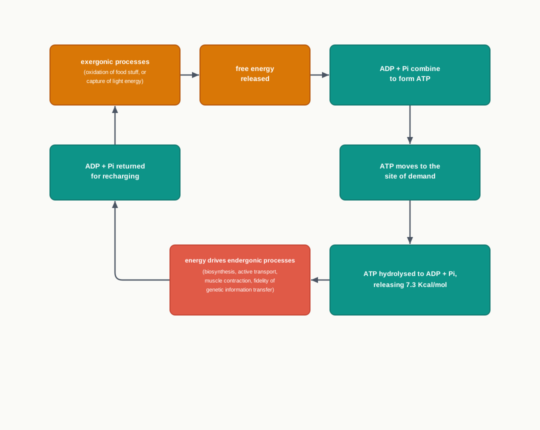
Fig. 4.19 – The ATP cycle.
ATP can be converted into adenosine monophosphate (AMP) and PPi (pyrophosphate) while donating its energy in some other processes, e.g., in the production of light flashes by the firefly. So there are two ways of drawing on the same molecule: removing one phosphate to give ADP and Pi, or removing two at once to give AMP and PPi, which releases a larger amount of energy in a single step.
ATP and its consecutive hydrolysis products, ADP and AMP, are nucleotides, made up of an adenine (a purine base occurring in DNA and RNA), a ribose (a pentose sugar), and three, two and one phosphate group(s), respectively. Their structures are compared in Fig. 4.20.
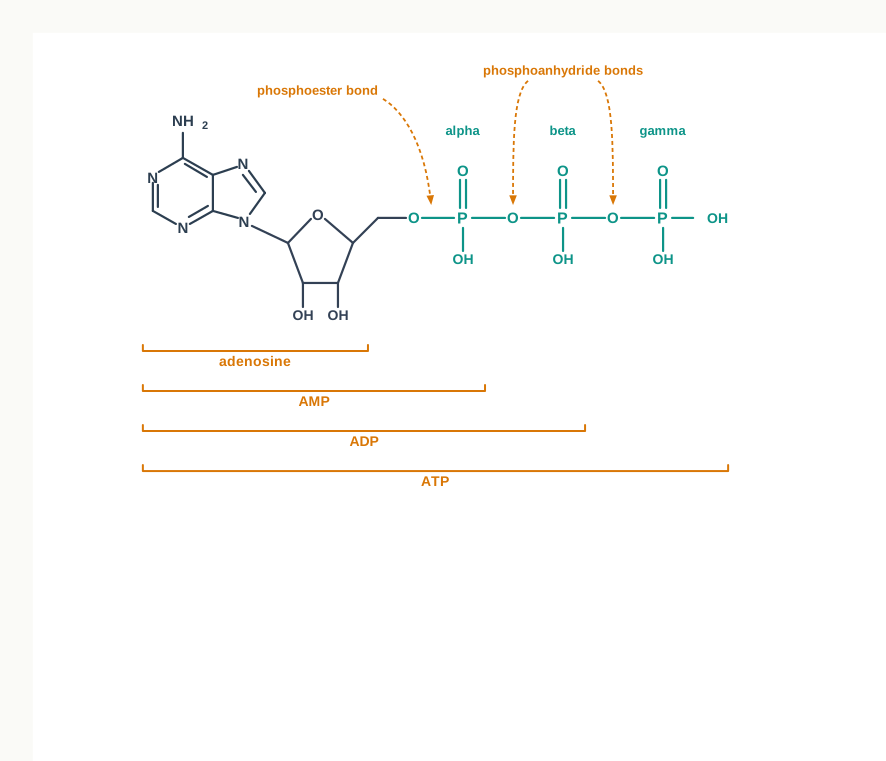
Fig. 4.20 – Structure of AMP, ADP and ATP.
Look carefully at the bonds labelled in Fig. 4.20. The phosphate closest to the sugar is joined by a phosphoester bond, while the phosphates joined to one another are linked by phosphoanhydride bonds. It is the breaking of these phosphoanhydride bonds that releases the large amount of free energy the cell uses, which is why the terminal phosphate groups of ATP matter so much.
4.3 Summary
Enzymes are biocatalysts that catalyse the biochemical reactions of the living system, working both inside the body and in a test tube, and emerging unchanged at the end of each reaction.
All enzymes are proteins except for a small group of catalytic RNA molecules called ribozymes. Many enzymes need a cofactor — either a coenzyme or a metal ion — and the complete active form is the holoenzyme, of which the protein part alone is the apoenzyme.
Enzymes are classified by the type of reaction they catalyse. The I.U.B. scheme recognises six major classes — oxidoreductases, transferases, hydrolases, lyases, isomerases and ligases — with translocase added more recently as a seventh.
Each enzyme has an active site into which the substrate molecule fits precisely. The ‘lock and key’ hypothesis postulated that the substrate fits precisely into the lock of the enzyme. This hypothesis has now been modified.
The modern ‘induced fit’ hypothesis does not regard the active site as a rigid structure but as a flexible one, which modifies its shape to fit the substrate molecule precisely.
Enzymes are highly specific. Their specificity may be group, absolute, stereochemical (optical) or geometrical.
Various factors like temperature, pH, substrate concentration, and the presence of inhibitors and activators influence the rate of enzyme catalysed reactions. Each enzyme has its own optimum temperature and optimum pH, and its velocity reaches a ceiling once the enzyme is saturated with substrate.
Enzyme activity is measured as work done rather than as weight. One enzyme unit converts 1 micromole of substrate per minute under standard conditions, and one katal converts 1 mole per second. Activity per milligram of protein gives the specific activity, which measures purity.
Enzymes lower the activation energy of the reaction they catalyse. They do not alter \(\Delta G\) and therefore cannot change the equilibrium of a reaction — they only speed up its attainment.
Simple single substrate enzyme catalysed reactions can be described by Michaelis-Menten kinetics, which gives a hyperbolic graph in terms of substrate concentration and initial velocity. \(K_m\) is the substrate concentration at which half of the maximum velocity is reached.
Enzymes are also affected by the presence of inhibitors, like competitive, non-competitive and uncompetitive inhibitors, which slow down the rate of reaction or stop it completely. Inhibition may be reversible or irreversible.
Allosteric enzymes do not obey Michaelis-Menten kinetics. They give a sigmoidal curve, carry a regulatory site in addition to the active site, and act as the key regulators of metabolic pathways.
Bioenergetics deals with the transformation and use of energy by the living cell.
There are two laws of thermodynamics. The first law states that energy can neither be created nor destroyed, but can be converted into other forms of energy.
The second law of thermodynamics states that the entropy or disorder of the universe is always increasing.
Combining the two laws of thermodynamics describes the free energy (G) change or the performance of work, through the relation \(\Delta G = \Delta H - T\Delta S\).
The photosynthetic organisms derive free energy from sunlight, whereas chemotrophs obtain free energy by the oxidation of food stuff.
This free energy is used to complete the cellular processes.
This free energy is partly converted into ATP, which is also known as the universal currency of free energy, and which transfers energy from exergonic to endergonic processes.
During energy releasing processes (breakdown), ATP is synthesised from ADP and inorganic phosphate (Pi), while in energy requiring processes ATP is broken into ADP and Pi, releasing 7.3 Kcal/mol.
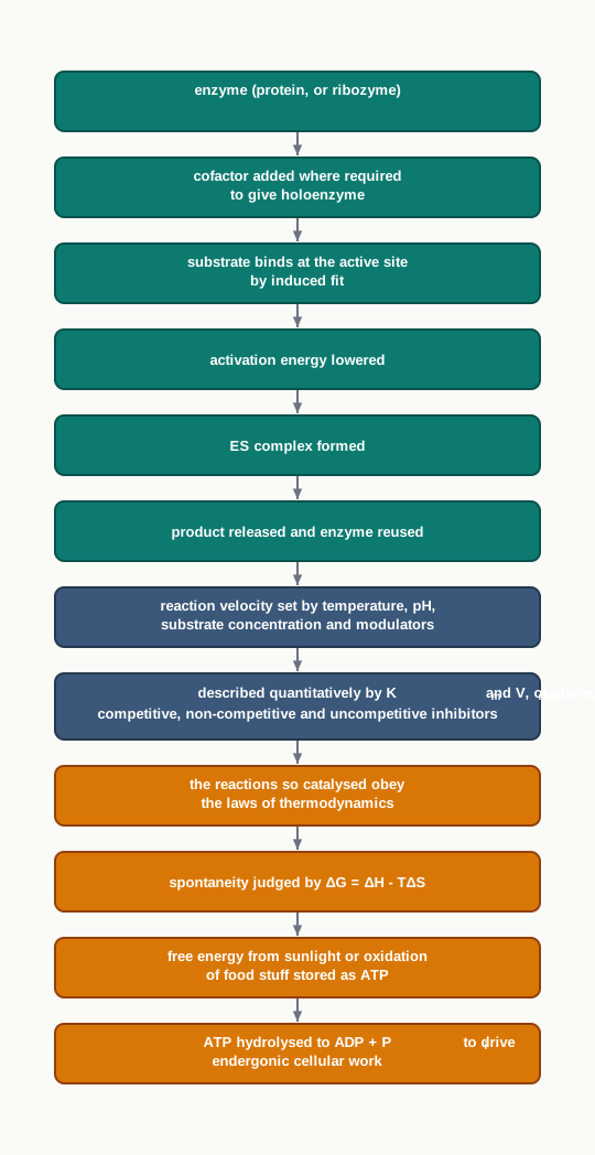
Fig. 4.21 – Chapter 4 at a glance.
4.4 Key Terms
Enzyme — A biocatalyst, almost always a protein, that greatly increases the rate of a specific biochemical reaction and emerges from it chemically unchanged.
In vivo — Inside the living body or living cell.
In vitro — Outside the living body, in a test tube or other reaction vessel.
Substrate — The molecule on which an enzyme acts and which it converts into product.
Ribozyme — A catalytic RNA molecule, the one recognised exception to the rule that all enzymes are proteins.
Denaturation — The loss of an enzyme’s folded three-dimensional structure, which destroys the active site and hence its catalytic activity.
Cofactor — A non-protein partner, either a coenzyme or a metal ion, required by many enzymes for catalytic activity.
Coenzyme — A complex organic cofactor, mostly derived from vitamins, which takes part in catalysis transiently and carries specific functional groups between enzymes.
Holoenzyme — The complete, catalytically active enzyme, consisting of the protein part together with its cofactor.
Apoenzyme — The protein component alone of a cofactor-requiring enzyme, which is inactive until the cofactor joins it.
Prosthetic group — A coenzyme or metal ion that is tightly bound to the enzyme protein through a covalent bond.
Oxidoreductases — I.U.B. class 1 enzymes, which catalyse oxidation–reduction reactions involving transfer of electrons.
Transferases — I.U.B. class 2 enzymes, which catalyse the transfer of groups from one molecule to another.
Hydrolases — I.U.B. class 3 enzymes, which catalyse hydrolytic reactions by transferring functional groups to water.
Lyases — I.U.B. class 4 enzymes, which catalyse the addition or removal of groups to form double bonds.
Isomerases — I.U.B. class 5 enzymes, which transfer groups within a molecule to yield isomeric forms.
Ligases — I.U.B. class 6 enzymes, which join two molecules by condensation coupled through ATP hydrolysis.
Translocase — The recently added seventh I.U.B. class, comprising enzymes that transfer ions or molecules across a membrane.
Isoenzymes (isozymes) — Different molecular forms of an enzyme in the same species, tissue or cell that catalyse the same reaction but differ in amino acid composition and physicochemical properties.
Active site — The small pocket or cleft, built from portions of the folded polypeptide chain, where the substrate binds and catalysis occurs.
Lock and Key Model — Emil Fischer’s 1894 proposal that the active site is pre-shaped and structurally complementary to the substrate, which fits it as a key fits a lock.
Induced Fit Hypothesis — Daniel Koshland’s 1958 proposal that the active site is flexible and becomes complementary to the substrate only after binding, like a glove shaped by a hand.
Group specificity — The ability of an enzyme to act on several different but closely related substrates.
Absolute specificity — The ability of an enzyme to act on only one particular substrate.
Stereochemical (optical) specificity — The ability of an enzyme to act on only one of two stereochemical forms of a substrate.
Geometrical specificity — The ability of an enzyme to distinguish between the cis and trans forms of a substrate.
Optimum temperature — The temperature at which a given enzyme catalyses its reaction at the maximum rate.
Optimum pH — The pH at which the activity of a particular enzyme is at its maximum.
Modulators — Substances that bind an enzyme and change its activity, decreasing it as inhibitors or increasing it as activators.
Enzyme unit (U) — The amount of enzyme that converts 1 micromole of substrate into product per minute under standard conditions.
Katal (kat) — The SI-based unit of enzyme activity, being the amount of enzyme that converts 1 mole of substrate into product per second.
Specific activity — The units of enzyme activity per milligram of enzyme protein, used as a measure of the purity of an enzyme preparation.
Transition state — The high-energy, strained arrangement of atoms through which a substrate must pass on its way to becoming product.
Activation energy (\(\Delta G^A\)) — The difference in free energy between the transition state and the substrate; enzymes work by lowering it.
Enzyme–substrate (ES) complex — The weakly bonded intermediate formed when substrate binds at the active site, which then decomposes to give product and free enzyme.
Michaelis-Menten equation — The relation \(v_0 = \dfrac{V_{max}[\text{S}]}{K_m + [\text{S}]}\), which describes the kinetics of a simple single-substrate enzyme reaction.
Michaelis constant (\(K_m\)) — The substrate concentration at which the reaction velocity is half of the maximum velocity.
Maximum velocity (\(V_{max}\)) — The highest reaction velocity attainable at a given enzyme concentration, reached when all enzyme molecules are saturated with substrate.
Enzyme inhibitor — A substance that decreases the rate of an enzyme catalysed reaction.
Irreversible inhibition — Inhibition in which the inhibitor binds very tightly to the enzyme and does not dissociate from it.
Reversible inhibition — Inhibition in which the inhibitor dissociates rapidly from the enzyme–inhibitor complex.
Competitive inhibition — Reversible inhibition in which an inhibitor resembling the substrate competes for the same active site, raising \(K_m\) while leaving \(V_{max}\) unchanged.
Non-competitive inhibition — Reversible inhibition in which the inhibitor binds at a site other than the active site, lowering \(V_{max}\) without affecting \(K_m\).
Uncompetitive inhibition — Reversible inhibition in which the inhibitor binds only to the enzyme–substrate complex, altering both \(K_m\) and \(V_{max}\).
Allosteric enzyme — An enzyme that does not obey Michaelis-Menten kinetics, gives a sigmoidal velocity curve, and carries a regulatory site in addition to its active site.
Regulatory site — The site on each subunit of an allosteric enzyme to which regulatory molecules bind reversibly, altering the enzyme’s affinity for substrate.
Bioenergetics — The study of the transformation and use of energy by living cells.
Thermodynamics — The branch of physical chemistry that deals with energy changes, without reference to the mechanism or speed of a process.
System — The matter within a defined region that is under study.
Surroundings — The matter constituting the rest of the universe outside the system.
First law of thermodynamics — Energy can neither be created nor destroyed but only converted into other forms, so the total energy of the universe remains constant.
Internal energy (\(\Delta E\)) — The energy content of a system, whose change depends only on the initial and final states and not on the path taken.
Entropy (S) — The degree of randomness or disorder of a system, which increases as the system becomes more disordered and is maximum at equilibrium.
Second law of thermodynamics — The entropy of the universe is always increasing, and a process occurs spontaneously only if the entropies of system and surroundings together increase.
Enthalpy (\(\Delta H\)) — The heat content of a system at constant pressure.
Free energy change (\(\Delta G\)) — The part of a system’s energy change that is theoretically available to perform useful work, given by \(\Delta G = \Delta H - T\Delta S\).
Closed system — A system that can exchange energy, but not matter, with its surroundings.
Chemotrophs — Non-photosynthetic organisms that obtain free energy by the oxidation of food stuff.
Exergonic process — A process that releases free energy.
Endergonic process — A process that consumes free energy.
Adenosine triphosphate (ATP) — The nucleotide made of adenine, ribose and three phosphate groups that serves as the universal currency of free energy in cells.
Adenosine diphosphate (ADP) — The nucleotide with two phosphate groups, formed when ATP donates energy and releases inorganic phosphate.
Adenosine monophosphate (AMP) — The nucleotide with a single phosphate group, formed when ATP donates energy and releases pyrophosphate.
Inorganic phosphate (Pi) — The free phosphate group released on hydrolysis of ATP to ADP, and reused in resynthesising ATP.
Pyrophosphate (PPi) — The two-phosphate unit released when ATP is converted into AMP.
Phosphoanhydride bond — The bond joining one phosphate group of ATP to the next, whose breakage releases the large free energy the cell uses.
Phosphoester bond — The bond joining the alpha phosphate group to the ribose sugar of the nucleotide.
4.5 Points to Ponder
Enzymes work equally well in vivo and in vitro. What does this single fact tell you about the nature of life processes, and why does it matter so much to an industry that wants to run biological reactions inside a steel vessel rather than inside a cell?
Most of an enzyme molecule is not part of its active site. Is that bulk wasted, or is there a reason a catalyst needs to be so much larger than the reaction it performs?
The lock and key model was not discarded when the induced fit model appeared; it was modified. Think about what this says about how scientific models are treated in biology — when is a model replaced, and when is it merely refined?
Cofactors and coenzymes allow a protein to do chemistry that its twenty amino acids cannot manage alone. Consider what limits this places on what proteins can achieve unaided, and how a shortage of a single vitamin can affect many different reactions at once.
An enzyme lowers the activation energy but cannot alter \(\Delta G\). If a reaction is thermodynamically unfavourable, no enzyme will make it happen. How, then, does a cell manage to carry out unfavourable reactions such as building macromolecules or transporting ions against a gradient?
Competitive inhibition can be overcome by adding more substrate, while non-competitive inhibition cannot. If you were designing a drug that must keep working even when the body’s level of the natural substrate rises sharply, which kind of inhibitor would you want, and what risks might come with that choice?
Allosteric enzymes give a sigmoidal curve rather than a hyperbola, and they sit at the control points of metabolic pathways. Reflect on why a switch-like response is more useful for regulation than a gradual one, and where else in biology you have met such all-or-none behaviour.
The chapter states that biological systems do not follow the second law, and also that entropy increases after the death and decomposition of every organism. Hold these two statements together and work out what exactly is being claimed about a living organism and its surroundings.
Enzymes are measured by activity rather than by weight, and purity is judged by specific activity. Why would weighing a protein sample tell a biotechnologist so little about its usefulness?
ATP is called a currency rather than a fuel. Follow that comparison as far as it will go — what does a cell gain from a single standard denomination, and where does the comparison begin to break down?
4.6 NCERT Questions
In order to catalyse a reaction, an enzyme is required to
- be saturated with substrate
- decrease the activation energy
- increase the equilibrium constant
- increase the activation energy
Pepsin is a gastric enzyme. Does it have an acidic or alkaline optimum pH? What happens to pepsin when it enters the duodenum?
What is the relationship between vitamins and enzyme co-factors?
What is the effect of temperature, pH, and substrate concentration on catalytic activity of enzyme?
The rate determining step of Michaelis-Menten kinetics is
- the complex dissociation of ES complex
- the complex formation
- the product formation
- the product degradation
Define \(K_m\) and its significance.
What is meant by one unit of enzyme?
What is specific activity of an enzyme?
Briefly describe first and second laws of thermodynamics.
Define entropy. What is the relationship between free energy and entropy?
Why ATP is called universal energy currency?
4.7 Objective Questions
All enzymes are proteins except
- isoenzymes
- ribozymes
- holoenzymes
- apoenzymes
The molecular weight of enzymes ranges from about
- 200 to 2000 Dalton
- 2000 to more than one million Dalton
- 20 to 200 Dalton
- one million to ten million Dalton
The protein component of a cofactor-requiring enzyme is called
- holoenzyme
- prosthetic group
- apoenzyme
- coenzyme
A coenzyme or metal ion tightly bound to the enzyme protein through a covalent bond is called a
- prosthetic group
- substrate
- modulator
- holoenzyme
The coenzyme derived from niacin (vitamin B3) and involved in the transfer of hydride is
- FAD
- coenzyme A
- NAD
- tetrahydrofolate
The metal ion that serves as a cofactor for DNA polymerase is
- \(\text{Zn}^{2+}\)
- \(\text{Mg}^{2+}\)
- \(\text{Ni}^{2+}\)
- \(\text{K}^+\)
Enzymes that catalyse the condensation of two molecules coupled through ATP hydrolysis belong to the I.U.B. class
- hydrolases
- lyases
- ligases
- isomerases
The class of enzymes added to the original six classes of the I.U.B. scheme is
- transferases
- translocase
- oxidoreductases
- isomerases
In human beings, lactate dehydrogenase (LDH) exists in
- two isozyme forms
- three isozyme forms
- four isozyme forms
- five isozyme forms
Which of the following is NOT one of the non-covalent interactions involved in enzyme–substrate binding?
- hydrogen bonds
- hydrophobic interactions
- peptide bonds
- Van der Waals forces
The induced fit hypothesis was proposed by
- Emil Fischer in 1894
- Daniel Koshland in 1958
- Leonor Michaelis in 1913
- Gibbs in 1878
D-amino acid oxidase acts only on D-amino acids and not on their mirror-image forms. This illustrates
- group specificity
- geometrical specificity
- stereochemical specificity
- absolute specificity
For most enzymes, the optimum temperature lies between
- 20-25°C
- 30-35°C
- 40-45°C
- 55-60°C
The optimum pH of pepsin is about
- pH 1-2
- pH 4-5
- pH 6-8
- pH 10-11
One katal is equivalent to
- \(6 \times 10^{5}\) IU
- \(6 \times 10^{6}\) IU
- \(6 \times 10^{7}\) IU
- \(6 \times 10^{8}\) IU
Specific activity of an enzyme preparation is expressed as
- units of activity per millilitre of solution
- units of activity per milligram of enzyme protein
- micromoles of substrate per second
- moles of product per milligram of substrate
Enzymes increase the rate of a reaction by
- increasing \(\Delta G\) of the reaction
- lowering the activation energy \(\Delta G^A\)
- shifting the equilibrium towards the product
- raising the free energy of the transition state
When \([\text{S}] = K_m\), the initial velocity \(v_0\) of a Michaelis-Menten enzyme is
- \(V_{max}\)
- \(V_{max}/2\)
- \(2V_{max}\)
- zero
Penicillin kills bacteria because it acts as
- a competitive inhibitor of cyclooxygenase
- an irreversible inhibitor of transpeptidase
- an allosteric activator of a bacterial enzyme
- a coenzyme for cell wall synthesis
In competitive inhibition,
- \(V_{max}\) is lowered and \(K_m\) is unchanged
- \(V_{max}\) is unchanged and \(K_m\) is increased
- both \(V_{max}\) and \(K_m\) are unchanged
- both \(V_{max}\) and \(K_m\) are lowered
An inhibitor that binds only to the enzyme–substrate complex and not to the free enzyme produces
- competitive inhibition
- non-competitive inhibition
- uncompetitive inhibition
- irreversible inhibition
A plot of \(v_0\) against \([\text{S}]\) for an allosteric enzyme is
- a rectangular hyperbola
- a straight line through the origin
- a bell-shaped curve
- a sigmoidal curve
According to the first law of thermodynamics, the change in the energy of a system depends on
- the path of the transformation only
- the initial and final states only
- the speed of the process
- the entropy of the surroundings only
A system that can exchange energy but not matter with its surroundings is called
- an open system
- a closed system
- an isolated system
- an equilibrium system
The Gibbs free energy function is expressed as
- \(\Delta G = \Delta H + T\Delta S\)
- \(\Delta G = \Delta H - T\Delta S\)
- \(\Delta G = T\Delta S - \Delta H\)
- \(\Delta G = \Delta E - P\Delta V\)
The energy released when ATP is converted into ADP and Pi is
- 3.7 Kcal/mol
- 7.3 Kcal/mol
- 17.3 Kcal/mol
- 73 Kcal/mol
ATP is converted into AMP and pyrophosphate during
- muscle contraction
- active transport across the membrane
- the production of light flashes by the firefly
- the synthesis of ATP from ADP
Assertion (A): An enzyme cannot alter the equilibrium of a biochemical reaction. Reason (R): An enzyme lowers the activation energy \(\Delta G^A\) but leaves \(\Delta G\) of the reaction unchanged.
Assertion (A): Denaturation of an enzyme destroys its catalytic activity even though its amino acids are still present. Reason (R): The active site is built from residues that are brought together only when the polypeptide chain folds correctly.
Assertion (A): A deficiency of a single vitamin can slow down many different reactions in the cell at the same time. Reason (R): Most coenzymes are derived from vitamins, and a coenzyme serves as a carrier of a specific functional group for many enzymes.
Assertion (A): Competitive inhibition can be overcome by increasing the substrate concentration. Reason (R): The competitive inhibitor and the substrate compete for the same active site on the enzyme.
Assertion (A): Non-competitive inhibition lowers \(V_{max}\) but does not change \(K_m\). Reason (R): The non-competitive inhibitor effectively reduces the concentration of active enzyme, while the enzyme molecules that remain free behave normally.
Assertion (A): Taq DNA polymerase remains active at temperatures that would destroy most human enzymes. Reason (R): The optimum temperature of an enzyme depends on the stability of its folded three-dimensional structure.
Assertion (A): Allosteric enzymes are well suited to act as control points of metabolic pathways. Reason (R): Their sigmoidal kinetics allows a small change in substrate concentration to produce a large change in reaction velocity.
Assertion (A): Free energy, rather than entropy, is used as the criterion of spontaneity for biochemical reactions. Reason (R): Changes in entropy of a reaction cannot be measured, and the entropy change of the surroundings would also have to be known.
Assertion (A): ATP is described as the universal currency of free energy. Reason (R): ATP transfers free energy from exergonic processes to endergonic processes in the cell.
4.8 Check Your Concepts
Enzymes catalyse reactions both in vivo and in vitro. Explain why this property is essential for the use of enzymes in industry, and what would follow if enzymes worked only inside living cells.
All enzymes are proteins except ribozymes. What does this exception tell you about the real requirement for catalytic activity in a biological molecule?
A purified enzyme is stored at a pH far from its optimum for several days and then returned to its optimum pH, but very little activity is recovered. Explain why.
Distinguish between an apoenzyme and a holoenzyme, and explain why an apoenzyme alone cannot catalyse a reaction.
Compare a coenzyme with a prosthetic group with respect to the nature of binding and behaviour during catalysis.
A laboratory sets up a DNA-copying reaction with a correct buffer, correct temperature and pure DNA polymerase, but forgets to add magnesium salt. Predict the outcome and justify your answer using the idea of a cofactor.
Match the following coenzymes with their precursor vitamins.
| Column I | Column II |
|---|---|
| a. FAD | i. folic acid |
| b. NAD | ii. riboflavin |
| c. coenzyme A | iii. niacin |
| d. tetrahydrofolate | iv. pantothenic acid |
- Match the following enzymes with their metal ion cofactors.
| Column I | Column II |
|---|---|
| a. urease | i. \(\text{Mn}^{2+}\) |
| b. arginase | ii. \(\text{K}^+\) |
| c. carbonic anhydrase | iii. \(\text{Ni}^{2+}\) |
| d. pyruvate kinase | iv. \(\text{Zn}^{2+}\) |
The I.U.B. classifies enzymes by the type of reaction they catalyse rather than by the organism they come from. Give two reasons why this basis of classification is more useful for a biochemist.
Assign each of the following reactions to its I.U.B. class and justify the choice: (i) transfer of electrons from one molecule to another, (ii) splitting of a bond using water, (iii) joining of two molecules coupled to ATP hydrolysis, (iv) movement of an ion across a membrane.
Isoenzymes catalyse the same reaction but differ in amino acid composition. Explain how this difference makes it possible for a laboratory to separate them, and why an organism benefits from having them.
The active site occupies only a small part of an enzyme. Explain the function served by the rest of the molecule.
The bonds holding a substrate at the active site are all weak, non-covalent interactions. Explain why the use of a strong covalent bond instead would be a disadvantage for a catalyst.
Draw and label a diagram of the interaction between an enzyme and its substrate according to (i) the lock and key model and (ii) the induced fit model. Mark clearly the one feature that distinguishes the two.
“The induced fit model explains enzyme specificity better than the lock and key model.” Examine this statement, using the idea of conformational change on substrate binding.
Compare group specificity and absolute specificity, and give one example each of stereochemical specificity and geometrical specificity from the chapter.
The curve of enzyme activity against temperature is bell-shaped. Explain what causes the rise on the left of the peak and what causes the fall on the right, and state why the two limbs have different explanations.
An enzyme from a hot-spring bacterium works at 100°C, while a human enzyme is destroyed well below that. What structural difference accounts for this, and why is such an enzyme so valuable in biotechnology?
Explain why the optimum pH of pepsin is very different from that of most enzymes of higher organisms, and relate your answer to where the enzyme functions.
A student increases the substrate concentration in a reaction tube step by step and finds that the velocity stops rising after a point. Explain this observation in terms of enzyme saturation, and state which quantity of the Michaelis-Menten equation has been reached.
Arrange the following steps of a catalytic cycle in the correct sequence: (i) release of product, (ii) formation of the enzyme–substrate complex, (iii) binding of substrate at the active site, (iv) conversion of substrate into product, (v) free enzyme available for the next substrate molecule.
Explain why enzyme activity is expressed in enzyme units or katals rather than in units of mass, and how specific activity is used to judge whether a purification step has worked.
“Enzymes speed up the attainment of equilibrium but cannot alter it.” Explain this statement using the difference between \(\Delta G\) and \(\Delta G^A\).
Draw a labelled energy profile diagram for a reaction with and without an enzyme. Mark the transition state, the activation energy in both cases, and \(\Delta G\), and use the diagram to explain why \(\Delta G\) is the same in both curves.
Interpret the graph of \(v_0\) against \([\text{S}]\) for a Michaelis-Menten enzyme. State what the axes represent, describe how \(V_{max}\) and \(K_m\) are read from it, and explain what a low \(K_m\) tells you about the enzyme.
Two enzymes act on the same substrate. Enzyme X has a low \(K_m\) and enzyme Y has a high \(K_m\). Which enzyme will work more efficiently when the substrate is scarce, and why?
Compare and contrast reversible and irreversible inhibition, using penicillin as an example of one of them.
Penicillin blocks a bacterial enzyme but does not harm human cells. Explain the basis of this selectivity, and state why selectivity is the central goal in designing an inhibitor as a drug.
A researcher measures \(K_m\) and \(V_{max}\) for an enzyme with and without an inhibitor. In trial 1, \(V_{max}\) is unchanged and \(K_m\) rises. In trial 2, \(V_{max}\) falls and \(K_m\) is unchanged. In trial 3, both change. Identify the type of inhibition in each trial and justify each answer.
Explain why a competitive inhibitor can never form an ESI complex, whereas a non-competitive inhibitor can.
Explain why increasing the substrate concentration actually favours the action of an uncompetitive inhibitor instead of relieving the inhibition.
Compare the kinetics of a normal Michaelis-Menten enzyme with that of an allosteric enzyme, and explain why the sigmoidal shape suits an enzyme placed at the control point of a pathway.
Explain why thermodynamics can tell us that a reaction is possible but not how long it will take, and identify which part of this chapter deals with the second question.
Glucose burnt in a flame and glucose oxidised through cellular pathways release the same total energy. Explain this using the path-independence of the first law, and state what the cell gains by releasing the energy slowly.
A living cell builds ordered macromolecules from disordered precursors, which lowers its entropy. Explain how this is reconciled with the second law of thermodynamics.
Explain why entropy is not used as a practical criterion of spontaneity for biochemical reactions, and what advantage the free energy function offers instead.
In the equation \(\Delta G = \Delta H - T\Delta S\), explain the physical meaning of each term and state which part of the energy change is unavailable for doing work.
Name any four cellular processes that consume free energy, and explain why each of them would not occur on its own without an input of energy.
Explain the difference between exergonic and endergonic processes, and describe the role ATP plays in linking the two.
Draw and label the structure of ATP. Mark the adenine, the ribose, the alpha, beta and gamma phosphate groups, the phosphoester bond and the phosphoanhydride bonds, and state which bonds release the large free energy used by the cell.
Distinguish between the hydrolysis of ATP to ADP and Pi and its conversion to AMP and PPi, and give the example from the chapter in which the second route is used.
A poison stops the synthesis of ATP from ADP and Pi, although the oxidation of food stuff continues normally. Explain why the cell still fails to carry out its energy-requiring processes.
4.9 Case-Based Questions
A student sets up four test tubes containing the same amount of a digestive enzyme and the same substrate. The tubes are kept at 10°C, 37°C, 45°C and 80°C respectively, and everything else is identical. After twenty minutes, the tube at 37°C shows the most product, the tube at 45°C slightly less, the tube at 10°C very little, and the tube at 80°C none at all. The student then cools the 80°C tube back to 37°C and waits another twenty minutes, but still finds no product.
- Sketch the shape of the curve you would expect if reaction velocity were plotted against temperature for this enzyme, and mark the optimum temperature on it.
- Explain why the tube at 10°C gave very little product, and why this reason is different from the reason for the tube at 80°C.
- Why did cooling the 80°C tube back to 37°C fail to restore activity?
- Name one enzyme mentioned in this chapter that would still have worked in the 80°C tube, and state the property that allows it to do so.
A biotechnology laboratory is purifying an enzyme from a bacterial culture. At the start, the crude extract shows a total activity of 900 units and contains 300 mg of protein. After two purification steps, the sample shows 720 units of activity and contains 24 mg of protein. The team is pleased with the result even though the total activity has fallen.
- Calculate the specific activity of the crude extract and of the purified sample.
- Explain why the team regards this as a successful purification although some total activity has been lost.
- Why is enzyme activity measured in enzyme units rather than by weighing the protein sample?
- State the fixed conditions that must be kept identical when the two activity measurements are made, and explain why this matters.
A pharmaceutical team is testing two candidate molecules, P and Q, against the same target enzyme. With molecule P, the measured \(V_{max}\) is unchanged but the \(K_m\) rises sharply; when the substrate concentration is raised to a very high level, the enzyme reaches its normal maximum velocity. With molecule Q, the \(V_{max}\) falls while the \(K_m\) stays the same, and raising the substrate concentration does not help at all.
- Identify the type of inhibition caused by P and by Q, giving a reason for each.
- Where on the enzyme does each molecule bind, and which complexes can form in each case?
- The target enzyme acts on a substrate whose concentration in the body rises steeply after a meal. Which of the two candidates would be the more dependable drug, and why?
- Sketch, on one set of axes, the \(v_0\) against \([\text{S}]\) curves expected for the uninhibited enzyme and for the enzyme with molecule Q, labelling the axes and the constants that differ.
A patient’s diet has been extremely poor for several months and is deficient in more than one B-group vitamin. Laboratory tests show that several unrelated reactions in the patient’s cells are running slowly at the same time — some involving the transfer of electrons, others involving the transfer of amino groups. The treating doctor prescribes vitamins in milligram quantities rather than in grams.
- Explain, using the idea of coenzymes, why a shortage of vitamins can slow down several unrelated reactions at once.
- Name the coenzyme derived from riboflavin and the one derived from pyridoxine, and state the group each transfers.
- Why are vitamins needed in such small amounts, unlike food materials such as carbohydrate?
- Would the protein parts of the affected enzymes still be present in the patient’s cells? Name the form in which such an enzyme exists without its cofactor.
A metabolic pathway converts compound A into compound E through four enzyme-catalysed steps. The enzyme catalysing the first step gives a sigmoidal curve when initial velocity is plotted against substrate concentration, and it also carries a second binding site on each subunit, distinct from its active site. The final product E binds reversibly at this second site and slows the enzyme down. The remaining three enzymes of the pathway give ordinary hyperbolic curves.
- What kind of enzyme catalyses the first step? Give two features from the passage that identify it.
- Explain why the cell places such an enzyme at the first step of the pathway rather than at the last.
- Why is a sigmoidal response more useful for control than a hyperbolic one?
- The binding of E at the second site is described as reversible. Why is reversibility important for the cell?
A cell suspension is supplied with glucose and oxygen, and measurements confirm that the oxidation of food stuff is proceeding normally and releasing energy. A chemical is then added which specifically blocks the formation of ATP from ADP and inorganic phosphate. Within a short time, active transport across the cell membrane stops, biosynthesis halts and the cells die, even though the energy-releasing reactions are still running.
- Explain why the cells die although free energy is still being released.
- Classify the oxidation of glucose and the active transport of ions as exergonic or endergonic, and state which one normally pays for the other.
- Why is ATP described as a currency rather than as a fuel?
- In what form is the energy released by oxidation lost when ATP cannot be made?
A student reads that pepsin has an optimum pH of about 1-2 and works in the stomach, while several enzymes in the small intestine work best around pH 6-8. The student wonders what becomes of pepsin once the partly digested food, together with the enzyme, is pushed out of the stomach into the duodenum, where the contents are much less acidic.
- State whether pepsin has an acidic or an alkaline optimum pH, and relate this to the compartment in which it works.
- Predict what happens to the activity of pepsin in the duodenum, and explain the reason in terms of the charges on the amino acid side chains of the active site.
- What does this example tell you about the relationship between an enzyme and the compartment of the body in which it is found?
- Sketch the general shape of a graph of enzyme activity against pH and mark the optimum pH on it.
4.10 Practice With Data
An enzyme preparation converts 45 micromoles of substrate into product in 9 minutes under standard conditions. Calculate the activity of the preparation in enzyme units (U).
A purified enzyme sample shows a total activity of 240 U and contains 8 mg of enzyme protein. Calculate its specific activity, and state what this figure measures.
During a purification, the following measurements are obtained.
| Stage | Total activity (U) | Total protein (mg) |
|---|---|---|
| Crude extract | 1200 | 400 |
| After salt precipitation | 1000 | 100 |
| After column chromatography | 800 | 16 |
- Calculate the specific activity at each stage. (ii) State by what factor the enzyme has been purified from the crude extract to the final stage. (iii) Explain why the total activity falls while the specific activity rises.
An enzyme sample has an activity of 0.25 katal. Using the relation \(1\ \text{kat} = 6 \times 10^{7}\ \text{IU}\), express this activity in international units.
An enzyme shows an activity of \(1.2 \times 10^{7}\) IU. Express this activity in katals, using \(1\ \text{kat} = 6 \times 10^{7}\ \text{IU}\).
For a single-substrate enzyme, \(V_{max} = 80\) micromoles per minute and \(K_m = 4\ \text{mmol L}^{-1}\). Using \(v_0 = \dfrac{V_{max}[\text{S}]}{K_m + [\text{S}]}\), calculate \(v_0\) at substrate concentrations of (i) \(4\ \text{mmol L}^{-1}\), (ii) \(12\ \text{mmol L}^{-1}\) and (iii) \(36\ \text{mmol L}^{-1}\). Comment on the trend in your three answers.
An enzyme has \(K_m = 5\ \text{mmol L}^{-1}\). At what substrate concentration will the initial velocity be exactly half of \(V_{max}\)? Justify your answer from the Michaelis-Menten equation without doing any calculation.
The following initial velocities were recorded for an enzyme at a fixed enzyme concentration.
| \([\text{S}]\) (mmol L\(^{-1}\)) | 1 | 2 | 5 | 10 | 20 | 40 |
|---|---|---|---|---|---|---|
| \(v_0\) (µmol min\(^{-1}\)) | 17 | 29 | 50 | 67 | 80 | 89 |
- Plot \(v_0\) against \([\text{S}]\) and describe the shape of the curve. (ii) Estimate \(V_{max}\) from the plateau region. (iii) Using your value of \(V_{max}\), estimate \(K_m\) from the graph. (iv) State what your value of \(K_m\) tells you about this enzyme’s behaviour when substrate is scarce.
- An enzyme is studied in the absence and in the presence of two different inhibitors, giving the values below.
| Condition | \(K_m\) (mmol L\(^{-1}\)) | \(V_{max}\) (µmol min\(^{-1}\)) |
|---|---|---|
| No inhibitor | 3 | 100 |
| With inhibitor X | 9 | 100 |
| With inhibitor Y | 3 | 40 |
| With inhibitor Z | 1.5 | 50 |
- Identify the type of inhibition produced by X, Y and Z. (ii) For each, state whether the inhibition can be relieved by adding excess substrate. (iii) Calculate \(v_0\) for the uninhibited enzyme and for the enzyme with inhibitor Y at \([\text{S}] = 3\ \text{mmol L}^{-1}\).
- The activity of an enzyme was measured at different temperatures, all other conditions being constant.
| Temperature (°C) | 10 | 20 | 30 | 40 | 45 | 55 | 70 |
|---|---|---|---|---|---|---|---|
| Relative activity (%) | 15 | 34 | 68 | 97 | 100 | 42 | 0 |
- Plot the data and identify the optimum temperature. (ii) Explain the rise in activity from 10°C to 45°C. (iii) Explain the fall from 45°C to 70°C, making clear why the explanation differs from that in part (ii). (iv) Predict what the reading at 70°C would be if the tube were cooled back to 45°C, and give a reason.
A closed system absorbs 250 J of heat from its surroundings and performs 90 J of work on them. Using \(\Delta E = Q - W\), calculate the change in internal energy of the system, and state whether the internal energy has risen or fallen.
For a biochemical reaction at 310 K, \(\Delta H = -30\ \text{kJ mol}^{-1}\) and \(\Delta S = +60\ \text{J K}^{-1}\ \text{mol}^{-1}\) for the system. Using \(\Delta G = \Delta H - T\Delta S\), calculate \(\Delta G\) and state whether the reaction is spontaneous. (Take care to convert the units of \(\Delta S\) before substituting.)
For another reaction at 300 K, \(\Delta H = +15\ \text{kJ mol}^{-1}\) and \(\Delta S = +20\ \text{J K}^{-1}\ \text{mol}^{-1}\). (i) Calculate \(\Delta G\). (ii) Calculate the temperature at which \(\Delta G\) would become zero. (iii) State what happens to the spontaneity of the reaction above that temperature.
A muscle cell hydrolyses \(3.0 \times 10^{-3}\) mol of ATP to ADP and Pi during a short burst of contraction. Taking the energy released as 7.3 Kcal/mol, calculate the total energy made available.
An endergonic process in a cell requires 36.5 Kcal of free energy. Using the value of 7.3 Kcal/mol for the conversion of ATP into ADP and Pi, calculate the number of moles of ATP that must be hydrolysed to supply this energy, assuming complete transfer.