Chapter 5: Cellular Processes
This chapter is long enough to require splitting to ensure no diagrams are cut off. This is Part A, covering up to the end of section 5.2.6. Please reply to continue, and I will process Part B and automatically merge and renumber the complete chapter for you.
In the early 1960s, the biologist Sydney Brenner went looking for an animal simple enough to be understood completely — every cell, every division, every fate. He settled on *Caenorhabditis elegans*, a transparent soil roundworm about a millimetre long. Because the worm is see-through, John Sulston was able to sit at a microscope and follow individual cells as the fertilised egg divided again and again, drawing up a complete family tree of the animal, cell by cell.

What that tree revealed surprised everyone. The adult hermaphrodite worm carries 959 body cells — but the developing embryo produces more than that. During normal development, 131 cells quietly die. And they are not random casualties. The same 131 cells die, in the same order, at the same moments, in worm after worm after worm.

Death, in other words, was scheduled. Robert Horvitz went on to identify the genes that carry out this orderly self-destruction, and closely related genes were later found in human cells. In 2002, the three scientists shared a Nobel Prize for showing that a cell dying on cue is as much a part of building an organism as a cell dividing.

That is the theme of this chapter. Cells listen, cells burn fuel, cells divide, cells specialise, cells crawl — and sometimes, on instruction, cells die.

<img src="images/fig5.0.png" onerror="this.onerror=null; this.src='images/fig5.0.svg';" alt="Fig. 5.0">

::: {.figure-caption}
Fig. 5.0 – Cellular processes in C. elegans.
:::


::: {.callout-note .box-curiosity icon=false title="Curiosity Corner"}

Q1. Thousands of chemical messages float past a cell every second. How does a cell know which message is meant for it?
Q2. Why does a cell that dies as part of normal development cause no swelling or redness, while a cell killed by injury does?
Q3. Most of your ATP is made in the mitochondria. Why is oxygen needed only right at the end of the process?
Q4. Every cell in your body carries the same DNA. Why, then, is a neuron so different from a red blood cell?
Q5. Cells have no legs and no fins. How does a cell actually move from one place to another inside the body?

:::


Keep these five questions in mind as you read. By the end of this chapter, you will be able to answer each one of them yourself.

## 5.1 Cell Signaling


A cell is never alone. Whether it is a bacterium in pond water or a nerve cell in your spinal cord, it sits in an environment that is constantly changing, and it must keep track of those changes as they happen. Both prokaryotic and eukaryotic cells receive information from their surroundings, interpret it, and respond to it in real time.

The signals a cell picks up are of many kinds. Light, heat, sound and touch are all physical signals that cells can sense. Alongside these, cells produce chemical signals of their own and release them into the fluid outside the cell, called the extracellular milieu. A neighbouring cell that picks up such a chemical acts on the message it carries.

This exchange of information is not a minor housekeeping activity. During the development of an embryo, the fate of a cell — whether it will become part of a muscle, a nerve or the skin — is decided by the signalling pathways it is exposed to. A cell that receives one set of signals becomes one thing; the same cell exposed to a different set becomes another.

##### Signals From Inside and Outside the Body {.unnumbered .unlisted}


Not every signal a cell responds to is made by the body itself. Cells also respond to 'external' signals — molecules that were never synthesised by any cell of our body, such as a drug, a plant chemical or a toxin. Taken together, this means cells are capable of sensing a very wide variety of signals, from within and from outside.

##### The Receptor Decides {.unnumbered .unlisted}


There is one strict condition, though. A cell can respond to a particular signal only if it possesses the corresponding receptor for that signal. Without the receptor, the message goes past the cell unread.

A receptor is a protein. It may sit on the cell surface, embedded in the plasma membrane, or it may lie inside the cell — in the cytoplasm or even in the nucleus. Where it sits depends on the kind of messenger it is built to catch: water-soluble messengers cannot cross the membrane and so are caught at the surface, while small fat-soluble messengers slip through the membrane and are caught inside.

The chemical messenger to which a receptor responds is called a ligand. The association between a receptor and its corresponding ligand is highly specific. Each receptor recognises the shape and chemistry of one particular ligand, in much the same way a lock accepts only one key. So a cell will respond to a chemical messenger if it bears the corresponding receptor for it, and not otherwise.

::: {.callout-note .box-definition icon=false title="Definition"}

*A receptor is a protein, located either on the cell surface or inside the cytoplasm or the nucleus, that binds a specific chemical messenger and allows the cell to respond to it.*

:::


::: {.callout-note .box-definition icon=false title="Definition"}

*A ligand is the chemical messenger to which a particular receptor responds; the binding between a ligand and its receptor is highly specific.*

:::


<img src="images/fig5.1.png" onerror="this.onerror=null; this.src='images/fig5.1.svg';" alt="Fig. 5.1">

::: {.figure-caption}
Fig. 5.1 – Specificity of receptor–ligand binding.
:::


::: {.callout-note .box-curiosity icon=false title="Curiosity Corner" collapse="true"}

**Q1. Thousands of chemical messages float past a cell every second. How does a cell know which message is meant for it?**

A. It does not have to sort through them. A cell can read only those messages for which it carries a matching receptor, and each receptor binds only its own specific ligand. Every other molecule drifts past without being recognised. Specificity of the receptor–ligand pair, not any decision by the cell, is what filters the traffic.

:::


##### What Happens When a Ligand Binds {.unnumbered .unlisted}


Transmission of a chemical message from one cell to another begins with the binding of the ligand to its receptor. That binding changes the shape of the receptor protein — a conformational change. The receptor is now in a different three-dimensional form from the one it had a moment earlier.

This change of shape is the whole point of the event. A shape change on the outer part of the receptor is passed on to the part of the receptor facing the inside of the cell, and that in turn switches on a message relay system within the cell. One molecule activates the next, which activates the next, and so on.

::: {.callout-note .box-sidebar icon=false title="Exam Corner" collapse="true"}

**Q. Why does the cell bother with a relay of many molecules instead of letting the ligand act directly?**

A. Two reasons. First, most ligands never enter the cell at all, so the message has to be handed over at the membrane. Second, a relay amplifies the signal — one activated receptor can switch on many molecules of the next component, so a few ligand molecules outside can produce a large response inside.

:::


The end result of this relay is a set of important changes in the activities inside the cell — a gene may be switched on, an enzyme may be activated, the cell may begin to divide, or it may begin to move. The full sequence, from arrival of the signal to the cell's response, is summarised in Fig. 5.2.

<img src="images/fig5.2.svg" onerror="this.onerror=null; this.src='images/fig5.2.png';" alt="Fig. 5.2">

::: {.figure-caption}
Fig. 5.2 – Steps of cell signalling.
:::


::: {.callout-note .box-incident icon=false title="Real Incident / Discovery" collapse="true"}

In the 1950s and 1960s, the American scientist Earl Sutherland was trying to work out how the hormone adrenaline makes liver cells break down their stored glycogen. He found that adrenaline itself never entered the cell. Instead, its arrival at the cell surface caused a small molecule to be produced inside the cell, and it was this internal molecule — cyclic AMP — that switched on the machinery for glycogen breakdown. Sutherland had uncovered the idea of a relay system inside the cell, and he received the Nobel Prize in Physiology or Medicine in 1971 for the work.

:::


Cells send and receive signals in more than one way. Depending on how far apart the sender cell and the recipient cell are, signalling is broadly classified into three categories: paracrine, autocrine and endocrine.

##### Paracrine Signaling {.unnumbered .unlisted}


In paracrine signalling, communication between cells takes place over relatively short distances. The sender cell releases a chemical message into the extracellular space, and the recipient cell sitting close by senses it almost instantly. Because the distance is small, the response is quick.

This type of signalling is seen commonly in the communication of neurons. At a synapse, the tip of one nerve cell releases a neurotransmitter into the very narrow gap separating it from the next cell, and receptors on the other side pick it up within milliseconds. The message never has to travel far, which is exactly why nervous signalling is so fast.

##### Autocrine Signaling {.unnumbered .unlisted}


Many times, a cell that secretes a ligand also possesses receptors specific for that same ligand. Such a cell, in effect, sends a message to itself. This type of signalling is referred to as autocrine signalling.

Cancer cells provide a striking example. Cancer cells are characterised by uncontrollable growth, and therefore they require a greater amount of growth factors for their proliferation. Unlike normal cells, cancer cells do not depend on external growth factors supplied by other cells. Instead, they are capable of synthesising their own growth factors and also possess the receptors specific for them.

Follow the consequence of that arrangement. A normal cell divides only when another cell instructs it to; the instruction can be withheld. A cancer cell issues its own instruction and immediately receives it, so there is no longer any outside control over how often it divides. This is one reason such growth is so difficult to stop, and it is also why several modern cancer medicines are designed to block the growth-factor receptor itself rather than the cell as a whole — if the receptor cannot receive the message, the loop is broken. The drug trastuzumab, used in certain breast cancers, works on exactly this principle by binding to a growth-factor receptor on the tumour cell surface.

##### Endocrine Signaling {.unnumbered .unlisted}


Endocrine signalling, also called long-distance signalling, requires the ligand to be synthesised by the cell and released into the bloodstream, so that it can travel to the recipient or target cell far away. Hormones generally exhibit this form of signalling.

Insulin is a familiar example. It is made in the pancreas, enters the blood, and acts on muscle, liver and fat cells all over the body — cells that carry the insulin receptor. Cells without that receptor ignore the hormone completely, even though it reaches them too. Endocrine signalling is slower than paracrine signalling because the messenger must travel through the circulation, but it reaches a far larger number of cells.

::: {.callout-note .box-compare icon=false title="Compare Table" collapse="true"}

Paracrine vs Autocrine vs Endocrine signalling
| Feature | Paracrine | Autocrine | Endocrine |
|---|---|---|---|
| Distance covered | Short, between neighbouring cells | None — the same cell | Long, across the body |
| Route taken by ligand | Extracellular space | Extracellular space, back to the same cell | Bloodstream |
| Sender and receiver | Different cells, close together | The same cell | Different cells, far apart |
| Speed of response | Very fast | Fast | Slower |
| Example | Communication of neurons | Growth factors made by cancer cells | Hormones |

:::


::: {.callout-note .box-sidebar icon=false title="Exam Corner" collapse="true"}

**Q. A hormone circulating in the blood reaches every cell of the body. Why then does it act only on a few?**

A. Because only some cells carry the receptor for that hormone. Reaching a cell and acting on a cell are two different things — the receptor, not the blood supply, decides which cells are the target cells.

:::


##### How This Topic Is Tested {.unnumbered .unlisted}


Questions from this section are usually application-based rather than recall-based. A common assertion–reason item pairs a statement about a hormone reaching all cells with a reason about receptor specificity. Comparison questions asking you to distinguish autocrine from paracrine signalling appear regularly, and case-based passages often describe a cell that fails to respond to a signal and ask you to explain why — where the expected answer is the absence of the corresponding receptor, not the absence of the signal.

## 5.2 Metabolic Pathways


Every activity of a living cell costs energy. Building a protein costs energy, pumping ions across a membrane costs energy, and moving a muscle costs energy. Metabolism is the process through which living organisms take in and utilise the free energy required to carry out their life processes.

On the basis of how they obtain this free energy, living organisms are of two types: phototrophs and chemotrophs.

Phototrophs use the energy of sunlight to convert simple molecules, which contain less energy, into more complex molecules, which are energy rich and serve as fuel to perform life processes. Phototrophs are photosynthetic organisms such as plants and some bacteria; they transform light energy into chemical energy.

Heterotrophs, such as animals, obtain energy indirectly from plants through their food. In chemotrophs, the energy is obtained by oxidising chemical compounds, which may be organic or inorganic.

##### How Energy Is Actually Transferred {.unnumbered .unlisted}


Energy uptake in organisms is done by coupling the exergonic reactions of nutrient oxidation to the endergonic processes required to maintain the living state. An exergonic reaction releases free energy; an endergonic one consumes it. Coupling simply means the two are linked, so that the energy released by one is not wasted as heat but is captured and handed over to the other.

Central to all these energy transactions is the energy currency called ATP. (The details of how ATP stores and releases energy are given in section 4.2, bioenergetics.) Think of ATP the way you think of money: a cell does not spend glucose directly on its work, it first converts glucose into ATP, and then spends ATP.

In metabolism, there are interlinked biochemical reactions that begin with a particular molecule and convert it into some other molecule or molecules in a carefully defined fashion. Nothing about this is random — each step is catalysed by a specific enzyme, and the product of one step is the starting material of the next.

The energy so released is utilised for various processes within the cell, such as the creation of gradients, the movement of molecules across membranes, the conversion of chemical energy into mechanical energy, and the powering of reactions that result in the synthesis of biomolecules.

::: {.callout-note .box-definition icon=false title="Definition"}

*Metabolism is the set of interlinked, enzyme-catalysed chemical reactions through which an organism takes in and uses free energy to carry out its life processes.*

:::


##### Metabolic Pathways and Their Two Classes {.unnumbered .unlisted}


The synthesis and breakdown of biomolecules is accomplished through a number of steps inside the living system. These steps collectively constitute a metabolic pathway. Metabolic pathways can broadly be classified into two classes: anabolic pathways and catabolic pathways.

::: {.callout-note .box-definition icon=false title="Definition"}

*A metabolic pathway is an ordered series of enzyme-catalysed steps by which one biomolecule is converted into another inside a living system.*

:::


##### Anabolic pathways {.unnumbered .unlisted}


In these pathways, larger and more complex molecules are synthesised from small molecules. Anabolic pathways are endergonic, which means they consume energy. Reactions that require energy, such as the synthesis of glucose, fats, protein or DNA, are called anabolic reactions or anabolism.

$$\text{Useful energy} + \text{Small molecules} \xrightarrow{\ \text{Anabolism}\ } \text{Complex molecules}$$

##### Catabolic pathways {.unnumbered .unlisted}


These pathways involve the breakdown of larger molecules. These are exergonic reactions, which means they release energy, and they produce reducing equivalents and ATP. Reducing equivalents are electron-carrying molecules such as NADH and FADH₂ — they hold the electrons stripped from the fuel, and later hand them over for ATP production.

$$\text{Fuel (carbohydrate, protein, fats)} \xrightarrow{\ \text{Catabolism}\ } \mathrm{CO_2} + \mathrm{H_2O} + \text{Useful energy}$$

The useful forms of energy that are produced in catabolism are utilised in anabolism, to generate complex structures from simple ones, or energy-rich states from energy-poor ones. The two classes are therefore not independent; they are two halves of one economy, one earning and the other spending.

::: {.callout-note .box-compare icon=false title="Compare Table" collapse="true"}

Anabolic vs Catabolic pathways
| Feature | Anabolic pathway | Catabolic pathway |
|---|---|---|
| Direction of change | Small molecules to larger, complex molecules | Large molecules broken down to smaller ones |
| Energy relationship | Endergonic — consumes energy | Exergonic — releases energy |
| ATP | Uses ATP | Produces ATP |
| Reducing equivalents | Consumed | Produced |
| Example | Synthesis of glucose, fats, protein or DNA | Oxidation of carbohydrate, protein and fat to $\mathrm{CO_2}$ and $\mathrm{H_2O}$ |

:::


::: {.callout-note .box-sidebar icon=false title="Exam Corner" collapse="true"}

**Q. If catabolism releases energy, why does the cell not simply let the fuel burn all at once?**

A. Because energy released in a single burst would appear almost entirely as heat and would be lost. By breaking the fuel down in many small enzyme-catalysed steps, the cell captures the energy a little at a time in ATP and reducing equivalents, in a form it can actually spend.

:::


### 5.2.1 Overview of carbohydrate metabolism


In animals, the metabolic fuel for most of the tissues is glucose. Glucose is metabolised into pyruvate through glycolysis, a pathway you will study step by step in section 5.2.4.

What happens to that pyruvate depends entirely on whether oxygen is available. Under aerobic conditions, that is, in the presence of oxygen, pyruvate enters the mitochondrial matrix, where it is converted into acetyl CoA and takes part in the citric acid cycle to complete the oxidation of glucose to $\mathrm{CO_2}$ and $\mathrm{H_2O}$, as shown in Fig. 5.3. This oxidation is linked to the formation of ATP through the process of oxidative phosphorylation.

Under anaerobic conditions, that is, in the absence or lack of $\mathrm{O_2}$, pyruvate is converted into lactic acid instead. The same starting molecule therefore has two very different destinations, decided by one factor: the supply of oxygen.

<img src="images/fig5.3.svg" onerror="this.onerror=null; this.src='images/fig5.3.png';" alt="Fig. 5.3">

::: {.figure-caption}
Fig. 5.3 – Overview of carbohydrate metabolism.
:::


Look carefully at Fig. 5.3 and notice something important: this is not a single straight road. Glucose is drawn down a central line, but at almost every intermediate there is a side branch leading somewhere else in the cell's chemistry. Glycolysis is therefore not only an energy pathway; it is also a supply depot for building materials.

The metabolic intermediates of glycolysis also take part in other metabolic processes, such as:

(i) In the synthesis of glycogen and its storage in animals.

(ii) In the pentose phosphate pathway, which is a source of reducing equivalent (NADPH) for fatty acid synthesis, and a source of ribose for nucleotides and nucleic acid synthesis.

(iii) The triose phosphate generates the glycerol moiety of triacylglycerol.

(iv) Acetyl CoA is the precursor for the synthesis of fatty acids and cholesterol. Cholesterol then synthesises all other steroids in animals.

(v) Pyruvate and intermediates of the citric acid cycle give rise to the carbon skeleton for amino acid synthesis.

(vi) When glycogen reserves are depleted, such as in starvation conditions, the non-carbohydrate precursors such as lactic acid, amino acids and glycerol can synthesise glucose through the process of gluconeogenesis.

::: {.callout-note .box-sidebar icon=false title="Exam Corner" collapse="true"}

**Q. Why does the body go to the trouble of making glucose from lactic acid and amino acids during starvation, instead of simply burning fat?**

A. Because some tissues cannot use fat. Brain, heart and red blood cells depend heavily on glucose, and the brain cannot be left without fuel. Gluconeogenesis exists to protect these tissues when stored glycogen has run out.

:::


### 5.2.2 Overview of lipid metabolism


Some vital tissues, such as the brain, heart and red blood cells, are exclusively dependent on glucose. In the fasting state, when glucose is limiting, the less glucose-dependent tissues such as muscles, liver and other tissues alternatively use a fuel other than glucose, as shown in Fig. 5.4. This arrangement makes sense as a strategy: the tissues that can switch fuels do so, leaving the scarce glucose for the tissues that cannot.

<img src="images/fig5.4.svg" onerror="this.onerror=null; this.src='images/fig5.4.png';" alt="Fig. 5.4">

::: {.figure-caption}
Fig. 5.4 – Overview of lipid metabolism.
:::


This alternative fuel is long-chain fatty acids, which are either taken from the diet or synthesised from acetyl CoA derived from carbohydrate or amino acids. Fatty acids may be oxidised to acetyl CoA through the β-oxidation pathway, or esterified with glycerol to synthesise triacylglycerol (fat), which is the main fuel reserve in the adipose tissue of animals.

Notice in Fig. 5.4 that the arrows between fatty acids and triacylglycerol run both ways. Esterification stores fat away; lipolysis releases it again. Whether a cell is storing or releasing depends on whether the body has just eaten or has been fasting.

Following are the three fates of acetyl CoA formed by the β-oxidation pathway.

(i) It is oxidised to $\mathrm{CO_2}$ and $\mathrm{H_2O}$ through the citric acid cycle.

(ii) It is a precursor for the synthesis of other lipids such as cholesterol. Cholesterol then synthesises all other steroids, including hormones and bile pigments.

(iii) It is used to synthesise ketone bodies — acetone, acetoacetate and 3-hydroxy butyrate — which are an alternative fuel for the liver and some other tissues in prolonged fasting.

::: {.callout-note .box-sidenote icon=false title="Side Note" collapse="true"}

Ketone bodies are the reason a person on a prolonged fast or a very low-carbohydrate diet may have a faintly sweet, fruity smell on the breath. That odour comes from acetone, one of the three ketone bodies listed above, leaving the body through the lungs.

:::


### 5.2.3 Overview of amino acid metabolism


Since amino acids are the building blocks of proteins, they are required for protein synthesis. There are 20 standard amino acids.

Some of these are non-essential amino acids, because they are synthesised in the body from metabolic intermediates by the process of transamination, shown in Fig. 5.5. The remaining ones are essential amino acids, which must be supplied in the diet as they are not synthesised in the body. The word "essential" here does not mean more important — it means essential in the diet.

<img src="images/fig5.5.svg" onerror="this.onerror=null; this.src='images/fig5.5.png';" alt="Fig. 5.5">

::: {.figure-caption}
Fig. 5.5 – Overview of amino acid metabolism.
:::


In transamination, the amino nitrogen is transferred from one amino acid to a carbon skeleton to form other amino acids. Nothing is discarded in this step — the nitrogen is simply moved from a molecule where it is not needed to one where it is.

In the process of deamination, by contrast, the amino nitrogen is excreted as urea. Deamination is therefore the disposal route, and it is used when the nitrogen is genuinely surplus.

::: {.callout-note .box-definition icon=false title="Definition"}

*Transamination is the transfer of the amino nitrogen from one amino acid to a carbon skeleton, forming a different amino acid.*

:::


::: {.callout-note .box-definition icon=false title="Definition"}

*Deamination is the removal of the amino nitrogen from an amino acid, after which the nitrogen is excreted as urea.*

:::


The carbon skeletons that remain after transamination can play the following roles:

(i) Oxidised to $\mathrm{CO_2}$ via the citric acid cycle.

(ii) Used to synthesise glucose through gluconeogenesis.

(iii) Form ketone bodies, which may be oxidised or used for fatty acid synthesis.

Some amino acids take part in the synthesis of other biomolecules, such as hormones of plants and animals, purines, pyrimidines, and neurotransmitters.

::: {.callout-note .box-sidebar icon=false title="Exam Corner" collapse="true"}

**Q. Carbohydrates and fats are stored as glycogen and triacylglycerol. What is the storage form of amino acids?**

A. There is none. The body has no dedicated protein store, so surplus amino acids must either be converted into carbohydrate or fat, or have their nitrogen removed by deamination and excreted as urea. This is why an excess of dietary protein cannot simply be kept in reserve.

:::


Some of the important metabolic pathways are described and detailed in the following sections.

### 5.2.4 Glycolysis


Glycolysis is a universal catalytic pathway found in all living cells. It is also known as the Embden-Meyerhof-Parnas (EMP) pathway, and its full sequence is shown in Fig. 5.6. It is a major pathway of carbohydrate metabolism, and all the enzymes involved are present in the cytosol.

The fact that glycolysis is present in every living cell, and that it needs no membrane-bound organelle to work, tells us something about its age: it is one of the oldest pieces of chemistry that living things possess.

<img src="images/fig5.6.svg" onerror="this.onerror=null; this.src='images/fig5.6.png';" alt="Fig. 5.6">

::: {.figure-caption}
Fig. 5.6 – The steps of glycolysis.
:::


###### Step 1: Phosphorylation of glucose {.unnumbered .unlisted}


The pathway starts with the phosphorylation of glucose, a 6-carbon molecule, to glucose-6-phosphate, which is catalysed by the enzyme hexokinase. ATP is the phosphate donor; its γ-phosphoryl group is transferred to glucose.

Ask why the cell spends an ATP at the very first step of an energy-yielding pathway. The answer is that the phosphate group carries a negative charge, and a charged molecule cannot slip back out through the membrane. Phosphorylation traps the sugar inside the cell and commits it to the pathway.

This reaction is irreversible, and the enzyme hexokinase is inhibited allosterically by its own product, glucose-6-phosphate. Allosteric inhibition means that the product binds to the enzyme at a site different from the active site and alters its catalytic activity.

::: {.callout-note .box-definition icon=false title="Definition"}

*Allosteric inhibition is the reduction of an enzyme's catalytic activity when a molecule binds to a site on the enzyme other than the active site.*

:::


This feedback arrangement is a sensible piece of design. If glucose-6-phosphate is already piling up, there is no point converting more glucose, and the product itself applies the brake.

Hexokinase can also phosphorylate sugars other than glucose, such as fructose, galactose, mannose and so on. Liver cells also contain an isoenzyme of hexokinase called glucokinase, which can phosphorylate only glucose. (Isoenzymes — different protein forms catalysing the same reaction — are discussed in detail in section 4.1.)

Glucose-6-phosphate is an important intermediate of carbohydrate metabolism, because it is formed in glycolysis, gluconeogenesis (the formation of glucose from non-carbohydrate molecules), the pentose phosphate pathway, glycogenesis (the synthesis of glycogen) and glycogenolysis (the breakdown of glycogen). It is a junction where five pathways meet.

###### Steps 2 and 3: Isomerisation and the second phosphorylation {.unnumbered .unlisted}


Glucose-6-phosphate is then converted into fructose-6-phosphate, also a 6-carbon molecule, by the enzyme phosphoglucose isomerase, which catalyses an aldose-ketose isomerisation reaction.

Fructose-6-phosphate then undergoes another phosphorylation by the enzyme phosphofructokinase (PFK), forming fructose-1,6-bisphosphate. Similar to hexokinase, PFK also catalyses an irreversible reaction and undergoes allosteric regulation. This second ATP is the cell's real commitment to glycolysis, and PFK is accordingly the pathway's main control point.

###### Step 4: Splitting the six-carbon sugar {.unnumbered .unlisted}


Fructose-1,6-bisphosphate is cleaved by the enzyme aldolase into two triose phosphates, each a 3-carbon molecule: glyceraldehyde-3-phosphate and dihydroxyacetone phosphate. This splitting is where the name of the pathway comes from — glycolysis literally means the splitting of sugar.

The two trioses synthesised are interconverted by the enzyme triose phosphate isomerase. Only glyceraldehyde-3-phosphate goes forward in the pathway, so this interconversion matters: it makes sure that neither of the two halves of the original glucose is wasted.

###### Steps 5 and 6: The first energy harvest {.unnumbered .unlisted}


Oxidation of glyceraldehyde-3-phosphate to 1,3-bisphosphoglycerate occurs by the enzyme glyceraldehyde-3-phosphate dehydrogenase, which is an NAD-dependent enzyme. The electrons removed in this oxidation are picked up by $\mathrm{NAD^+}$, which becomes NADH.

In the next reaction, phosphate is transferred from 1,3-bisphosphoglycerate to ADP, forming ATP and 3-phosphoglycerate, a 3-carbon molecule, by the enzyme phosphoglycerate kinase. This phosphorylation of ADP to form ATP is called substrate-level phosphorylation, because the phosphate comes directly from a substrate molecule and not from a proton gradient.

::: {.callout-note .box-definition icon=false title="Definition"}

*Substrate-level phosphorylation is the formation of ATP by direct transfer of a phosphate group from a substrate molecule to ADP.*

:::


Since two molecules of triose phosphates are synthesised per molecule of glucose, two ATP molecules are formed at this stage per molecule of glucose undergoing glycolysis. From this step onward, every reaction happens twice for each glucose.

###### Steps 7 to 10: Rearrangement and the second energy harvest {.unnumbered .unlisted}


The next step of glycolysis involves isomerisation of 3-phosphoglycerate, a 3-carbon molecule, into 2-phosphoglycerate, also a 3-carbon molecule, by the enzyme phosphoglycerate mutase.

The subsequent step involves a dehydration reaction converting 2-phosphoglycerate into phosphoenol pyruvate by the enzyme enolase, which requires either $\mathrm{Mg^{2+}}$ or $\mathrm{Mn^{2+}}$ for its activity. Removing a water molecule here rearranges the electrons in the molecule so that its remaining phosphate group becomes very easy to transfer — which is exactly what the next enzyme exploits.

The phosphoenol pyruvate, by the enzyme pyruvate kinase, is converted into pyruvate, a 3-carbon molecule, during which phosphate is transferred to ADP to form ATP. This is again substrate-level phosphorylation.

###### Where glycolysis is controlled {.unnumbered .unlisted}


Out of the 10 glycolytic reactions, three reactions are exergonic and therefore irreversible. These reactions are catalysed by the regulatory enzymes hexokinase, PFK and pyruvate kinase, and are therefore the major sites of regulation of glycolysis.

There is a general principle here worth carrying to other pathways. A reversible step cannot be used as a control point, because the pathway can simply run backwards around it. An irreversible step is a one-way gate, and a one-way gate is the only sensible place to put a lock.

::: {.callout-note .box-sidebar icon=false title="Exam Corner" collapse="true"}

**Q. If the reaction catalysed by hexokinase is irreversible, how does the liver ever make free glucose again during gluconeogenesis?**

A. It does not reverse the hexokinase reaction. Gluconeogenesis bypasses each of the three irreversible steps using different enzymes, which is also why the two pathways can be regulated independently of each other.

:::


###### Energy yield by glycolysis {.unnumbered .unlisted}


The net reaction in the transformation of one molecule of glucose into two pyruvate molecules is:

$$\text{Glucose} + 2\,\mathrm{ATP} + 2\,\mathrm{NAD^+} + 4\,\mathrm{ADP} + 4\,\mathrm{P_i} \longrightarrow 2\,\text{Pyruvate} + 2\,\mathrm{ADP} + 2\,\mathrm{NADH} + 2\,\mathrm{H^+} + 4\,\mathrm{ATP} + 2\,\mathrm{H_2O}$$

Here $\mathrm{P_i}$ stands for inorganic phosphate, and $\mathrm{NAD^+}$ and NADH are the oxidised and reduced forms of the same coenzyme.

Thus four molecules of ATP are generated in the conversion of glucose into two molecules of pyruvate. The net ATP production is two, as two ATP molecules are utilised during the process. Read the equation and the sentence together: the gross yield is 4, the investment is 2, and the profit is 2.

::: {.callout-note .box-numerical icon=false title="Numerical 5.1" collapse="true"}

**A muscle cell metabolises 30 molecules of glucose completely to pyruvate by glycolysis. Calculate (a) the number of ATP molecules consumed, (b) the number of ATP molecules produced, (c) the net ATP gain, and (d) the number of NADH molecules formed.**

:::


###### Fate of pyruvate {.unnumbered .unlisted}


There are two major fates of the pyruvate formed during glycolysis. One is anaerobic, in the absence of oxygen, and the other is aerobic, in the presence of oxygen. In the anaerobic fate, the pyruvate is metabolised to lactate, which is called homolactic fermentation, or it may be metabolised into ethanol, which is called alcoholic fermentation. Under aerobic conditions, pyruvate is metabolised into $\mathrm{CO_2}$ and $\mathrm{H_2O}$ through the citric acid cycle, also called the TCA cycle.

All the steps of glycolytic reactions from glucose to pyruvate are similar in most organisms and most types of cells, but the fate of pyruvate is different, as shown in Fig. 5.7.

<img src="images/fig5.7.svg" onerror="this.onerror=null; this.src='images/fig5.7.png';" alt="Fig. 5.7">

::: {.figure-caption}
Fig. 5.7 – Fates of pyruvate.
:::


<img src="images/fig5.8.svg" onerror="this.onerror=null; this.src='images/fig5.8.png';" alt="Fig. 5.8">

::: {.figure-caption}
Fig. 5.8 – Deciding the fate of pyruvate.
:::


###### Fermentation {.unnumbered .unlisted}


For glycolysis to continue, $\mathrm{NAD^+}$, which the cells have in only limited quantities, must be recycled after its reduction to NADH by glyceraldehyde-3-phosphate dehydrogenase. Under anaerobic conditions, the $\mathrm{NAD^+}$ is replenished by the reduction of pyruvate in an extension of the glycolytic pathway.

This is the single most important idea about fermentation, and it is often missed. Fermentation is not carried out to make lactate or ethanol; those are simply the waste products. It is carried out to regenerate $\mathrm{NAD^+}$. If $\mathrm{NAD^+}$ ran out, step 5 of glycolysis would stop, and with it the cell's only remaining source of ATP.

###### Homolactic fermentation {.unnumbered .unlisted}


Under anaerobic conditions, pyruvate is reduced by NADH to lactate by the enzyme lactate dehydrogenase. Lactate dehydrogenase is also an isozyme. The overall process of anaerobic glycolysis in muscle may be represented as:

$$\text{Glucose} + 2\,\mathrm{ADP} + 2\,\mathrm{P_i} \longrightarrow 2\,\text{Lactate} + 2\,\mathrm{ATP} + 2\,\mathrm{H_2O} + 2\,\mathrm{H^+}$$

Much of the lactate produced by glycolysis is exported from the muscle cell to the liver, where it is reconverted to glucose. The muscle, in effect, borrows energy quickly during hard exercise and lets the liver settle the account afterwards.

###### Alcoholic fermentation {.unnumbered .unlisted}


Under anaerobic conditions in yeast, $\mathrm{NAD^+}$ is regenerated through the conversion of pyruvate to ethanol and $\mathrm{CO_2}$.

The reaction has two steps. The first reaction is the decarboxylation of pyruvate to form acetaldehyde and $\mathrm{CO_2}$ by the enzyme pyruvate decarboxylase, which contains the coenzyme TPP (thiamine pyrophosphate). The second reaction is a reduction of acetaldehyde into ethanol by the enzyme alcohol dehydrogenase. In this reaction NADH is oxidised to $\mathrm{NAD^+}$.

$$\text{Pyruvate} \xrightarrow[\ -\,\mathrm{CO_2}\ ]{\ \text{pyruvate decarboxylase}\ } \text{Acetaldehyde} \xrightarrow[\ \mathrm{NADH} \rightarrow \mathrm{NAD^+}\ ]{\ \text{alcohol dehydrogenase}\ } \text{Ethanol}$$

Fermentation results in the production of 2 ATP per glucose.

Both of these fermentations are used daily in industry. Alcoholic fermentation by yeast produces beverages and bioethanol fuel, and the carbon dioxide released in the same reaction is what makes bread dough rise. Lactic acid fermentation by bacteria is what turns milk into curd and sets the flavour of idli and dosa batter.

::: {.callout-note .box-compare icon=false title="Compare Table" collapse="true"}

Homolactic vs Alcoholic fermentation
| Feature | Homolactic fermentation | Alcoholic fermentation |
|---|---|---|
| End product | Lactate | Ethanol and $\mathrm{CO_2}$ |
| Number of steps from pyruvate | One | Two |
| Enzymes involved | Lactate dehydrogenase | Pyruvate decarboxylase, alcohol dehydrogenase |
| Coenzyme/prosthetic group named | — | TPP in pyruvate decarboxylase |
| Is $\mathrm{CO_2}$ released? | No | Yes |
| Occurs in | Contracting muscle, erythrocytes, some other cells and some microorganisms | Yeast |
| ATP per glucose | 2 | 2 |

:::


###### Aerobic breakdown of pyruvate {.unnumbered .unlisted}


Under aerobic conditions, pyruvate is transferred from the cytosol to the mitochondria, where it is converted to acetyl CoA by oxidative decarboxylation. This is an irreversible reaction, catalysed by a multienzyme complex known as the pyruvate dehydrogenase complex (PDH), which is found only in mitochondria.

$$\text{Pyruvate} + \text{CoA} + \mathrm{NAD^+} \longrightarrow \text{Acetyl CoA} + \mathrm{CO_2} + \mathrm{NADH} + \mathrm{H^+}$$

The coenzymes and prosthetic groups required in this reaction sequence are TPP, FAD (flavin adenine dinucleotide coenzymes), $\mathrm{NAD^+}$, and lipoamide.

This irreversible reaction is the link between glycolysis and the citric acid cycle. Because it cannot be reversed, animals cannot convert acetyl CoA back into pyruvate — and that is precisely why fatty acids, which yield only acetyl CoA, cannot be used to make glucose.

### 5.2.5 Citric acid cycle


The citric acid cycle is also known as Kreb's cycle or the Tricarboxylic acid (TCA) cycle, after the name of Hans Kreb, who discovered it for the first time. The full cycle is shown in Fig. 5.9.

It is a sequence of reactions in mitochondria that oxidise the acetyl moiety of acetyl CoA and reduce $\mathrm{NAD^+}$, which is then reoxidised through the electron transport chain, linked to the formation of ATP.

The citric acid cycle is the final common pathway for the oxidation of carbohydrates, lipids and proteins, because glucose, fatty acids and most amino acids are metabolised to acetyl CoA or to intermediates of the TCA cycle. Whatever you eat, its carbon skeleton eventually arrives here.

::: {.callout-note .box-incident icon=false title="Real Incident / Discovery" collapse="true"}

Hans Krebs worked out this cycle in the 1930s while working in England, using pigeon breast muscle, which is unusually rich in mitochondria and respires very rapidly. He noticed that adding small amounts of certain acids caused the tissue to consume far more oxygen than those acids could account for on their own — the sign of a catalytic cycle rather than a straight line. When he submitted the work for publication, the journal *Nature* declined it, and the paper appeared elsewhere. He was awarded the Nobel Prize in Physiology or Medicine in 1953.

:::


In eukaryotes, the reactions of the citric acid cycle take place inside mitochondria, in contrast with those of glycolysis, which occur in the cytosol. The enzymes that catalyse the reactions of the TCA cycle are located in the mitochondrial matrix, either free or attached to the inner mitochondrial membrane, where the enzymes and coenzymes of the electron transport chain are also found. This arrangement is efficient: the NADH produced by the cycle is generated a very short distance away from the machinery that will use it.

<img src="images/fig5.9.svg" onerror="this.onerror=null; this.src='images/fig5.9.png';" alt="Fig. 5.9">

::: {.figure-caption}
Fig. 5.9 – The citric acid cycle.
:::


###### Going round the cycle {.unnumbered .unlisted}


The cycle begins with a condensation reaction between two-carbon acetyl CoA and four-carbon oxaloacetate to form six-carbon citrate by the enzyme citrate synthase.

Citrate is isomerised into isocitrate, via the formation of a cis-aconitate intermediate, by the enzyme aconitase. The reason for this apparently pointless rearrangement is chemical: citrate has no hydroxyl group in a position that can be oxidised, and isocitrate does.

Isocitrate undergoes dehydrogenation to form oxalosuccinate by the enzyme isocitrate dehydrogenase. The enzyme isocitrate dehydrogenase is an isozyme present in three forms. One form is found in mitochondria and requires $\mathrm{NAD^+}$ for its activity. The other two forms use $\mathrm{NADP^+}$ and are found in both mitochondria and cytosol.

In the citric acid cycle, oxidation of the isocitrate takes place via the $\mathrm{NAD^+}$-dependent enzyme, which results in the formation of $\mathrm{NADH+H^+}$ along with the product oxalosuccinate.

Oxalosuccinate, a transient intermediate, remains bound to the enzyme and undergoes decarboxylation to form five-carbon α-ketoglutarate. That word "transient" is a hint worth noticing — the intermediate never floats free in the matrix, which is why it is easy to miss in a simplified diagram of the cycle.

α-ketoglutarate, in a coenzyme A mediated reaction, undergoes oxidative decarboxylation to form four-carbon succinyl CoA by the enzyme α-ketoglutarate dehydrogenase complex. In this step, another $\mathrm{NAD^+}$ gets reduced to $\mathrm{NADH+H^+}$ and another $\mathrm{CO_2}$ is liberated. This reaction is an irreversible reaction and favours the formation of succinyl CoA.

Count the carbons at this point. Two carbons entered as the acetyl group, and two carbons have now left as $\mathrm{CO_2}$. The molecule that started the cycle must therefore be regenerated exactly as it was, and the remaining reactions do precisely that.

Succinyl CoA is converted into succinate by the enzyme succinyl CoA synthetase. This is the only step in the citric acid cycle where substrate level phosphorylation takes place, in which a GDP is phosphorylated to GTP, which is then converted to ATP.

Succinate is then oxidised to form fumarate by the enzyme succinate dehydrogenase. This enzyme contains the coenzyme FAD and an iron-sulfur (Fe-S) protein; the FAD gets reduced to $\mathrm{FADH_2}$. In the citric acid cycle, succinate dehydrogenase is the only enzyme which remains attached to the inner membrane of the mitochondria, whereas the other enzymes remain free in the matrix.

That single structural fact will explain a number in the next section: because this enzyme sits in the membrane itself, the $\mathrm{FADH_2}$ it makes hands its electrons into the electron transport chain at a later point than NADH does.

Fumarate is then converted into malate by the enzyme fumarase. Malate is oxidised into oxaloacetate by the enzyme malate dehydrogenase. In this step $\mathrm{NAD^+}$ gets reduced to $\mathrm{NADH+H^+}$, and the oxaloacetate so regenerated is ready to accept the next molecule of acetyl CoA.

The citric acid cycle harvests the high energy electrons from acetyl CoA and uses these electrons to form NADH and $\mathrm{FADH_2}$. Very little ATP is made in the cycle itself — its real product is a load of electrons, carried away for use in the next stage.

The overall reaction of the TCA cycle is:

$$\text{Acetyl CoA} + 3\,\mathrm{NAD^+} + \mathrm{FAD} + \mathrm{GDP} + \mathrm{P_i} + 2\,\mathrm{H_2O} \longrightarrow 2\,\mathrm{CO_2} + 3\,\mathrm{NADH} + 3\,\mathrm{H^+} + \mathrm{FADH_2} + \mathrm{GTP} + \text{CoA}$$

::: {.callout-note .box-sidebar icon=false title="Exam Corner" collapse="true"}

**Q. The citric acid cycle needs no oxygen at any of its steps. Why then does it stop almost immediately when oxygen runs out?**

A. Because the cycle depends on a supply of $\mathrm{NAD^+}$ and FAD, and these are regenerated only when NADH and $\mathrm{FADH_2}$ hand their electrons to the electron transport chain — which ends by giving those electrons to oxygen. No oxygen means no regeneration, and the cycle halts for lack of oxidised coenzyme.

:::


### 5.2.6 Electron transport chain


The reduced coenzymes ($\mathrm{NADH+H^+}$ and $\mathrm{FADH_2}$) produced during the TCA cycle and various other catabolic pathways are oxidised through the electron transport chain (ETC) located in the inner mitochondrial membrane. These are subsequently utilised for the production of ATP through the ETC.

In oxidative phosphorylation, the electrons released during the reoxidation of NADH and $\mathrm{FADH_2}$ flow through a series of membrane proteins to generate a proton gradient across the membrane. These protons then flow back through ATP synthase to synthesise ATP from ADP and inorganic phosphate. Oxygen is utilised as the electron acceptor at the end of the electron transport chain, resulting in the formation of $\mathrm{H_2O}$, and thereby allowing the regeneration of $\mathrm{NAD^+}$ and FAD.

::: {.callout-note .box-definition icon=false title="Definition"}

*Oxidative phosphorylation is the synthesis of ATP that is driven by the flow of protons back across the inner mitochondrial membrane, the gradient itself having been created by electron transport from NADH and $\mathrm{FADH_2}$ to oxygen.*

:::


###### The four complexes {.unnumbered .unlisted}


The ETC involves a chain of four large protein complexes called NADH-Q-oxidoreductase (complex I), succinate-Q-reductase (complex II), Q-cytochrome C oxidoreductase (complex III), and cytochrome C oxidase (complex IV), as shown in Fig. 5.10.

These electron carriers are large transmembrane protein complexes with multiple oxidation-reduction centres, such as quinones, flavins, iron sulfur clusters, heme and copper ions. Succinate-Q-reductase (complex II), in contrast with the other three complexes, does not pump protons.

<img src="images/fig5.10.svg" onerror="this.onerror=null; this.src='images/fig5.10.png';" alt="Fig. 5.10">

::: {.figure-caption}
Fig. 5.10 – Complexes of the electron transport chain.
:::


Electrons are carried from complex I to complex III by the reduced form of coenzyme Q, also known as ubiquinone. Ubiquinone is a quinone which is lipid, and hence hydrophobic, and it diffuses quickly within the inner mitochondrial membrane. Being small and fat-soluble, it can shuttle sideways within the membrane in a way that the huge protein complexes cannot.

The electrons from $\mathrm{FADH_2}$ are generated through succinate-Q-reductase (complex II), and are then transferred to cytochrome-C-oxidoreductase (complex III). Then the electron from complex III is transferred to cytochrome-C-oxidase (complex IV), which is the final component in the ETC. This component catalyses the reduction of $\mathrm{O_2}$ into $\mathrm{H_2O}$.

<img src="images/fig5.11.svg" onerror="this.onerror=null; this.src='images/fig5.11.png';" alt="Fig. 5.11">

::: {.figure-caption}
Fig. 5.11 – Pathway of electron flow in the ETC.
:::


###### Oxidative phosphorylation (ATP synthesis) {.unnumbered .unlisted}


The flow of electrons from NADH or $\mathrm{FADH_2}$ to $\mathrm{O_2}$ through the four protein complexes located in the mitochondrial inner membrane leads to the pumping of protons out of the mitochondrial matrix side to the cytosolic side of the inner mitochondrial membrane, as shown in Fig. 5.12.

The resulting uneven distribution of protons generates a proton motive force (pmf), which consists of a pH gradient, with the matrix side basic, and a transmembrane electrical potential, with the matrix side negative. Note that the force has two components, not one: a difference in concentration and a difference in charge, both pulling protons in the same direction.

Synthesis of ATP takes place when protons flow back to the matrix side through an enzyme complex called ATP synthase. ATP synthase is best pictured as a turbine set into the membrane, turned by the returning stream of protons.

<img src="images/fig5.12.png" onerror="this.onerror=null; this.src='images/fig5.12.svg';" alt="Fig. 5.12">

::: {.figure-caption}
Fig. 5.12 – Proton pumping and ATP synthesis.
:::


::: {.callout-note .box-incident icon=false title="Real Incident / Discovery" collapse="true"}

For many years, biochemists searched for a high-energy chemical intermediate that would link electron transport to ATP synthesis, and no one could find it. In the 1960s the British biochemist Peter Mitchell proposed that there was no such intermediate at all — the link, he argued, was a gradient of protons across the membrane. The idea, called the chemiosmotic hypothesis, was resisted for years before the evidence settled the matter, and Mitchell received the Nobel Prize in Chemistry in 1978.

:::


###### Counting the ATP {.unnumbered .unlisted}


The flow of electrons through NADH-Q-oxidoreductase, Q-cytochrome-c-oxidoreductase and cytochrome-c-oxidase develops a gradient large enough to synthesise 1, 0.5 and 1 molecule of ATP, respectively.

Hence, 2.5 molecules of ATP are synthesised per molecule of NADH oxidised in the mitochondrial matrix, whereas only 1.5 molecules of ATP are made per molecule of $\mathrm{FADH_2}$ oxidised, because its electrons enter the electron transport chain at $\mathrm{QH_2}$ (complex III).

Trace the reason back to the previous section. $\mathrm{FADH_2}$ is produced by succinate dehydrogenase, which is complex II, and complex II does not pump protons. Electrons from $\mathrm{FADH_2}$ therefore skip the proton pumping done by complex I, and one full ATP's worth of gradient is never generated.

::: {.callout-note .box-sidenote icon=false title="Side Note" collapse="true"}

You may have seen the older figures of 3 ATP per NADH and 2 ATP per $\mathrm{FADH_2}$ in some books. Those were rounded estimates from earlier work. The values 2.5 and 1.5 used here are the currently accepted ones, and they are not whole numbers because the number of protons needed to turn ATP synthase does not divide evenly into the number of protons pumped.

:::


::: {.callout-note .box-numerical icon=false title="Numerical 5.2" collapse="true"}

**During the complete aerobic oxidation of one molecule of glucose, 10 molecules of NADH and 2 molecules of $\mathrm{FADH_2}$ are handed to the electron transport chain. Using the values given in this section, calculate the number of ATP molecules obtained from oxidative phosphorylation alone.**

:::


The citric acid cycle, along with oxidative phosphorylation, takes place in mitochondria and is the major source of ATP in aerobic organisms.

::: {.callout-note .box-curiosity icon=false title="Curiosity Corner" collapse="true"}

**Q3. Most of your ATP is made in the mitochondria. Why is oxygen needed only right at the end of the process?**

A. Because oxygen is not used to burn the fuel directly — it is used to collect the electrons after everything else has finished with them. Glycolysis and the citric acid cycle strip electrons from fuel molecules onto NADH and $\mathrm{FADH_2}$; the electron transport chain then passes those electrons from one complex to the next, pumping protons as it goes. At the very end, complex IV hands the spent electrons to oxygen, which becomes water. Oxygen's job is to be the final electron acceptor. Without it the chain backs up, NADH and $\mathrm{FADH_2}$ cannot be reoxidised, and the whole system stalls.

:::
5.4 Programmed Cell Death (Apoptosis)
During the embryonic stage of development, our fingers are joined together, giving our hands a web-like appearance. As development continues, gaps begin to appear between our fingers. If it were not so, we would have webbed hands and would not be able to hold things firmly.
But how are these gaps created? The answer is that the cells between our fingers undergo apoptosis. Nothing carves the gaps out from outside — the cells occupying those spaces remove themselves.
Apoptosis, also known as Programmed Cell Death, is a property of the cells which enables them to die during development. It is a highly controlled, energy-dependent process and does not occur randomly.
Pause on the phrase “energy-dependent”, because it sounds contradictory. A cell dying by apoptosis spends ATP to do so. This is not a cell collapsing for want of energy; it is a cell actively carrying out an orderly shutdown, and an orderly shutdown has to be paid for.
It also means that once a cell is committed to undergo apoptosis, it cannot reverse its fate. The decision is a one-way one.
Why an Organism Needs Cells to Die
Not only is apoptosis useful in normal development, but it also plays a major role in protecting us from diseases, including viral infections and cancers.
The protection works in a direct way. A virus can only multiply inside a living cell, so a cell that detects it has been infected and destroys itself takes the virus down with it before new virus particles can be released. Similarly, a cell whose DNA has been badly damaged is a potential future tumour, and removing it is safer than repairing it.
In certain cases where apoptosis does not work efficiently, unregulated growth and division of cells may occur, which could result in the formation of cancer. Cancer, seen from this angle, is not only a disease of too much cell division; it is also a disease of too little cell death.
This understanding has shaped treatment. Many chemotherapy drugs and radiation treatments do not kill tumour cells directly at all — they damage the cell enough to trigger its own apoptosis machinery, letting the cell finish the job itself.
What an Apoptotic Cell Looks Like
Apoptosis is characterised by certain morphological and physiological changes in the cell. These include DNA fragmentation, blebbing of the plasma membrane, breakdown of the nuclear envelope, and increased susceptibility of DNA to deoxyribonuclease (DNAase).
Blebbing means that the plasma membrane forms small bulges or blisters on its surface. Note that even while the cell is being dismantled, the membrane is never simply torn open — this detail turns out to matter a great deal.
Eventually, the entire cell splits up into small membrane-enclosed fragments, which are ultimately phagocytosed by the nearby macrophages, as shown in Fig. 5.13. Because every fragment stays wrapped in membrane, the contents of the dying cell are never spilled into the surrounding tissue. The neighbouring cells are not disturbed, and no immune alarm is raised.
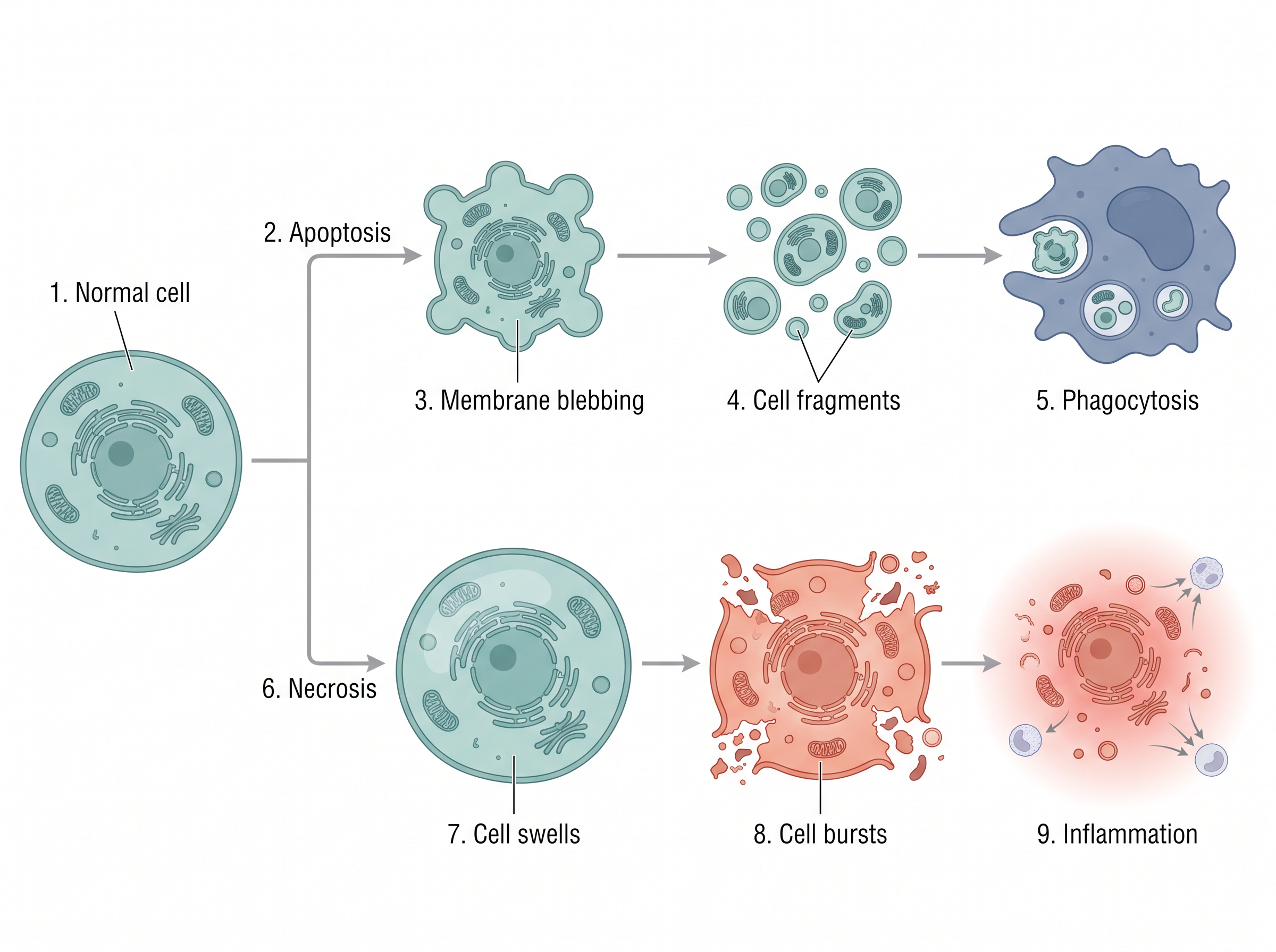
Fig. 5.13 – Morphological changes in apoptosis and necrosis.
Necrosis — The Other Kind of Cell Death
It is important to note that apoptosis is not the only mechanism by which cells die. Sometimes, when there is a physical injury, an immune response is triggered which results in inflammation of the injured area. In such conditions, the cells undergo necrosis, which results in unregulated digestion of cell fragments.
Essentially, necrosis is a traumatic, unplanned death of cells which occurs as a result of injuries or exposure to toxic substances.
Unlike apoptosis, necrosis is not a regulated process and does not involve DNA fragmentation or blebbing of the plasma membrane. The content of a cell which undergoes necrosis is released into the surrounding and causes inflammation.
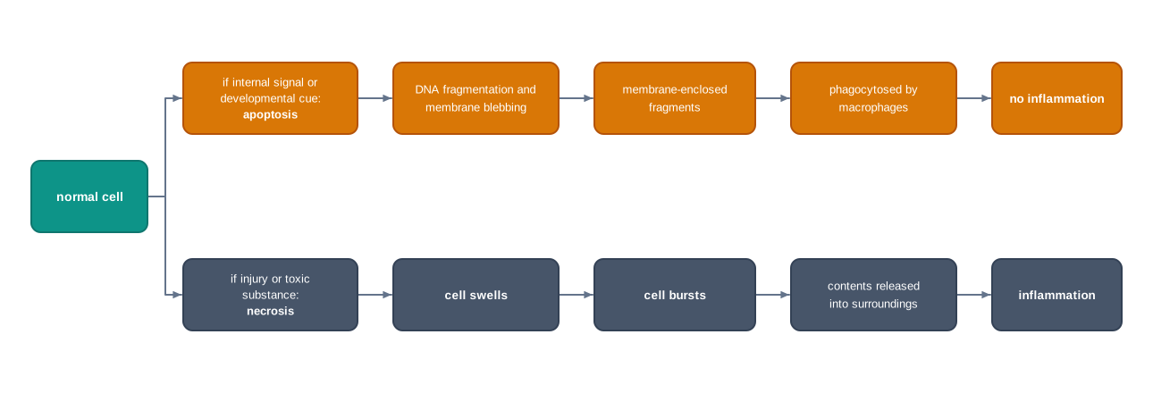
Fig. 5.14 – Two routes to cell death.
How This Section Is Tested
Apoptosis and necrosis are a classic compare-and-contrast pair, and the comparison appears in the NCERT exercises itself. Beyond that, expect assertion–reason items linking apoptosis to cancer — a common one pairs the statement that apoptosis protects against cancer with a reason about unregulated cell division — and case-based passages describing a tissue sample with either swollen, burst cells or shrunken, fragmented ones, asking you to identify which type of cell death has occurred and to justify the answer from the evidence given.
5.5 Cell Differentiation
You are aware that a multicellular organism consists of trillions of cells having exactly the same DNA. Yet, it is observed that a group of cells is different from another group of cells in terms of their structural and functional roles.
For instance, a neuron is specialised to conduct impulses, whereas RBCs perform the function of oxygen transport. Their shapes, their contents and their behaviour have almost nothing in common, and yet the instructions inside them are identical.
How are these distinctions created? The answer lies in the way genes are expressed in different cells. In other words, the unique combination of genes which are differentially expressed at a given time in different cells dictates their structure and function.
Think of the genome as a large book of instructions given to every cell. No cell reads the whole book. A nerve cell reads one set of chapters, a red blood cell reads another, and it is the selection of chapters read, not the book itself, that makes the two cells different.
From Unspecialised to Specialised
As development of an embryo proceeds, the unspecialised cells begin to acquire specific properties and gradually become specialised. The process by which an unspecialised cell becomes specialised, or differentiated, is known as cell differentiation.
A few examples of differentiated cells are epithelial cells, RBCs, WBCs, cardiac cells, neurons and muscle cells.
Stem Cells
It is important to note that not all of our body cells are differentiated. Some populations of unspecialised or undifferentiated cells, also known as stem cells, are found to be present in organisms even after attaining adulthood.
Stem cells are unspecialised cells which divide to produce two daughter cells, one of which remains a stem cell and the other one becomes differentiated. This arrangement is worth noticing, because it solves two problems at once — the tissue gets a new specialised cell, and the supply of stem cells is not used up.
The major sources of stem cells are embryos and adult tissues, the latter being called adult stem cells.
Embryonic Stem Cells
The embryonic stem cells can be derived from embryos which are in the early phase of development; usually a week old blastocyst is preferred. A blastocyst is the hollow ball of cells formed a few days after fertilisation, with a cluster of cells inside it called the inner cell mass.
It is a common practice to extract the inner cell mass from the blastocyst and culture the cells in a culture dish containing essential nutrients. In the total absence of any necessary stimulation to differentiate, these cells begin to divide while retaining the ability to differentiate into any type of cell.
That last condition is the key to the whole procedure. The cells stay unspecialised only as long as nothing instructs them to specialise, so the culture conditions must be kept free of differentiation signals if the cells are to be maintained as stem cells.
Adult Stem Cells
The adult stem cells, on the other hand, can be found in various tissues throughout the body, for example bone marrow, blood vessels, brain, skeletal muscles and liver.
In these tissues, the adult stem cells remain in a non-dividing, or quiescent, state until they are finally activated due to tissue injury. They are a reserve, held back until the tissue needs repair.
Adult stem cells are already used in routine medical treatment. In a bone marrow transplant, blood-forming stem cells from a healthy donor are given to a patient whose own marrow has failed or has been destroyed, as in leukaemia or thalassaemia. Once established in the recipient’s marrow, these cells go on producing all the types of blood cell for the rest of the patient’s life.
What Controls Differentiation
Cellular differentiation is controlled by major molecular processes involving signalling. The principal signalling molecules which aid in communication from one cell to another in the process of differentiation are known as growth factors.
You have already met the machinery involved in section 5.1. A growth factor is a ligand; it acts on cells that carry the matching receptor, and the conformational change in that receptor sets off the relay that ultimately alters which genes the cell expresses. Differentiation, in other words, is cell signalling with a change of identity as its outcome.
Cell Potency
The ability of stem cells to differentiate into other cell types is known as cell potency. Since stem cells can give rise to any type of cell, they are considered to be the most potent.
We can classify them on the basis of their potential to differentiate into 3 types.
1. Totipotent Cells
A totipotent cell is able to differentiate into all possible cell types in an organism. This ability of the cell is referred to as totipotency. Totipotent cells, thus, exhibit the highest differentiation potential.
Unlike pluripotent cells, totipotent cells can give rise to embryonic as well as extraembryonic cells. Examples of totipotent cells include a zygote formed after fertilisation, shown in Fig. 5.15, and asexual spores.
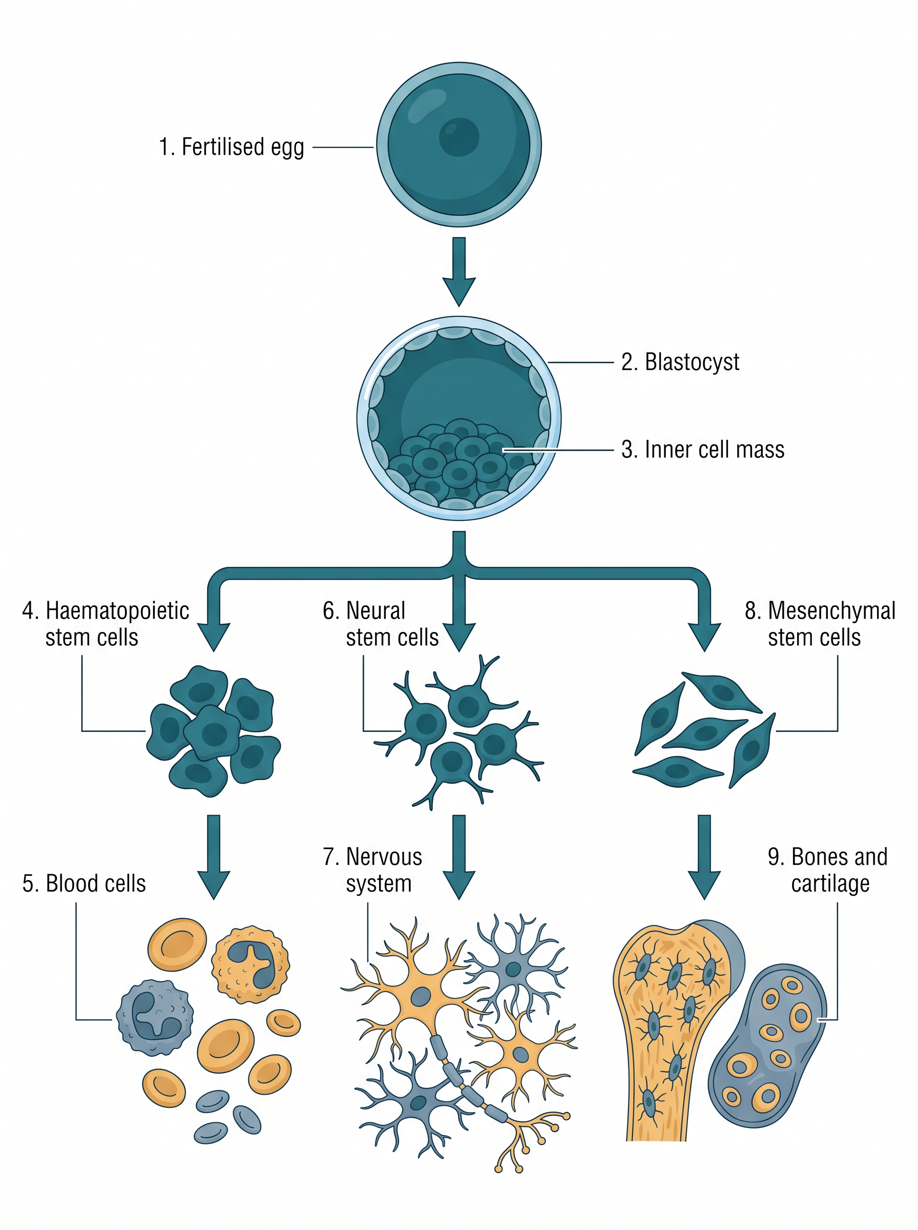
Fig. 5.15 – Differentiation of cells from a fertilised egg.
Totipotency is not limited to animals, and in plants it is put to direct practical use. A single cell taken from a carrot or a banana plant can, under suitable culture conditions, regenerate an entire plant — which is the basis of the tissue culture methods used to multiply disease-free planting material on a commercial scale.
2. Pluripotent Cells
These cells do not exhibit full potency, in the sense that they can get differentiated into most tissues of the body excepting extraembryonic tissues such as placenta or yolk sacs. Their differentiation potential is, thus, less than that of totipotent cells.
An example of pluripotent stem cells includes the cells derived from the inner cell mass. They essentially give rise to three germ layers, namely endoderm, mesoderm and ectoderm. These can further differentiate into most of the body tissues and organs.
3. Multipotent Cells
These cells have a limited range of tissues into which they can differentiate. Therefore, they have an even lower cell differentiation potential.
An excellent example of this cell type is the haematopoetic stem cells (HSCs). HSCs differentiate into a large number of blood cells including red blood cells (RBCs), white blood cells (WBCs) and platelets. Notice the limit, though — a haematopoietic stem cell produces many kinds of blood cell, but it will not produce a neuron or a liver cell.
Read the three types as a descending sequence: totipotent cells can make everything including the supporting tissues of the embryo, pluripotent cells can make the body but not those supporting tissues, and multipotent cells can make only one family of related cell types.
How This Topic Is Tested
This section is a favourite for comparison and reasoning questions. The NCERT exercises themselves ask you to compare totipotent and pluripotent cells and to explain how stem cells differ from blood cells in terms of potency. Expect assertion–reason items pairing a statement about all cells having the same DNA with a reason about gene expression, and case-based passages describing a stem cell line in culture and asking which potency class it belongs to, judged from the cell types it can produce. Diagram-based questions usually draw on Fig. 5.15.
5.6 Cell Migration
Cell migration refers to the movement of a cell, or of cells, from one place to another. It occurs in unicellular organisms like amoeba as well as in multicellular organisms like mammals, and in very different environments — from natural soil to experimental set-ups such as Petri dishes.
Cell migration occurs in an organism under different conditions, such as during embryogenesis to create new layers, during organogenesis and regeneration to generate different organs, for feeding requirements, and in immune responses.
Depending on the type of cell and the various conditions, there are different modes of cell migration. Cells can move as a single unit or in groups.
Cell migration varies with different intrinsic factors of a cell, such as the organisation of the cytoskeleton, that is, whether it is highly organised or not, the extracellular matrix, adhesion strength, and the migratory signals.
How a Cell Moves Without Legs
Cell migration involves several stages of highly coordinated and integrated signalling networks of events. These processes comprise polarisation, which means spatial differences in shape, structure and function within a cell, protrusion and adhesion, translocation of the cell body, and retraction of the rear, as shown in Fig. 5.16.
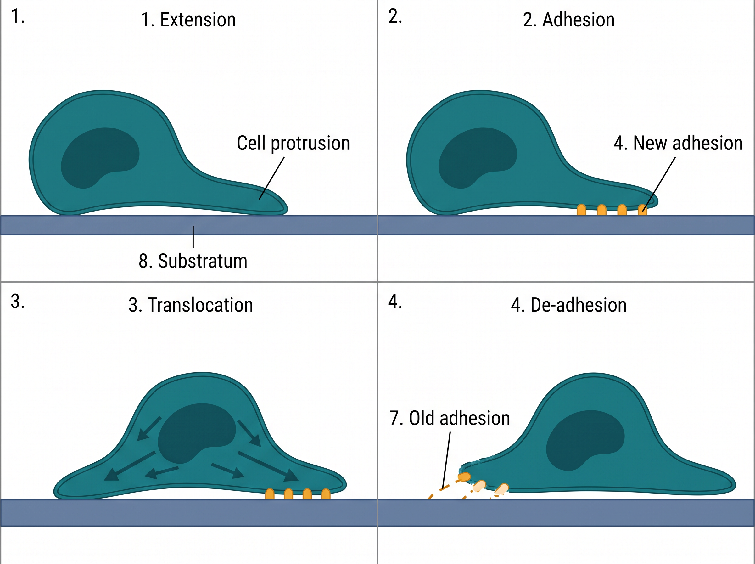
Fig. 5.16 – The four steps of cell migration.
Before reading the stages one by one, look at Fig. 5.16 as a whole. The cell reaches forward, grips at the front, hauls its body up to the new grip, and then lets go at the back. It is very close to the way a person climbs a rock face, and the order of the four moves cannot be changed.
- Foremost, cells develop polarisation by means of specialisation of the plasma membrane and the cell cortex. The polarity is reinforced by, and often even arises from, environments that offer a directional signal.
These directional signals can be chemical-based, electric field-based, mechanical force-based, or based on concentrations of the substrate. The end result is a definite cell front and a rear.
Without this step, movement would be pointless. A cell that pushed out equally in every direction would change shape but stay where it was; polarity is what turns activity into displacement.
During cell migration, the branching and polymerisation of actin filaments facilitate the extension of the leading edge. Actin subunits are added to the growing ends of the filaments just beneath the membrane at the front, and the lengthening filaments push the membrane outwards.
- To create a leading edge of the cell, protrusions — which are membrane extensions in the direction of cell migration, like pseudopodia — are required to be formed and extended.
This comprises three chief components: the spreading out of the plasma membrane, the development of a core backbone that holds the expanded plasma membrane, and the setting up of contact with the substratum, which provides a foothold for the migration of the cell body and supplies signals or cues that control actin polymerisation.
These protrusions attach to the substratum across which the cell is moving. Eventually, the lagging edge of the cell then separates from the substratum and is pulled back into the cell body.
Adhesions are sites of molecular communication between the cell and the substrate. They assemble and disassemble in reaction to extracellular signals and regulate cell migration.
During cell migration, adhesions assemble at the leading edge and disassemble at the trailing edge. However, in protruding areas of cells, as new adhesions form, they can disassemble, or they can stabilise and develop into more fully grown, well-built adhesions.
Hence, the extremely motile cells, particularly those in vivo, do not have the well-built adhesions that characterise less motile cells. The relationship is worth stating plainly: the firmer the grip, the slower the cell. A cell that anchors itself too strongly cannot release quickly enough to keep moving.
- For cells to move forward, adhesions at the rear end are required to disassemble and the trailing edge to retract, or else under tension the cell will be ripped apart.
For this, the termination of the adhesive contacts at the rear edge is attained through downregulation by phosphorylation through phosphatases, or merely by proteolysis of the adhesive contacts by protease enzymes.
Notice what would go wrong if this last step failed. The front of the cell would keep pulling forward while the back stayed anchored, and the cell would be stretched until it tore. Releasing at the rear is not a finishing touch — it is a requirement for survival during movement.
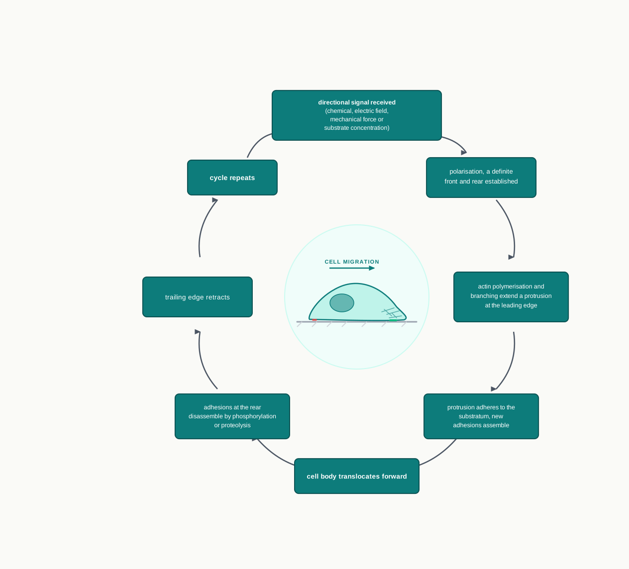
Fig. 5.17 – The cell migration cycle.
Role of Cell Migration
- Cell migration plays a fundamental role in the origin and development of the morphological characteristics of embryos during gastrulation, which is the formation of the embryonic layers in the embryo by migration of a large number of cells — endoderm, mesoderm and ectoderm — as well as during organogenesis, the formation of organs and tissues.
These migratory cells reach their ultimate destinations and undergo differentiation and produce the different organs and limbs. Migration and differentiation therefore work together: a cell must arrive at the right place before the signals available at that place can tell it what to become.
- Cell migration is a key element for maintaining homeostasis, which is the ability to maintain a steady internal environment in response to environmental changes of an individual.
For example, tissue regeneration and repair, and inflammation during injury or infection, are important events of homeostasis. Inflammation involves the migration of immune cells from the lymph nodes to the circulation, where they remain alert until an inflammatory reaction is triggered, which leads them to respond to injury or infection.
Therefore, failure of cells to migrate can cause severe life-threatening defects, for example autoimmune diseases and defective wound repair. A cut that will not heal and an immune system that cannot deliver its cells to the right place are, at the cellular level, the same kind of failure.
- Cell migration also plays an important role in metastasis, that is, the spread of cancer from one place to another. Due to the loss of cell-cell interactions and increased cell motility, the cancer cells develop invasive properties and migrate and spread from the primary site of tumour growth.
This explains why metastasis, rather than the original tumour, is what makes cancer so dangerous. A tumour confined to one site can often be removed surgically; cells that have already migrated cannot be found and removed in the same way. The same capacity for movement that builds an embryo and heals a wound, turned loose without control, is what allows a cancer to spread.
How This Topic Is Tested
Cell migration is usually tested through sequencing and application. Expect questions asking you to arrange the steps of migration in the correct order, or to label the four panels of Fig. 5.16. A frequent reasoning question asks what would happen if the adhesions at the rear failed to disassemble. Case-based passages often describe either a wound that heals poorly or a tumour that has spread to a second organ, and ask you to connect the observation to the cellular process behind it.
5.7 Summary
All cells respond to signals in their environment through specific receptors, which can either be located on the cell surface or be present inside the cytoplasm or the nucleus. A ligand is a chemical messenger which is specific to a receptor. Once bound to its ligand, the receptor undergoes conformational changes, allowing the signal to be interpreted and relayed inside the cell.
There are three types of signalling — paracrine signalling, over short distances between neighbouring cells; autocrine signalling, in which the cell responds to a ligand it has itself secreted; and endocrine signalling, in which the ligand travels through the bloodstream to a distant target cell.
Living organisms take up and utilise free energy through metabolism. Metabolic pathways are of two classes: anabolic pathways, which build complex molecules and consume energy, and catabolic pathways, which break molecules down and release energy.
Glucose acts as an important metabolic fuel in animals. The conversion of glucose into pyruvate occurs through glycolysis, a ten-step pathway present in the cytosol of all living cells, with a net gain of two ATP and two NADH per glucose.
Under anaerobic conditions, pyruvate is converted to lactate in homolactic fermentation, or to ethanol and CO₂ in alcoholic fermentation. The purpose of fermentation is to regenerate NAD⁺ so that glycolysis can continue.
Under aerobic conditions, pyruvate enters the mitochondrial matrix, is converted into acetyl CoA, and is oxidised through the citric acid cycle. The final products of the citric acid cycle are CO₂ and H₂O. The cycle is the final common pathway for the oxidation of carbohydrates, lipids and proteins.
The reduced coenzymes NADH+H⁺ and FADH₂ are oxidised through the electron transport chain in the inner mitochondrial membrane. The proton gradient created drives ATP synthesis through ATP synthase, a process called oxidative phosphorylation, with oxygen acting as the final electron acceptor.
Oxidation of fatty acids to acetyl CoA occurs through the β-oxidation pathway. Acetyl CoA may be oxidised in the citric acid cycle, used to make cholesterol and other steroids, or used to synthesise ketone bodies as an alternative fuel during prolonged fasting.
Transamination of amino acids involves the transfer of the amino nitrogen from an amino acid to a carbon skeleton to form other amino acids. Deamination involves excretion of the amino nitrogen as urea.
Photosynthesis is the process by which light energy gets converted into chemical energy. Photosynthesis occurs inside the chloroplast in plants and some photosynthetic bacteria.
During the light reactions, solar energy is captured by PSII and PSI, splitting of water occurs along with the release of oxygen, and ATP and NADPH+H⁺ are formed. When electrons flow from water to NADP⁺ the process is non-cyclic photophosphorylation; when they return to PSI, only ATP is made, and the process is cyclic photophosphorylation.
Dark reactions, or the Calvin cycle, involve assimilation of carbon into trioses and hexoses, leading to the synthesis of sugar. The cycle runs in three stages — fixation of CO₂ by RuBisCO, reduction using ATP and NADPH+H⁺, and regeneration of the acceptor ribulose 1,5-bisphosphate.
Some plants, such as maize, sorghum and sugarcane, follow the C4 pathway, in which CO₂ is first fixed by PEP carboxylase in mesophyll cells and released again in bundle sheath cells for the Calvin cycle. Their leaves show Kranz anatomy.
Cell division gives rise to two daughter cells which carry the same genetic makeup as the parent cell. Interphase and mitotic phase are the important phases of the cell cycle. Interphase is further divided into G1 phase, S phase and G2 phase. During interphase, the cell grows and replicates its genetic content.
M phase in mitosis consists of four stages: prophase, metaphase, anaphase and telophase. Cells that stop dividing leave the cycle at G1 and enter the quiescent stage, G0.
Unlike mitosis, meiosis has a single interphase which is followed by two nuclear divisions, named Meiosis I (reductional division) and Meiosis II (equational division). Prophase I passes through leptotene, zygotene, pachytene, diplotene and diakinesis, and crossing over during pachytene is a major source of variation.
M-phase is followed by cytokinesis, the division of the cytoplasm. Animal cells divide by a contractile ring of actin filaments, while plant cells form a cell plate that becomes the new cell wall.
Apoptosis refers to programmed cell death, which occurs as a part of an organism’s growth. It is a normal and highly regulated process. Necrosis, in contrast, is the unregulated death of cells following injury, and it causes inflammation.
The process by which an unspecialised cell achieves a specialised state is referred to as cell differentiation. It arises from the particular combination of genes expressed in a cell, not from any difference in the DNA itself.
Stem cells are undifferentiated and unspecialised cells. The ability of stem cells to become differentiated into other cell types is known as cell potency. Stem cells are characterised into three types depending on their potency: totipotent cells, pluripotent cells and multipotent cells.
Cell migration is the movement of a cell from one place to another. It is important at various stages of development of a living organism, such as embryogenesis, organogenesis, regeneration and immune responses, and its uncontrolled form underlies metastasis in cancer.
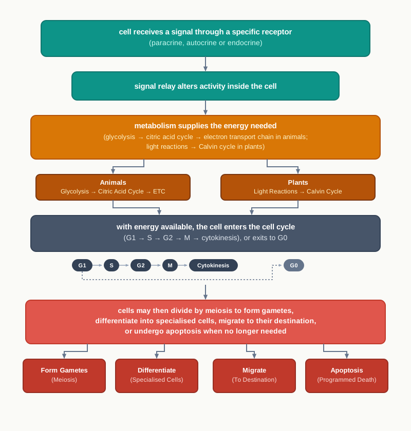
Fig. 5.18 – Summary of cellular processes and fates.
5.8 Key Terms
Receptor — A protein located on the cell surface or inside the cytoplasm or nucleus that binds a specific chemical messenger and allows the cell to respond to it.
Ligand — The chemical messenger to which a particular receptor responds, the binding between the two being highly specific.
Conformational change — The change in the three-dimensional shape of a receptor protein on binding its ligand, which starts the message relay inside the cell.
Paracrine signalling — Signalling over short distances, in which a chemical released into the extracellular space by one cell is sensed almost instantly by neighbouring cells.
Autocrine signalling — Signalling in which a cell secretes a ligand and also possesses the receptors specific for that same ligand.
Endocrine signalling — Long-distance signalling in which the ligand is released into the bloodstream and travels to a distant target cell.
Growth factor — A signalling molecule that carries messages between cells and controls their proliferation and differentiation.
Metabolism — The total set of interlinked, enzyme-catalysed reactions by which an organism takes in and uses free energy for its life processes.
Phototrophs — Photosynthetic organisms that use the energy of sunlight to convert simple molecules into energy-rich complex molecules.
Heterotrophs — Organisms such as animals that obtain energy indirectly from plants through their food.
Chemotrophs — Organisms that obtain energy by oxidising organic or inorganic chemical compounds.
Exergonic reaction — A reaction that releases free energy.
Endergonic reaction — A reaction that consumes free energy.
ATP — The energy currency of the cell, the common form in which energy released by catabolism is held and later spent.
Metabolic pathway — The ordered series of enzyme-catalysed steps by which one biomolecule is built up from, or broken down into, another.
Anabolic pathway — An energy-consuming pathway in which larger, complex molecules are synthesised from small ones.
Catabolic pathway — An energy-releasing pathway in which larger molecules are broken down, producing reducing equivalents and ATP.
Reducing equivalent — A carrier such as NADH or FADH₂ that holds high-energy electrons removed from a fuel molecule and later passes them on.
Glycolysis — The ten-step cytosolic pathway, found in all living cells, that converts one glucose molecule into two pyruvate molecules with a net gain of two ATP.
Gluconeogenesis — The formation of glucose from non-carbohydrate precursors such as lactic acid, amino acids and glycerol.
Glycogenesis — The synthesis of glycogen.
Glycogenolysis — The breakdown of glycogen.
Pentose phosphate pathway — A pathway branching from glucose metabolism that supplies NADPH for fatty acid synthesis and ribose for nucleotide and nucleic acid synthesis.
Allosteric inhibition — Reduction of an enzyme’s catalytic activity caused by a molecule binding at a site other than the active site.
Isoenzyme (isozyme) — One of several different forms of an enzyme that catalyse the same reaction, such as glucokinase in liver cells.
Substrate-level phosphorylation — Formation of ATP by direct transfer of a phosphate group from a substrate molecule to ADP.
Fermentation — The anaerobic extension of glycolysis in which pyruvate is reduced, regenerating the NAD⁺ needed for glycolysis to continue.
Homolactic fermentation — The anaerobic reduction of pyruvate to lactate by lactate dehydrogenase.
Alcoholic fermentation — The two-step anaerobic conversion of pyruvate into ethanol and CO₂ in yeast.
Pyruvate dehydrogenase complex (PDH) — The mitochondrial multienzyme complex that irreversibly converts pyruvate into acetyl CoA, linking glycolysis to the citric acid cycle.
β-oxidation — The pathway by which fatty acids are oxidised to acetyl CoA.
Ketone bodies — Acetone, acetoacetate and 3-hydroxy butyrate, made from acetyl CoA and used as an alternative fuel during prolonged fasting.
Transamination — Transfer of the amino nitrogen from an amino acid to a carbon skeleton, forming a different amino acid.
Deamination — Removal of the amino nitrogen from an amino acid, after which it is excreted as urea.
Essential amino acids — Amino acids that must be supplied in the diet because the body cannot synthesise them.
Non-essential amino acids — Amino acids that the body can synthesise from metabolic intermediates by transamination.
Citric acid cycle — The mitochondrial cycle, also called Kreb’s cycle or the TCA cycle, that oxidises the acetyl group of acetyl CoA and is the final common pathway for the oxidation of carbohydrates, lipids and proteins.
Electron transport chain (ETC) — The series of four protein complexes in the inner mitochondrial membrane through which reduced coenzymes are oxidised and protons are pumped.
Ubiquinone (coenzyme Q) — A hydrophobic quinone that carries electrons from complex I to complex III within the inner mitochondrial membrane.
Oxidative phosphorylation — The synthesis of ATP driven by protons flowing back across the inner mitochondrial membrane after electron transport has created the gradient.
Proton motive force (pmf) — The energy stored in the uneven distribution of protons across a membrane, consisting of a pH gradient and an electrical potential.
ATP synthase — The enzyme complex through which protons return down their gradient, synthesising ATP from ADP and inorganic phosphate.
Photosynthesis — The process in which light energy is trapped and converted into chemical energy, taking place in chloroplasts in plants and some photosynthetic bacteria.
Light reactions — The photochemical phase of photosynthesis in the thylakoid membranes, which captures solar energy, splits water and forms ATP and NADPH+H⁺.
Photosystem I (PSI) — The light-harvesting protein complex that responds to light of wavelength 700 nm.
Photosystem II (PSII) — The light-harvesting protein complex whose chlorophyll a responds to light of wavelength 680 nm.
Oxygen evolving complex — The protein complex on the luminal surface of the thylakoid membrane that catalyses the splitting of water.
Non-cyclic photophosphorylation — Light-driven ATP synthesis in which electrons travel from water through PSII and PSI to NADP⁺, also producing NADPH+H⁺ and O₂.
Cyclic photophosphorylation — Light-driven ATP synthesis in which electrons excited in PSI return to PSI, producing only ATP.
Calvin cycle (dark reactions) — The cyclic pathway in the stroma that fixes CO₂ and reduces it to sugar using the ATP and NADPH+H⁺ made in the light reactions.
CO₂ fixation (assimilation) — The conversion of gaseous carbon dioxide into stable organic molecules such as trioses and hexoses.
RuBisCO — Ribulose 1,5-bisphosphate carboxylase/oxygenase, the enzyme that fixes CO₂ onto ribulose 1,5-bisphosphate and possesses both carboxylase and oxygenase activity.
C3 pathway — The basic photosynthetic pathway, common to all plants, in which the first stable product is the three-carbon 3-phosphoglycerate.
C4 pathway — The pathway of plants such as maize, sorghum and sugarcane, in which CO₂ is first fixed by PEP carboxylase into the four-carbon oxaloacetate.
Kranz anatomy — The leaf structure of C4 plants in which mesophyll cells form a ring around bundle sheath cells that contain more chloroplasts.
Cell cycle — The ordered series of events, made up of interphase and M phase, through which a cell grows, replicates its DNA and divides.
Interphase — The period between two successive mitotic phases, comprising G1, S and G2, during which the cell grows and replicates its DNA.
G1 phase — The first gap phase, in which the cell is metabolically active and grows but does not replicate its genetic content.
S phase — The synthesis phase, in which nuclear DNA doubles from 2C to 4C and the centriole is duplicated, while the chromosome number stays unchanged.
G2 phase — The second gap phase, in which more proteins and organelles are synthesised and the cell prepares to enter M phase.
Quiescent stage (G0) — The inactive state entered by cells that leave the cell cycle at G1 and no longer divide, such as cardiac cells and neurons.
Centriole — The microtubule-organising structure, duplicated during S phase, that is required for proper segregation of chromosomes.
Karyokinesis — The division of the nucleus, made up of prophase, metaphase, anaphase and telophase.
Equational division — A division in which the daughter cells receive the same number of chromosomes as the parent cell.
Chromosome — The condensed form of chromatin, visible as a distinct body once the cell enters prophase.
Mitotic spindle — The structure of fibres, assembled as the centrioles move to opposite poles, along which chromosomes are moved.
Centromere — The region holding the two sister chromatids of a chromosome together.
Kinetochore — The small disc-shaped structure on the surface of the centromere that serves as the site of attachment of a chromosome to the spindle fibers.
Metaphase plate — The plane at the equator of the cell along which the chromosomes align during metaphase.
Cytokinesis — The physical division of the cytoplasm that separates a parent cell into two daughter cells.
Cell plate — The precursor of the new cell wall, formed during cytokinesis in plant cells.
Meiosis — The division producing four haploid cells from a diploid cell through a single interphase followed by two nuclear divisions.
Reductional division — A division, namely meiosis I, in which the chromosome number of the daughter cells is halved.
Synaptonemal complex — The structure assembled during zygotene that facilitates the pairing of homologous chromosomes.
Bivalent (tetrad) — The structure formed by a pair of synapsed homologous chromosomes.
Crossing over — The exchange of genetic material between non-sister chromatids of homologous chromosomes, producing recombination and variation.
Chiasmata — The crossover sites at which homologous chromosomes remain joined after the synaptonemal complex degenerates in diplotene.
Interkinesis — The short gap between the two meiotic divisions, during which no DNA replication takes place.
Apoptosis — Programmed cell death, a highly controlled, energy-dependent and irreversible process by which a cell dies during development.
Necrosis — The unplanned, traumatic death of cells following injury or toxic exposure, which releases the cell contents and causes inflammation.
Cell differentiation — The process by which an unspecialised cell acquires specific properties and becomes specialised, according to the genes expressed in it.
Stem cells — Unspecialised cells that divide to give two daughter cells, one remaining a stem cell and the other becoming differentiated.
Blastocyst — The early embryonic stage, about a week old, from which the inner cell mass is extracted to obtain embryonic stem cells.
Inner cell mass — The cluster of cells within the blastocyst that gives rise to pluripotent stem cells and to the three germ layers.
Adult stem cells — Stem cells present in tissues such as bone marrow, blood vessels, brain, skeletal muscles and liver, which stay quiescent until activated by tissue injury.
Cell potency — The ability of a stem cell to differentiate into other cell types.
Totipotent cells — Cells with the highest differentiation potential, able to form all cell types of the organism, including extraembryonic cells.
Pluripotent cells — Cells that can differentiate into most tissues of the body but not into extraembryonic tissues such as placenta or yolk sac.
Multipotent cells — Cells that can differentiate into only a limited range of related tissues, such as haematopoietic stem cells forming the various blood cells.
Induced Pluripotent Stem Cells (iPSC) — Pluripotent cells produced from adult tissue cells by expressing specific transcription factors, avoiding the need for embryos.
Cell migration — The movement of a cell or group of cells from one place to another, brought about by polarisation, protrusion, adhesion, translocation and retraction.
Polarisation — The establishment of spatial differences in shape, structure and function within a cell, giving it a definite front and rear.
Protrusion — A membrane extension, such as a pseudopodium, pushed out in the direction of migration by the polymerisation of actin filaments.
Adhesions — Sites of molecular communication between a cell and its substrate, which assemble at the leading edge and disassemble at the trailing edge.
Gastrulation — The formation of the embryonic layers — endoderm, mesoderm and ectoderm — by the migration of large numbers of cells.
Organogenesis — The formation of organs and tissues by migratory cells that reach their destinations and differentiate.
Homeostasis — The ability to maintain a steady internal environment in response to environmental changes.
Metastasis — The spread of cancer cells from the primary tumour site to other parts of the body, resulting from loss of cell-cell interactions and increased motility.
5.9 Points to Ponder
A cell can respond only to those signals for which it carries a receptor. What would happen to an organism if one of its tissues lost the receptor for a single hormone, while the hormone itself continued to be made normally?
Cancer cells make their own growth factors and also carry the receptors for them. Think about why a loop that begins and ends in the same cell is so much harder for the body to control than a signal sent between two different cells.
Catabolism releases energy and anabolism consumes it. Why does a living cell need to run both kinds of pathway at the same time, instead of completing one before starting the other?
Glycolysis needs no oxygen and no organelle, and it is found in every living cell that has been examined. What does this suggest about when in the history of life this pathway may have appeared?
Fermentation yields far less energy per glucose molecule than aerobic respiration, yet cells still carry it out. Under what circumstances is a low yield better than no yield at all?
Oxygen takes no part in glycolysis or in the citric acid cycle, and yet both stop within minutes when oxygen runs out. Reflect on what this tells you about how the pathways of a cell depend on one another.
The dark reactions of photosynthesis do not need light directly, but they normally take place in daylight. Is the name “dark reaction” a useful one? What would you call this phase instead, and why?
C4 plants spend extra energy fixing carbon twice over, and are still more efficient than C3 plants. In what kind of climate would you expect that extra expense to be worth paying, and in what kind would it not?
During S phase the amount of DNA in a cell doubles, but the chromosome number stays the same. Explain to yourself, in your own words, how both statements can be true at once.
Heart muscle cells and neurons enter the quiescent stage and rarely divide again. Consider what advantages a body might gain from having such cells stop dividing, given the cost of being unable to replace them after injury.
Meiosis halves the chromosome number, and crossing over reshuffles the genes. Which of these two outcomes would you consider more important for the survival of a species over many generations, and on what grounds?
Apoptosis is described as an energy-dependent process. Why should a cell need to spend ATP in order to die, and what would happen in a tissue if that energy were not available?
Every cell in your body carries the same DNA, yet a liver cell never behaves like a nerve cell even after years of division. What must be passed from a cell to its daughters, besides the DNA sequence itself?
Induced pluripotent stem cells can be made from a patient’s own adult tissue. Think about which of the concerns surrounding embryonic stem cells this technology removes, and which concerns it leaves untouched.
The same ability to migrate that builds an embryo and heals a wound also allows a cancer to spread. Is it possible to design a treatment that blocks the harmful migration without disturbing the useful kind? What would such a treatment have to distinguish between?
5.10 NCERT Questions
Give a comparative account of the following:
- Apoptosis and necrosis
- Autocrine and paracrine signaling
- Anabolic and catabolic pathways
- Totipotent and pluripotent cells
Explain how stem cells are different from blood cells in terms of potency.
How many mitotic divisions will produce 64 cells out of a single cell?
Match the following:
| Column I | Column II |
|---|---|
| a. Centromere | i. Reductional division |
| b. Kinetochore | ii. Holds the two sister chromatids together |
| c. Metaphase | iii. Pairing of homologous chromosomes |
| d. Zygotene | iv. Equational division |
| e. Pachytene | v. Assembly of homologous chromosomes on metaphase plate |
| f. Meiosis I | vi. Site of attachment of chromosomes to spindle fibers |
| g. Meiosis II | vii. Crossing over between homologous chromosomes occurs |
How is cell migration important for the development of an embryo?
What is the significance of citric acid cycle?
The first reaction in glycolysis is catalysed by which of the following enzymes:
- Glucokinase
- Phosphoglycerate kinase
- Phosphofructokinase
- Hexokinase
Which of the following statements regarding cell signaling is NOT true?
- In endocrine signaling, the ligand reaches the target cell through bloodstream after being synthesised in the extracellular space.
- Synaptic signaling is an example of paracrine signaling.
- Ligands bind to receptors in a non-specific manner.
- Cancer cells exhibit autocrine signaling.
Dark reaction in photosynthesis is named so because—
- It occurs independent of light energy
- It occurs in dark
- It cannot occur in daylight
- All of the above
Differentiate between cyclic and non-cyclic photophosphorylation.
Briefly describe Calvin cycle.
5.11 Objective Questions
A receptor may be located in all of the following places EXCEPT:
- On the cell surface
- In the cytoplasm
- In the nucleus
- In the extracellular fluid, freely floating
A cell fails to respond to a hormone circulating in the blood, although the hormone reaches the cell. The most likely reason is that the cell:
- Lacks the receptor for that hormone
- Has too little cytoplasm
- Is in the G0 stage
- Has an inactive nucleus
The type of signalling in which a cell secretes a ligand and also carries receptors for the same ligand is:
- Paracrine signalling
- Autocrine signalling
- Endocrine signalling
- Synaptic signalling only
Which of the following correctly describes an anabolic pathway?
- Exergonic, breaking large molecules into small ones
- Endergonic, building complex molecules from small ones
- Exergonic, producing reducing equivalents and ATP
- Endergonic, releasing CO₂ and H₂O
Glucose-6-phosphate is an intermediate in all of the following EXCEPT:
- Glycogenesis
- Pentose phosphate pathway
- Gluconeogenesis
- β-oxidation of fatty acids
The enzyme that converts glucose-6-phosphate into fructose-6-phosphate is:
- Phosphofructokinase
- Aldolase
- Phosphoglucose isomerase
- Triose phosphate isomerase
Which of the following is NOT one of the three regulatory enzymes of glycolysis?
- Hexokinase
- Phosphofructokinase
- Pyruvate kinase
- Enolase
The enzyme enolase requires which of the following ions for its activity?
- Mg²⁺ or Mn²⁺
- Fe²⁺ or Cu²⁺
- Na⁺ or K⁺
- Ca²⁺ only
The net gain of ATP per molecule of glucose converted to two molecules of pyruvate in glycolysis is:
- One
- Two
- Four
- Six
The coenzyme present in the enzyme pyruvate decarboxylase of alcoholic fermentation is:
- NAD⁺
- FAD
- TPP (thiamine pyrophosphate)
- Lipoamide
Substrate-level phosphorylation in the citric acid cycle takes place during the conversion of:
- Citrate to isocitrate
- Succinyl CoA to succinate
- Succinate to fumarate
- Malate to oxaloacetate
The only enzyme of the citric acid cycle that remains attached to the inner mitochondrial membrane is:
- Citrate synthase
- Fumarase
- Succinate dehydrogenase
- Malate dehydrogenase
Which complex of the electron transport chain does NOT pump protons?
- Complex I
- Complex II
- Complex III
- Complex IV
The number of ATP molecules synthesised per molecule of FADH₂ oxidised is:
- 1.0
- 1.5
- 2.5
- 3.0
In the proton motive force generated across the inner mitochondrial membrane, the matrix side is:
- Basic and negative
- Basic and positive
- Acidic and negative
- Acidic and positive
Chlorophyll a at photosystem II responds to light of wavelength:
- 600 nm
- 680 nm
- 700 nm
- 760 nm
Cyclic photophosphorylation produces:
- ATP, NADPH+H⁺ and O₂
- ATP and NADPH+H⁺ only
- ATP only
- NADPH+H⁺ only
The splitting of water during the light reaction is catalysed by a complex located:
- On the luminal surface of the thylakoid membrane
- In the stroma
- On the outer chloroplast membrane
- In the cytosol
In stage III of the Calvin cycle, the number of ATP molecules used to convert three molecules of ribulose-5-phosphate into ribulose-1,5-bisphosphate is:
- Two
- Three
- Six
- Nine
In C4 plants, the first acceptor of CO₂ and the cell in which it acts are:
- RuBisCO in bundle sheath cells
- RuBisCO in mesophyll cells
- PEP carboxylase in mesophyll cells
- PEP carboxylase in bundle sheath cells
At the end of S phase, a diploid cell that began with a DNA content of 2C has:
- 2C DNA and double the chromosome number
- 4C DNA and double the chromosome number
- 4C DNA and an unchanged chromosome number
- 2C DNA and an unchanged chromosome number
Cardiac cells and neurons are examples of cells that:
- Divide continuously throughout life
- Have exited the cell cycle at G1 into the quiescent stage
- Remain permanently in S phase
- Undergo meiosis instead of mitosis
The kinetochore is best described as:
- The plane along which chromosomes align at metaphase
- A disc-shaped structure on the surface of the centromere
- The structure that holds sister chromatids together
- A microtubule-organising centre at the poles
The synaptonemal complex is assembled during which substage of prophase I?
- Leptotene
- Zygotene
- Pachytene
- Diakinesis
During anaphase I of meiosis:
- Sister chromatids separate and move to opposite poles
- Homologous chromosomes separate while sister chromatids stay attached at their centromeres
- Chromosomes align at the equatorial plate
- The nuclear envelope reforms around each set of chromosomes
Which feature is present in apoptosis but absent in necrosis?
- Release of cell contents into the surroundings
- Inflammation of the surrounding tissue
- DNA fragmentation and membrane blebbing
- Swelling and bursting of the cell
Haematopoietic stem cells are an example of:
- Totipotent cells
- Pluripotent cells
- Multipotent cells
- Fully differentiated cells
The main advantage of induced pluripotent stem cell (iPSC) technology over embryonic stem cells is that it:
- Produces cells with greater potency than totipotent cells
- Avoids the need to obtain embryos and allows patient-matched cells
- Removes the need for any culture medium
- Produces cells that never divide
Arrange the steps of cell migration in the correct order:
- Adhesion → polarisation → translocation → retraction of the rear
- Polarisation → protrusion and adhesion → translocation of the cell body → retraction of the rear
- Translocation → polarisation → protrusion → adhesion
- Protrusion → retraction of the rear → adhesion → translocation
Assertion (A): A hormone released into the bloodstream acts only on certain cells of the body. Reason (R): Only some cells possess the receptor specific for that hormone.
Assertion (A): Hexokinase is inhibited by glucose-6-phosphate. Reason (R): Glucose-6-phosphate binds to the active site of hexokinase and competes with glucose.
Assertion (A): Fermentation allows glycolysis to continue in the absence of oxygen. Reason (R): The reduction of pyruvate regenerates NAD⁺, which is required by glyceraldehyde-3-phosphate dehydrogenase.
Assertion (A): The citric acid cycle stops soon after the oxygen supply is cut off. Reason (R): Oxygen is directly used as a substrate in one of the reactions of the citric acid cycle.
Assertion (A): Oxidation of one molecule of FADH₂ yields less ATP than oxidation of one molecule of NADH. Reason (R): Electrons from FADH₂ enter the electron transport chain at QH₂, bypassing one proton-pumping complex.
Assertion (A): The dark reactions of photosynthesis normally take place during the day. Reason (R): The dark reactions depend on the ATP and NADPH+H⁺ supplied by the light reactions.
Assertion (A): No oxygen is released during cyclic photophosphorylation. Reason (R): In cyclic electron flow the electron returns to PSI, so water need not be split to replace it.
Assertion (A): The DNA content of a cell doubles during S phase, but its chromosome number does not change. Reason (R): Each chromosome comes to consist of two sister chromatids joined at the centromere, and is still counted as one chromosome.
Assertion (A): Meiosis is a source of genetic variation among the offspring of sexually reproducing organisms. Reason (R): Crossing over occurs between non-sister chromatids of homologous chromosomes during pachytene.
Assertion (A): Apoptosis does not cause inflammation of the surrounding tissue. Reason (R): The apoptotic cell breaks into membrane-enclosed fragments that are phagocytosed before their contents can escape.
Assertion (A): A highly motile cell does not form the well-built adhesions seen in less motile cells. Reason (R): Adhesions at the rear must disassemble quickly for the cell to move forward without being torn apart.
5.12 Check Your Concepts
A cell is surrounded by dozens of different chemical messengers, yet it responds to only two or three of them. Explain what decides which messages the cell can act on.
Why is a conformational change in the receptor necessary for a signal to be transmitted, even though the ligand itself never enters the cell?
Neurons communicate over very short distances while hormones travel through the whole body. Compare paracrine and endocrine signalling in terms of distance, route, speed and the number of cells affected.
Explain why the autocrine loop of a cancer cell makes its growth much harder for the body to control than the growth of a normal cell that depends on external growth factors.
Catabolism releases energy and anabolism consumes it. Explain, with one example of each, why an organism must run both types of pathway at the same time.
All the enzymes of glycolysis are present in the cytosol, and the pathway requires neither oxygen nor any organelle. What does this tell you about which types of cell can carry out glycolysis?
Arrange the following intermediates of glycolysis in the correct order: 2-phosphoglycerate, glucose-6-phosphate, pyruvate, fructose-1,6-bisphosphate, 1,3-bisphosphoglycerate, phosphoenol pyruvate, fructose-6-phosphate, 3-phosphoglycerate, glyceraldehyde-3-phosphate. Name the enzyme catalysing the final step.
Hexokinase is inhibited by its own product. Explain how this kind of control benefits the cell, and predict what would happen if the inhibition were lost.
Liver cells contain glucokinase in addition to hexokinase. State one way in which the two enzymes differ, and suggest why a liver cell may need an enzyme specific for glucose.
Only three of the ten reactions of glycolysis are used as control points. Explain why irreversible reactions, and not reversible ones, serve as regulatory steps.
Fermentation yields no ATP beyond that already made in glycolysis. Why, then, does a muscle cell working without oxygen still convert pyruvate to lactate?
Compare homolactic fermentation and alcoholic fermentation with respect to end products, enzymes involved, release of CO₂ and the organisms in which each occurs.
The reaction catalysed by the pyruvate dehydrogenase complex is irreversible. Use this fact to explain why acetyl CoA obtained from fatty acids cannot be used by animals to make glucose.
The citric acid cycle is called the final common pathway for the oxidation of carbohydrates, lipids and proteins. Justify this description using the entry points of each of the three types of fuel.
Only one molecule of GTP is formed directly in one turn of the citric acid cycle, yet the cycle is described as the main energy-harvesting stage of respiration. Resolve this apparent contradiction.
Succinate dehydrogenase is the only citric acid cycle enzyme attached to the inner mitochondrial membrane. Explain why its position suits its function.
Draw and label a diagram of the citric acid cycle showing all the intermediates, and mark clearly the two steps at which CO₂ is released and the four steps at which a reduced coenzyme is formed.
Arrange the following components of the electron transport chain in the order in which electrons pass through them: cytochrome c, complex I, oxygen, complex III, ubiquinone, complex IV. State separately where electrons from FADH₂ enter this sequence.
Explain why the oxidation of NADH yields 2.5 ATP while the oxidation of FADH₂ yields only 1.5 ATP.
A chemical is added to isolated mitochondria that makes the inner membrane freely permeable to protons, without damaging any of the four complexes. Predict what will happen to electron transport and to ATP synthesis, and explain your reasoning.
Oxygen is not used in glycolysis or in the citric acid cycle, yet both stop when oxygen runs out. Explain the connection.
Match the following:
| Column I | Column II |
|---|---|
| a. Hexokinase | i. Splitting of water in the thylakoid lumen |
| b. RuBisCO | ii. Reduces O₂ to H₂O |
| c. ATP synthase | iii. Phosphorylation of glucose |
| d. Oxygen evolving complex | iv. Fixation of CO₂ onto ribulose 1,5-bisphosphate |
| e. Cytochrome c oxidase | v. Synthesis of ATP as protons flow down their gradient |
Study Fig. 5.19 again and explain how the transfer of electrons from PSII to PSI results in the accumulation of protons inside the thylakoid lumen.
A plant is illuminated with light of 700 nm only. Predict whether NADPH+H⁺ and oxygen will be produced, and explain your answer in terms of the two photosystems.
Explain why the name “dark reaction” is misleading, and state the evidence from the chapter that supports your explanation.
Six molecules of glyceraldehyde-3-phosphate are formed in stage II of the Calvin cycle, but only one leaves the cycle. Explain what happens to the remaining five and why this step is essential for the cycle to continue.
RuBisCO possesses both carboxylase and oxygenase activity. Use this fact to explain why concentrating CO₂ in the bundle sheath cells makes C4 plants more efficient than C3 plants.
Bundle sheath cells are rich in RuBisCO but lack PEP carboxylase, while mesophyll cells of C4 plants contain PEP carboxylase. Explain how this division of labour makes the C4 pathway possible, and what would happen if PEP carboxylase were also present in the bundle sheath cells.
A cell at the beginning of G1 has 20 chromosomes and a DNA content of 2C. State, with reasons, the chromosome number and DNA content of the cell at the end of S phase, during metaphase, and in each daughter cell after cytokinesis.
Centrioles are duplicated during S phase, along with the DNA. Explain what would go wrong in the following M phase if this duplication failed.
Why must the nuclear membrane disappear during prophase? What would happen to the chromosomes if it remained intact?
At metaphase, the two kinetochores of a chromosome are attached to spindle fibers from opposite poles. Explain how this arrangement guarantees that each daughter cell receives an identical set of chromosomes.
Identify the stage of mitosis represented by each of the panels of Fig. 5.20, and give one observable feature that allowed you to identify each one.
Compare cytokinesis in an animal cell and a plant cell, explaining why the difference between them arises.
Arrange the substages of prophase I in the correct order and state, for each one, the single most important event that takes place: diakinesis, pachytene, leptotene, diplotene, zygotene.
Crossing over occurs during pachytene, but chiasmata become visible only in diplotene. Explain this delay.
Meiosis has a single interphase followed by two nuclear divisions. Explain how the absence of DNA replication during interkinesis makes the four final cells haploid.
Explain why meiosis I is called reductional division while meiosis II is called equational division, referring to what separates at anaphase in each case.
Apoptosis is described as an energy-dependent process. Explain why a dying cell needs to spend ATP, and how this distinguishes apoptosis from necrosis.
A tissue section is examined and the cells are seen to be swollen and burst, with inflammation in the surrounding area. Identify the type of cell death, and justify your answer using two observations from the section.
Explain how a failure of apoptosis can contribute to the formation of cancer, even if the rate of cell division in the tissue is normal.
All cells of the body carry identical DNA, and yet a differentiated cell never behaves like its neighbour of a different type. Explain what makes them different, and describe one piece of evidence from the chapter showing that the genome of a specialised cell is still complete.
A researcher wishes to maintain embryonic stem cells in culture without letting them specialise. What condition must the culture satisfy, and what would happen if growth factors were added to the dish?
Compare embryonic stem cells and adult stem cells with respect to their source, their potency and their state in the tissue before they are activated.
Arrange the following in decreasing order of differentiation potential and justify the order: haematopoietic stem cell, zygote, cell of the inner cell mass.
Explain why cells produced by iPSC technology are unlikely to be rejected when transplanted back into the same patient.
Label the four panels of Fig. 5.16 and describe, in one sentence each, what the cell is doing in each step.
Explain why a cell that forms very strong, well-built adhesions with its substratum is a poorly migrating cell.
What would happen to a migrating cell if the adhesions at its rear failed to disassemble while the leading edge continued to move forward? Explain your answer in terms of the tension developed.
A student sets up a scratch assay but forgets to wash the dish after making the scratch, and also photographs the culture at a different region of the dish at each time point. Identify the two errors and explain how each one would affect the measurement of migration.
Cell migration builds the embryo, heals wounds and also allows cancer to spread. Explain how one and the same cellular ability can be both essential and dangerous, using one example of each.
5.13 Case-Based Questions
A patient has normal levels of a hormone circulating in the blood, but the tissue that this hormone normally acts on shows no response at all. Tests confirm that the hormone is being synthesised correctly and reaches the tissue through the circulation. Other tissues of the body respond normally to their own hormones.
- Suggest the most likely reason why this one tissue fails to respond, even though the hormone reaches it.
- Name the type of signalling described in this passage and state one feature that identifies it.
- Explain why raising the dose of the hormone would probably not correct the problem.
A tumour sample is examined in a laboratory. The tumour cells are found to be producing a growth factor and, on the same cells, receptors specific for that growth factor are also detected. Normal cells of the same tissue produce neither. The cells divide continuously in culture even when no growth factors are added to the medium.
- Identify the type of signalling shown by these tumour cells and justify your choice.
- Explain why these cells continue to divide in a medium containing no added growth factors.
- Suggest one point in this signalling loop at which a drug could act to slow the growth of the tumour, and explain how it would work.
An athlete runs a 400 m race at full speed. Towards the end of the race the oxygen supply to her leg muscles falls well below what the muscles are consuming. Analysis of the muscle tissue immediately after the race shows a rise in lactate. Blood samples taken over the next hour show lactate being carried away from the muscles to the liver.
- Name the pathway operating in the muscle during the last part of the race and the enzyme that produces lactate.
- Explain why the muscle converts pyruvate to lactate rather than allowing pyruvate to accumulate.
- State what happens to the lactate that reaches the liver, and name the process involved.
- Calculate the net ATP yield per glucose molecule in the muscle during this period, and state how it compares with the yield under aerobic conditions.
In an experiment, mitochondria are isolated from liver tissue and supplied with pyruvate, ADP, inorganic phosphate and oxygen. ATP synthesis is measured. In a second set of tubes, a substance is added that blocks the flow of electrons at complex IV. Oxygen consumption stops immediately, and ATP synthesis falls to almost nothing, although complexes I, II and III are undamaged.
- Explain why blocking complex IV stops oxygen consumption.
- Explain why ATP synthesis falls even though ATP synthase itself has not been affected.
- Predict what will happen to the amounts of NADH and FADH₂ inside these mitochondria, and explain the effect this will have on the citric acid cycle.
A crop scientist grows two cereal crops side by side in an open field in a hot region. Crop A is maize and Crop B is a C3 cereal. Over the season, Crop A produces considerably more dry matter per unit area than Crop B, although both receive the same water, nutrients and sunlight. Leaf sections of Crop A show mesophyll cells arranged in a ring around bundle sheath cells that are packed with chloroplasts.
- Name the leaf anatomy seen in Crop A and state its two identifying features.
- Name the first CO₂ acceptor and the first stable product in each of the two crops.
- Explain, in terms of the properties of RuBisCO, why Crop A achieves a higher photosynthetic efficiency.
A cell biologist follows a population of cultured cells through one complete cell cycle. She measures the DNA content of the cells at intervals and finds that it stays constant for several hours, then doubles over the next few hours, then stays constant again before the cells divide. Chromosome counts made just before division show the same number of chromosomes as at the start. A small proportion of the cells in the same dish never show any increase in DNA content at all.
- Identify the three phases of interphase represented by the three parts of the DNA measurement, and state what happens in each.
- Explain why the chromosome number is unchanged even though the DNA content has doubled.
- Suggest what has happened to the small group of cells whose DNA content never increases, and name the stage they are in.
Two tissue samples are taken from a patient. In sample X, the cells are shrunken and have broken up into small membrane-enclosed fragments, many of which have been taken up by nearby macrophages; the surrounding tissue looks normal. In sample Y, the cells are swollen, several have burst, their contents lie in the surrounding tissue, and the area is inflamed. The patient’s history records a crush injury to the region from which sample Y was taken.
- Identify the type of cell death seen in each sample and give two reasons for each identification.
- Explain why inflammation is seen around sample Y but not around sample X.
- State one situation in normal development in which the type of cell death seen in sample X is essential.
A hospital treats a patient whose bone marrow has been destroyed by disease. Cells capable of forming all the types of blood cell are taken from a matched donor and given to the patient. In a separate research programme in the same hospital, scientists working on a heart condition need pluripotent cells to model the disease in culture. They decide to reprogramme skin cells taken from the affected patients rather than obtain cells from early embryos.
- Name the type of stem cell used in the bone marrow treatment and state its potency class, giving a reason.
- Name the technology used in the research programme and explain how it produces pluripotent cells without using embryos.
- State one ethical concern that this choice of technology avoids, and one safeguard that would still apply to research using human cells.
A researcher grows two batches of cultured cells until each forms a continuous sheet, then draws a fine sterile tip across each sheet to make a clean gap. Batch P is grown in ordinary medium. Batch Q is grown in medium containing a compound that interferes with the assembly of adhesions. Photographs taken at 0, 12 and 24 hours show that the gap in Batch P narrows steadily, while the gap in Batch Q has barely changed after 24 hours.
- Name the technique being used and state the principle on which it works.
- Explain, using the steps of cell migration, why the cells of Batch Q fail to close the gap.
- The gap in Batch P measured 900 µm at 0 hours and 270 µm at 24 hours. Calculate the percentage of the gap closed and the average rate of closure in µm per hour.
- Suggest why a compound of the kind used on Batch Q is of interest to cancer researchers.
5.14 Practice With Data
A yeast culture metabolises 120 molecules of glucose to pyruvate by glycolysis. Using the net reaction given in the chapter, calculate (i) the total number of ATP molecules produced, (ii) the number of ATP molecules consumed, (iii) the net ATP gain, and (iv) the number of NADH molecules formed.
During the complete aerobic oxidation of one molecule of glucose, 10 molecules of NADH and 2 molecules of FADH₂ are delivered to the electron transport chain. Taking 2.5 ATP per NADH and 1.5 ATP per FADH₂, calculate (i) the ATP obtained from oxidative phosphorylation alone, and (ii) the percentage of this total that comes from FADH₂.
One turn of the citric acid cycle produces 3 NADH, 1 FADH₂, 1 GTP and 2 CO₂. For the complete oxidation of one molecule of glucose, two turns of the cycle occur. Calculate, for these two turns, (i) the number of CO₂ molecules released, (ii) the number of reduced coenzyme molecules formed, and (iii) the total ATP equivalents, counting GTP as equivalent to 1 ATP and using the values of 2.5 and 1.5 ATP given in the chapter.
A muscle cell working anaerobically produces 2 ATP per glucose, while the same cell working aerobically obtains far more from the same molecule. If the cell needs 900 molecules of ATP for a particular burst of contraction, calculate how many molecules of glucose it must consume anaerobically to meet this requirement, and state how many molecules of lactate will be formed in doing so.
The number of cells produced from a single cell after \(n\) successive mitotic divisions is given by \(N = 2^n\), where \(N\) is the number of cells formed and \(n\) is the number of divisions. Calculate (i) \(N\) when \(n = 7\), (ii) the value of \(n\) required to produce 1024 cells, and (iii) the number of additional divisions needed to go from 64 cells to 256 cells.
A tissue culture is started with 25 cells. Every cell divides once every 20 hours, and no cell dies. Calculate (i) the number of cells present after 80 hours, and (ii) the time required for the culture to reach 400 cells.
A diploid animal cell has 16 chromosomes and a DNA content of 2C at the beginning of G1. Copy and complete the table below.
| Stage | Number of chromosomes | DNA content |
|---|---|---|
| Beginning of G1 | 16 | 2C |
| End of S phase | ||
| Metaphase of mitosis | ||
| Each daughter cell after mitotic cytokinesis | ||
| Each cell at the end of meiosis I | ||
| Each cell at the end of meiosis II |
A population of 60 diploid germ cells in an organism completes meiosis. Calculate (i) the number of haploid cells produced, and (ii) the number of nuclear divisions that took place in total. State also how many rounds of DNA replication occurred.
A cell biologist measures the duration of the phases of the cell cycle in a cultured cell line and obtains the values shown below.
| Phase | Duration (hours) |
|---|---|
| G1 | 10 |
| S | 7 |
| G2 | 4 |
| M | 1 |
Calculate (i) the total duration of one cell cycle, (ii) the percentage of the cycle spent in interphase, and (iii) the percentage of the cycle during which the DNA content of the cell is 4C.
In the Calvin cycle, the fixation of 3 molecules of CO₂ consumes 6 ATP and 6 NADPH+H⁺ in stage II and a further 3 ATP in stage III, and yields a net gain of 1 molecule of glyceraldehyde-3-phosphate. Using these figures, calculate (i) the ATP and NADPH+H⁺ needed per molecule of CO₂ fixed, and (ii) the CO₂, ATP and NADPH+H⁺ needed to produce the 2 molecules of glyceraldehyde-3-phosphate required to build one molecule of glucose.
In the light reaction, two molecules of water are oxidised to generate one molecule of O₂ for every four electrons sent from PSII to PSI, as shown by the reaction \(2\text{H}_2\text{O} \longrightarrow 4\text{H}^+ + 4e^- + \text{O}_2\). Calculate (i) the number of water molecules that must be split to release 18 molecules of O₂, (ii) the number of electrons transferred in doing so, and (iii) the number of protons released into the thylakoid lumen from this splitting.
Using the basic reaction of photosynthesis, \(6\text{CO}_2 + 12\text{H}_2\text{O} \longrightarrow \text{C}_6\text{H}_{12}\text{O}_6 + 6\text{O}_2 + 6\text{H}_2\text{O}\), calculate the number of molecules of CO₂ consumed and of O₂ released when a leaf synthesises 45 molecules of glucose. State also the number of water molecules consumed.
Two cell lines are compared in a scratch assay. The width of the cell-free gap is measured at fixed intervals, as shown below.
| Time (hours) | Gap width in Line A (µm) | Gap width in Line B (µm) |
|---|---|---|
| 0 | 1000 | 1000 |
| 8 | 760 | 920 |
| 16 | 500 | 850 |
| 24 | 250 | 790 |
Calculate (i) the percentage of the gap closed by each line at 24 hours, (ii) the average rate of closure in µm per hour for each line, and (iii) state, with a reason drawn from the data, which line is the more migratory.
- A sample of adipose tissue is analysed during prolonged fasting, and three fates of the acetyl CoA formed by β-oxidation are recorded. Of every 100 molecules of acetyl CoA produced, 62 enter the citric acid cycle, 25 are used to synthesise ketone bodies and the remainder are used for cholesterol synthesis. Calculate (i) the number of molecules used for cholesterol synthesis, (ii) the percentage of acetyl CoA not entering the citric acid cycle, and (iii) the number of CO₂ molecules released by the acetyl CoA that does enter the cycle, given that each turn releases 2 CO₂.