Chapter 7: Basic Processes
This chapter contains a very large number of diagrams (over 40, including flowcharts) and is too long to fully expand in a single pass without risking the response being cut off. I am proactively splitting it here.
Below is Part A, which covers everything from the start of the chapter up through the end of section 7.3.4. It has been fully expanded and temporarily given its own local numbering sequence (Fig. 7.0 to 7.22).
Please reply to continue, and I will process Part B, merge both halves seamlessly, and build the final, correctly numbered master file for the complete chapter.
In 1922, a boy was born in a small village in Punjab, into a family of very modest means where his father worked as a village clerk. There was no laboratory anywhere near him, and very few children in the village went to school at all. That boy was Har Gobind Khorana, and he would grow up to help read the oldest message on Earth — the instructions written inside every living cell.

Khorana studied at Punjab University in Lahore, then worked in Britain, in Canada and finally in the United States. By the early 1960s, biologists knew that DNA carried the instructions for making proteins, but nobody knew how those instructions were written. Khorana approached the problem like a chemist rather than a biologist. He built artificial RNA chains in which the bases repeated in a known, deliberate pattern, added them to cell-free mixtures that could still make protein, and then looked at which amino acids turned up in the chain that came out. Piece by piece, the code gave way.

In 1968 he shared the Nobel Prize in Physiology or Medicine with Marshall Nirenberg and Robert Holley for interpreting the genetic code. Later, he became the first person to build a working gene from scratch in the laboratory.

<img src="images/fig7.0.png" onerror="this.onerror=null; this.src='images/fig7.0.svg';" alt="Fig. 7.0">

::: {.figure-caption}
Fig. 7.0 – The flow of information from DNA to protein.
:::


::: {.callout-note .box-curiosity icon=false title="Curiosity Corner"}

Q1. How did scientists prove that DNA, and not protein, is the material that carries the instructions of life?
Q2. A single human cell holds nearly two metres of DNA. How is all of it packed into a nucleus far too small to see?
Q3. DNA has only four bases, but proteins are built from twenty amino acids. How can four letters spell out twenty different words?
Q4. Every cell in your body carries exactly the same genes, so why does a nerve cell behave so differently from a cell lining your gut?
Q5. Mistakes creep into DNA almost every day. Why, then, are we not riddled with errors?

:::


Keep these five questions in mind as you read. By the end of this chapter, you will be able to answer every one of them yourself.

## 7.1 DNA as the Genetic Material


In the previous chapter you studied that characters or traits pass from parents to offspring through **genes**. You also know that these genes sit on **chromosomes**, and that chromosomes are made of two kinds of chemical substances — nucleic acids and proteins.

That much was settled long ago. But a much harder question stayed open for decades: what *is* a gene, chemically? Which of the two substances in the chromosome actually carries the instructions that decide a trait? This was one of the biggest challenges before the scientific community.

The answer came only after a series of careful experiments, and it was this: **deoxyribonucleic acid (DNA)** determines the trait or feature of any organism, except in a few viruses. In those few viruses the genetic material is RNA instead — an exception worth remembering, because examiners often frame it as an assertion–reason item.

##### Why the Question Was So Difficult {.unnumbered .unlisted}


The credit for the discovery of DNA goes to **Johann Friedrich Miescher**. He was the first person to isolate an acidic substance from the nuclei of pus cells, and he named it **nuclein**. What he had in his hands was a mixture containing both DNA and protein.

Because both of these chemical components — nucleic acid (mainly DNA) and protein — were present in the chromosome and in the nucleus, both became possible candidates for the genetic material. There was no obvious way to choose between them.

If anything, protein looked like the stronger candidate at first. Proteins are built from twenty different amino acids, so they can be arranged in an enormous number of ways — exactly what you would expect of a molecule that has to store a great deal of information. DNA, by contrast, has only four different bases and looked far too repetitive and monotonous to carry the blueprint of a whole organism.

So the nature of the genetic material remained unknown for a long time. Gradually, experiments carried out with microorganisms by different investigators produced results that tilted the evidence firmly in favour of DNA.

::: {.callout-note .box-incident icon=false title="Real Incident / Discovery" collapse="true"}

Miescher worked in the second half of the nineteenth century and needed a rich source of cells with large nuclei. He obtained it from the pus-soaked bandages discarded by a nearby surgical clinic — the white blood cells in pus gave him exactly the nuclei he wanted. He had no idea that the sticky acidic material he extracted from them would turn out to be the carrier of heredity.

:::


::: {.callout-note .box-definition icon=false title="Definition"}

*Genetic material is the chemical substance in a cell that stores the instructions for the organism's traits and passes those instructions on to the next generation. In all cellular organisms this substance is DNA; in a few viruses it is RNA.*

:::


### 7.1.1 Discovery of the Transforming Principle


In 1928, a British medical officer named **Frederick Griffith** made an observation while trying to develop a vaccine against pneumonia. The disease is caused by the bacterium *Streptococcus pneumoniae* (also called *Diplococcus pneumoniae*) in mammals. It causes pneumonia in human beings and is normally lethal in mice.

Griffith identified two different strains, that is, two varieties of this bacterium — a **virulent** (disease-causing) strain and a **non-virulent** (harmless) strain.

In the virulent strain, each bacterium is surrounded by a **polysaccharide capsule**. Because of this coat, when the bacteria are grown on an agar plate the colony looks smooth and shiny, and the strain is therefore called the **smooth strain (S)**. The non-virulent strain lacks the polysaccharide coat and produces a rough-looking colony, so it is called the **rough strain (R)**. The S type bacteria kill mice by causing pneumonia.

It helps to understand *why* the capsule matters. The polysaccharide capsule is slippery and chemically unfamiliar to the animal's defence cells, so capsuled bacteria escape being engulfed and destroyed. Without the capsule, the bacteria are cleared quickly and cause no disease.

::: {.callout-note .box-compare icon=false title="Compare Table" collapse="true"}

S strain vs R strain of *Streptococcus pneumoniae*

:::


| Feature | S strain (smooth) | R strain (rough) |
|---|---|---|
| Polysaccharide capsule | Present | Absent |
| Appearance of colony on agar | Smooth | Rough |
| Virulence | Virulent (disease-causing) | Non-virulent (harmless) |
| Effect on injected mice | Mice develop pneumonia and die | Mice show no ill effects |

###### The Four Experiments {.unnumbered .unlisted}


Griffith made a series of experiments with S and R type bacteria, shown together in Fig. 7.1. When he injected **live S bacteria** into mice, the mice developed pneumonia and died. When he infected mice with **R type bacteria**, the mice showed no ill effects at all.

<img src="images/fig7.1.png" onerror="this.onerror=null; this.src='images/fig7.1.svg';" alt="Fig. 7.1">

::: {.figure-caption}
Fig. 7.1 – Griffith's transformation experiment.
:::


The results of these first two experiments confirmed that the polysaccharide coat present in the S type bacteria was apparently necessary for virulence. Notice that Griffith needed both experiments, not one. The first shows that S kills; the second shows that a bacterium of the *same species* without a capsule does not. Only the pair together points to the capsule.

In order to understand the matter further, Griffith killed some virulent S bacteria by boiling them, and injected these heat-killed bacteria into mice. As he expected, the mice survived. Dead bacteria cannot multiply, so they cannot cause disease.

Then came the result nobody expected. Mice died of pneumonia when they were injected with a **mixture of heat-killed S bacteria and live R bacteria** — even though neither of these, given alone, had harmed a mouse.

Examination of the blood and tissue fluid of the dead mice revealed the presence of **live S type bacteria**. Since the heat-killed S cells could not come back to life, these living capsuled bacteria had to have come from somewhere else.

::: {.callout-note .box-sidebar icon=false title="Exam Corner" collapse="true"}

**Q. Why did Griffith heat-kill the S bacteria instead of simply filtering them out of the mixture?**

A. Heating destroys the bacteria's ability to grow and divide, but it does not destroy every molecule inside them. This is exactly what makes the experiment informative — the dead cells could no longer cause disease themselves, yet something inside them was still chemically intact and still able to act on the live R cells. Filtering would simply have removed the S material altogether, and nothing would have happened.

:::


Based on the above observation, Griffith concluded that the R-strain bacteria must have taken up what he called a **'transforming principle'** from the heat-killed S bacteria. This allowed them to 'transform' into smooth-coated bacteria and become virulent. He called the phenomenon **transformation**.

::: {.callout-note .box-definition icon=false title="Definition"}

*Transformation is the transfer of genetic material from one cell to another, which alters the genetic make-up of the recipient cell.*

:::


<img src="images/fig7.2.svg" onerror="this.onerror=null; this.src='images/fig7.2.png';" alt="Fig. 7.2">

::: {.figure-caption}
Fig. 7.2 – Griffith's logical reasoning.
:::


Griffith had proved that something material passes from a dead cell to a living cell and permanently changes it. But he could not say what that something was made of. The nature of the transforming substance still needed to be determined. That is the exact question the next set of scientists set out to answer.

### 7.1.2 Biochemical Characterisation of Transforming Principle


Three scientists — **Oswald T. Avery, Colin Macleod and Maclyn McCarty** — conducted a series of experiments to identify Griffith's transforming principle, and it was confirmed in **1944** that the transforming agent is DNA. Their design is summarised in Fig. 7.3.

In designing the experiment, they focused on the three main components of the smooth strain of bacteria, that is, **DNA, RNA and protein**. Their strategy was elegant in its simplicity: instead of asking "which molecule causes transformation?", they asked "which molecule, when destroyed, *stops* transformation?"

They prepared an extract of the heat-killed smooth strain of the bacteria, from which lipids and carbohydrates were removed. This step already narrows the field — it rules out the capsule sugars themselves as the carrier of the instruction. The remaining components of the extract, containing proteins, RNA and DNA, were retained for the experiment by dividing the extract into three parts.

These extracts were then treated separately with hydrolytic enzymes: **ribonuclease (RNase)**, **deoxyribonuclease (DNase)** and **protease**, which degrade RNA, DNA and protein respectively. Each enzyme-treated extract was then tested for its transforming ability by transferring it into three different cultures of the rough strain of bacteria.

<img src="images/fig7.3.png" onerror="this.onerror=null; this.src='images/fig7.3.svg';" alt="Fig. 7.3">

::: {.figure-caption}
Fig. 7.3 – Confirmation of the transforming principle.
:::


The result was clear-cut. Transformation of the rough strain into the smooth strain was observed in those cultures to which the **RNase-treated and protease-treated extracts** were added, and *not* in the culture to which the **DNase-treated extract** was added.

Read that result carefully, because the logic runs backwards from what students often expect. Destroying RNA did not stop transformation, so RNA was not the carrier. Destroying protein did not stop transformation, so protein was not the carrier either. Only when DNA was destroyed did transformation fail — so DNA must be the molecule doing the job. These results established beyond doubt that it is DNA which acts as the likely transforming principle.

<img src="images/fig7.4.svg" onerror="this.onerror=null; this.src='images/fig7.4.png';" alt="Fig. 7.4">

::: {.figure-caption}
Fig. 7.4 – Workflow identifying DNA as the transforming principle.
:::


::: {.callout-note .box-sidenote icon=false title="Side Note" collapse="true"}

Avery's result was among the most important findings of twentieth-century biology, yet it was received cautiously at the time. Many biologists still found it hard to believe that a molecule with only four bases could carry so much information, and continued to suspect that a trace of contaminating protein was responsible. Avery never received a Nobel Prize. It took further evidence, from a completely different organism, before the scientific community accepted the conclusion.

:::


### 7.1.3 The Hershey – Chase Experiment


Later on, yet another experiment — conducted by **Alfred Hershey and Martha Chase in 1952** with **T2 bacteriophages** — provided evidence in favour of DNA as the genetic material. A bacteriophage is simply a virus that infects bacteria.

The virus T2 bacteriophage infects *Escherichia coli* bacteria and contains **DNA surrounded by a protein coat**. When it infects a bacterial cell, it attaches onto the outer surface of the cell and then injects its DNA into the cell.

This peculiar life style is what makes T2 such a useful tool. The phage is made of only two things — DNA and protein — and it physically separates them during infection, leaving one outside and sending the other inside. If you can find out which of the two goes in, you have found out which one carries the instructions for making new viruses.

In a series of experiments with the T2 bacteriophage and *E. coli*, the purpose was therefore to establish which component is responsible for the multiplication of phage particles — DNA or protein.

###### Labelling the Two Components Separately {.unnumbered .unlisted}


To identify the two components easily, the T2 bacteriophages were initially grown with colonies of *E. coli* in a medium containing **radioactive phosphorus ($^{32}$P)** and **radioactive sulfur ($^{35}$S)** separately, as shown in Fig. 7.5. This led to the labelling of one set of bacteriophages with radioactive phosphorus ($^{32}$P) and the other set with radioactive sulphur ($^{35}$S).

::: {.callout-note .box-sidebar icon=false title="Exam Corner" collapse="true"}

**Q. Why were phosphorus and sulfur chosen as labels, and not carbon or nitrogen?**

A. Because the label has to mark one molecule and only one. Carbon, hydrogen, oxygen and nitrogen are all present in both DNA and protein, so labelling any of them would light up both molecules and tell you nothing. Phosphorus is present in the phosphate backbone of DNA but is essentially absent from protein, while sulfur is present in certain amino acids but is absent from DNA. Each element therefore tags exactly one of the two candidates.

:::


::: {.callout-note .box-technique icon=false title="Technique" collapse="true"}

Radioactive isotope labelling
Principle: A radioactive isotope of an element behaves chemically just like the normal isotope, so a cell incorporates it into its molecules in the usual way — but it can afterwards be detected by the radiation it emits. If the element is unique to one type of molecule, that molecule alone becomes traceable.
Steps: Grow the organism in a medium containing the radioactive isotope → the isotope is built into the molecule of interest → allow the labelled material to take part in the process being studied → measure where the radioactivity ends up.
Used for: Following a particular molecule through a biological process. In the Hershey–Chase experiment it was used to follow phage DNA and phage protein separately during infection of *E. coli*.

:::


###### Blending, Spinning and Reading the Result {.unnumbered .unlisted}


The $^{35}$S and $^{32}$P labelled T2 phages were now inoculated into two separate cultures of unlabelled *E. coli* bacterial colonies. After infection, the bacterial cultures were agitated in a blender. The purpose of blending was to remove any remaining phage and phage parts from the outside of the bacterial cells.

This step is easy to skip over, but the experiment fails without it. The empty protein coats stay stuck to the bacterial surface after injection. If they were not shaken off, they would travel with the bacteria at the next stage and their $^{35}$S would be counted as though it had entered the cell.

The mixture from the blender was then centrifuged to separate the bacteria, which are heavier and collect in the **pellet** at the bottom, from the lighter phage debris, which stays suspended in the **supernatant** above.

<img src="images/fig7.5.png" onerror="this.onerror=null; this.src='images/fig7.5.svg';" alt="Fig. 7.5">

::: {.figure-caption}
Fig. 7.5 – The Hershey–Chase experiment.
:::


Now read the result. The pellets of bacterial culture which showed radioactivity were those infected with phages having **radioactive DNA**. In contrast, in the culture infected with $^{35}$S bacteriophage, radioactivity was observed in the **supernatant**.

This indicates that proteins did not enter the bacteria from the phage. It was therefore concluded that the material which enters into the bacterial cell, that is, the **DNA**, can be the genetic material.

<img src="images/fig7.6.svg" onerror="this.onerror=null; this.src='images/fig7.6.png';" alt="Fig. 7.6">

::: {.figure-caption}
Fig. 7.6 – Step-by-step workflow of the Hershey–Chase experiment.
:::


::: {.callout-note .box-compare icon=false title="Compare Table" collapse="true"}

The three classic experiments on the genetic material

:::


| Feature | Griffith (1928) | Avery, Macleod and McCarty (1944) | Hershey and Chase (1952) |
|---|---|---|---|
| Organism used | *Streptococcus pneumoniae* in mice | *Streptococcus pneumoniae* in culture | T2 bacteriophage and *E. coli* |
| Main tool | Live and heat-killed strains | Hydrolytic enzymes (RNase, DNase, protease) | Radioactive labelling ($^{32}$P, $^{35}$S), blender, centrifuge |
| What it showed | Some material transfers between cells and transforms them | The transforming material is DNA | Only DNA enters the host cell during phage infection |
| What it could not show | The chemical nature of the transforming principle | Convinced only part of the scientific community | How DNA stores the information |

::: {.callout-note .box-curiosity icon=false title="Curiosity Corner" collapse="true"}

**Q1. How did scientists prove that DNA, and not protein, is the material that carries the instructions of life?**

A. Through three experiments, each answering the weakness of the one before it. Griffith showed that some material passes from heat-killed S bacteria into live R bacteria and permanently makes them virulent, but he could not say what it was. Avery, Macleod and McCarty destroyed RNA, protein and DNA one at a time in an extract of heat-killed S cells and found that transformation stopped only when DNA was destroyed. Hershey and Chase then labelled phage DNA with $^{32}$P and phage protein with $^{35}$S, and found that only the phosphorus label — that is, only the DNA — got inside the bacterial cell during infection.

:::


###### What Was Still Missing {.unnumbered .unlisted}


Though the above experiments provided strong evidence in favour of DNA as the genetic material, it was still not clear *how* the DNA molecule is the repository of genetic information. Knowing that a molecule carries a message is not the same as knowing how the message is written into it.

Subsequent studies made by **Erwin Chargaff, Maurice Wilkins, Rosalind Franklin, James Watson and Francis Crick** led to the discovery of the structure of DNA, and this clarified how DNA can encode large amounts of information. That structure has already been described in Chapter 3.

Once the structure was known, three further questions opened up, and they form the backbone of the rest of this chapter: how such a long molecule is packed inside a cell, how it is copied faithfully, and how the message written in it is read out to build a protein.

## 7.2 Prokaryotic and Eukaryotic Gene Organisation


By now it is well understood that traits get inherited from parents to offspring as a 'gene unit', and that DNA is the genetic material in all organisms except some viruses, where the genetic material is RNA.

This settles *what* the genetic material is, but it immediately raises the next question. How is the gene actually organised inside a cell? Is this organisation similar in prokaryotes and in eukaryotes, and how does it function at the molecular level?

The question is not merely academic. A prokaryotic cell and a eukaryotic cell face the same physical problem — a DNA molecule enormously longer than the space available to hold it — but they have solved that problem in two quite different ways. Understanding those two solutions is what this section is about.

##### Gene Organisation in Prokaryotes {.unnumbered .unlisted}


There is no membrane-bound nucleus in prokaryotes. Their genetic material, which is circular double stranded DNA, is present in a specialised region within the cell called the **nucleoid region**. The nucleoid is not an organelle; it is simply the part of the cytoplasm where the DNA is concentrated.

Since the DNA molecule is very large and circular, the obvious question follows: how is such a large circular DNA accommodated within the very small space of the cell? The organisation and packaging of the genetic material takes place through an important mechanism that makes this possible.

In prokaryotes, the genome is composed of one double-stranded DNA molecule in the form of a **loop** or **circle**, that is, double stranded circular DNA. Some prokaryotes also have separate smaller loops of DNA, in addition to the main nucleoid DNA, called **plasmids**. Plasmids are not essential for the normal growth of the cell.

::: {.callout-note .box-definition icon=false title="Definition"}

*A plasmid is a small, circular, double-stranded DNA molecule that exists in a bacterial cell separately from the main nucleoid DNA and is not essential for the normal growth of the cell.*

:::


Plasmids may be dispensable to the bacterium, but they are anything but dispensable to biotechnology. Because a plasmid is small, circular and able to replicate on its own inside a host cell, it can be cut open, given a piece of foreign DNA, and put back into a bacterium — which is exactly how recombinant human insulin is manufactured today. You will meet plasmids again, in far more detail, when you study vectors and gene cloning.

##### Supercoiling {.unnumbered .unlisted}


One way in which prokaryotes compress their DNA into a smaller space is through **supercoiling**. Imagine twisting a rubber band so that it forms tiny coils. Now twist it even further, so that the original coils fold over one another and form a condensed ball. When this type of twisting happens to a bacterial genome, it is known as supercoiling. The effect is dramatic: the same length of DNA now occupies a far smaller volume.

The direction of the twisting matters. Prokaryotic (bacterial) genomes can be **negatively supercoiled**, meaning that the DNA is twisted in the opposite direction to that of the double helix, or **positively supercoiled**, meaning that the DNA is twisted in the same direction as the double helix. **Most bacterial genomes are negatively supercoiled during normal growth.**

There is a good reason for this preference. Negative supercoiling twists the helix *against* its own winding, which puts the two strands under a strain that makes them easier to pull apart. Since the strands have to separate before the DNA can be copied or read, a negatively supercoiled genome is a genome kept ready for work. A positively supercoiled molecule would be wound even tighter and would resist opening.

As shown in Fig. 7.7, when the axis of the double helix is itself coiled on itself, it forms a new helix — a superhelix — and this superhelix is usually called a super coil.

<img src="images/fig7.7.svg" onerror="this.onerror=null; this.src='images/fig7.7.png';" alt="Fig. 7.7">

::: {.figure-caption}
Fig. 7.7 – Supercoiling of DNA in a prokaryote.
:::


::: {.callout-note .box-definition icon=false title="Definition"}

*Supercoiling is the coiling of the axis of the DNA double helix upon itself to form a superhelix, which condenses the molecule into a much smaller volume.*

:::


Supercoiling alone is not the whole answer. **Multiple proteins** act together to condense the prokaryotic DNA. In particular, **HLU** (histone like protein) is the most abundant protein in the nucleoid.

::: {.callout-note .box-sidebar icon=false title="Exam Corner" collapse="true"}

**Q. Bacteria have no histones, so why do they need proteins in the nucleoid at all?**

A. Because twisting alone is unstable. A supercoiled loop will spontaneously relax again unless something holds it in shape. Proteins such as HLU bind to the DNA and hold the loops and coils in place, so the compact arrangement is maintained while the cell goes about its normal business.

:::


##### Gene Organisation in Eukaryotes {.unnumbered .unlisted}


Eukaryotes have a well-defined membrane-bound nucleus, within which the genome is present in the form of **double stranded linear DNA molecules**. Note the two differences from prokaryotes straightaway: the DNA is linear rather than circular, and it is enclosed within a nucleus.

Since eukaryotic DNA is too long, it employs a different packaging strategy to accommodate it inside the nucleus. Supercoiling by itself would not be enough for a molecule of this length.

At the most basic level, DNA is wrapped around basic proteins known as **histones** to form structures called **nucleosomes**. The histones are composed of **basic amino acids**, and eight different histone molecules associate to form an **octamer**.

Here the chemistry does the work. The DNA is acidic and negatively charged, because of the presence of phosphate groups in its backbone. The histones are basic, and therefore positively charged. Opposite charges attract, so the DNA is wrapped tightly around the basic core of the histone octamer to form a spherical bead-like structure called the **nucleosome**.

::: {.callout-note .box-sidebar icon=false title="Exam Corner" collapse="true"}

**Q. What would happen if histones were acidic instead of basic?**

A. The packaging would fail at the first step. Two negatively charged molecules repel each other, so acidic histones would push the DNA away instead of gathering it in. The basic nature of histones is not an incidental detail — it is the very property that allows DNA to be wound onto them.

:::


Nucleosomes then appear as **beads on a string**, as seen in Fig. 7.8. One nucleosome is linked to the next one with the help of a small segment of DNA called **linker DNA**.

<img src="images/fig7.8.png" onerror="this.onerror=null; this.src='images/fig7.8.svg';" alt="Fig. 7.8">

::: {.figure-caption}
Fig. 7.8 – Packaging of the eukaryotic gene.
:::


The **core histone octamer** of each nucleosome consists of eight histone molecules — two each of four different histone types: **H2A, H2B, H3 and H4**. However, **H1** is not a part of the histone core assembly. It binds independently with the linker DNA and is hence called the **linker histone**. Only a single H1 histone is present on each linker DNA, and H1 is slightly larger than the other histones.

::: {.callout-note .box-definition icon=false title="Definition"}

*A nucleosome is the basic unit of eukaryotic chromatin packaging, consisting of a core of eight histone molecules (two each of H2A, H2B, H3 and H4) around which double-stranded DNA is wrapped, with adjacent nucleosomes joined by linker DNA.*

:::


##### Levels of Packaging {.unnumbered .unlisted}


Although nucleosomes may look like extended "beads on a string" under an electron microscope, they appear differently in living cells. In such cells, nucleosomes (**10 nm** diameter) stack up against one another in an organised manner with multiple levels of packaging, and are compacted into a fibre (**30 nm** diameter).

These 30 nm fibres then form a series of loops, which fold back on themselves for additional compacting with a scaffold (**300 nm** diameter). At the metaphase stage, the chromosomes are condensed to the maximum, are approximately **700 nm** in width, and are found in association with scaffold proteins.

<img src="images/fig7.9.svg" onerror="this.onerror=null; this.src='images/fig7.9.png';" alt="Fig. 7.9">

::: {.figure-caption}
Fig. 7.9 – Levels of DNA packaging in a eukaryotic cell.
:::


This is why the metaphase chromosome is the form you see under a light microscope in a dividing cell, and why you cannot see individual chromosomes in a resting cell. Condensation is not permanent; the DNA must be unpacked again before it can be copied or transcribed. Packaging and unpackaging are therefore continuous processes tied to the cell's activity.

::: {.callout-note .box-curiosity icon=false title="Curiosity Corner" collapse="true"}

**Q2. A single human cell holds nearly two metres of DNA. How is all of it packed into a nucleus far too small to see?**

A. By packing it in stages rather than all at once. The 2 nm double helix is first wound around histone octamers to give 10 nm nucleosomes, these stack into a 30 nm fibre, the fibre is thrown into loops anchored on a protein scaffold to give a 300 nm structure, and at metaphase this condenses further into a chromosome about 700 nm wide. Each stage multiplies the compaction of the stage before it, so an enormous length of DNA ends up inside a nucleus while still remaining accessible when a gene has to be read.

:::


::: {.callout-note .box-technique icon=false title="Technique" collapse="true"}

Electron microscopy of chromatin
Principle: A beam of electrons has a far shorter wavelength than visible light, so an electron microscope can resolve structures only a few nanometres across — well below the limit of a light microscope.
Steps: Gently isolate chromatin fibres from the nucleus → spread the fibres on a support film → stain or shadow them with a heavy metal so they scatter electrons → image the preparation in the electron microscope.
Used for: Seeing structures too small for light microscopy. It was electron microscopy that first revealed the beads-on-a-string appearance of chromatin, and so revealed the nucleosome.

:::


##### What Is a Gene? The Bead-on-a-String Confusion {.unnumbered .unlisted}


The total DNA content in one haploid set of chromosomes, as well as the organelle DNA, is called a **genome**.

::: {.callout-note .box-definition icon=false title="Definition"}

*A genome is the total DNA content present in one haploid set of chromosomes of an organism, together with its organelle DNA.*

:::


As we see in the case of viruses or bacteria, the size of the genome is comparatively much smaller than that of a eukaryotic genome. Eukaryotic genomes are much more complex than prokaryotic genomes, and plant genomes are even more complex than any other eukaryotic genomes. The estimated size of the genome in various groups of living organisms is shown in Fig. 7.10. In most of the eukaryotes, a major part of the genome remains unexpressed.

<img src="images/fig7.10.svg" onerror="this.onerror=null; this.src='images/fig7.10.png';" alt="Fig. 7.10">

::: {.figure-caption}
Fig. 7.10 – Genome size variations in various groups of living organisms.
:::


Read Fig. 7.10 carefully, because it is a favourite source of data-interpretation questions. The scale is logarithmic, so each step along the axis is a ten-fold increase, not a fixed addition. Notice also that the bars are ranges rather than points — a group such as plants spans several orders of magnitude — and that genome size does not track the complexity of the organism in any simple way.

::: {.callout-note .box-numerical icon=false title="Numerical 7.1" collapse="true"}

**Express a genome of $4.6 \times 10^{6}$ base pairs in kilobases and in megabases, using the relations $1\ \text{kb} = 1000\ \text{bp}$ and $1\ \text{Mb} = 1000\ \text{kb}$. Then state, using Fig. 7.10, which group of organisms a genome of this size most likely belongs to.**

:::


In the early 1940s, cytological investigations of the chromatin fibre revealed a somewhat bead-on-a-string-like structure through electron microscopy, shown in Fig. 7.11, and it was readily concluded that each bead perhaps represents a gene.

<img src="images/fig7.11.png" onerror="this.onerror=null; this.src='images/fig7.11.svg';" alt="Fig. 7.11">

::: {.figure-caption}
Fig. 7.11 – Beads on string structure of chromatin.
:::


Later investigations revealed that this conclusion was wrong. Each bead is a **nucleosome**, containing a core of histone octamer and a double stranded DNA of **146 bp**, and the string between two beads is the **linker DNA**. It was also established that each nucleosome with its linker region involves approximately **200 bp**.

This cannot be considered to be a gene, as the size of genes in many cases has to be much bigger than 200 nucleotides. The simple reason is that many proteins have more than 100 amino acid residues, and their corresponding regulatory genes cannot be less than three times of the same — this follows from the triplet nature of the codon, since three bases are needed to specify one amino acid.

::: {.callout-note .box-numerical icon=false title="Numerical 7.2" collapse="true"}

**If each nucleosome along with its linker DNA accounts for approximately 200 bp, estimate the number of nucleosomes required to package one haploid human genome of about 3,000 Mb. Express your answer in scientific notation.**

:::


It has now become evident that a **gene is the segment of DNA with a specific promoter region, where RNA polymerase can bind and transcribe mRNA**. The transcribed mRNA then gets involved in the process of translation. The mechanism is the same for all organisms, from virus to bacteria, plants and animals.

::: {.callout-note .box-definition icon=false title="Definition"}

*A gene is a segment of DNA carrying a specific promoter region, to which RNA polymerase binds in order to transcribe mRNA; that mRNA is then translated into a polypeptide.*

:::


This raises a further question that runs through the rest of the chapter: are the genes of a virus, a bacterium or a higher organism similar in their structure and function?

##### Coding and Non-coding DNA in Eukaryotes {.unnumbered .unlisted}


In most of the eukaryotes, a major part of the genome is not expressed and remains as **non-coding sequence**. It has also been observed that in eukaryotic gene expression, the size of the active mRNA involved in the process of polypeptide chain synthesis is much smaller than the primary transcript — in other words, the cell transcribes more than it eventually uses.

In fact, many of the eukaryotic genes, for example the **β-globin gene** of haemoglobin, undergo the process of **splicing** after transcription. In splicing, a few interspersed segments of the primary transcript are excised — these are the **introns** — and the remaining portions of the RNA transcript, the **exons**, are joined together to form mRNA. You will study splicing in detail as part of gene expression in Section 7.4.

A eukaryotic cell has two types of genome: (i) the **nuclear genome** and (ii) the **organellar genome**.

##### Nuclear Genome {.unnumbered .unlisted}


In eukaryotes, the majority of the DNA is found in the nucleus and is known as **nuclear DNA**.

In prokaryotes, most of the genome is composed of coding DNA sequences, while in eukaryotic genomes the coding regions make up a relatively very small part of the total genome. For example, the size of the human genome is about **3,000 Mb**, or **3 billion base pairs** of DNA, and is estimated to have more than **20,000 genes**, which constitutes approximately **2%** of the total genome.

In the non-coding region of the genome, there are sequences which are repeated thousands to several million times in the form of **tandem arrays** — that is, copies lying one after another along the DNA. The size and number of these repetitive DNA sequences in the genome varies significantly, and it is precisely this variation between individuals that DNA fingerprinting exploits in forensic and paternity testing.

##### Organellar Genomes {.unnumbered .unlisted}


In addition to nuclear DNA, a few membrane-bound cellular organelles like the **chloroplast** and **mitochondria** contain organelle-specific DNA.

Organelle genomes are mostly circular double stranded DNAs and are present in multiple copies in each organelle, as shown in Fig. 7.12. Notice how closely this description matches the prokaryotic genome — circular, double stranded, without histone packaging — a resemblance that is a favourite starting point for questions on the endosymbiotic origin of these organelles.

<img src="images/fig7.12.png" onerror="this.onerror=null; this.src='images/fig7.12.svg';" alt="Fig. 7.12">

::: {.figure-caption}
Fig. 7.12 – Nuclear and organellar DNA.
:::


These organelle genomes replicate in a **semi-conservative** fashion and are inherited separately from the nuclear genome. Organelle DNA contains genes that are required for organelle-specific functions, and are usually **uniparental** — inherited into the next generation through the female gametes.

Because mitochondrial DNA passes only down the maternal line and does not get reshuffled the way nuclear DNA does, it is widely used to trace maternal ancestry and to identify human remains when nuclear DNA is too degraded to work with.

::: {.callout-note .box-compare icon=false title="Compare Table" collapse="true"}

Prokaryotic vs Eukaryotic gene organisation

:::


| Feature | Prokaryotes | Eukaryotes |
|---|---|---|
| Nucleus | Absent; DNA lies in the nucleoid region | Well-defined membrane-bound nucleus |
| Shape of genomic DNA | Circular, double stranded | Linear, double stranded |
| Extra DNA molecules | Plasmids may be present | Organellar DNA in mitochondria and chloroplasts |
| Main packaging mechanism | Supercoiling, aided by proteins such as HLU | Nucleosomes formed with histones, then 30 nm fibre, loops and scaffold |
| Histones | Absent (histone-like proteins present) | Present (H2A, H2B, H3, H4 in the core; H1 as linker histone) |
| Proportion of coding DNA | Most of the genome is coding | Coding regions form a very small part of the genome |
| Genome size | Comparatively small | Much larger and more complex |

::: {.callout-note .box-compare icon=false title="Compare Table" collapse="true"}

Nuclear genome vs Organellar genome

:::


| Feature | Nuclear genome | Organellar genome |
|---|---|---|
| Location | Nucleus | Mitochondria and chloroplasts |
| Shape | Linear DNA molecules | Mostly circular DNA molecules |
| Copy number | Two copies per gene in a diploid cell | Multiple copies in each organelle |
| Mode of inheritance | From both parents | Usually uniparental, through female gametes |
| Genes carried | Genes for the whole range of cell functions | Genes required for organelle-specific functions |

::: {.callout-note .box-sidenote icon=false title="Side Note" collapse="true"}

Notice that "one chromosome is made up of only one DNA molecule". Students often picture a chromosome as a bundle of many DNA pieces, but a single unbroken DNA molecule runs from one end of a chromosome to the other. What changes between interphase and metaphase is not the number of DNA molecules but how tightly that one molecule is folded.

:::


## 7.3 DNA Replication


When the three-dimensional structure of DNA was proposed by **James Watson and Francis Crick in 1953**, the feature that most excited biologists was the **complementary relationship of the bases** of the two polynucleotide chains.

The reason for that excitement is worth pausing over. A molecule that stores information is useful only if it can also be copied, and copied exactly, every time a cell divides. Watson and Crick could see that the structure they had proposed carried the copying mechanism inside itself. If adenine always pairs with thymine and cytosine always pairs with guanine, then each strand already contains all the information needed to rebuild its partner.

Watson and Crick therefore immediately suggested a model in which the basis for copying the genetic information is **complementarity**. The details of DNA structure have already been dealt with in Unit II, Chapter 2.

According to the model proposed by them, the two strands of DNA separate during replication, each strand serving as a **template** for the synthesis of a new complementary strand, because of the specificity of base pairing — that is, thymine with adenine, and cytosine with guanine.

Thus the parental duplex DNA forms two identical daughter duplexes, each of which consists of **one parental strand and one newly synthesised daughter strand**, as shown in Fig. 7.13. This form of DNA replication is called **semiconservative replication**.

<img src="images/fig7.13.svg" onerror="this.onerror=null; this.src='images/fig7.13.png';" alt="Fig. 7.13">

::: {.figure-caption}
Fig. 7.13 – Semiconservative mode of DNA replication.
:::


::: {.callout-note .box-definition icon=false title="Definition"}

*Semiconservative replication is the mode of DNA replication in which the parental duplex gives rise to two identical daughter duplexes, each containing one intact parental strand and one newly synthesised daughter strand.*

:::


The word "semiconservative" is exact and worth reading literally. Half of each new molecule is conserved from the parent, and half is newly built. The evidence for this mode of replication was provided by **Mathew Messelson and Franklin Stahl in 1958**.

### 7.3.1 Messelson and Stahl Experiment


The difficulty facing Messelson and Stahl was a practical one. All DNA looks alike. To find out whether a new molecule contained an old strand, they first had to make old strands physically distinguishable from new ones.

To distinguish between the old and the new strand, Messelson and Stahl used two isotopes of nitrogen — $^{14}$N, the common form, and $^{15}$N, a rare, heavy form. Nitrogen was the right element to choose because every purine and pyrimidine base of DNA contains nitrogen, so any nitrogen supplied to the cell ends up written into the DNA itself.

Note that $^{15}$N is *not* radioactive. It is simply heavier than ordinary nitrogen. That single difference in mass is enough, because it makes the whole DNA molecule slightly denser, and density can be measured.

###### Growing Cells with Heavy DNA {.unnumbered .unlisted}


They grew *Escherichia coli* bacteria in a medium containing the heavier isotope of nitrogen as the **sole nitrogen source**. Since the bacteria had no other nitrogen available, every new base they made had to be built with $^{15}$N.

After several generations, all the *E. coli* cells had $^{15}$N incorporated into the purine and pyrimidine bases of their DNA. Their DNA was now uniformly heavy.

By using **density gradient centrifugation**, they observed that the DNA extracted from *E. coli* grown in $^{15}$N produced a single band at the lower side of the centrifuge tube, while the DNA extracted from bacterial cells grown in $^{14}$N medium formed a band closer to the top.

This indicated that the DNA of *E. coli* cells grown in $^{15}$N medium was denser than that of bacteria grown in a medium containing the lighter isotope, $^{14}$N, as shown in Fig. 7.14. The method had passed its first test: it could tell heavy DNA from light DNA.

::: {.callout-note .box-technique icon=false title="Technique" collapse="true"}

Density gradient centrifugation
Principle: Molecules in a mixture can be separated on the basis of their density. If a very dense salt solution is spun at extremely high speed, the salt ions themselves pile up towards the bottom of the tube, creating a continuous density gradient; any molecule placed in that gradient settles at the level where its own density matches the density of the surrounding solution.
Steps: Fill the centrifuge tube with a heavy salt solution of caesium chloride (CsCl) together with the sample whose density is to be measured → spin the tube in an ultracentrifuge at very high speed for several days → the enormous artificial force generated causes the Cs ions to migrate towards the bottom of the tube, creating a gradient with high density at the bottom and low density at the top → the DNA molecules float or sink through the gradient until their density matches that of the salt, and settle there as a sharp band.
Used for: Separating molecules of slightly different density. It was this technique that allowed Messelson and Stahl to distinguish $^{15}$N-containing DNA from $^{14}$N-containing DNA and so establish semiconservative replication.

:::


###### Switching the Cells to Light Nitrogen {.unnumbered .unlisted}


Messelson and Stahl then transferred the *E. coli* bacteria from the $^{15}$N medium to the $^{14}$N medium, and collected DNA at various time intervals as the bacterial cells multiplied. *E. coli* divides after every **20 minutes**, which makes it possible to sample the culture generation by generation.

From this moment onwards, every new nucleotide the bacteria made had to be built from light nitrogen, while all the DNA they already possessed was heavy. Any new molecule would therefore carry a record of how much old material it had retained.

The DNA extracted from *E. coli* cells after the first round of division (**generation I**), when analysed by using caesium chloride salt in density gradient centrifugation, produced a **single band**, but at a position **intermediate** between the bands of heavy DNA ($^{15}$N) and light DNA ($^{14}$N).

Stop and consider what this single result already rules out. If replication had kept the two parental strands together and made a completely new duplex alongside them, generation I would have shown two separate bands — one fully heavy and one fully light — and no intermediate band at all. A single intermediate band means every molecule in the culture contained the same mixture of old and new material.

When DNA was extracted from *E. coli* cells after a second round of replication in $^{14}$N medium (**generation II**), **two bands of equal intensity** appeared in the centrifuge tube — one in the intermediate position, and the other at the position expected of DNA that contained only $^{14}$N DNA.

When samples of DNA were collected after additional rounds of replication, they produced two bands. The bands representing light DNA became progressively **thicker**, but the band at the intermediate position **remained unchanged**.

<img src="images/fig7.14.svg" onerror="this.onerror=null; this.src='images/fig7.14.png';" alt="Fig. 7.14">

::: {.figure-caption}
Fig. 7.14 – The Messelson–Stahl experiment.
:::


::: {.callout-note .box-sidebar icon=false title="Exam Corner" collapse="true"}

**Q. Why did the intermediate band stay the same thickness generation after generation, while the light band kept getting thicker?**

A. Because only the original heavy parental strands can form hybrid molecules, and their number never increases. There were a fixed number of $^{15}$N strands at the start, and each one keeps being handed down to exactly one daughter molecule in every round. So the amount of hybrid DNA stays constant. Meanwhile every round adds more and more entirely light molecules, so that band keeps thickening.

:::


::: {.callout-note .box-numerical icon=false title="Numerical 7.3" collapse="true"}

**A culture of E. coli with fully $^{15}$N-labelled DNA is transferred to $^{14}$N medium and allowed to replicate for four generations. Assuming semiconservative replication and that E. coli divides every 20 minutes, calculate (a) the total time taken, (b) the total number of DNA molecules formed from one original molecule, and (c) the ratio of hybrid ($^{15}$N–$^{14}$N) molecules to fully light ($^{14}$N–$^{14}$N) molecules at the end.**

:::


### 7.3.2 Interpretation of Results by Messelson and Stahl


After the first round of replication, each daughter DNA duplex was a **hybrid**, having one heavy strand containing $^{15}$N from the parent and one light strand containing $^{14}$N from the medium. This is precisely why the DNA banded at a density halfway between the two extremes.

When this hybrid duplex replicated, its two strands separated and went into different daughter molecules. The heavy template strand formed another hybrid duplex DNA, while the lighter strand formed a light DNA duplex. That is why generation II gave one intermediate band and one light band, of equal intensity.

Thus, the Messelson and Stahl experiment clearly confirmed the prediction of the Watson and Crick model — that DNA replicates in a **semi-conservative** manner.

<img src="images/fig7.15.svg" onerror="this.onerror=null; this.src='images/fig7.15.png';" alt="Fig. 7.15">

::: {.figure-caption}
Fig. 7.15 – Logical interpretation of the Messelson–Stahl results.
:::


::: {.callout-note .box-incident icon=false title="Real Incident / Discovery" collapse="true"}

The Messelson–Stahl experiment is often described as one of the most beautiful experiments in the history of biology. The reason is not the equipment, which was ordinary, but the design. Three competing possibilities existed for how DNA might be copied, and this one experiment separated them all in a single photograph of bands in a tube — no complicated interpretation was needed, because each possibility predicted a visibly different pattern.

:::


**Taylor and colleagues in 1958** also conducted similar experiments in *Vicia faba*, the broad bean, by using **radioactive thymidine** to detect the distribution of newly synthesised DNA in the chromosomes. Their work extended the conclusion beyond bacteria: they also proved that DNA in chromosomes replicates semiconservatively. Since thymidine is used only for building DNA and not RNA, it labels newly made DNA specifically.

### 7.3.3 The Machinery of Replication


In order to carry out the replication of a double stranded DNA molecule, it must first **unwind**, so that the strands are separated and the bases are exposed. A base that is still paired to its partner cannot pair with an incoming nucleotide.

Both DNA strands act as a template for the assembly of complementary bases to form new polynucleotide strands, which are **anti-parallel** to the template strand. Several proteins and enzymes are required for unwinding the double helix and for the synthesis of the new strands of DNA.

The key enzymes and proteins involved in the replication of DNA, in both prokaryotes and eukaryotes, include the following.

###### (i) DNA Polymerases {.unnumbered .unlisted}


These are the main enzymes of replication, as they are responsible for synthesising the new DNA strands. They synthesise new DNA strands in the **5'→3' direction**, by catalysing the formation of a **phosphodiester bond** between the 3′ hydroxyl group at the growing end of the DNA chain and the 5′ phosphate group of the incoming **deoxyribonucleoside triphosphate (dNTP)**.

In prokaryotes, there are three kinds of DNA polymerases — **DNA polymerase I, II and III**.

**DNA polymerase III** is the main enzyme of DNA replication. It synthesises the new strand in the 5'→3' direction, and by its **3'→5' exonuclease activity** it is able to correct errors in the growing chain, by removing mis-incorporated nucleotides in the 3'→5' direction. An exonuclease is simply an enzyme that removes nucleotides from the end of a chain, and this backwards-facing activity is what gives the enzyme its proofreading ability.

**DNA polymerase I** has 5'→3' polymerisation activity, in addition to both 5'→3' and 3'→5' exonuclease activity. By its 5'→3' exonuclease activity, it removes the RNA primers laid down by primase — a job you will see it perform in Section 7.3.4.

**DNA polymerase II** is involved in the repair of DNA.

In eukaryotes, multiple DNA polymerases are involved in the synthesis of new strands and in the repair of damaged DNA.

::: {.callout-note .box-compare icon=false title="Compare Table" collapse="true"}

The three prokaryotic DNA polymerases

:::


| Feature | DNA polymerase I | DNA polymerase II | DNA polymerase III |
|---|---|---|---|
| Polymerisation activity | 5'→3' | Present | 5'→3' |
| Exonuclease activity | Both 5'→3' and 3'→5' | — | 3'→5' |
| Main role | Removes RNA primers and fills the gap with DNA | Repair of DNA | Main enzyme of DNA replication |

###### (ii) Primase {.unnumbered .unlisted}


The drawback of DNA polymerase is that it **cannot initiate** the synthesis of a new strand of DNA. It can only add to something that already exists. It needs an **RNA primer**, that is, an existing segment of nucleotides that provides a **3′ OH group** to which it can add a new nucleotide.

The **primase**, a DNA-dependent RNA polymerase, synthesises a short RNA oligonucleotide chain, about **10–12 nucleotides long**, called the **RNA primer**, in order to get DNA replication started.

::: {.callout-note .box-sidebar icon=false title="Exam Corner" collapse="true"}

**Q. Why does the cell lay down a primer made of RNA rather than of DNA?**

A. Because an RNA primer can be recognised as temporary. DNA polymerase cannot start a chain on its own, so some enzyme that *can* start from scratch must lay the first few nucleotides — and that enzyme is an RNA-making enzyme. Making the primer out of RNA also flags it as foreign material within a DNA strand, so it can later be found, removed and replaced with proper DNA. If the primer were made of DNA, there would be no way to tell it apart from the rest of the strand, and any errors in it would be kept permanently.

:::


::: {.callout-note .box-definition icon=false title="Definition"}

*A primer is a short chain of nucleotides, made of RNA and laid down by primase, which supplies the free 3′ OH group that DNA polymerase requires in order to begin adding nucleotides.*

:::


###### (iii) Helicases {.unnumbered .unlisted}


In order to generate single strand templates, the DNA double helix must unwind. **DNA helicase** breaks the hydrogen bonds that exist between the bases of the two strands of a DNA molecule, by using energy in the form of **ATP**.

Note that helicase breaks only the hydrogen bonds *between* the two strands. The much stronger phosphodiester bonds *along* each strand are left untouched, so neither strand is broken.

###### (iv) Topoisomerase {.unnumbered .unlisted}


As the two strands of DNA separate, torsional strain causes the DNA helix to coil up, forming a knot in front of the replication fork. This is the same effect you see if you hold a twisted rope at one end and pull its strands apart at the other — the twist does not disappear, it simply piles up further along.

**Topoisomerases** relieve the tension by **nicking and religating** the DNA that holds the helix in its coiled and supercoiled structure. In other words, the enzyme cuts the strand, lets the accumulated twist spin out, and seals the cut again.

###### (v) Single Strand Binding (SSB) Proteins {.unnumbered .unlisted}


After the unwinding of DNA by helicase, the single stranded nucleotide chains have a tendency to form hydrogen bonds again and **reanneal** — that is, to snap back together into a double helix.

**SSB proteins** attach tightly to the exposed single stranded DNA and stabilise them in the single stranded form, so that replication can take place. Without them, the helicase would be undoing its own work continuously.

###### (vi) DNA Ligase {.unnumbered .unlisted}


The **DNA ligase** enzyme joins the newly synthesised DNA fragments by forming a **phosphodiester bond** between the 3′OH group and the 5' phosphate group. It is the sealing enzyme — it makes no new nucleotides of its own, but it closes the breaks left in the sugar–phosphate backbone.

::: {.callout-note .box-sidebar icon=false title="Exam Corner" collapse="true"}

**Q. What would happen to replication if all the proteins were removed from a cell but the DNA and nucleotides were left behind?**

A. Nothing would happen at all. The double helix would not unwind, because there would be no helicase; the strands would not stay apart, because there would be no SSB proteins; no chain could be started, because there would be no primase; no nucleotide could be joined, because there would be no DNA polymerase; and no fragment could be sealed, because there would be no ligase. DNA does not copy itself — it is copied, by an entire team of enzymes.

:::


### 7.3.4 Mechanism of DNA Replication


###### Where Replication Begins {.unnumbered .unlisted}


In prokaryotes, replication usually starts at a specific site on the DNA sequence known as the **origin of replication (*ori*)**, shown in Fig. 7.16. In bacteria, DNA is circular and double stranded. It has a **single replication origin** — in *E. coli* it is called ***oriC*** — and replication ends at a specific site known as the **terminus**.

<img src="images/fig7.16.svg" onerror="this.onerror=null; this.src='images/fig7.16.png';" alt="Fig. 7.16">

::: {.figure-caption}
Fig. 7.16 – Bacterial chromosomes have a single point of origin.
:::


::: {.callout-note .box-definition icon=false title="Definition"}

*The origin of replication (ori) is the specific site on a DNA molecule at which replication is initiated.*

:::


**Initiator proteins** bind to the origin of replication and unwind a short section of DNA. Further separation of the two strands, on both sides of this initial opening, is brought about by **helicase**, by breaking the hydrogen bonds present between the bases of the two nucleotide strands.

**Replication forks** are formed on both sides, and they move away from the origin in opposite directions. This mode of replication is called **bidirectional replication**. The advantage is straightforward: opening the circle from a single point in both directions halves the time needed to copy the whole molecule.

As DNA synthesis requires a single stranded template, **single strand binding proteins** stabilise the single stranded DNA by binding to it. A **topoisomerase** reduces the torsional strain generated in front of the replication fork as a result of the unwinding of the DNA strands, as shown in Fig. 7.17.

<img src="images/fig7.17.svg" onerror="this.onerror=null; this.src='images/fig7.17.png';" alt="Fig. 7.17">

::: {.figure-caption}
Fig. 7.17 – The machinery of DNA replication.
:::


###### How a New Strand Is Built {.unnumbered .unlisted}


**DNA polymerase III** synthesises a new DNA strand in the **5'→3' direction, antiparallel to the template strand**, as shown in Fig. 7.18. DNA polymerase cannot initiate DNA synthesis on a bare template; rather, it can only add a nucleotide to the 3'OH group of a primer strand.

The **primase** synthesises an RNA primer, a short stretch of about **10–12 nucleotides** long, in the 5'→3' direction on the template strand in each replication fork at the origin. The RNA primer provides the 3'OH group to which DNA polymerase adds nucleotides.

After the formation of the RNA primer on the template strand oriented in the 5'→3' direction in the replication fork, DNA polymerase III elongates the polynucleotide strand by catalysing the formation of a phosphodiester bond between the 3'OH group present at the growing end of the DNA strand and the **5' α phosphate group** of the incoming deoxyribonucleoside triphosphate (dNTP).

In each step, the **β and γ phosphates** of the incoming dNTP are cleaved, and the resulting nucleotide — a nucleotide monophosphate — is added to the 3'-OH group of the growing nucleotide strand.

<img src="images/fig7.18.svg" onerror="this.onerror=null; this.src='images/fig7.18.png';" alt="Fig. 7.18">

::: {.figure-caption}
Fig. 7.18 – Addition of a nucleotide by DNA polymerase.
:::


The three phosphates on the incoming nucleotide are not decoration. Breaking off two of them releases the energy that drives the joining reaction forward, which is why the substrate is a triphosphate and not a simple nucleotide.

###### Leading Strand and Lagging Strand {.unnumbered .unlisted}


Here the antiparallel arrangement of DNA creates a genuine problem. DNA polymerase can build only in the 5'→3' direction, but the two template strands at a fork run in opposite directions. Both cannot therefore be copied in the same convenient way.

On the template strand oriented in the **3'→5' direction**, the new strand is synthesised **continuously** in the 5'→3' direction, and is called the **leading strand**, as shown in Fig. 7.19. This template happens to point the right way, so the polymerase can simply follow the fork as it opens.

<img src="images/fig7.19.svg" onerror="this.onerror=null; this.src='images/fig7.19.png';" alt="Fig. 7.19">

::: {.figure-caption}
Fig. 7.19 – Continuous and discontinuous synthesis of DNA.
:::


Synthesis of new DNA strands on both template strands in the replication fork takes place **simultaneously**. On the template strand oriented in the **5'→3' direction** in the replication fork, replication proceeds in the direction *opposite* to that of the movement of the replication fork.

On this strand, the primase synthesises an RNA primer at the replication fork. DNA polymerase III adds nucleotides to the 3′OH end of the primer and synthesises a short stretch of DNA of about **1000–2000 nucleotides**. As unwinding proceeds, another RNA primer is formed, which is elongated by DNA polymerase.

Synthesis on this template therefore takes place **discontinuously**, in the form of short segments called **Okazaki fragments**, named after **R. Okazaki**, who along with his colleagues first identified them. Each Okazaki fragment begins with an RNA primer. The DNA strand synthesised discontinuously is called the **lagging strand**.

::: {.callout-note .box-definition icon=false title="Definition"}

*Okazaki fragments are the short stretches of DNA, each beginning with an RNA primer, that are synthesised discontinuously on the lagging strand template during DNA replication.*

:::


Since during DNA replication one strand is synthesised continuously (the leading strand) and the other strand is synthesised discontinuously (the lagging strand), replication is said to be **semi-discontinuous**.

Thus, the leading strand has only **one RNA primer** at its 5' end, while the lagging strand has **multiple RNA primers**, equal in number to the number of Okazaki fragments.

::: {.callout-note .box-compare icon=false title="Compare Table" collapse="true"}

Leading strand vs Lagging strand

:::


| Feature | Leading strand | Lagging strand |
|---|---|---|
| Orientation of its template | 3'→5' | 5'→3' |
| Direction of synthesis relative to fork movement | Same direction as the moving fork | Opposite to the movement of the fork |
| Mode of synthesis | Continuous | Discontinuous, in Okazaki fragments |
| Number of RNA primers required | One, at the 5' end | Many, one for each Okazaki fragment |
| Role of DNA ligase | Not needed to join fragments | Needed to seal the fragments together |

###### Joining the Fragments {.unnumbered .unlisted}


The question that arises here is: how are these Okazaki fragments converted into a continuous DNA strand?

**DNA polymerase I**, by its **5'→3' exonuclease activity**, removes the nucleotides of the RNA primer and replaces them with complementary DNA nucleotides by its **5'→3' polymerisation activity**. The enzyme therefore does two things at once — it chews out the RNA ahead of it and lays down DNA behind it.

Even after this, one break still remains in the sugar–phosphate backbone between the end of one fragment and the start of the next. **DNA ligase** joins the Okazaki fragments by catalysing the formation of phosphodiester bonds between them, and the lagging strand becomes continuous at last.

<img src="images/fig7.20.svg" onerror="this.onerror=null; this.src='images/fig7.20.png';" alt="Fig. 7.20">

::: {.figure-caption}
Fig. 7.20 – Maturation of the lagging strand.
:::


###### Termination of Replication {.unnumbered .unlisted}


The exact details of the termination process are not clear, but it is known that a specific **termination site** is located roughly **opposite to the origin of replication** on the circular DNA molecule. This position makes sense — two forks travelling away from a single origin in opposite directions around a circle will meet on the far side.

Replication is terminated whenever the two replication forks meet, as shown in Fig. 7.21. A **termination protein** binds to the termination site and blocks the movement of helicase, thus stopping the replication fork and preventing further DNA replication.

<img src="images/fig7.21.svg" onerror="this.onerror=null; this.src='images/fig7.21.png';" alt="Fig. 7.21">

::: {.figure-caption}
Fig. 7.21 – Termination of replication in circular DNA.
:::


###### Replication in Eukaryotes {.unnumbered .unlisted}


Eukaryotic DNA replication resembles bacterial replication in many respects — the same enzymes, the same 5'→3' rule, the same leading and lagging strands. The differences arise from the shape and size of the DNA involved.

Eukaryotes have **multiple chromosomes** containing **linear, double stranded, long DNA molecules**. Instead of a single origin, each eukaryotic chromosome has **multiple origins**, and the replication forks are **bidirectional**. Replication can start simultaneously from all the origins, as shown in Fig. 7.22.

<img src="images/fig7.22.svg" onerror="this.onerror=null; this.src='images/fig7.22.png';" alt="Fig. 7.22">

::: {.figure-caption}
Fig. 7.22 – Eukaryotic chromosomes have multiple points of origin.
:::


The reason for multiple origins is a matter of arithmetic. A eukaryotic chromosome carries far more DNA than a bacterial one, and if it had to be copied from a single origin, a cell would need an impossibly long time to divide. By starting at many points at once and letting the growing bubbles merge, the cell copies an enormous molecule in a workable span of time.

Several kinds of DNA polymerases are found in eukaryotes, involved in both replication and repair.

::: {.callout-note .box-compare icon=false title="Compare Table" collapse="true"}

Prokaryotic vs Eukaryotic DNA replication

:::


| Feature | Prokaryotes | Eukaryotes |
|---|---|---|
| Shape of DNA replicated | Circular | Linear |
| Number of origins per DNA molecule | One (*oriC* in *E. coli*) | Multiple origins per chromosome |
| Direction of fork movement | Bidirectional | Bidirectional |
| DNA polymerases involved | Three kinds — I, II and III | Several kinds, for replication and repair |
| Mode of replication | Semiconservative and semi-discontinuous | Semiconservative and semi-discontinuous |

::: {.callout-note .box-numerical icon=false title="Numerical 7.4" collapse="true"}

**A bacterial chromosome contains $4.0 \times 10^{6}$ base pairs and is replicated bidirectionally from a single origin. If each replication fork moves at 1000 base pairs per second, calculate the time in minutes required to replicate the entire chromosome. State clearly how the bidirectional movement affects your answer.**

:::
This chapter is long enough to split into two parts to ensure it fully expands without cutting off. I have expanded Part A (ending just before Section 7.9) below. Please reply to continue, and I will then expand Part B and automatically merge and renumber the entire chapter into a single continuous file.
7.7 Gene Mutation
You have understood the fact that traits or characters in an organism are regulated or controlled by genes, which are a part of the DNA of the chromosomes. Traits are faithfully inherited in the form of coded information through these genes present on the DNA or chromosomes, from parents to the offspring.
All the mechanisms and processes such as DNA replication, transcription, and the distribution of chromosomes during the process of cell division — mitosis or meiosis — are extremely precise and accurate, under the control of specific enzymes.
Yet there are possibilities that some error may occur during these molecular processes, leading to changes in the chromosomal organisation and in the molecular structure of the DNA carrying genes that are to be transmitted. No copying system, however careful, is perfect.
What Is a Mutation?
These changes are broadly classified as mutation, or sudden changes in the genetic material.
Thus, it is now clear that changes may take place in the carrier of genetic information, that is, in DNA or in the chromosome, both in somatic cells and in germinal cells. However, such a change, unless it occurs in germinal cells and gets inherited into the offspring, has no significance from the point of view of inheritance.
The reason is straightforward. A somatic cell dies with the individual, so a change in it goes no further. Only a germinal cell forms gametes, and only through gametes can a change be passed to the next generation. A mutation in a somatic cell may still matter to the individual — it can, for instance, lead to cancer — but it does not enter the hereditary line.
Chromosomal Mutations
Let us now examine the different categories of changes or modifications that may happen.
Among eukaryotes, the chromosomes present in a germinal cell carry the hereditary information from parents to offspring. Hence, any change either in the structure of a chromosome — a chromosomal aberration — or in the overall number of chromosomes — the ploidy — is categorised as a chromosomal mutation.
Changes in Chromosome Structure
An aberration may take place either due to the loss of a part, or the addition of some part, in a chromosome. Even the rearrangement of a chromosomal segment, either within a chromosome or between two chromosomes, is categorised as a chromosomal aberration.
Many extraneous factors like ionising radiations or some chemicals may induce such aberrations in chromosomes. Such an agent is called a mutagen.
You will appreciate that all such rearrangements can be identified by either a specific chromosome staining technique called banding, or by fluorescence in situ hybridisation (FISH), which you will study later in Unit V.
Changes in Chromosome Number
Similarly, there may be some exceptional situations in which the overall number of chromosomes — which normally remains constant from one generation to the other — may get changed, either by an increase or by a decrease of one or both the homologues. Such a situation is described in the category of aneuploidy.
You will find later that aneuploidy is responsible for different types of hereditary syndromes observed in human beings.
Likewise, a change in number may occur by the multiplication of the complete haploid set, in such a way that the number increases to 3n, 4n or even more. This is called polyploidy.
Keep the distinction firmly in mind, because the two words are easily confused. Aneuploidy alters the count by ones and twos; polyploidy alters it by whole sets.
Gene or Point Mutation
You may be thinking now that if such a change at the chromosomal level may take place or be induced, there may be a possibility that changes in the genetic material, that is, DNA or RNA, may also occur at the molecular level.
A bacterium has only a circular DNA, and each of the chromosomes of a eukaryote contains DNA, and all of these are involved in the process of making a copy of themselves by the process of replication, before mitosis or meiosis.
Any error happening during replication, or by other means, may alter the reading frame of the code of one or the other gene, and may alter the genetic code, and thus may affect the trait encoded by the gene. Such a change in the genetic material at the molecular level is categorised as gene mutation or point mutation.
Sickle cell anaemia is one such example of point mutation, in which the substitution of one nucleotide results in the formation of abnormal sickle haemoglobin in the human red blood cell, and consequently the disease.
Consider what that sentence means in proportion. The human genome contains about three billion base pairs, and a change in a single one of them is enough to distort the shape of the red blood cell, block small blood vessels and cause a lifelong illness. Sickle cell anaemia is a significant public health concern in several parts of India, and screening programmes exist precisely because the underlying change is so small and so precisely known.
Extraneous agents (mutagens) — physical (ionising radiation, UV rays), chemical, or biological (viruses) — may induce gene mutations. In this section we will focus our attention mainly on gene mutation.
| Feature | Chromosomal mutation | Gene (point) mutation |
|---|---|---|
| Level at which the change occurs | Whole chromosome | A single gene, within the DNA sequence |
| Nature of the change | Change in chromosome structure or number | Addition, deletion or substitution of one or a few nucleotides |
| Categories | Chromosomal aberration, aneuploidy, polyploidy | Frame shift mutation, transition, transversion |
| How it is detected | Banding, FISH | DNA sequencing and protein analysis |
| Example | Aneuploidy causing hereditary syndromes | Sickle cell anaemia |
Types of Change at the Molecular Level
It is now clear that alteration in the genetic material, that is, DNA — or RNA in the case of a few viruses — may occur during the molecular processes. It has been observed that there are some intrinsic properties of the molecule or of the process that may result in changes in the DNA or the gene, from the point of view of structural organisation at the molecular level.
These changes can be categorised in three different groups, that is, addition, deletion and substitution of one or a few nucleotides.
Addition and Deletion: The Frame Shift
Among these, the two types of changes, namely addition and deletion, change the entire reading frame of the nucleotide sequence on the DNA molecule.
The impact of such a change can be understood by the fact that the changed coding of DNA may change its expression during RNA transcription, and ultimately during polypeptide chain synthesis. Obviously, the protein synthesised by such a modified gene may not be a normal one, or the specific protein may not be synthesised at all.
Recall from Section 7.6 that the ribosome moves along the mRNA exactly three nucleotides at a time, and from Section 7.5 that the code is non-overlapping. Both facts mean that the message is read in fixed groups of three from a fixed starting point. Insert or remove a single base, and every group after that point is regrouped.
A short illustration makes this clear.
Original mRNA: 5'-AUG UUU GGC AAA-3' → Met – Phe – Gly – Lys
After insertion of one G: 5'-AUG GUU UGG CAA A-3' → Met – Val – Trp – Gln
Only one base was added, yet every amino acid after the first has changed. This is why a frame shift mutation is usually far more damaging than a substitution.
Substitution: Transition and Transversion
The change in a gene may also happen due to substitution or replacement of a single nucleotide.
The replacement of a nucleotide may take place in which a purine base is replaced by another purine base. The same may happen for a pyrimidine base too — a pyrimidine replaced by another pyrimidine. Such a substitution type of mutation is known as transition.
Similarly, a purine base may be replaced in a gene by a pyrimidine base, or vice versa, which is called transversion, as shown in Fig. 7.23.
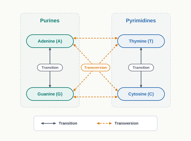
Fig. 7.23 – Substitution mutations: transition and transversion.
| Feature | Transition | Transversion |
|---|---|---|
| Nature of the base change | Purine replaced by purine, or pyrimidine replaced by pyrimidine | Purine replaced by pyrimidine, or pyrimidine replaced by purine |
| Examples of the change | A ↔︎ G, or T ↔︎ C | A ↔︎ T, A ↔︎ C, G ↔︎ T, G ↔︎ C |
| Change in ring structure | The ring structure of the base is preserved | The ring structure of the base is altered |
| Effect on reading frame | Reading frame unchanged | Reading frame unchanged |
Note the last row carefully. Neither type of substitution shifts the reading frame — only one codon is altered — which is why a substitution often causes a milder change than an addition or deletion. Sometimes, because the genetic code is degenerate, the altered codon still specifies the same amino acid and no change appears in the protein at all.
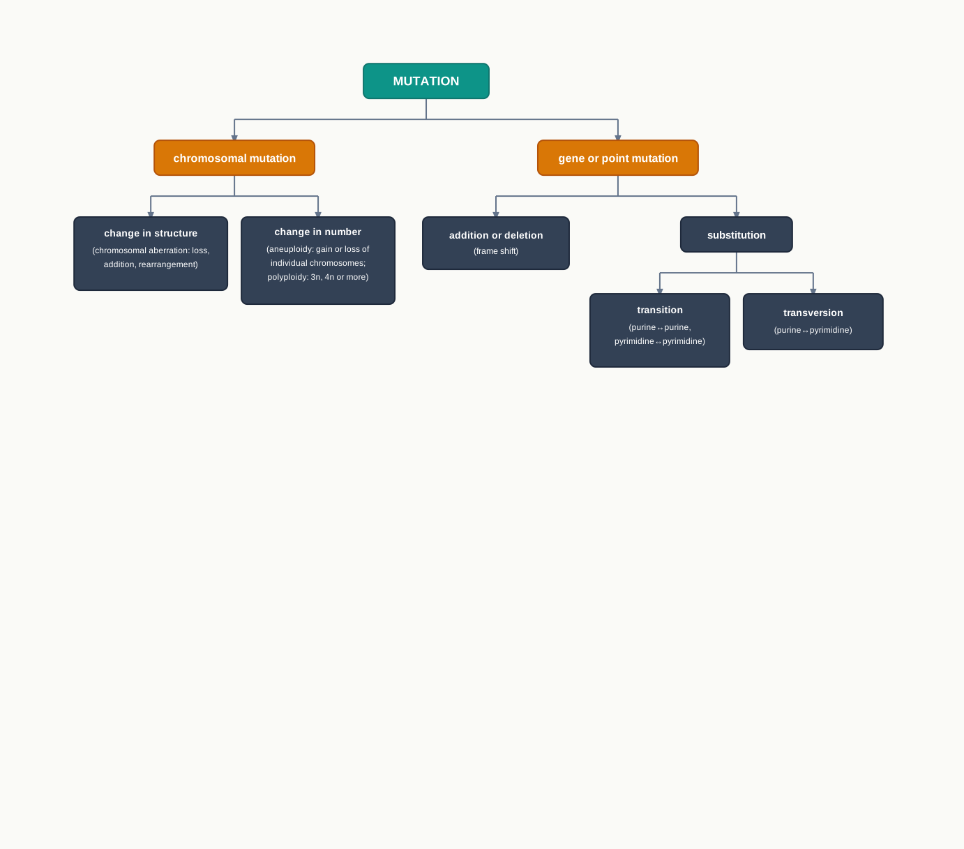
Fig. 7.24 – Classification of mutations.
7.7.1 Molecular Mechanism of Mutation
As discussed earlier, mutations may happen spontaneously, due to the intrinsic properties of the molecule, or they may even be induced by external agents called mutagens.
Spontaneous Mutations
DNA carries the genetic information in the form of nucleotides. Among these nucleotides, there are functional groups present either in the C=O or the C–NH₂ form, normally called the keto or amino form respectively.
However, in these forms of nitrogenous bases, a hydrogen atom may shift from one atom of the molecule to another atom within the base. Such a phenomenon is known as a tautomeric shift, and it results in a temporary phase of the nitrogen base called either the enol (C–OH) form or the imino (C=NH) form.
These rare tautomeric forms of a nitrogenous base have a changed property to pair with a different nucleotide in the DNA molecule. The base has not been replaced by anything; it has merely rearranged internally, and that is enough to change which partner it accepts.
Therefore, at the time of replication, when the imino form of guanine is present in the DNA, it makes a complementary pairing with thymine, whereas the former would have normally paired with cytosine.
During the next replication cycle, the place where thymine was incorrectly added may normally pair with cytosine, leading to the substitution of a \(G \equiv C\) pair in the gene by an \(A = T\) pair, as shown in Fig. 7.25. We have already discussed the consequence of a single base substitution in the case of sickle cell anaemia.
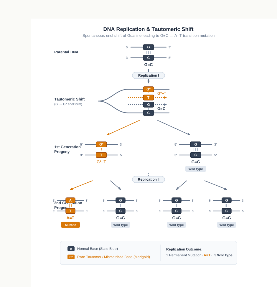
Fig. 7.25 – Spontaneous substitution mutation via tautomeric shift.
Follow Fig. 7.25 across the generations rather than reading it as a single event. The mutation is not complete after one round of replication — at that stage the molecule still carries an abnormal pairing. Only after a second round of replication does one of the daughter molecules carry a permanent, ordinary \(A = T\) pair where a \(G \equiv C\) pair once stood. The error becomes fixed in the DNA only when it has been copied twice.
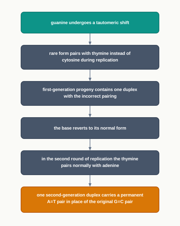
Fig. 7.26 – Fixation of a spontaneous substitution.
Induced Mutations
It is after the use of Hermann J. Muller’s experiments of inducing mutation using X-rays that a new area of induction of mutation using various external agents was opened.
These mutagens fall into the following categories:
- Physical — Radiations like X-ray, UV rays, etc.
- Chemical — Alkylating agents like Mustard gas and ethyl methane sulfonate (EMS); base analogs like 5-bromouracil or 2-aminopurine; and deaminating agents like nitrous acid, etc.
In order to understand the mechanism of induction of mutation by an external agent, let us try with a few examples.
How Physical Mutagens Act
Any cell, especially those responsible for the formation of gametes, when it gets exposed to the ionising radiation of X-rays, may undergo breakage of different bonds present within the molecule.
In case such a breakage occurs in the phosphodiester bond of DNA, it may result in the loss of a few nucleotides in it. This may result in a frame shift mutation due to the deletion of one or a few nucleotides.
The consequence of such a change in the reading frame of the genes is understandable from what you read earlier in this section — it may lead to a wrong RNA transcription, and to non-expression or altered expression of the gene.
Even non-ionising radiation like UV rays may lead to the excitation of an electron within the DNA molecule. These excited molecules may become more reactive, and may lead to the induction of deletion or substitution type of mutation.
Notice the difference in mechanism between the two. Ionising radiation carries enough energy to break bonds outright. UV radiation does not; it merely excites electrons and makes the molecule chemically reactive, so the damage that follows is a chemical consequence rather than a direct physical break.
How Chemical Mutagens Act
Base analogs. A base analog is a molecule that resembles a normal base closely enough to be mistaken for it. 5-bromouracil, when present in its keto form, may get incorporated preferentially in the DNA as a complementary base against adenine.
However, in the eventuality of its conversion into the tautomeric enol form, 5-bromouracil may pair with a guanine nucleotide, resulting in the substitution of an \(A = T\) pair into a \(G \equiv C\) pair, as shown in Fig. 7.27.
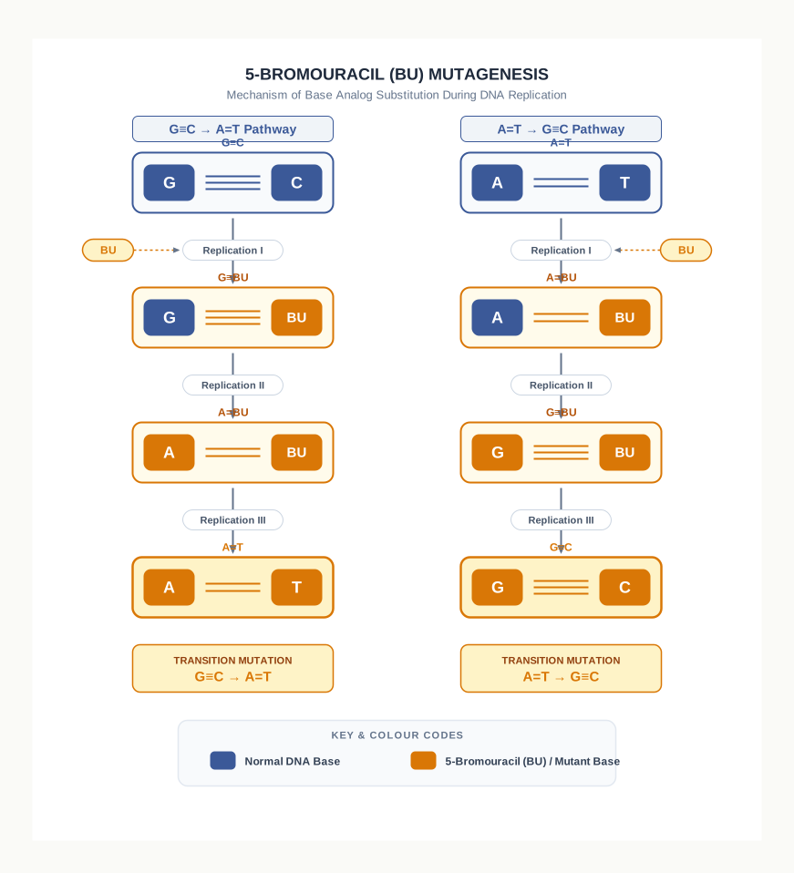
Fig. 7.27 – Induced substitution mutation by 5-bromouracil.
Compare Fig. 7.27 with Fig. 7.25 and you will see that the underlying mechanism is the same in both — a tautomeric shift that changes the pairing behaviour of a base. The difference is only in origin. In Fig. 7.25 the shift occurs in a normal base of the cell’s own DNA and the mutation is spontaneous; in Fig. 7.27 it occurs in a foreign molecule that the cell has been tricked into incorporating, and the mutation is induced.
Alkylating agents. An alkylating agent like EMS ethylates a DNA nucleotide either at the nitrogen at the 7th position or the oxygen at the 6th position. Such an alkylation alters the pairing property of the base.
For example, the ethylated guanine nucleotide pairs with thymine, which means a \(G \equiv C\) pair in DNA will be mutated to an \(A = T\) pair.
Many such chemical mutagens induce mutation by the process of altered pairing property. This is the common thread running through this entire subsection: whether the cause is a spontaneous tautomeric shift, an incorporated base analog, or a chemically modified base, the outcome is the same — a base that pairs with the wrong partner during replication.
| Feature | Physical mutagens | Chemical mutagens |
|---|---|---|
| Examples | X-rays, UV rays | Mustard gas, EMS, 5-bromouracil, 2-aminopurine, nitrous acid |
| Mode of action | Break bonds directly (ionising) or excite electrons and make the molecule reactive (non-ionising) | Get incorporated in place of a normal base, or chemically modify an existing base |
| Typical type of mutation caused | Deletion and frame shift, also substitution | Mainly substitution, through altered pairing property |
7.8 DNA Repair
From the above account on mutation, it may perhaps be understood that the rate of mutation — either spontaneous or induced — must be very high. Tautomeric shifts occur constantly, ultraviolet light falls on us every day, and replication runs through millions of base pairs at speed.
But such a high rate of mutation is not observed. Also, a very high rate of mutation is not expected, considering the fact that the genetic material gets transferred in a stable way from one generation to the next. If errors accumulated at the rate at which they arise, no species could remain recognisably itself for long.
Why Mutation Rates Stay Low
The understanding of the molecular processes in various organisms has revealed that mechanisms exist so that most of the errors that occur are corrected.
This is the crucial point of this section. The low mutation rate of living organisms is not the result of errors being rare. Errors are common. The low rate is the result of a second layer of machinery whose entire job is to find those errors and put them right.
Many of such mechanisms have been identified in the bacterial system E. coli. Out of these we will see a few, in order to understand the mechanism.
Excision Repair
Excision repair is a mechanism in which altered or modified bases in the DNA are removed, or excised, sequentially — by first identifying the altered base and subsequently removing it by enzymatic binding.
The gap thus created is eventually filled by a DNA polymerase.
Notice the general strategy before looking at the details, because both forms of excision repair follow the same four movements: recognise the damage, remove the faulty portion, replace it using the undamaged strand as template, and reseal the backbone. The undamaged complementary strand is what makes repair possible at all — it holds an intact copy of the information that has been lost.
The following are the two important excision repair mechanisms.
Base Excision Repair (BER)
In BER, an altered or damaged base is replaced by a correct base.
DNA nucleotides can be damaged by oxidation, alkylation and deamination, which result in incorrect base pairing leading to mutations. You have already met alkylation in Section 7.7, as the way in which an agent like EMS alters the pairing property of guanine — BER is the cell’s answer to exactly that kind of chemical injury.
The enzyme DNA glycosylase identifies the damaged bases and removes them. It does not cut the backbone; it detaches only the faulty base from its sugar.
The gap region so created is termed the AP site — the apurinic or apyrimidine site — meaning a position in the strand where the sugar and phosphate remain but the base is missing.
In the next step, an endonuclease cleaves this region to yield a free 3’-OH and 5’-PO₄ group, as shown in Fig. 7.28a. This step matters for a specific reason: DNA polymerase cannot begin work without a free 3’-OH group, so the endonuclease is effectively preparing the site for the polymerase.
DNA polymerase then adds the correct bases, and through its exonuclease activity removes the old damaged bases.
Finally, DNA ligase seals the remaining gap by the formation of a phosphodiester bond — the same sealing role you saw it perform on the lagging strand during replication.
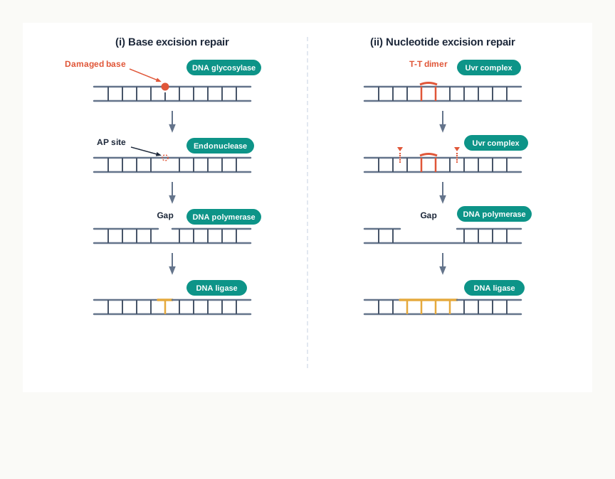
Fig. 7.28a – Excision repair mechanisms: (i) base excision and (ii) nucleotide excision.
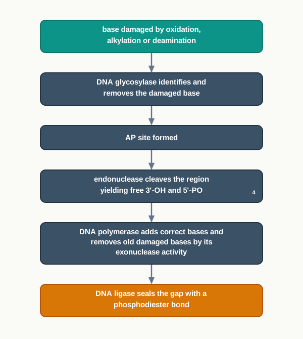
Fig. 7.29 – Base excision repair pathway.
Nucleotide Excision Repair (NER)
In the case of NER, a comparatively large portion of the altered part of DNA is repaired. Where BER replaces a single faulty base, NER removes a whole stretch of nucleotides.
DNA is susceptible to radiations like UV radiation, and this may lead to the formation of pyrimidine dimers (T-T dimers), which distort the DNA helix.
Understand why a dimer is so serious. Two adjacent thymines become covalently joined to each other, so they can no longer pair with the adenines opposite them. The helix bulges at that point. A replication fork or an RNA polymerase arriving at this bulge is obstructed, so the damage blocks both replication and transcription until it is removed.
There is a mechanism in the cell by which these dimers are removed. It is facilitated by a protein complex called UVr, as shown in Fig. 7.28a.
Initially, a component of the UVr complex scans the DNA for the presence of T-T dimers that distort the DNA helix. Note that the complex does not need to recognise the chemical damage itself; it recognises the distortion of the helix, which is a far simpler signal and works for many different kinds of bulky damage.
After identification, other components of UVr cleave the phosphodiester bond four nucleotides downstream and eight nucleotides upstream to the DNA damage site. Two cuts are made, one on each side of the lesion, so that the damaged stretch can be lifted out as a single piece.
In the next step, the other component of the UVr complex, which has DNA helicase property, unwinds the DNA and removes the cleaved nucleotide fragment.
Finally, DNA polymerase I adds the nucleotides in the 5’-3’ region, and the gap gets sealed by DNA ligase.
In human beings, an inherited failure of nucleotide excision repair causes the condition known as xeroderma pigmentosum. Affected individuals cannot remove the pyrimidine dimers produced by sunlight, so the damage accumulates and they develop severe skin lesions and skin cancers from an early age. The disease demonstrates, in the clearest possible way, that repair is not an optional refinement but a requirement for survival.
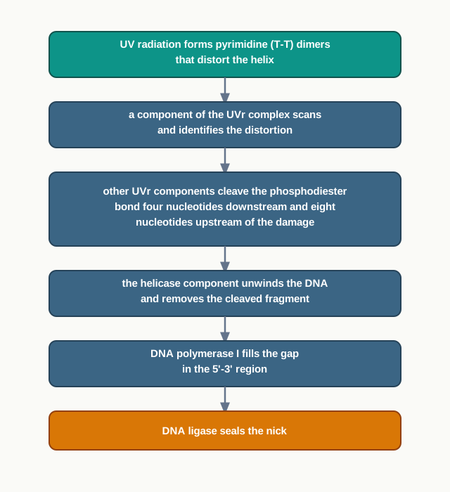
Fig. 7.30 – Nucleotide excision repair pathway.
Mismatch Repair (MMR)
During DNA replication, sometimes an incorrect nucleotide gets incorporated. Here nothing is chemically damaged at all — both bases are perfectly normal, they are simply the wrong pair.
There exists a mechanism by which such a mismatch is corrected, through different types of proteins, namely MutH, MutL, MutS and MutT.
These four proteins recognise and cleave the replicated DNA segment about 1,000 nucleotides from the point of mismatch. The gap thus created is filled by DNA polymerase, followed by the joining of the nick by DNA ligase.
This process is also important for proof-reading during replication.
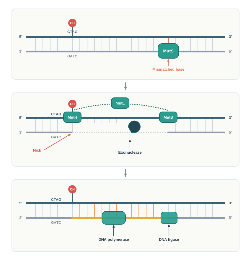
Fig. 7.28b – Mismatch repair mechanism.
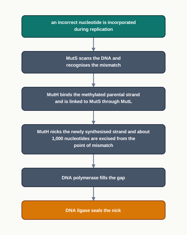
Fig. 7.31 – Mismatch repair pathway.
| Feature | Base Excision Repair (BER) | Nucleotide Excision Repair (NER) | Mismatch Repair (MMR) |
|---|---|---|---|
| Type of fault corrected | A single altered or damaged base | A large helix-distorting lesion such as a T-T dimer | A normal but incorrectly paired nucleotide |
| Usual cause of the fault | Oxidation, alkylation, deamination | UV radiation | Error during DNA replication |
| Recognition step | DNA glycosylase identifies the damaged base | UVr complex detects the distortion of the helix | MutS recognises the mismatch |
| Extent of DNA removed | A single base, then the baseless nucleotide | The fragment cut four nucleotides downstream and eight upstream of the lesion | About 1,000 nucleotides from the point of mismatch |
| Key proteins involved | DNA glycosylase, endonuclease, DNA polymerase, DNA ligase | UVr complex, DNA polymerase I, DNA ligase | MutH, MutL, MutS, MutT, DNA polymerase, DNA ligase |
| Common final steps | Gap filled by DNA polymerase, nick sealed by DNA ligase | Same | Same |
Read the last row of the table carefully. However different the three pathways look at the recognition stage, they converge on the same two enzymes at the end — a polymerase to rebuild and a ligase to seal. This is a good illustration of a general principle in molecular biology: the cell rarely invents new machinery when existing machinery will do.
Other Repair Mechanisms
A few other DNA repair mechanisms have also been observed and studied, both in prokaryotes and in eukaryotes. You will study them in your higher classes.
However, based on the account of mutation in Section 7.7 and of the repair mechanisms in this section, the stable nature of genetic information can now be appreciated, alongside its ability to undergo changes.
The two sections belong together and should be read as a pair. Mutation supplies the variation on which evolution depends; repair keeps that variation rare enough for a species to remain stable. Neither alone would be workable. A genome that never changed could not evolve, and a genome that changed freely could not be inherited.