Chapter 8: Genetic Disorder
In 1866, an English physician named John Langdon Down published a careful description of a group of children who shared a strikingly similar set of features — a flat facial profile, a small mouth, a single deep crease across the palm. They were not related to one another. They came from different families, different towns, different backgrounds. Yet they looked more like each other than like their own brothers and sisters. Down could describe the pattern precisely, but he could not explain it. Neither could anyone else, for almost a hundred years.
The answer arrived only after biologists learned to count. By the mid-1950s it was firmly established that a normal human cell carries 46 chromosomes. In 1959, a team in Paris that included Jérôme Lejeune, Marthe Gautier and Raymond Turpin examined cells from children with the condition Down had described. The count came to 47. Chromosome number 21 was present three times instead of twice.
There was nothing faulty about the genes on that extra chromosome. Every one of them was an ordinary human gene, spelt correctly. The problem was arithmetic — one copy too many. Fig. 8.0 captures that moment of counting, when a pattern of physical features finally became a pattern of chromosomes.
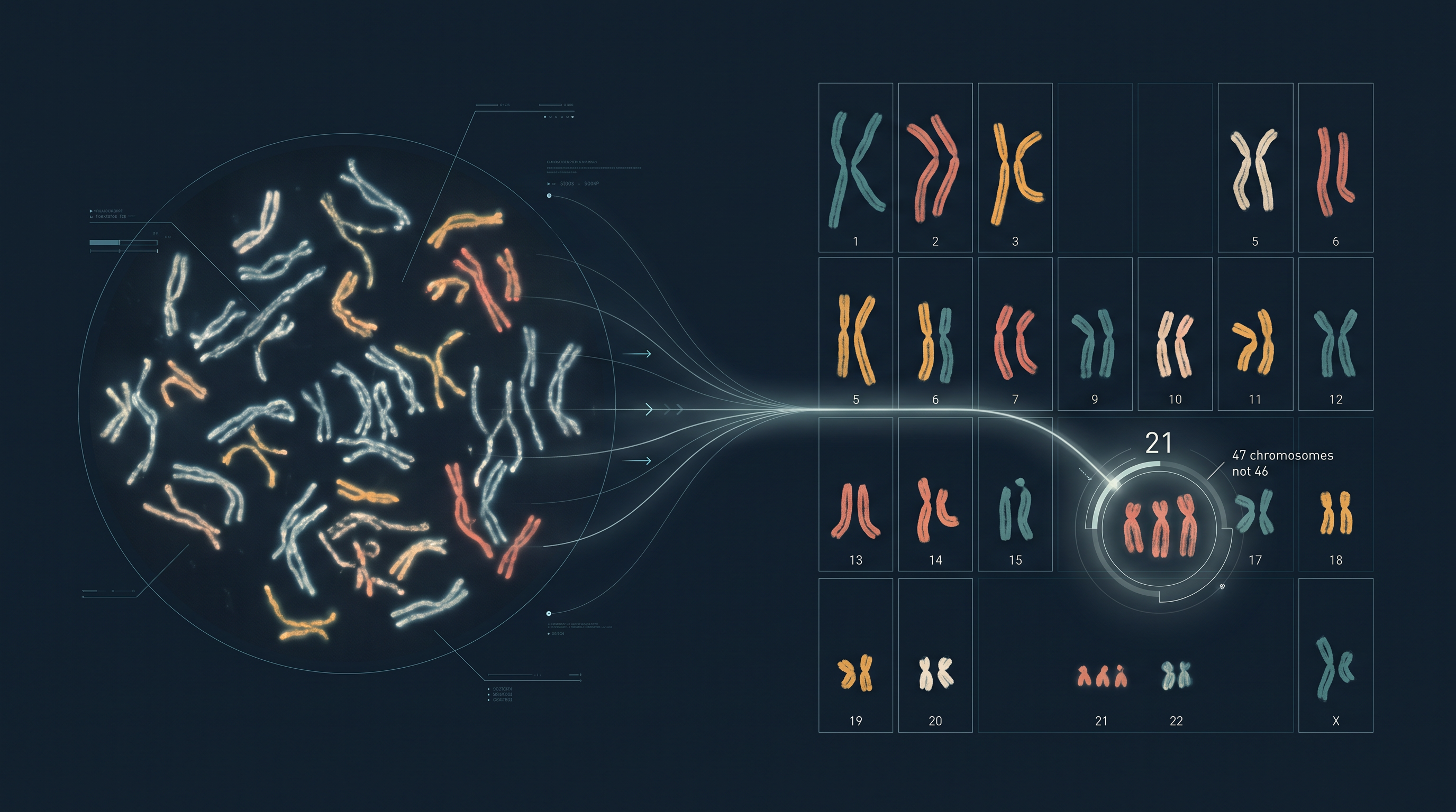
Fig. 8.0 – Sorting chromosomes into a karyogram.
Each of these questions is answered somewhere in this chapter. Keep them in mind as you read — you will meet the answers as the ideas are built up, section by section.
8.1 Chromosomal Abnormalities and Syndromes
Every cell in your body carries a complete set of instructions for building and running you, and those instructions are packed into structures called chromosomes. A normal human body cell carries 46 chromosomes, arranged as 23 pairs. One member of each pair came from the mother and the other from the father, which is why a body cell is described as diploid and written as \(2n\).
You have met these terms in earlier classes, so it is worth fixing them firmly before going further. A haploid cell, such as a sperm or an ovum, carries only one copy of each chromosome (\(n = 23\) in humans). A diploid cell carries two copies of each (\(2n = 46\)). Fertilisation restores the diploid number by bringing two haploid sets together.
Chromosomes are remarkably stable structures, but they are not indestructible. In certain situations — exposure to environmental radiation, something taken in through food, or internal genetic conditions — chromosomes may suffer damage, or their number may change.
Two Ways a Chromosome Set Can Change
The changes fall into two broad categories, and the distinction between them is important. A chromosome may keep its correct number but change its shape or internal arrangement. Alternatively, every chromosome may be perfectly normal in structure, yet there may be too many or too few of them.
A change in the structure of a chromosome is called a structural chromosomal abnormality, also known as an aberration. A change in the number of chromosomes is called a numerical chromosomal abnormality.
Counting the Chromosomes
Numerical changes are described by comparing the affected cell with the normal diploid number. When one chromosome of a pair is absent, the condition is called monosomy for that chromosome, and the cell is written as \(2n - 1\). An example is monosomy of chromosome 1, where only one copy of chromosome 1 is present instead of two.
When a chromosome is present in three copies instead of two, the condition is called trisomy, written as \(2n + 1\). An example is trisomy of chromosome X.
Notice that in both monosomy and trisomy, only one particular chromosome has gone wrong. The rest of the set is untouched. Because of this, monosomy and trisomy both come under the broad category of aneuploidy.
There is a second, very different kind of numerical change. Here nothing is missing or added piecemeal — instead, the entire set of chromosomes is multiplied. A cell with three complete sets would carry \(69 = 23 \times 3\) chromosomes, and a cell with four complete sets would carry \(92 = 23 \times 4\). This condition is called polyploidy.
Polyploidy on Your Plate
Polyploidy is not always a disorder. In plants it is common, and plant breeders have deliberately used it. The artificial breeding of plants has produced several polyploid varieties that we eat every day.
Bread wheat, for example, has six sets of chromosomes and is therefore hexaploid. Cabbages and mustards are tetraploids, carrying four sets. Banana and apple are triploid, with three sets of chromosomes. Strawberry and sugar cane are octoploid, with eight sets of chromosomes.
This raises an obvious question, and answering it teaches something important about how chromosomes actually work. Why should a plant thrive with six full sets while a human being is seriously affected by a single extra chromosome?
The reason lies in balance. In polyploidy, every gene on every chromosome is multiplied by the same factor. The proportions between genes stay exactly as they were, so the cell’s chemistry remains balanced even though everything has been scaled up. In aneuploidy, only one chromosome changes its copy number. All the genes on that one chromosome are now produced in the wrong proportion relative to the thousands of genes on the other chromosomes, and it is this imbalance — not the extra genetic material by itself — that disturbs development.
Whether the change is structural or numerical, the consequences can be significant. Both kinds of change can alter the phenotype of an individual in the form of diseases or syndromes, and the rest of this section examines the most important examples of each.
8.1.1 Structural chromosomal abnormalities
Structural abnormalities arise when a chromosome breaks and the broken pieces are not restored correctly. There are four main types, and it helps to keep in mind what each one does to the length of the chromosome and to the order of genes along it, as shown in Fig. 8.1.
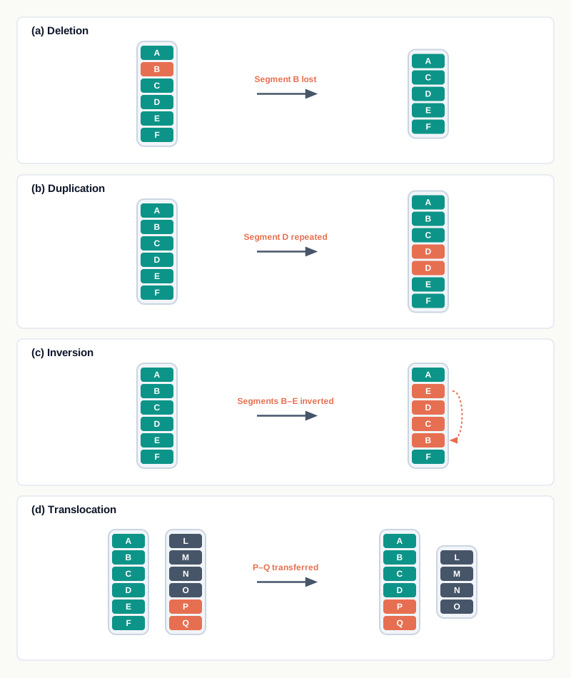
Fig. 8.1 – Structural chromosomal abnormalities: (a) deletion, (b) duplication, (c) inversion, and (d) translocation.
1. Deletion — In deletion, a segment of a chromosome breaks away, leading to shortening of the chromosome [Fig. 8.1(a)]. The genes carried on that lost segment are simply gone. For example, retinoblastoma, a cancer of the eye, is caused due to deletion of a portion of chromosome 13. Sometimes both ends of a chromosome are deleted, and the two cut ends then join to each other. The chromosome closes into a circle, and such a chromosome is called a ring chromosome.
2. Duplication — Duplication refers to a situation in which a segment of the chromosome gets repeated, resulting in a longer chromosome [Fig. 8.1(b)]. The genes on that segment are now present in extra copies. This can lead to conditions such as Charcot-Marie-Tooth disease, caused due to duplication of genes on chromosome 17.
3. Inversion — In inversion, a segment of the chromosome breaks away, completely reverses itself, and reattaches with the chromosome. Here the overall length of the chromosome remains the same, but the orientation of the genes in that segment is reversed by 180 degrees [Fig. 8.1(c)]. For example, RCAD syndrome is caused by inversion of a segment of chromosome 17.
4. Translocation — In translocation, a segment of a chromosome breaks away and reattaches itself with another chromosome — that is, the piece moves to a chromosome it does not belong to. Translocation comes in two forms, and telling them apart is important.
If there is a mutual exchange of segments between two chromosomes, so that each gives a piece and each receives a piece, it is called reciprocal translocation. An example is Burkitt’s lymphoma, where exchange of materials happens between chromosomes 8 and 14.
If a segment of a chromosome breaks away and attaches with another chromosome without any mutual exchange — a one-way move — it is called Robertsonian translocation. Because two chromosomes effectively fuse into one, this may result in a decrease of the chromosome number of the cell [Fig. 8.1(d)].
When Rearrangement Leads to Cancer
Two of the examples above — retinoblastoma and Burkitt’s lymphoma — are cancers, and this is not a coincidence. Genes that control when a cell is allowed to divide are among the most sensitive to being lost, duplicated or moved. If a segment carrying a brake on cell division is deleted, the brake is gone. If a growth-promoting gene is carried next to a powerful switch on another chromosome by a translocation, it may be driven far harder than it should be. The result in either case is a cell that divides when it should not.
8.1.2 Numerical chromosomal abnormalities
Before looking at the individual conditions, two words used throughout this section need to be separated clearly, because students often use them interchangeably in exams.
The term syndrome generally refers to a group of symptoms which consistently occur together, or a condition characterised by a set of associated symptoms. The features travel as a package — which is precisely why the pattern was recognisable long before its cause was known.
A disease, on the other hand, refers to an abnormal physiological response to internal or external factors. Fever caused due to microbes is an example of a disease.
Reading a Karyotype
All three conditions described below are identified by examining the chromosome set of the individual, so the technique used deserves attention before the conditions themselves.
The result is written in a short standard notation. In 47, XX, +21 the first number is the total chromosome count, the letters give the sex chromosomes present, and the +21 records one extra copy of chromosome 21. Once you can read this notation, a karyotype line tells you the whole story in a few characters.
1. Down’s Syndrome
Incidence: Occurs in approximately 1 per 800 live births.
Chromosomal basis: Down syndrome is a genetic condition that arises due to the presence of an extra chromosome 21. Here, chromosome 21 is repeated thrice — trisomy 21 — instead of showing up twice as in a normal individual. The karyotype of Down syndrome is represented as 47, XX, +21 in females and 47, XY, +21 in males [Fig. 8.2(a)].

Fig. 8.2 – Karyograms for (a) Down syndrome and (b) Klinefelter’s syndrome.
The trisomic condition is usually caused by an error in the process of cell division called non-disjunction, that is, the inability of chromosomes to separate at the time of cell division. Normally, the two members of a chromosome pair are pulled to opposite poles so that each gamete receives exactly one. If they fail to separate, both travel to the same pole. One gamete then carries two copies of that chromosome and the other carries none, and the whole sequence is summarised in Fig. 8.3.
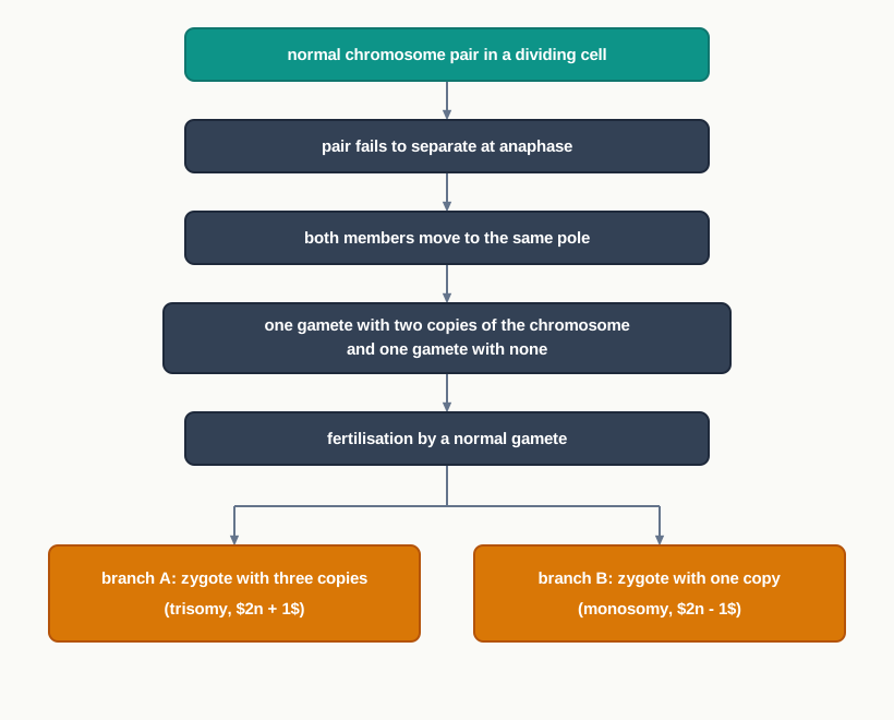
Fig. 8.3 – Non-disjunction and its outcome.
The possibility of having a Down’s syndrome baby in the family increases with the maternal age. It has been reported that more than 85% of Down syndrome babies are born to mothers over 35 years of age at the time of pregnancy. This is the reason chromosomal screening is routinely offered during pregnancies in this age group.
Clinical symptoms: Some of the distinguishing features of Down’s syndrome are a flat face, slanting eyes, a small mouth, a protruding tongue, a flattened nose, a short neck, short arms and legs, a single deep crease across the palm, low IQ, stunted growth, muscular hypotonia (reduced muscle tone) and under-developed gonads. Down’s syndrome babies also show breathing, heart or hearing problems.
Diagnosis and Treatment: Down syndrome is usually diagnosed by finding an extra chromosome 21 in the karyotype. There is no single standard treatment protocol for Down syndrome. Treatments are tailored to the specific set of conditions presented by each individual. At an early age, children with Down’s syndrome can benefit from speech therapy, physiotherapy and taking nutritional supplements.
The effect of such care shows clearly in survival figures. In the early 1900s, on an average, individuals with Down’s syndrome used to live until age 9. Now, with advances in diagnostic and treatment technologies, life expectancy has increased up to 60 and even longer.
2. Klinefelter’s Syndrome
Incidence: Occurs in approximately 1 out of 1000 newborn males.
Chromosomal basis: The genotype is 47, XXY, and the condition affects males. The extra chromosome is not transmitted genetically — that is, a Klinefelter newborn cannot have a Klinefelter father. It arises instead from the inability of the X chromosome to detach itself from the pair during meiosis, at the time of gamete formation. Fertilisation of an XX ovum with a Y sperm produces an XXY zygote.
Read that last sentence carefully, because it explains why the condition appears fresh in each affected individual rather than running through a family. The error is an accident of gamete formation, not an instruction copied from a parent.
Clinical Symptoms: Children with Klinefelter’s syndrome are unusually tall for their age, have reduced facial and body hair, smaller testes, enlarged breasts and a coarse voice [Figs. 8.2(b) and 8.4].
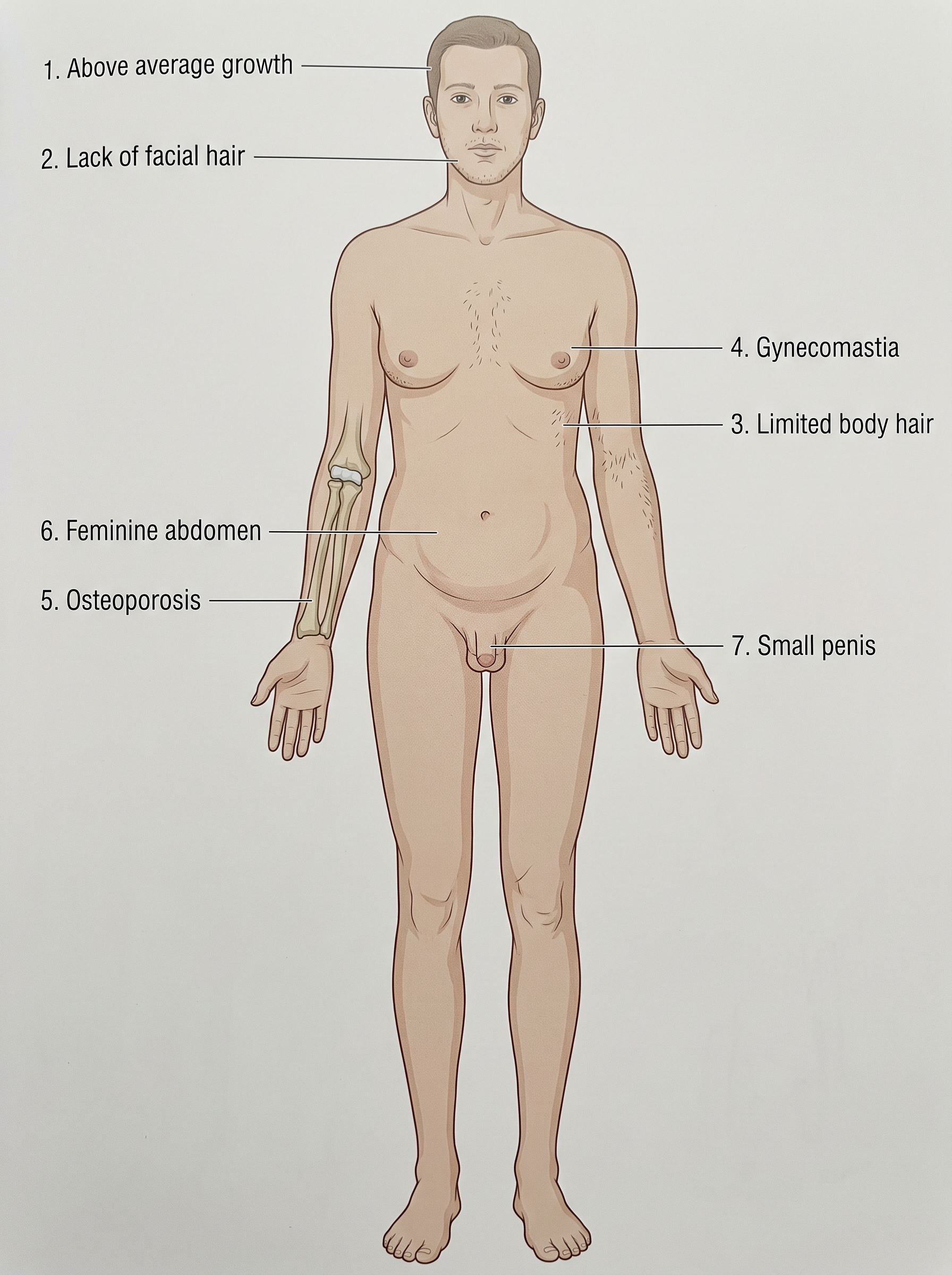
Fig. 8.4 – Features of Klinefelter’s syndrome.
Diagnosis and Treatment: One of the most frequent methods used to diagnose Klinefelter syndrome is the Barr body test of a buccal smear. Normally, no Barr body appears in a male buccal smear. However, in Klinefelter’s syndrome one Barr body shows up, indicating the presence of an extra X chromosome.
To understand why this simple test works, recall that in a cell with more than one X chromosome, all but one X is switched off and condenses into a small dark mass at the edge of the nucleus. That mass is the Barr body. A normal male has a single X, so nothing is left over to be switched off and no Barr body forms; a person with two X chromosomes has one Barr body. Counting Barr bodies therefore counts X chromosomes indirectly, without any need for full karyotyping.
At the time of birth, babies with Klinefelter’s syndrome differ only a little from other newborn babies. However, as age increases the differences become noticeable, especially at the time of puberty, when the effect of the extra X chromosome on hormone production begins to show.
People with Klinefelter’s syndrome are often treated with testosterone to develop masculine features. They also need to be psychologically counselled to control depression leading to aggression. Note that this treatment manages the effects of the condition; it does not remove the extra chromosome, which is present in every cell.
3. Turner’s Syndrome
Incidence: Occurs in 1 in 2,500 newborn girls, and is frequently observed in miscarriages and still births.
Chromosomal basis: Turner’s syndrome affects females and arises due to a missing X chromosome in the affected female. This is called monosomy X, and the karyotype is represented as 45, X.
A cell division error during meiosis of an ovum results in one ovum with no X chromosome and another with two X chromosomes. The ovum with no X chromosome fuses with a sperm carrying one X chromosome to generate the 45, X condition. Mothers with Turner syndrome cannot pass the condition on to their daughters — that is, this condition is not inherited.
Clinical symptoms: Turner’s syndrome is diagnosed by the following features — short stature, webbed neck (the neck skin is unusually loose and can be pulled several centimetres away from the neck), small breasts, low-set ears (the ears are placed below the normal position), and swollen hands and feet. Furthermore, the ovaries are under-developed and menstrual periods are usually absent (Fig. 8.5).
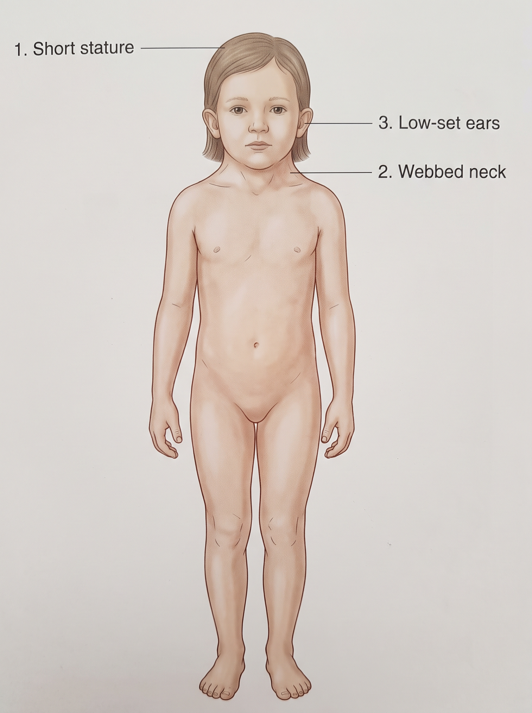
Fig. 8.5 – Features of Turner’s syndrome.
Diagnosis and treatment: Prenatal chromosomal diagnosis usually happens through amniocentesis or chorionic villus sampling. In amniocentesis, a small sample of the amniotic fluid surrounding the foetus is withdrawn; it contains foetal cells that can be grown and karyotyped. In chorionic villus sampling, a tiny piece of the developing placental tissue is taken instead, which allows testing somewhat earlier in pregnancy. The overall workflow is summarised in Fig. 8.6.
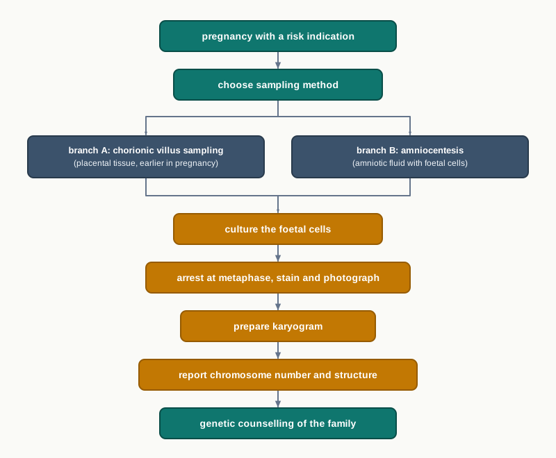
Fig. 8.6 – Prenatal chromosomal diagnosis workflow.
At puberty, the first test performed is that of the Barr body of a buccal smear. The absence of a Barr body in a female smear is the first indicator, and it prompts a detailed investigation to follow up the condition.
As with the other syndromes described here, there is no permanent cure, because every cell of the body carries the altered chromosome number. However, growth and ovarian functions can be strengthened by the controlled administration of the hormones androgen and estrogen.
8.2 Monogenic Disorders and Pedigree Mapping
In the previous section, the fault lay with whole chromosomes — an extra one, a missing one, or a piece in the wrong place. This section moves to a much finer scale. Here every chromosome is present in the right number and the right shape, and the error sits inside a single gene.
A monogenic disease is caused by an error in a single gene. According to current estimates, over 10,000 human diseases are estimated to be monogenic, affecting millions of individuals worldwide.
What the Faulty Gene Does Decides the Disease
The nature of the disease, and its signs and symptoms, depend on the functions performed by the modified or defective gene. This is the single most useful idea in this section. If the gene normally makes a protein that carries oxygen, the disease will involve oxygen supply. If it makes a protein needed for blood clotting, the disease will involve bleeding. If it makes a protein that gives muscle fibres their strength, the disease will involve muscle weakness. Asking “what does this protein normally do?” will get you most of the way to predicting the symptoms.
These diseases are inherited according to Mendel’s Laws, which you have studied earlier — the laws describing how alleles segregate during gamete formation and recombine at fertilisation. That is why the appearance of a monogenic disease in a family follows predictable ratios rather than occurring at random.
In some cases, however, the mutation can be spontaneous. It arises fresh in the individual, and in such cases we will not get any previous family history of the disease. A clean family history therefore does not rule out a genetic disorder.
There is also variation in how many different errors in the same gene can cause the same illness. There may be a single mutation in one gene causing a specific disease, as in sickle cell anemia. Alternatively, there may be multiple types of mutation in one gene all producing the same disease, as in cystic fibrosis, where more than 200 different types of mutation can occur in one gene. This difference matters in the laboratory: a test for sickle cell anemia can look for one known change, while a test for cystic fibrosis must screen for many.
Four Patterns of Inheritance
The single-gene or monogenic diseases can be classified into the following categories according to the inheritance pattern:
- Autosomal recessive
- Autosomal dominant
- X-linked recessive
- X-linked dominant
Two questions decide which category a disease falls into. First, is the faulty gene on an autosome (any of the 22 non-sex chromosome pairs) or on the X chromosome? Second, does one faulty copy alone produce the disease (dominant), or are two faulty copies needed (recessive)?
Pedigree Analysis
For diagnosis of an inherited genetic disease, one has to understand the concept of pedigree analysis. Pedigree analysis is the process of interpretation of information displayed as a family tree. If more than one person in a family is involved with a disease, then pedigree analysis can be done.
Because a family tree has to be read at a glance, specific symbols are used to indicate different aspects of a pedigree, as shown in Fig. 8.7. A square represents a male and a circle represents a female. A filled (shaded) square or circle represents an affected individual. A horizontal line joining a square and a circle indicates mating. The offspring are drawn below, in birth order, hanging from that line. Generations are numbered with Roman numerals, so that I and II are the first and second generations, and individual offspring within a generation are numbered as II-1, II-2 and so on. Identical twins are shown as two symbols branching from a single point with a connecting bar between them, while non-identical twins branch from a single point without that bar.
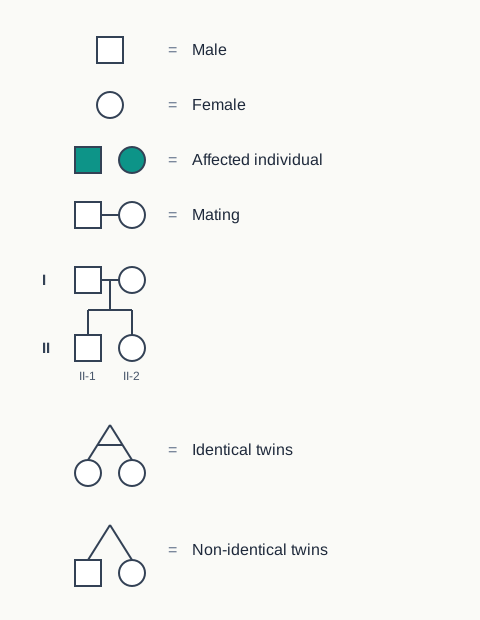
Fig. 8.7 – Standard pedigree symbols.
The logic used in that last step can be set out as a decision sequence, given in Fig. 8.8.
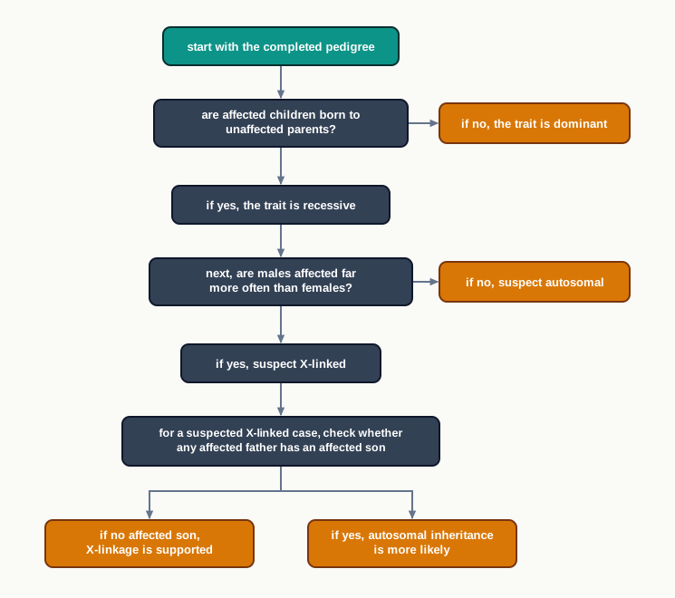
Fig. 8.8 – Deciding the inheritance pattern from a pedigree.
Autosomal Recessive Disorder
The word ‘recessive’ indicates that 2 copies of the gene are needed to have the trait, and in the case of a mutated gene, to have the disorder. Out of the 2 copies, one gene is inherited from the father and one from the mother.
If an individual carries one defective recessive gene and one normal gene, then he or she will be a carrier and will not develop the disease. The single normal copy is enough to make sufficient working protein, so the person is healthy — but the faulty allele is still there and can be passed to a child.
Carriers are far more common than most students expect. On the basis of statistical projection, it is estimated that every human being carries about 5 or more defective recessive genes which can cause a genetic disease. These normally stay hidden, because the chance that two unrelated people carry a fault in the same gene is small.
The disease phenotype of a recessive disorder is due to homozygosity of the recessive allele, and the unaffected phenotype is secondary to the corresponding dominant allele. In plain words: two faulty copies produce the disease, and one working copy is enough to prevent it.
Sickle Cell Anemia
This can be explained with the example of sickle cell anemia, which is an autosomal recessive disease. Sickle cell disease is caused by a mutation in the hemoglobin-β gene found on chromosome 11.
The mutation results in a defective haemoglobin (Hb). Haemoglobin is the protein inside red blood cells that picks up oxygen in the lungs and releases it in the tissues. After giving up oxygen, these defective Hb molecules cluster together, resulting in the formation of rod-like structures. These rigid rods push against the cell membrane from inside, so the red blood cells become stiff and assume a sickle shape (Fig. 8.9).
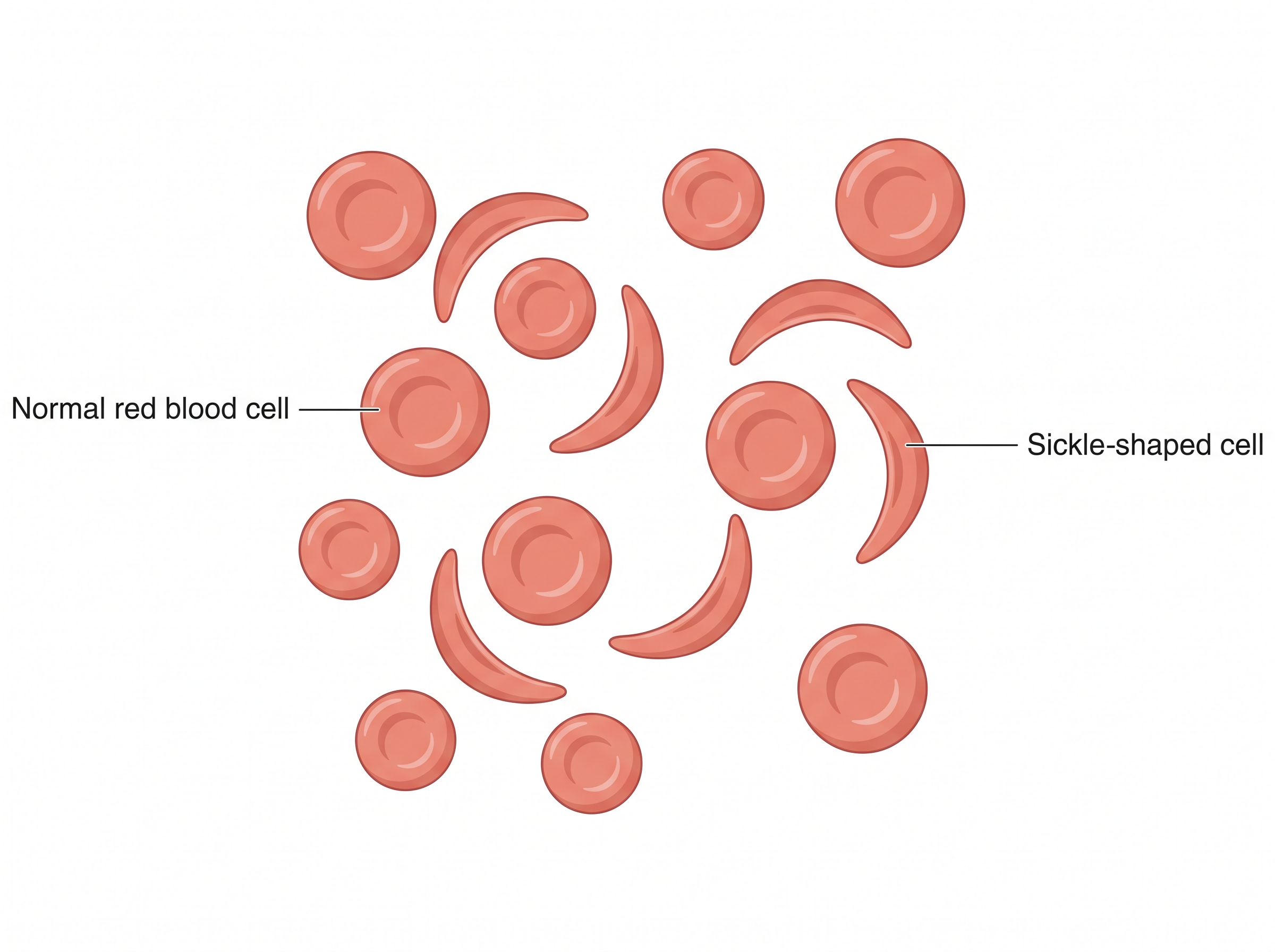
Fig. 8.9 – Red blood cells in sickle cell anaemia.
Notice how directly the symptoms follow from that shape change. A normal red blood cell is round and flexible, and squeezes easily through the narrowest capillaries. A stiff, sickled cell cannot. It jams in small vessels, blocking blood flow and causing pain, and it is destroyed by the body sooner than a normal cell, which is why the condition is an anaemia.
Sickle cell anemia is determined by an allele which we can designate as s, and the normal condition by S. The person affected by the disease will have a genotype of s/s, and unaffected individuals will be either S/S or S/s.
We can make a projected pedigree of the disease assuming that both parents are carriers, S/s (Fig. 8.10). Each parent can pass on either S or s, so four combinations are possible in the children: S/S, S/s, S/s and s/s. The expected genotypic ratio is therefore
\[S/S : S/s : s/s = 1 : 2 : 1\]
This means that from two carrier parents, on an average one child in four is expected to have normal red blood cells (S/S), two in four are expected to be carriers like the parents (S/s), and one in four is expected to have sickle cell disease (s/s). The chance of an affected child is \(\frac{1}{4}\), or 25 per cent, in each pregnancy — and it is a fresh 25 per cent every time, not a quota that gets used up.
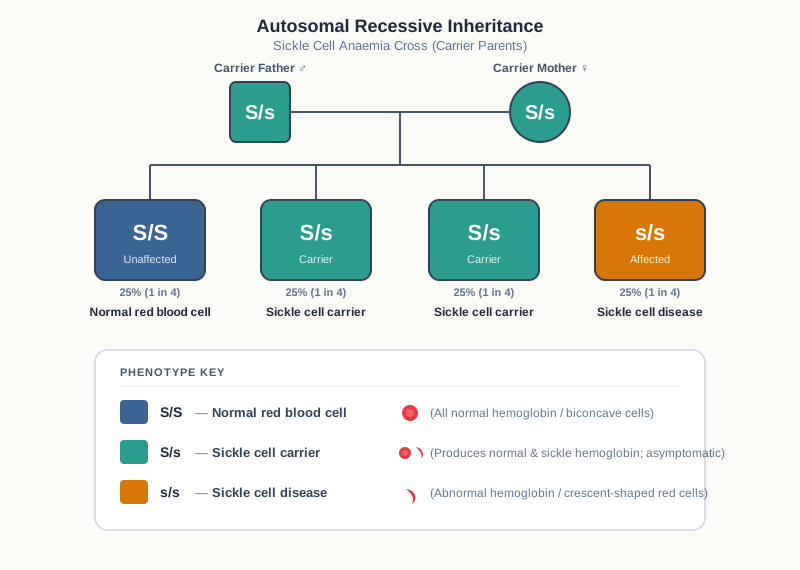
Fig. 8.10 – Inheritance of sickle cell anaemia.
Sickle cell anemia is particularly common among people whose ancestors originated from sub-Saharan Africa, South America, Cuba, Central America, Saudi Arabia, India, and Mediterranean countries. In India, it is common among people of the Deccan plateau of central India, with a smaller focus in the north of Kerala and Tamil Nadu. This geographical clustering is the reason India runs targeted screening programmes in these regions rather than testing the whole population uniformly.
Other Autosomal Recessive Diseases
Other examples of autosomal recessive diseases include cystic fibrosis, Tay-Sachs disease and phenylketonuria. In each case, the symptoms follow directly from the job the faulty protein was supposed to do.
Individuals with cystic fibrosis produce mucus that is abnormally thick and sticky. Thin, watery mucus normally traps dust and microbes and is cleared away; thick mucus instead sits and blocks. It can damage different organs, especially the lungs, resulting in chronic infections. Recall that cystic fibrosis is the disease mentioned earlier in which more than 200 different types of mutation in the same gene can produce the same illness.
Tay-Sachs disease is due to the absence of an enzyme called hexosaminidase A. This enzyme normally breaks down a particular fatty substance. Without it, the fatty substance accumulates in nerve cells, particularly affecting the brain. It is a fatal disease that manifests in childhood. One in 27 persons of European Ashkenazi Jewish origin carries the Tay-Sachs gene.
Phenylketonuria is caused by a mutation in the phenylalanine hydroxylase gene. This enzyme normally converts the amino acid phenylalanine into another substance, so when it is missing, the result is an increase in phenylalanine in the blood.
Autosomal Dominant Disorder
In this kind of inheritance, the normal allele is recessive and the abnormal allele is dominant. Only one faulty copy is needed to produce the disease, which is why an affected person almost always has an affected parent, and why the disorder appears in every generation of a pedigree instead of skipping.
A rare autosomal dominant disorder, achondroplasia, may be considered as an example. It leads to a type of dwarfism in the affected individuals (Fig. 8.11).
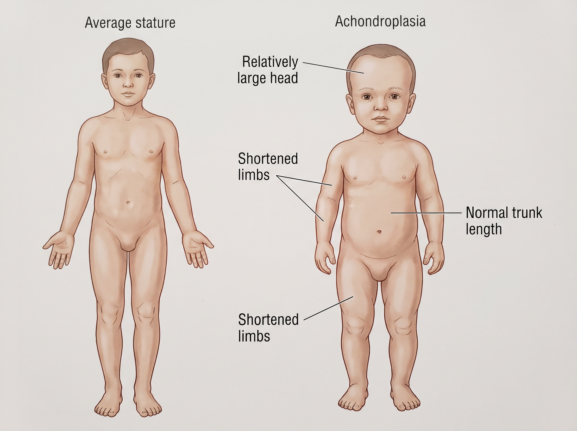
Fig. 8.11 – Achondroplasia causing dwarfism.
In this condition, normal people have the genotype d/d, affected individuals with mild disease have D/d, and severely affected individuals have D/D, which is often lethal. So most of the surviving cases of achondroplasia are heterozygotes.
This is an important exception to the usual expectation that “dominant” simply means one copy is enough. Here, one copy causes the mild condition and two copies are usually fatal — the effect grows with the number of faulty copies. Board questions often use this example to check whether a student has understood dominance as a description of what one allele does in the presence of the other, rather than as a fixed rank.
Huntington’s disease is another example of a rare autosomal dominant disorder, which affects the nervous system.
X-linked Recessive Disorder
In X-linked recessive inheritance, the affected gene lies on the X chromosome. In the mother (XX), the affected gene remains on one of her two X chromosomes. As a result she becomes a carrier, because her second, normal X chromosome supplies the working protein. Usually only males (XY) are affected by the disorder (Fig. 8.12).
The reason is straightforward once you follow the chromosomes. A male has only one X chromosome. There is no second X to compensate, so a single faulty allele on his one X is enough to produce the disease. He is neither homozygous nor heterozygous for X-linked genes — he simply has one copy.
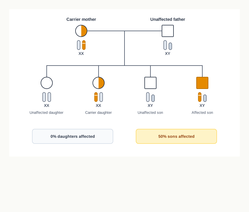
Fig. 8.12 – Inheritance of an X-linked recessive disorder.
In the progeny, men pass the Y chromosome on to their sons and their X chromosome to their daughters. So a man who is affected will not pass it on to his sons, but all his daughters will be carriers. This “no father-to-son transmission” rule is the single most reliable clue for spotting X-linked recessive inheritance in a pedigree.
Read Fig. 8.12 carefully, because the probabilities are frequently misquoted. From a carrier mother and an unaffected father, each son has a 50% chance of being affected and each daughter has a 50% chance of being a carrier.
Some examples of X-linked recessive disorders are haemophilia and Duchenne Muscular Dystrophy. Red-green colour blindness, in which a person cannot distinguish red from green, is another familiar X-linked recessive condition and follows exactly the same pattern — far more common in males, transmitted through carrier mothers, never passed from father to son.
Haemophilia is a bleeding disorder associated with mutation in the coagulation factor VIII gene (type A) or the coagulation factor IX gene (type B). Mutations in coagulation factor genes result in the production of an abnormal version of coagulation factor VIII or IX, or a reduced amount of one of these proteins. Blood clotting is a chain of reactions in which each factor activates the next, so a single missing link breaks the whole chain. The altered or missing coagulation factor cannot effectively complete the blood clotting process, leading to spontaneous bleeding or increased bleeding tendencies.
Duchenne Muscular Dystrophy (DMD) is caused by mutation of the dystrophin gene, resulting in reduction or absence of dystrophin, or the presence of an abnormal protein. Dystrophin normally acts as a supporting anchor inside muscle fibres, absorbing the stress of contraction. Dystrophin abnormality or deficiency leads to dystrophy, that is, degeneration of muscle, making the muscles more fragile and weak.
X-linked Dominant Disorder
In this type of inheritance, the faulty allele on the X chromosome is dominant, so a single copy is enough to cause the disorder in either sex.
Affected males pass on the mutated dominant gene to all their daughters, but to none of their sons (Fig. 8.13a). The reason is the same rule seen earlier: a father gives his X chromosome to every daughter and his Y chromosome to every son, so a fault carried on his X reaches all his daughters and no son at all.
In the case of an affected female married to an unaffected male, the condition is passed on to half of their sons and half of their daughters (Fig. 8.13b). She has two X chromosomes, only one of which carries the mutation, and each child receives one X from her at random.
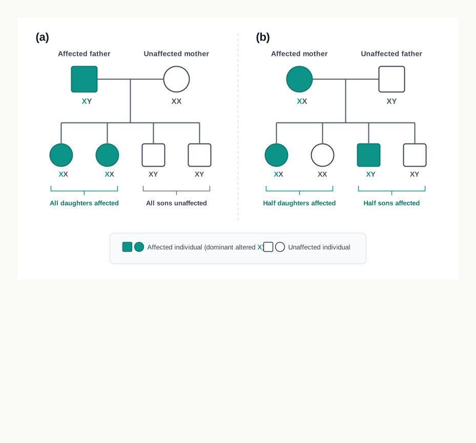
Fig. 8.13 – Inheritance of an X-linked dominant disorder through (a) an affected father and (b) an affected mother.
Examples of such disorders are hypophosphatemia, a type of vitamin D resistant rickets, and Alport syndrome, which is associated with progressive hearing loss and kidney disease.
8.3 Polygenic Disorders
The disorders in the previous section could each be traced to one gene. That made them relatively easy to describe: identify the gene, identify the protein it makes, and the symptoms follow. Most of the illnesses that fill hospital wards, however, do not behave like that at all. Heart disease, high blood pressure and diabetes run in families, yet no single gene can be blamed for them.
A polygenic disorder is caused by the defect or combined action of more than one gene. The classical examples of polygenic disease include hypertension, coronary heart disease and diabetes.
As the pathogenesis proceeds, such diseases depend on the simultaneous association of several genes, and therefore they cannot be explained as monogenic diseases. No single allele is either necessary or sufficient. Each of the genes involved makes only a small contribution to the risk, and the disease appears when enough of these small contributions accumulate — usually alongside diet, physical activity, body weight, smoking and age.
Why Many Genes Behave Differently from One
This has a practical consequence for how such diseases are studied and predicted. A monogenic disease sorts people into clear categories — affected, carrier, unaffected — and a pedigree can often predict the outcome for the next child. A polygenic disease produces a continuous range instead. Blood pressure and blood sugar do not come in two varieties; they come as a spread of values, and the line between “normal” and “disease” has to be fixed by agreement on a number.
Diabetes Mellitus
Diabetes mellitus is an important example of polygenic disease. It is a heterogeneous group of disorders characterised by a persistently high blood sugar level, or hyperglycemia. The word “heterogeneous” is doing real work in that sentence: several different underlying faults can all end in the same raised blood sugar reading.
To see why the blood sugar rises, recall what insulin does. Insulin is needed for transporting glucose from the blood to the inside of cells, such as muscle cells, for producing energy and maintaining cell function. If insulin is missing, or if the cells stop responding to it, glucose stays in the blood instead of entering the cells. The paradox of diabetes is that the blood is full of fuel while the cells are starved of it.
There are two most common forms of diabetes: Type 1 diabetes (T1D, previously known as insulin dependent diabetes or IDDM) and Type 2 diabetes (T2D, previously known as non-insulin-dependent diabetes or NIDDM).
Type 1 diabetes is caused by the destruction of the beta cells of the pancreas due to an immunological abnormality — the body’s own defence system attacks the very cells that make insulin. This type constitutes approximately 10% of all cases of diabetes. Since the source of insulin itself has been destroyed, lifelong treatment with insulin injection is needed for these cases.
Type 2 diabetes is the commonest form of the disease and represents about 90% of cases. It is caused by impaired insulin secretion from the beta cells of the pancreas, and also by peripheral insulin resistance, in which the tissues of the body respond poorly to the insulin that is present. Here the beta cells are not destroyed; they simply cannot keep up with a demand that has risen because the tissues have stopped listening.
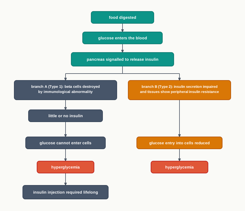
Fig. 8.14 – How blood glucose rises in the two forms of diabetes.
Hypertension
Hypertension, or a persistent increase in blood pressure, is a major risk factor for renal disease, heart disease and stroke involving the brain. It is a leading cause of global mortality and morbidity.
Blood pressure is written as two numbers. The systolic value is the pressure in the arteries when the heart contracts and pushes blood out; the diastolic value is the pressure that remains when the heart relaxes between beats. Both are measured in millimetres of mercury, written as mm Hg. Because a polygenic disease has no natural dividing line, the categories below are defined by agreed cut-off values.
Normal blood pressure (BP) is \(<120\) (systolic) \(/<80\) (diastolic) mm Hg. Elevated BP is \(120\text{–}129/<80\) mm Hg. Hypertension stage 1 is \(130\text{–}139\) or \(80\text{–}89\) mm Hg, and hypertension stage 2 is \(\ge 140\) or \(\ge 90\) mm Hg.
Read the small words in that table carefully, because they change the meaning entirely. For the normal and elevated categories the two conditions are joined by “and”, so both numbers must be low. For the two stages of hypertension they are joined by “or”, so a single high value is enough to place a person in that stage even if the other value is acceptable.
Sustained high pressure matters because it works on the arteries continuously, year after year. The vessel walls thicken and stiffen in response, and the heart has to work harder against the raised resistance. This is why hypertension is described as a risk factor rather than a disease of one organ — the damage shows up wherever the arteries matter most, which is in the kidneys, the heart and the brain.
Coronary Heart Disease
The heart muscle works without rest, and it receives its own blood supply through the coronary arteries. Coronary Heart Disease (CHD) develops due to the narrowing of the coronary artery, which supplies blood to the heart muscles, by the gradual build-up of fatty material on its wall (Fig. 8.15).
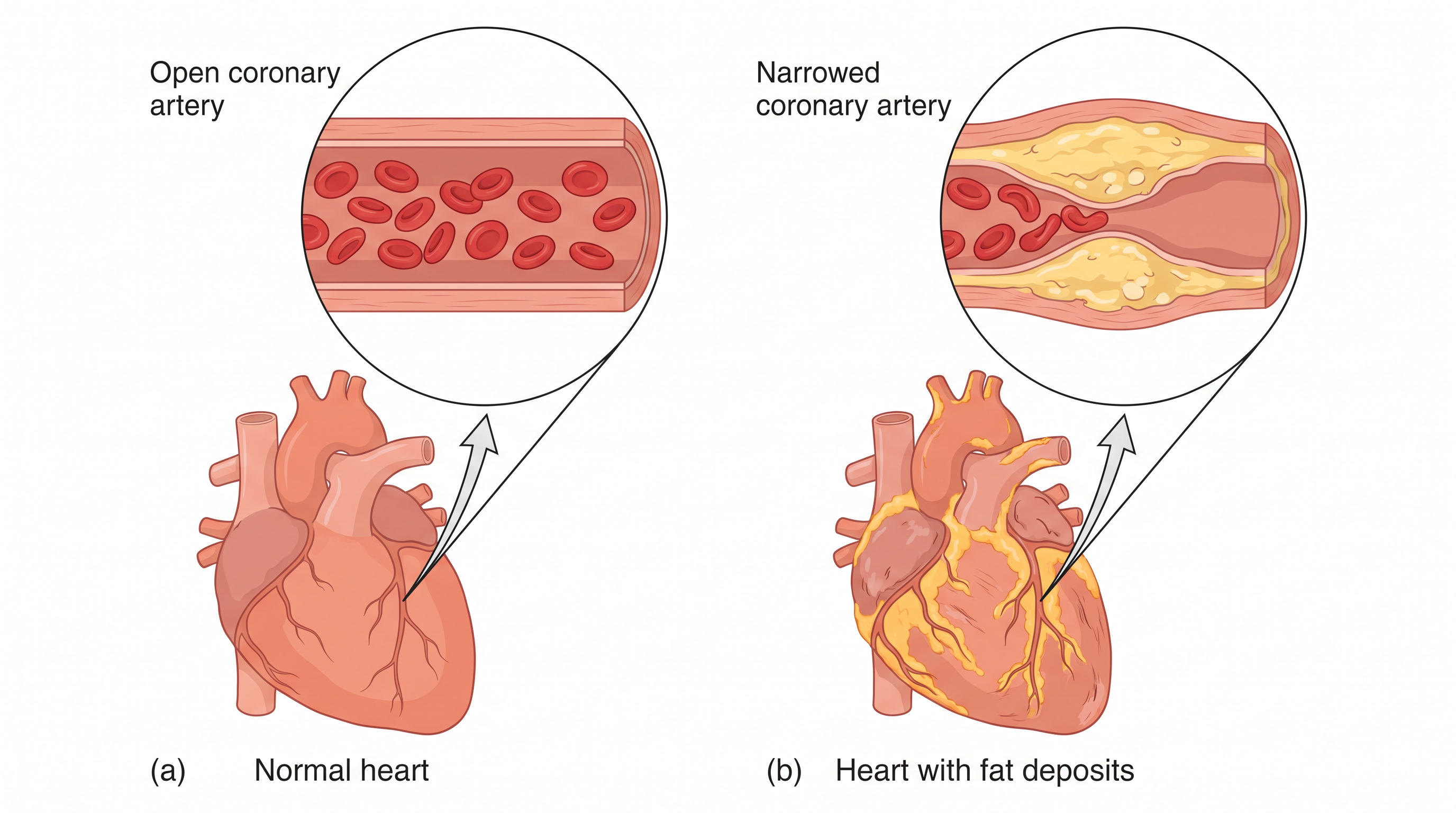
Fig. 8.15 – Coronary artery: (a) normal and (b) narrowed by atherosclerosis.
This accumulation of fatty material on the wall of the artery is called atherosclerosis. Compare the two insets in Fig. 8.15 — in the normal artery the red blood cells occupy a wide open channel, while in the diseased artery the plaque projects inwards and leaves only a narrow passage for the same volume of blood.
Due to the narrowing of the coronary artery by atherosclerosis, it cannot supply oxygen-rich blood to the heart muscle, which needs it for its continuous activity. This phenomenon of reduction of blood supply is called ischemia. That is why coronary heart disease was previously called ischaemic heart disease.
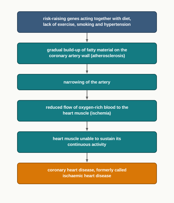
Fig. 8.16 – Development of coronary heart disease.
Notice how the three conditions in this section connect to one another rather than standing apart. Hypertension damages arterial walls and speeds up atherosclerosis. Long-standing high blood sugar in diabetes damages the same vessels. The genes involved overlap, and so do the lifestyle factors. This clustering is characteristic of polygenic disease and is the reason prevention advice for all three is so similar.
When Inheritance Does Not Come From the Nucleus
All the disorders discussed so far — chromosomal, monogenic and polygenic — involve genes carried on the chromosomes inside the nucleus. There is one further route by which a disease can be inherited, and it follows a rule of its own.
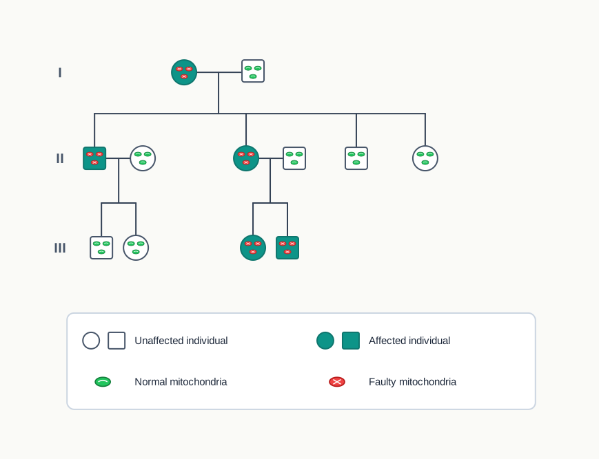
Fig. 8.17 – Mitochondrial inheritance in a family.
8.4 Summary
Haploid cells have only one copy of each chromosome, while diploid cells have two copies of the same chromosome.
Any deviation in which one or a few chromosomes are either absent or present in multiple copies is called aneuploidy.
In the polyploidy condition, the entire set of chromosomes is multiplied.
A syndrome is a specific collection of signs or symptoms suggesting a particular disease, while a disease is a broader term referring to an abnormal physiological response to internal or external factors.
Symptoms are subjective — they are what the patient feels and reports. Signs are objective — they are what an examiner can observe or measure.
Structural chromosomal abnormalities can be caused by four types of change: deletion, duplication, inversion and translocation.
Monogenic disease is caused by a modification in a single gene. Such diseases are classified according to their inheritance pattern into autosomal recessive, autosomal dominant, X-linked recessive and X-linked dominant.
A polygenic disorder is caused by the defect in, or the combined action of, more than one gene.
Mitochondrial inheritance and mitochondrial diseases are caused by a defect or mutation in the genes coding for the critical enzymes present in the mitochondria.
Down syndrome occurs due to trisomy 21, and is characterised by features such as a moon-like face, protruding tongue, muscular hypotonia, a palmar crease and so on.
Klinefelter syndrome occurs due to the presence of an extra X chromosome in males, and is characterised by tall height, enlarged breasts, coarse voice, hypogonadism and so on.
Turner syndrome occurs due to the absence of one X chromosome in females, and is characterised by short stature, webbed neck, small breasts, absence of menstruation and so on.
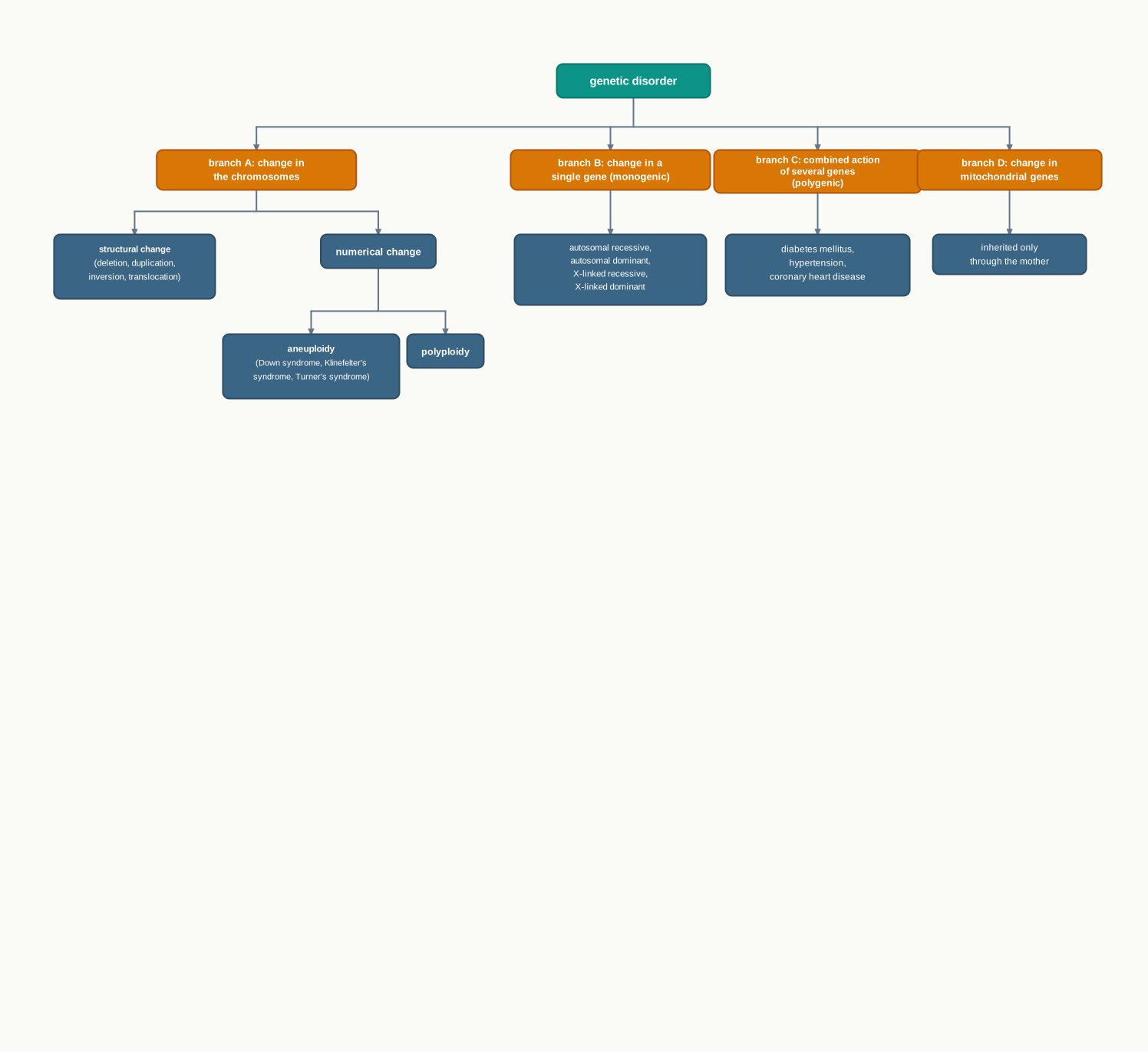
Fig. 8.18 – Classification of genetic disorders.
8.5 Key Terms
Chromosome — A thread-like structure inside the nucleus that carries the genes of an organism.
Haploid — A cell that carries only one copy of each chromosome, as in a sperm or an ovum.
Diploid — A cell that carries two copies of each chromosome, one from each parent.
Structural chromosomal abnormality (aberration) — A change in the physical structure or internal arrangement of a chromosome.
Numerical chromosomal abnormality — A change in the number of chromosomes present in a cell.
Monosomy — The condition in which one chromosome of a pair is absent, written as \(2n - 1\).
Trisomy — The condition in which a chromosome is present in three copies instead of two, written as \(2n + 1\).
Aneuploidy — The condition in which one or a few individual chromosomes are absent or present in extra copies.
Polyploidy — The condition in which the entire set of chromosomes is multiplied, giving three, four or more complete sets.
Deletion — A structural abnormality in which a segment of a chromosome breaks away and is lost, shortening the chromosome.
Ring chromosome — A chromosome formed when both ends are deleted and the remaining cut ends join to each other in a circle.
Duplication — A structural abnormality in which a segment of a chromosome is repeated, making the chromosome longer.
Inversion — A structural abnormality in which a chromosome segment breaks, reverses through 180 degrees and rejoins the same chromosome.
Translocation — A structural abnormality in which a segment of one chromosome breaks away and attaches to another chromosome.
Reciprocal translocation — A translocation in which two chromosomes mutually exchange segments with each other.
Robertsonian translocation — A translocation in which a segment attaches to another chromosome without mutual exchange, which may reduce the chromosome number of the cell.
Syndrome — A group of symptoms that consistently occur together, or a condition characterised by a set of associated symptoms.
Disease — An abnormal physiological response of the body to internal or external factors.
Symptom — A subjective feature of illness, that is, what the patient feels and reports.
Sign — An objective feature of illness, that is, what an examiner can observe or measure.
Karyotype — The complete chromosome set of an individual, described by number and structure, and written in a short standard notation such as 47, XY, +21.
Karyogram — The photographed chromosomes of a cell arranged in numbered pairs for examination.
Non-disjunction — The failure of chromosomes to separate at the time of cell division, so that one daughter cell receives an extra chromosome and the other receives one fewer.
Down syndrome — A condition arising from trisomy of chromosome 21, with a karyotype of 47, XX, +21 or 47, XY, +21.
Klinefelter’s syndrome — A condition of males arising from an extra X chromosome, with the genotype 47, XXY.
Barr body — The dark, condensed mass formed in a cell by an inactivated X chromosome, whose number is always one less than the number of X chromosomes present.
Turner’s syndrome — A condition of females arising from a missing X chromosome, that is, monosomy X, with the karyotype 45, X.
Amniocentesis — A prenatal test in which a sample of amniotic fluid containing foetal cells is withdrawn for chromosomal examination.
Chorionic villus sampling — A prenatal test in which a small piece of developing placental tissue is taken for chromosomal examination.
Monogenic (single-gene) disease — A disorder caused by an error or mutation in one single gene.
Pedigree analysis — The interpretation of a family tree in order to trace how a trait or disease has been inherited across generations.
Carrier — An individual with one defective recessive allele and one normal allele, who does not show the disease but can pass the allele on.
Autosomal recessive disorder — A disease that appears only when both copies of a gene on an autosome are defective.
Sickle cell anaemia — An autosomal recessive disease caused by a mutation in the hemoglobin-β gene on chromosome 11, in which red blood cells become stiff and sickle-shaped after releasing oxygen.
Cystic fibrosis — An autosomal recessive disease in which abnormally thick and sticky mucus damages organs, especially the lungs.
Tay-Sachs disease — A fatal childhood disease caused by the absence of the enzyme hexosaminidase A, leading to fatty accumulation in nerve cells.
Phenylketonuria — An autosomal recessive disease caused by a mutation in the phenylalanine hydroxylase gene, raising phenylalanine levels in the blood.
Autosomal dominant disorder — A disease in which a single faulty copy of a gene on an autosome is enough to produce the condition.
Achondroplasia — A rare autosomal dominant disorder leading to a type of dwarfism, in which most surviving cases are heterozygotes (D/d).
Huntington’s disease — A rare autosomal dominant disorder affecting the nervous system.
X-linked recessive disorder — A disease caused by a recessive faulty gene on the X chromosome, usually affecting males while females act as carriers.
Haemophilia — An X-linked recessive bleeding disorder caused by mutation in the coagulation factor VIII gene (type A) or the factor IX gene (type B).
Duchenne Muscular Dystrophy (DMD) — An X-linked recessive disorder caused by mutation of the dystrophin gene, leading to degeneration and weakness of muscle.
Colour blindness — An X-linked recessive condition, commoner in males, in which the affected person cannot distinguish certain colours such as red and green.
X-linked dominant disorder — A disease caused by a dominant faulty gene on the X chromosome, which an affected father passes to all his daughters and none of his sons.
Polygenic disorder — A disease caused by the defect in, or the combined action of, more than one gene.
Diabetes mellitus — A heterogeneous group of disorders characterised by a persistently high blood sugar level.
Hyperglycemia — A persistently raised level of glucose in the blood.
Insulin — The hormone that transports glucose from the blood into cells such as muscle cells for producing energy.
Insulin resistance — A condition in which the tissues of the body respond poorly to the insulin that is present.
Hypertension — A persistent increase in blood pressure, defined as a reading of \(\ge 140\) or \(\ge 90\) mm Hg at stage 2.
Atherosclerosis — The accumulation of fatty material on the inner wall of an artery, which narrows the vessel.
Ischemia — The reduction of blood supply, and therefore of oxygen supply, to a tissue or organ.
Coronary Heart Disease (CHD) — Disease resulting from narrowing of the coronary artery by atherosclerosis, formerly called ischaemic heart disease.
Mitochondrial inheritance — The transmission of genes carried in the circular DNA of mitochondria, which pass to the offspring only from the mother.
8.6 Points to Ponder
A polyploid plant may carry six or eight complete sets of chromosomes and still grow perfectly well, while a human being with one extra chromosome develops a syndrome. What does this tell you about whether cells care more about the quantity of genetic material or about the balance between genes?
In an inversion, not a single gene is lost and not a single gene is gained, yet a disorder can still result. In what different ways can a gene be damaged without its sequence being removed?
A karyotype can tell a family the chromosome number of an unborn child within days. It cannot tell them what that child will enjoy, achieve or become. How far should a test result be allowed to shape a decision it cannot fully inform?
The Barr body test counts X chromosomes indirectly and costs almost nothing, while karyotyping counts them directly and costs a great deal more. When is a cheap indirect test the better choice, and when is it not enough?
Down syndrome, Klinefelter’s syndrome and Turner’s syndrome are all genetic conditions, yet none of them is inherited from an affected parent in the ordinary sense. What does this suggest about the difference between “genetic” and “hereditary”?
Every human being is estimated to carry about five or more defective recessive genes, and almost none of us ever finds out. Under what circumstances does this hidden load suddenly become visible in a family?
The sickle cell allele causes a serious disease in the homozygous state, yet it has persisted for thousands of years in particular regions of the world. Can an allele be harmful and useful at the same time, and what would decide which it is?
In achondroplasia, two copies of the dominant allele are more damaging than one, and the surviving affected individuals are almost all heterozygotes. Does this fit your earlier understanding of what “dominant” means? What might need adjusting?
Huntington’s disease usually appears only after the age at which people have had children. How might the timing of a disease, quite apart from its severity, affect how long it survives in a population?
In X-linked recessive inheritance, an affected father never passes the condition to his sons. Trace the chromosomes through the cross yourself and satisfy yourself that this is a consequence of the mechanism rather than a rule to be memorised.
Blood pressure and blood sugar exist as a continuous range of values, so the boundary between health and disease has to be fixed by agreement on a number. What is gained by drawing such a line, and what is lost?
Diabetes, hypertension and coronary heart disease share genes, share risk factors and often occur together in the same person. Should they be thought of as three separate diseases or as three faces of one process?
A father with a faulty mitochondrial gene cannot pass it to any of his children, while a mother passes her mitochondria to all of hers. What does this single asymmetry tell you about where a gene sits inside a cell?
For the chromosomal syndromes described in this chapter there is no permanent cure, and treatment aims at managing the effects instead. Why does the location of the fault — in every cell of the body — make cure so much harder than management?
Genetic information about one person is unavoidably information about that person’s parents, siblings and children. Who, then, has a legitimate claim to see a genetic test result, and who decides?
8.7 NCERT Questions
Define the following terms: dominant, recessive, homozygous, heterozygous, phenotype and genotype.
Describe the origin, symptoms and treatment of Down syndrome.
Describe the origin, symptoms and treatment of Klinefelter syndrome.
Describe the origin, symptoms and treatment of Turner syndrome.
Describe the various structural chromosomal abnormalities.
8.8 Objective Questions
The condition in which one chromosome of a pair is absent is represented as:
- \(2n + 1\)
- \(2n - 1\)
- \(3n\)
- \(n\)
A cell containing 92 chromosomes, that is \(23 \times 4\), is an example of:
- trisomy
- monosomy
- polyploidy
- aneuploidy
Bread wheat is hexaploid. The number of chromosome sets it therefore carries is:
- three
- four
- six
- eight
Which of the following is octoploid?
- Banana
- Apple
- Cabbage
- Sugar cane
Retinoblastoma is caused by deletion of a portion of chromosome:
- 11
- 13
- 17
- 21
Charcot-Marie-Tooth disease is caused by duplication of genes on chromosome:
- 8
- 11
- 17
- 21
In an inversion, the orientation of the genes in the affected segment is reversed by:
- 90 degrees
- 180 degrees
- 270 degrees
- 360 degrees
In Burkitt’s lymphoma, exchange of material takes place between chromosomes:
- 8 and 13
- 8 and 14
- 13 and 17
- 14 and 21
The type of translocation that may result in a decrease in the chromosome number of the cell is:
- reciprocal translocation
- Robertsonian translocation
- inversion
- duplication
The karyotype 47, XY, +21 represents:
- a female with Down syndrome
- a male with Down syndrome
- a male with Klinefelter’s syndrome
- a female with Turner’s syndrome
The incidence of Down syndrome is approximately:
- 1 per 800 live births
- 1 in 1000 newborn males
- 1 in 2,500 newborn girls
- 1 in 27 individuals
The genotype associated with Klinefelter’s syndrome is:
- 45, X
- 46, XY
- 47, XXY
- 47, XX, +21
The number of Barr bodies expected in a buccal smear from a male with Klinefelter’s syndrome is:
- zero
- one
- two
- three
Turner’s syndrome is an example of:
- trisomy
- monosomy
- polyploidy
- reciprocal translocation
In Turner’s syndrome, growth and ovarian functions can be strengthened by the controlled administration of:
- insulin
- testosterone alone
- androgen and estrogen
- coagulation factor VIII
Sickle cell disease is caused by a mutation in the hemoglobin-β gene found on chromosome:
- 8
- 11
- 13
- 17
The genotype of a sickle cell carrier is written as:
- S/S
- S/s
- s/s
- D/d
Tay-Sachs disease is due to the absence of the enzyme:
- phenylalanine hydroxylase
- hexosaminidase A
- dystrophin
- coagulation factor IX
Most surviving cases of achondroplasia have the genotype:
- d/d
- D/d
- D/D
- s/s
Haemophilia type B is associated with mutation in the gene for:
- coagulation factor VIII
- coagulation factor IX
- dystrophin
- hemoglobin-β
Alport syndrome, associated with progressive hearing loss and kidney disease, is an example of a disorder that is:
- autosomal recessive
- autosomal dominant
- X-linked recessive
- X-linked dominant
Type 1 diabetes constitutes approximately what proportion of all cases of diabetes?
- 10%
- 25%
- 50%
- 90%
Hypertension stage 2 is defined as a blood pressure of:
- \(\ge 120\) or \(\ge 80\) mm Hg
- \(\ge 130\) or \(\ge 80\) mm Hg
- \(\ge 140\) or \(\ge 90\) mm Hg
- \(\ge 160\) or \(\ge 100\) mm Hg
The accumulation of fatty material on the wall of an artery is called:
- ischemia
- atherosclerosis
- hypotonia
- hyperglycemia
In the offspring, the mitochondrial genes are inherited from:
- the father only
- the mother only
- both parents equally
- neither parent
Assertion: A male with Klinefelter’s syndrome shows one Barr body in a buccal smear, although a normal male shows none. Reason: The number of Barr bodies in a cell is always one less than the number of X chromosomes present in it.
Assertion: A plant may live normally with six complete sets of chromosomes, while a human being is seriously affected by a single extra chromosome. Reason: In polyploidy the copy number of every gene rises together, so the balance between genes is preserved, whereas in aneuploidy only one chromosome changes its copy number.
Assertion: A man affected by haemophilia never transmits the condition to his sons. Reason: A father transmits his Y chromosome to his sons and his X chromosome to his daughters.
Assertion: Two completely healthy parents can have a child affected by sickle cell anaemia. Reason: A carrier with the genotype S/s produces enough normal haemoglobin to remain healthy, but can still pass on the s allele.
Assertion: A mother with Turner’s syndrome cannot pass the condition on to her daughters. Reason: Turner’s syndrome arises from a cell division error during meiosis of the ovum rather than from an allele inherited from a parent.
Assertion: Most individuals surviving with achondroplasia are heterozygous for the condition. Reason: The homozygous dominant genotype D/D is often lethal.
Assertion: Coronary heart disease was previously known as ischaemic heart disease. Reason: Narrowing of the coronary artery by atherosclerosis reduces the supply of oxygen-rich blood to the heart muscle.
Assertion: A pedigree cannot reliably predict whether a particular child will develop Type 2 diabetes. Reason: Type 2 diabetes is a polygenic disorder in which several genes make small contributions alongside diet, weight and physical activity.
Assertion: An inversion can cause a disorder even though no genetic material is lost or gained. Reason: In an inversion the overall length of the chromosome remains the same.
8.9 Check Your Concepts
Why does the addition of a whole extra set of chromosomes affect a plant far less than the addition of a single extra chromosome affects a human being? Answer in terms of the balance between genes rather than the quantity of genetic material.
An inversion neither removes nor adds any genetic material, yet it can still cause a disorder. Give three distinct reasons why this is possible.
Compare and contrast reciprocal translocation and Robertsonian translocation under the following headings: direction of transfer, presence of mutual exchange, and effect on the chromosome number of the cell.
Draw a chromosome carrying the segments A, B, C, D, E and F. Using this chromosome, illustrate deletion, duplication, inversion and translocation, and label each drawing. In each case, state whether the overall length of the chromosome increases, decreases or stays the same.
What happens if a chromosome pair fails to separate at the time of cell division? Trace the consequence through both gametes formed, and state the chromosome constitution of the two possible zygotes that result when each is fertilised by a normal gamete.
What does this chapter report about the relationship between maternal age and the birth of a baby with Down syndrome, and how does that reported relationship influence the way prenatal screening is offered?
The following steps of karyotyping are given in the wrong order. Arrange them correctly and give a one-line reason for the position of the first and the last step: (i) stain to produce a banding pattern; (ii) sort the chromosomes into numbered pairs; (iii) collect dividing cells from a blood sample; (iv) photograph the chromosome spread; (v) arrest cell division at metaphase.
A student examines a buccal smear taken from a healthy adult male and reports the presence of one Barr body. Give two possible explanations for this result, and state which confirmatory test should be carried out before any conclusion is drawn.
Why is the Barr body test described as an indirect count of X chromosomes? State the arithmetic rule that connects the number of Barr bodies to the number of X chromosomes, and use it to predict the count for a normal female, a normal male and a female with Turner’s syndrome.
There is no permanent cure for Down syndrome, Klinefelter’s syndrome or Turner’s syndrome. Explain why the location of the fault makes cure so difficult, and state what the treatments described in the chapter actually achieve.
Match the karyotype in Column A with the condition in Column B.
| Column A | Column B |
|---|---|
| a. 45, X | i. Klinefelter’s syndrome |
| b. 47, XXY | ii. normal female |
| c. 47, XX, +21 | iii. Turner’s syndrome |
| d. 46, XX | iv. Down syndrome in a female |
Distinguish between a syndrome and a disease, and support your answer with one example of each taken from this chapter.
Why can an autosomal recessive disease skip several generations in a family, while an autosomal dominant disease usually appears in every generation?
A couple, both carriers of sickle cell anaemia, already have three unaffected children. Explain why this does not reduce the risk of the disease in their next child, and state what the expected ratio of genotypes among their children is.
Arrange the following events in the correct sequence, starting from the gene: (i) red blood cell becomes stiff and sickle-shaped; (ii) mutation in the hemoglobin-β gene; (iii) defective haemoglobin molecules cluster into rod-like structures; (iv) defective haemoglobin gives up oxygen in the tissues; (v) defective haemoglobin is produced.
Why do the symptoms of sickle cell anaemia become worse during heavy exercise, at high altitude or during an infection? Base your answer on the exact point in the process at which sickling occurs.
More than 200 different types of mutation can occur in the gene responsible for cystic fibrosis, whereas sickle cell anaemia results from a single mutation. What difficulty does this create for anyone designing a diagnostic test for cystic fibrosis?
In achondroplasia, individuals with the genotype D/d survive while those with D/D usually do not. Explain what this tells you about the meaning of the word “dominant”, and why most surviving affected individuals are heterozygotes.
In a family pedigree, a disorder appears only in males, none of the affected males has an affected son, and the mothers of the affected males are unaffected. Identify the most likely mode of inheritance and justify your answer using two separate observations from the description.
A student draws a pedigree using circles for males, squares for females, and shading for carriers rather than for affected individuals. List the errors against the conventions given in Fig. 8.7, and state one wrong conclusion a reader could draw from this pedigree.
Compare X-linked recessive and X-linked dominant inheritance by considering what happens to the daughters and to the sons of an affected father in each case.
Why does Type 1 diabetes require lifelong insulin injection, while Type 2 diabetes does not always begin with insulin treatment? Answer by comparing the underlying defect in each case.
Explain why hypertension, diabetes mellitus and coronary heart disease so often occur together in the same person, and why the prevention advice for all three is broadly similar.
Arrange the following in the correct order: (i) reduced supply of oxygen-rich blood to the heart muscle; (ii) gradual build-up of fatty material on the coronary artery wall; (iii) coronary heart disease; (iv) narrowing of the coronary artery. Then name the process described in step (ii) and the process described in step (i).
A father is affected by a disease caused by a faulty mitochondrial gene, but none of his children is affected. Explain why. Then state two features of a family tree that would let you distinguish mitochondrial inheritance from X-linked recessive inheritance.
Why does the severity of a mitochondrial disease differ from one affected person to another, and why are the brain, heart, kidneys, muscle and liver affected most often?
8.10 Case-Based Questions
A paediatrician examines a newborn baby who has a flat facial profile, a protruding tongue, a short neck and a single deep crease across each palm. The baby also has reduced muscle tone and is being investigated for a heart problem. The mother was 38 years old at the time of the pregnancy. A blood sample is sent to the cytogenetics laboratory, and the report returns as 47, XY, +21.
- Name the condition and explain what each part of the reported karyotype tells you.
- Name the error in cell division that usually produces this condition, and describe how it leads to a zygote with three copies of a chromosome.
- The parents ask whether there is a treatment that will remove the extra chromosome. Frame a scientifically accurate reply, and mention the forms of support described in this chapter that would benefit the child from an early age.
- The mother’s age is noted prominently in the case file. What does the chapter report about maternal age and this condition?
A boy of seventeen is brought to a clinic because he is much taller than his classmates, has very little facial and body hair, and has developed enlarged breasts. His parents mention that as an infant he appeared no different from other babies, and that the changes became noticeable only around puberty. The clinician orders a buccal smear, which shows one Barr body, and a karyotype is then requested.
- What condition does the buccal smear result suggest, and what karyotype would you expect the laboratory to report?
- Explain the principle on which the buccal smear test works, and why a normal male smear shows no Barr body.
- Why did the differences become apparent only at puberty rather than at birth?
- The boy’s father asks whether he passed the condition on to his son. Explain, with reasons, what you would tell him.
A girl of fourteen is brought to a hospital because she is much shorter than other girls of her age and has not begun to menstruate. On examination the clinician notes loose skin at the sides of the neck that can be pulled several centimetres away, ears set below the usual position, and swollen hands and feet. A buccal smear is taken and no Barr body is found.
- Name the condition indicated, write its karyotype, and state whether it is a monosomy or a trisomy.
- Explain how the absence of a Barr body in a female smear becomes the first indicator of this condition, and what investigation should follow.
- The girl’s mother is anxious that she may have transmitted the condition. What does the chapter say about whether this condition is inherited, and how does it arise?
- Which hormones are used to strengthen growth and ovarian function in such cases, and why is this described as management rather than cure?
A screening camp is organised in a district of the Deccan plateau in central India. Blood samples from 600 young adults are tested, and a number of participants are found to carry one copy of the sickle cell allele although they are entirely healthy. Two of the participants, who are engaged to be married, are both found to be carriers. They are referred for genetic counselling.
- Write the genotypes of the two participants, and using a cross, work out the expected genotypes and phenotypes of their children.
- Explain why both of them are healthy despite carrying the allele, and what the term used for such individuals is.
- This district was chosen for the camp rather than a district picked at random. Give the reason, using the distribution described in the chapter.
- The counsellor gives the couple the results and the risk figures, but does not tell them what decision to take. Give one argument in support of this approach and one argument that could be raised against it.
A family history is recorded in a haematology clinic. A man reports frequent, prolonged bleeding after minor injuries, and tests show a reduced amount of coagulation factor VIII. His mother and his two sisters have never had any bleeding problem. His mother’s brother had the same condition. The man himself has one son and one daughter, neither of whom has shown any bleeding tendency so far.
- Name the condition and its mode of inheritance, and justify your answer using two observations from the family history.
- Explain, at the level of the protein, why the patient bleeds for longer than normal.
- State what you would expect the genetic status of the man’s son and daughter to be, and give the reason in terms of which chromosome each received from him.
- Draw the pedigree of this family using the standard symbols, shading the affected individuals and marking those who must be carriers.
At a community health camp, a 52-year-old man is examined. His blood pressure is recorded as 146/88 mm Hg on two separate readings. His fasting blood sugar is persistently raised, and he is told he has Type 2 diabetes. His father had a heart attack at the age of 60, and his elder brother is on treatment for high blood pressure. The man works at a desk, walks very little, and has gained considerable weight in the last ten years.
- Classify his blood pressure reading using the categories given in the chapter, and state which of the two values decided your answer.
- These conditions run in his family, yet no single faulty gene can be identified. Explain what kind of disorder this is and why a pedigree cannot predict it in the way a pedigree predicts haemophilia.
- Explain the basic defect in Type 2 diabetes, and why it differs from the defect in Type 1 diabetes.
- The man is warned that he is at risk of coronary heart disease. Describe, step by step, how the process leading to that disease develops in the coronary artery.
A woman has a disorder involving muscle weakness and poor exercise tolerance. All four of her children show the same disorder, although the severity differs a great deal between them — one is severely affected, two are moderately affected, and one has only mild symptoms. Her husband is entirely healthy. Investigation shows a mutation in a gene coding for an enzyme of energy production that is located in the circular DNA of the mitochondria.
- Name the mode of inheritance and explain why all four children are affected while the father is healthy.
- Explain why the severity of the disorder varies so much between children of the same parents.
- If the affected woman’s son later marries an unaffected woman, what would you expect for their children? Give the reason.
- State two features of this family tree that would rule out X-linked recessive inheritance.
8.11 Practice With Data
A human somatic cell normally contains 46 chromosomes. Calculate the chromosome number in a human cell showing (i) monosomy of chromosome 1, (ii) trisomy of chromosome X, (iii) three complete sets of chromosomes, and (iv) four complete sets of chromosomes. For each case, state whether it is an example of aneuploidy or of polyploidy, and write the general expression (\(2n - 1\), \(2n + 1\), and so on) that applies.
A flowering plant has a basic set of 7 chromosomes. Calculate the total chromosome number in a diploid, a triploid, a tetraploid, a hexaploid and an octoploid individual of this species. Then state which of the crop plants mentioned in this chapter corresponds to each of the last four ploidy levels.
A large maternity hospital records 6,400 live births in one year, of which 3,200 are male and 3,200 are female. Using the incidence figures given in this chapter, estimate the expected number of babies born that year with (i) Down syndrome, (ii) Klinefelter’s syndrome, and (iii) Turner’s syndrome. Which condition is expected to be the least frequent at this hospital, and why can the observed number for Turner’s syndrome be lower than the figure you calculate?
Among individuals of European Ashkenazi Jewish origin, 1 in 27 persons carries the Tay-Sachs gene. In a community gathering of 1,350 such individuals, estimate the number of carriers present. Express this proportion as a percentage, correct to one decimal place.
A man and a woman are both carriers of sickle cell anaemia.
| Gametes | S | s |
|---|---|---|
| S | ||
| s |
- Complete the cross above and write the genotypes obtained. (ii) State the expected genotypic ratio and the expected phenotypic ratio among their children. (iii) Express, as a percentage, the chance that a child of this couple will be a carrier. (iv) Calculate the probability that all three of their children will be unaffected by the disease.
- A carrier mother marries an unaffected father, and the disorder in question is X-linked recessive.
- Write the genotypes of both parents. (ii) Set out the cross and list the four expected classes of offspring. (iii) State the percentage of sons expected to be affected and the percentage of daughters expected to be affected. (iv) Now calculate the percentage of all the children, taken together, who are expected to be affected, and explain why this figure differs from the one quoted for the sons alone in Fig. 8.12.
Consider two separate families in which an X-linked dominant disorder is present. In family A the father is affected and the mother is unaffected. In family B the mother is affected and the father is unaffected. For each family, calculate the percentage of daughters and the percentage of sons expected to be affected, and state which of the two families shows the sharper difference between the sexes.
The blood pressure readings of five adults are given below, each taken twice on separate days.
| Person | Reading 1 (mm Hg) | Reading 2 (mm Hg) |
|---|---|---|
| P | 116/74 | 118/78 |
| Q | 126/78 | 124/76 |
| R | 134/86 | 136/82 |
| S | 128/94 | 130/92 |
| T | 148/86 | 152/90 |
- Classify each person using the categories given in this chapter. (ii) For persons S and T, state which of the two values decided the category and explain why. (iii) Person Q asks whether the reading is normal. Frame an accurate answer using the correct category name.
A survey of a city finds 24,000 people living with diabetes mellitus. Using the proportions given in this chapter, estimate how many of them are likely to have Type 1 diabetes and how many are likely to have Type 2 diabetes. If insulin injection is required lifelong by all Type 1 patients, calculate the number of people in this survey who fall in that group, and state one reason why the true number requiring insulin would be higher than this figure.
In a family affected by a mitochondrial disorder, the proportion of faulty mitochondria measured in muscle tissue is given below.
| Sibling | Faulty mitochondria (%) |
|---|---|
| W | 18 |
| X | 42 |
| Y | 67 |
| Z | 85 |
- Arrange the four siblings in the expected order of increasing severity of symptoms, and give the reason for your ordering. (ii) Calculate the percentage of normal mitochondria in each sibling. (iii) Explain why muscle tissue rather than skin was chosen for this measurement.
- The cytogenetics laboratory reports the following observations for five individuals.
| Individual | Total chromosome number | Sex chromosomes | Barr bodies in buccal smear |
|---|---|---|---|
| 1 | 46 | XX | 1 |
| 2 | 46 | XY | 0 |
| 3 | 47 | XXY | 1 |
| 4 | 45 | X | 0 |
| 5 | 47 | XX | 1 |
- Identify the condition, if any, in each individual, and write the full karyotype notation for individuals 3, 4 and 5, given that the extra chromosome in individual 5 is chromosome 21. (ii) State the arithmetic rule connecting the Barr body count to the number of X chromosomes, and verify it against the data for individuals 1 to 4. (iii) Explain why the Barr body count alone cannot distinguish individual 5 from individual 1.